2024
|
wileyonlinelibrary.com/journal/fsb2 The FASEB Journal. 2020;34:2024–2040.R E S E A R C H A R T I C L E
Impairment of DHA synthesis alters the expression of neuronal
plasticity markers and the brain inflammatory status in mice
Emanuela Talamonti
1,2|
Valeria Sasso
3|
Hoi To
1|
Richard P. Haslam
4|
Johnathan A. Napier
4|
Brun Ulfhake
5|
Karin Pernold
5|
Abolfazl Asadi
2|
Tara Hessa
1|
Anders Jacobsson
2|
Valerio Chiurchiù
6,7|
Maria Teresa Viscomi
81Department of Biochemistry and Biophysics, Stockholm University, Stockholm, Sweden
2Department of Molecular Biosciences, The Wenner-Gren Institute, Stockholm University, Stockholm, Sweden 3Laboratory of Experimental Neurorehabilitation, IRCCS Santa Lucia Foundation, Rome, Italy
4Department of Plant Science, Rothamsted Research, Harpenden, UK 5Department of Neuroscience, Karolinska Institute, Stockholm, Sweden 6Department of Medicine, Campus Bio-Medico University of Rome, Rome, Italy
7Laboratory of Resolution of Neuroinflammation, IRCCS Santa Lucia Foundation, Rome, Italy 8Istituto di Istologia ed Embriologia, Università Cattolica del S. Cuore, Rome, Italy
Valerio Chiurchiù and Maria Teresa Viscomi are equally senior authors.
Abbreviations: Arc1, activity-regulated cytoskeleton-associated protein 1; BDNF, brain-derived neurotrophic factor; DHA, docosahexaenoic acid; Egr-1, early growth response protein 1; ELOVLs, elongases; Elovl2, elongation of long-chain fatty acids 2; EPA, alpha-linolenic acid-derived eicosapentaenoic acid; GFAP, glial fibrillary acidic protein; Iba1, ionized calcium-binding adapter molecule 1; PUFA, ω-3 polyunsaturated fatty acids; SPMs, specialized pro-resolving mediators; TMEM-119, transmembrane protein 119.
Correspondence
Emanuela Talamonti, Department of Biochemistry and Biophysics, Stockholm University, Stockholm 106 91, Sweden. Email: [email protected]
Valerio Chiurchiù, Department of Medicine, University Campus bio-Medico, Via Alvaro del Portillo 21, Rome 00123, Italy. Email: [email protected] Maria Teresa Viscomi, Istituto di Istologia ed Embriologia, Università Cattolica del Sacro Cuore, Largo Francesco Vito 1, Rome 00168, Italy.
Email: [email protected] Funding information
Fondazione Italiana Sclerosi Multipla (FISM), Grant/Award Number: FISM2017/R/8; RCUK | Biotechnology and Biological Sciences Research Council (BBSRC), Grant/Award Number: BBS/
Abstract
Docosahexaenoic acid (DHA) is a ω-3 fatty acid typically obtained from the diet or endogenously synthesized through the action of elongases (ELOVLs) and desaturases. DHA is a key central nervous system constituent and the precursor of several mol-ecules that regulate the resolution of inflammation. In the present study, we questioned whether the impaired synthesis of DHA affected neural plasticity and inflammatory status in the adult brain. To address this question, we investigated neural and inflam-matory markers from mice deficient for ELOVL2 (Elovl2−/−), the key enzyme in DHA
synthesis. From our findings, Elovl2−/− mice showed an altered expression of markers
involved in synaptic plasticity, learning, and memory formation such as Egr-1, Arc1, and BDNF specifically in the cerebral cortex, impacting behavioral functions only marginally. In parallel, we also found that DHA-deficient mice were characterized by an increased expression of pro-inflammatory molecules, namely TNF, IL-1β, iNOS, caspase-1 as well as the activation and morphologic changes of microglia in the ab-sence of any brain injury or disease. Reintroducing DHA in the diet of Elovl2−/− mice
reversed such alterations in brain plasticity and inflammation. Hence, impairment of
This is an open access article under the terms of the Creative Commons Attribution License, which permits use, distribution and reproduction in any medium, provided the original work is properly cited.
systemic DHA synthesis can modify the brain inflammatory and neural plasticity sta-tus, supporting the view that DHA is an essential fatty acid with an important role in keeping inflammation within its physiologic boundary and in shaping neuronal func-tions in the central nervous system.
K E Y W O R D S
anti-inflammatory molecules, brain plasticity, microglia, omega-3, PUFA
1
|
INTRODUCTION
The main ω-3 polyunsaturated fatty acid (PUFA) of the brain is docosahexaenoic acid (DHA), which is a key structural com-ponent of neuronal membrane phospholipids and plays an es-sential role in brain development and function.1,2 DHA cannot
be synthesized de novo in mammals and need to be synthesized from the essential dietary fatty acid α-linolenic acid (ALA) through a series of elongation and desaturation reactions per-formed by distinct enzymes residing in the endoplasmic retic-ulum.3 Indeed, DHA biosynthesis requires three elongation
steps, the last two of which are catalyzed by elongation of long-chain fatty acids 2 (ELOVL2), an enzyme that is significantly expressed in liver, testis, retina and central nervous system (CNS), tissues that are particularly rich in DHA.4-6
DHA is essential for CNS functions, including neurite out-growth and synaptogenesis,7 brain-derived neurotrophic factor
(BDNF)-induced neuronal survival,8 synaptic integrity and
neu-rotransmission9 as well as protection from neuroinflammatory
processes, and cognitive decline.10 Indeed, a DHA deficient
diet is involved in the pathophysiology of memory impairment and several mood disorders such as anxiety and depressive-like behaviors.11-15 DHA deficiency in the CNS is also paralleled
by an increased expression of pro-inflammatory cytokines and a modification of neuronal plasticity-related gene expression, including the early growth response protein 1 (Egr-1) and the activity-regulated cytoskeleton-associated protein (Arc-1),16
ul-timately associated with learning and memory formation.17,18
Such evidence prompted the investigation of several clinical tri-als with DHA on neurodevelopmental disorders, including au-tism and attention-deficit/hyperactivity disorder.19 Despite the
fact that there is a growing number of trials testing the efficacy of n-3 PUFAs in the treatment of different psychiatric disorders, only a few studies investigated the efficacy of DHA alone on cognitive functions. Moreover, the small number of trials on cognitive impairment in psychiatric disorders shows doubtful and/or contrasting results that need further investigations.20,21
Moreover, the role of DHA in inflammation is already consolidated and is receiving greater attention mainly due to the discovery that it serves as a metabolic precursor for a new genus of anti-inflammatory molecules, that is, resolvins, neuroprotectins, and maresins, that are involved in the spon-taneous but active process of resolution of inflammation,21,22
which mainly involves myeloid cells such as macrophages and glial cells.23,24 Indeed, we have recently shown that the
impair-ment of systemic DHA synthesis in Elovl2−/− mice, delineates
an immunophenotypical alteration of M1/M2 macrophages, with M1 being more pro-inflammatory and M2 less protec-tive, supporting the view that DHA has a relevant role in con-trolling inflammatory responses.25 Given that DHA plays a
key role in CNS function and plasticity and is a potent anti-in-flammatory mediator, we sought to systematically investigate whether Elovl2−/− mice, characterized by a systemic
impair-ment of endogenous DHA synthesis, displayed neuronal plas-ticity dysfunctions and brain inflammation and whether such alterations could be restored upon DHA supplementation.
2
|
MATERIAL AND METHODS
2.1
|
Ethics statement for experimental
animals
This study was approved by the Institutional Review Board on Animal Studies of Stockholm University and by the Animal Ethics Board of the North Stockholm region, and performed in accordance with national guidelines and regulations for the care and use of laboratory animals in agreement with the guidelines of the European Communities Council Directive 2010/63/EU for the care and use of laboratory animals. All efforts were made to minimize the number of animals used and their suffering.
2.2
|
Animals and tissue processing
Elovl2−/− mice were generated as described previously26 and
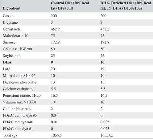
backcrossed into the 129S2/Sv strain for five generations. All animals were housed at room temperature and maintained on a 12 hours light/dark cycle. About 20-25 weeks old male or female mice were fed standard chow DHA-free diet (10% kcal fat, D12450H, Research Diets, New Brunswick, NJ, USA) or DHA-enriched (10% kcal fat, 1% DHA, D13021002, Research Diets, New Brunswick, NJ, USA) for 3 months, according to the experimental groups. Dietary fatty acid composition and other nutrients are shown in Tables 1 and 2. All animals were fed ad libitum and had free access to water. At the end of the
E/C/000I0420; Svenska Forskningsrådet Formas (Swedish Research Council Formas), Grant/Award Number: IB13-0026
study, animals were euthanized with CO2 and killed by
cervi-cal dislocation. Brains were removed immediately, dissected, and stored in liquid nitrogen or in 4% paraformaldehyde.
2.3
|
HPLC analysis of acyl-CoA fatty acids
Brain tissues were ground to a powder and 6 mg extracted27
for reverse-phase LC (Agilent 1200 LC system; Gemini C18 column, 2 mm inner diameter, 150 mm with 5 mm particles) with electrospray ionisation tandem mass spectrometry (multi reaction monitoring, MRM) on a QTRAP 4000 (SCIEX) in-strument in positive ion mode. LC-MS/MS MRM analysis followed the methods previously described.28 A total of 68
acyl-CoA fatty acids (14:0 to 34:6) were tracked using indi-vidual MRM mass pairs. For identification and calibration, standard acyl-CoA esters with acyl chain lengths from C14 to C20 were purchased from Sigma as free acids or lithium salts.
2.4
|
Western blotting
Whole brains and the cerebral cortex were collected, ho-mogenized, and proteins were extracted in Ripa buffer (Milli-Q Water, Tris hydrochloride 0.05 M pH 7.4, 0.001 M potassium chloride, 0.0015 M magnesium chloride, 0.001 M EDTA, 0.001 M dithiothreitol, 0.005 M sodium fluoride, 0.001 M sodium metavanadate, 0.1% SDS, 10% Na-DOC,1% Triton X-100, 1X protease inhibitor cocktail (Sigma, P8340), for 20 minutes on ice, and then were cen-trifuged for 15 minutes at 4°C (14 000 rpm). Supernatants were collected and transferred in a 1.5 mL vial. The total protein content of the resulting supernatant was quantified by the Bradford's colorimetric assay (Bio-Rad, Milan, Italy). Each protein sample was separated by SDS-polyacrylamide gel electrophoresis and transferred to a nitrocellulose mem-brane. Membranes were saturated with 5% dried nonfat milk and incubated overnight at 4 degrees C with specific
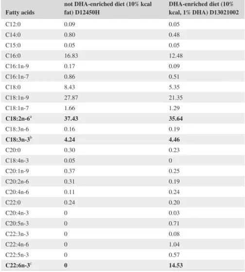
Fatty acids not DHA-enriched diet (10% kcal fat) D12450H DHA-enriched diet (10% kcal, 1% DHA) D13021002
C12:0 0.09 0.05 C14:0 0.80 0.48 C15:0 0.05 0.05 C16:0 16.83 12.48 C16:1n-9 0.17 0.09 C16:1n-7 0.86 0.51 C18:0 8.43 5.35 C18:1n-9 27.87 21.35 C18:1n-7 1.66 1.29 C18:2n-6a 37.43 35.64 C18:3n-6 0.16 0.19 C18:3n-3b 4.24 4.46 C20:0 0.30 0.23 C18:4n-3 0.05 0 C20:1n-9 0.37 0.25 C20:2n-6 0.31 0.19 C20:4n-6 0.11 0.24 C22:0 0.24 0.20 C20:4n-3 0 0.03 C20:5n-3 0 0.71 C22:3n-3 0 0.08 C22:4n-6 0 1.04 C22:5n-3 0 0.57 C22:6n-3c 0 14.53
Note: Fatty acids expressed as % of total fatty acids of control diet (10% kcal fat, D12450H, Research Diets,
New Brunswick, NJ, USA) and DHA-enriched diet (10% kcal fat, 1% DHA, D13023002, Research Diets, New Brunswick, NJ, USA).
aLA (linoleic acid). bALA (α-linolenic acid). cDHA (docosahexaenoic acid).
primary antibodies. Membranes were then incubated with the appropriate horseradish peroxidase-conjugated second-ary antibodies. Immunoreactive bands were detected using an enhanced chemiluminescence kit (ECL; Amersham Biosciences). The relative levels of immunoreactivity were determined by densitometry using the software ImageQuant 5.0. Samples were incubated with the following primary antibodies: rabbit anti-Caspase 1 (1:1000, Cell Signaling, Danvers, MA, USA); monoclonal mouse anti-Arc (1:200, Santa Cruz Biotechnology, Dallas, TX, USA), rabbit anti-Iba1 (1:500, Wako, Japan), rabbit anti-GFAP (1:1000, #AB5804 Merck Millipore, Burlington, MA, USA), mouse anti-actin (1:1000, Cell Signaling), mouse anti-Egr-1 (1:500, Santa Cruz Biotechnology), and mouse anti-BDNF (1:500, Immunological Science, Rome, Italy). Densities of protein bands in the Western blots were measured with ImageJ and mean ratios between proteins and actin were reported as a percentage of control values.
2.5
|
qRT-PCR
Total RNA was isolated with TRI Reagent (Sigma-Aldrich) and total RNA was isolated following the manufacturer's pro-cedure. For real-time PCR, 500 ng total RNA was reverse
transcribed using random hexamer primers, deoxynucleoside triphosphates, MultiScribe reverse transcriptase, and RNase inhibitor (Applied Biosystems, Foster City, CA). cDNA sam-ples were diluted 1:10 and aliquots of 2 μl per reaction were run in duplicate. Thermal cycling conditions were 2 minutes at 50°C, 10 minutes at 95°C, and 40 cycles of 15 s at 95°C and 1 minutes at 60°C, followed by the melting curve analy-sis on a Bio-Rad CFX Connect Real-Time system. Primers used for qRT-PCR were caspase-1, inducible nitric oxide synthase (iNOS), interleukin 1β (IL-1β), tumor necrosis fac-tor (TNF), activity-regulated cytoskeleton-associated protein 1 (Arc-1), early growth response protein 1 (Egr-1), brain-de-rived neurotrophic factor (BDNF), and glial fibrillary acidic protein (GFAP). Actin and transcription factor IIB (TFIIB) were used as housekeeping genes for quantity normalization. For primers’ sequences see Table 3.
2.6
|
Behavioral testing
Behavioral testing took place during a fixed time window from 1 PM to 5 PM, depending on a number of tests. This is toward the end of the light cycle (6 AM to 6 PM). About 28 mice at 6-10 months of age were used for behavioral test-ing. Males and females were always tested separately and all
Ingredient Control Diet (10% kcal fat) D12450H DHA-Enriched Diet (10% kcal fat, 1% DHA) D13021002
Casein 200 200 L-cystine 3 3 Cornstarch 452.2 452.2 Maltodextrin 10 75 75 Sucrose 172.8 172.8 Cellulose, BW200 50 50 Soybean oil 25 25 DHA 0 10 Lard 20 10 Mineral mix S10026 10 10 Dicalcium phosphate 13 13 Calcium carbonate 5.5 5.5 Potassium citrate, 1H20 16.5 16.5 Vitamin mix V10001 10 10 Choline bitartrate 2 2
FD&C yellow dye #5 0.04 0 FD&C red dye #40 0.01 0.025 FD&C blue dye #1 0 0.025
Total (g) 1055.5 1055.05
Note: Diet composition expressed in gram of mass for each ingredient, of control diet (10% kcal fat, D12450H,
Research Diets, New Brunswick, NJ, USA) and DHA-enriched diet (10% kcal fat, 1% DHA, D13023002, Research Diets, New Brunswick, NJ, USA) formulated by Research Diets, New Brunswick, NJ, USA.
maintained on a standard diet. In all nonautomated test re-cordings, two observers were present. Observers were placed in a fixed position in each test, for example, on opposing sides of a mesh grid. Prior to testing, all animals had at least one week of acclimatization following rehousing or arrival to the animal house. For the open field activity, ambulation levels as well as anxiety were examined with the open field test described in,29 using a square area with light grey floor
and clear Plexiglass walls (50 × 50 × 38 cm surrounded by to frames of light beams; at 3 cm above the arena floor to detect movements and at 6 cm above the arena floor to detect rear-ing events (ActiMot system, TSE systems, Germany). The light level in the center of the arena floor was adjusted to 150-200 lux. Explorative behavior was recorded for 90 minutes. During this period the following behavioral characteristics were evaluated: a) ambulation; b) rearing; c) immobility. For the novel object recognition (NOR) experiments, mice (n = 7 per experimental group) were acclimatized in a 29 × 29 cm square cage (arena) made of yellow “plastic” with 35 cm high walls for 5 minutes just prior the training. During the training trial, two identical plastic objects with particular shapes and colors were presented. The mouse was allowed to explore both objects for a cumulative time of 10 seconds (cut-off time 15 min). After a 24 hours inter-trial interval (ITI), one of the familiar objects was replaced by a novel object, with a differ-ent shape and color, to test for memory retdiffer-ention. The mouse was again allowed to explore both objects for a cumulative time of 10 seconds (cut-off time 15 min) and exploration of an object was defined as pointing the nose to the object (within 1 cm distance from the object) and/or touching the object with the nose. Analysis was based on the time spent exploring the novel and the familiar objects.29 For the Barnes
maze test, we tested only female animals. This test occurs on a circular platform (Ø125 cm, elevated 70 cm above the floor) with 36 escape holes ringed around the center of the platform. Bright overhead lighting creates an aversive stimu-lus, encouraging the animal to escape the light through the Target Escape Hole, which is attached to an escape box.
Visual cues placed around the maze act as spatial cues. In this test, we used a shortened protocol developed by Attar et al,30
with five training trials over 2 days followed by the probe trial on the third day. This protocol emphasizes the early learning phase and is more sensitive with respect to discrete spatial learning difficulties/memory consolidation than the standard protocol with 15 training trials. The only aversive stimulus used was light (approx. 400-500 lux in the center of the arena). All tests were recorded with a high-resolution video camera at 15 frames per second and analyzed with the Ethovision XT 11 software (Noldus IT, The Netherlands).
2.7
|
Histology and immunohistochemistry
Mice were perfused transcardially with 20 mL of saline fol-lowed by 20 mL of 4% paraformaldehyde in phosphate buffer (PB; 0.1 M; pH 7.4) under anesthesia induced by i.p. injec-tion of sodium pentobarbital (60 mg/kg). Each brain was re-moved immediately, postfixed in the same fixative for 2 hours and, after three washes in PB, transferred to 30% sucrose in PB solution at 4°C until they sank. Brains were cut into four series of 30 μm-thick transverse sections by means of a freez-ing microtome and were collected in PB. The sections were incubated overnight with a cocktail of primary antibodies, one of this including rabbit anti-GFAP (1:1000; #Z0334, Dako Agilent, Santa Clara, CA, USA), rabbit anti-Iba1 (1:700; Wako, Japan), goat anti-Iba1 (1:400; Abcam, Cambridge, MA, USA), rabbit anti-TMEM-119 (1:400; Abcam), goat anti-IL-1β (1:200; R&D Systems, Minneapolis, MN, USA), and goat anti-TNF (1:200; R&D Systems, Minneapolis, MN, USA). All primary antibody solutions were prepared in PB and 0.3% Triton X-100 and were incubated overnight. Afterward, sections were incubated for 2 hours at RT with a cocktail of secondary antibodies, including Alexa Fluor 488 conjugated donkey anti-mouse (1:200; Thermo Fisher Scientific, Waltham, MA USA), Alexa Fluor 555 conjugated donkey anti-rabbit (1:200), Alexa Fluor 488 conjugated
TABLE 3 Murine primers used for the quantitative RT-PCR
Primers Forward Reverse
Casp1 GGACATCCTTCATCCTCAGAAACA TTTCTTTCCATAACTTCTGGGCTTT
iNOS TCTCCCTTTCCTCCCTTCTT CTTCAGTCAGGAGGTTGAGTTT
IL-1β TGCCACCTTTTGACAGTGATG AAGGTCCACGGGAAAGACAC
TNF CCACATCTCCCTCCAGAAA CTTCTGCCAGTTCCACGTC
Egr-1 AGCGAACAACCCTATGAGCA ATAACTCGTCTCCACCATCCGC
BDNF TGGCATCTACCCACACACTTT TCAGTTTGTTCGGCTCCACT
GFAP GGCCCTGAGAGAGATTCGC ACCGATACCACTCCTCTGTGCT
Arc-1 ACGATCTGGCTTGGTCATTCTGG AGGTTCCCTGAGCATGTCTGCTT
TFIIB TGGAGATTTGTCCACCATGA GAATTGCCAAACTCATCAAAACT
donkey anti-rabbit (1:200), and Alexa Fluor 555 conjugated donkey anti-goat (1:200). Sections were also counterstained with Neuro-Trace 640⁄660 deep-red Fluorescent Nissl Stain (1:200; Thermo Fisher Scientific). After further washes in PBS, the sections were mounted using an anti-fade medium (Fluoromount; Sigma, Milan, Italy) and examined under a confocal laser-scanning microscope (Zeiss CLSM700, Oberkochen Germany). The specificity of immunohisto-chemical labeling was confirmed by the omission of primary antibodies and the use of normal serum instead (negative controls). Confocal acquisition of the brain area of interest (motor cortex and somatosensory cortex) was identified by a 10 × objective (Plan-Apochromat, Zeiss; NA = 0.30; zoom factor 0.5) and captured through a confocal laser-scanning microscope (CLSM700; Zeiss, Oberkochen, Germany). The following acquisition settings were used: image format 1024 × 1024; image size 1279.1 × 1279.1 μm; Airy Units 1.0 producing an optical section thickness of 5.2 μm; pixel dwell time 12.61 μs. The confocal image acquisitions were performed so that all samples were captured using consist-ent settings for laser power and detector gain. Images were exported in the TIFF format, contrast and brightness were adjusted only during image processing for counting to ensure that fluorescence intensity of Iba1 staining falls within the linear range for each sample to provide uniform visualization of cells. Final plates were composed with Adobe Illustrator CS3.
2.8
|
Quantitative analysis of microglia
To assess microglia and astrocytes proliferation in the cerebral cortex (motor cortex and sensorimotor cortex), quantitative analyses were performed off-line on confo-cal images acquired through the 10x objective at the 0.7 zoom factor. Specifically, all labeled cells in two rectangu-lar boxes (100 µm width × 200 µm length) randomly posi-tioned in five regularly spaced sections were counted. Iba1 or GFAP-immunolabeled cells were digitally marked and recorded, and data were stored in an archive as previously described.31
To assess microglial morphological features, quantitative analyses were performed on-line on sections stained with Iba1 and counterstained with Neuro-Trace to identify the areas of interest. Sections were visualized using an optical microscope (DMLB, Leica, Wetzlar, Germany) equipped with a motorized stage and a camera connected to soft-ware (Neurolucida 7.5, MicroBrightField). Iba1 positive cells were reconstructed under a Zeiss Axio Plan objective (NEOFLUAR × 100 objective with a numerical aperture of 1.3 under oil immersion). The only microglia that displayed intact processes unobscured by background labeling or other cells were included in reconstructions. Microglia were traced
throughout the entire thickness of the section (30 μm) to ac-quire morphometric analysis of the cell body (cross-sectional area and perimeter) and the processes ramification (number of branch nodes and intersections) (Table 4). To account for changes in the cell's complexity in relation to distance from the cell soma, Sholl analysis was performed for each cell. Concentric circles (radii) were spaced 10 μm apart, origi-nating from the soma. The number of branch points (nodes), processes that intersected the radii, was measured as a func-tion of the distance from the cell soma for each radius.24,32
Data were collected and stored for statistical analysis. Fifteen cells per animal were randomly selected for a total of 90 cells per group (N = 6 mice/group) and included for analysis. All quantitative analyses were conducted blind to the animal's ex-perimental group assignment.
2.9
|
ELISA assay
The cytokine content was determined by standard two-site sandwich ELISAs, using the available commercial kits for IL-1β (detection limit 2 pg/mL) (eBioscience, ready-set-go), according to manufacturer's instructions.
2.10
|
Statistical analysis
Data were expressed as mean ± SEM and were analyzed by means of Prism 4 software (GraphPad Software, San Diego, CA). Differences between two or multiple groups were ana-lyzed by means of the Student's t test or one-way ANOVA followed by Bonferroni post hoc test or Tukey's test. A P value <.05 was considered significant. Since there were no gender-related differences, data from male and female mice were pooled together. For each experiment, except for behav-ioral studies, 40% males and 60% females were randomized between the groups.
TABLE 4 Summary of microglia morphology measures Unit of measure Sampling Interpretation Perimeter of the cell
body µm 15 cells per animal Cell body size Cross-sectional area
of the cell body µm
2 15 cells per
animal Cell body size Processes
intersection (with each radius)
Number (per
radius) 15 cells per animal Cell ramification Branch points
(nodes) for each radius
Number (per
3
|
RESULTS
3.1
|
DHA deficiency affects fatty acid
composition in adult brains
To directly link PUFA synthesis in the brain with alterations in brain plasticity and inflammation, we profiled for the first time the acyl-CoA composition of various fatty acids in the brain tissues of Elovl2−/− (KO) and wild-type (WT) mice
using targeted metabolite monitoring via liquid chromatog-raphy in combination with mass spectrometry (LC-MS/MS) (Figure 1A). Such lipidomic analysis allowed us to detect several fatty acids derived both from alpha-linolenic acid (n-3) and linoleic acid (n-6), namely the metabolic pool in which the Elovl enzymes work. In particular, although most fatty acids were detectable in both WT and KO mice (Figure 1A,B), we observed a significant accumulation of alpha- linolenic acid-derived eicosapentaenoic acid (EPA) (20:5n-3) and docosapentaenoic acid (DPA) (22:5n-3) coupled with, as expected, markedly reduced the levels of DHA (22:6n-3) in the brain of KO mice (Figure 1C). Interestingly, since Elovl2 is also involved in the elongation of linoleic acid-de-rived n-6 fatty acids, KO mice displayed a general, yet not significant, accumulation of linoleic acid itself (18:2n-6) and dihomo-γ-linoleic acid (20:3n-6), and a significant ac-cumulation of docosatetraenoic acid (20:5n-6) (Figure 1C). Furthermore, when looking at other n-3 and n-6 fatty acids, such as 20:3n-3, 20:4n-3 or 22:4n-6, we did not observe any significant change between WT and KO brain tissues
(Table 5). Similarly, also several saturated fatty acids, such as arachidic acid (20:0), behenic acid (22:0), and lignoceric acid (24:0), were unchanged between WT and KO brains, with the exception of stearic acid (18:30) that was significantly reduced in KO brains (Table 5).
3.2
|
DHA deficiency affects synaptic
plasticity genes
Next, we questioned whether several markers of brain plastic-ity were affected following impaired DHA synthesis. To do so, we analyzed the expression of several well-known plasticity
FIGURE 1 Biosynthesis of fatty acids in wild-type (WT) and Elovl2−/− (KO) mice brains. A-B, Representative spectra of WT and KO
mice fed standard chow diet (no DHA) and determined by extraction, derivatization, and LC-MS/MS analysis of acyl-etheno-CoAs fatty acids. C, Composition of key n-6 and n-3 fatty acids in the brain of WT and KO. Results shown are means ± SEM from seven animals per group. *P < .05, **P < .01, and ***P < .001
TABLE 5 Composition of other n-6, n-3, and saturated fatty acids in the brain of WT and KO
Acyl-CoA fatty
acid WT (μmol/mg) KO (μmol/mg) C12:0 15.98 ± 0.89 14.21 ± 0.59 C14:0 5.73 ± 0.55 6.06 ± 0.68 C15:0 1.01 ± 0.17 1.13 ± 0.22 C16:0 9.55 ± 1.65 11.07 ± 1.23 C16:1n-9 3.74 ± 0.80 3.86 ± 0.75 C16:1n-7 0.66 ± 0.04 0.85 ± 0.08 C18:0 4.45 ± 0.98 4.72 ± 0.95
Note: Results shown are means ± SEM from seven animals per group.
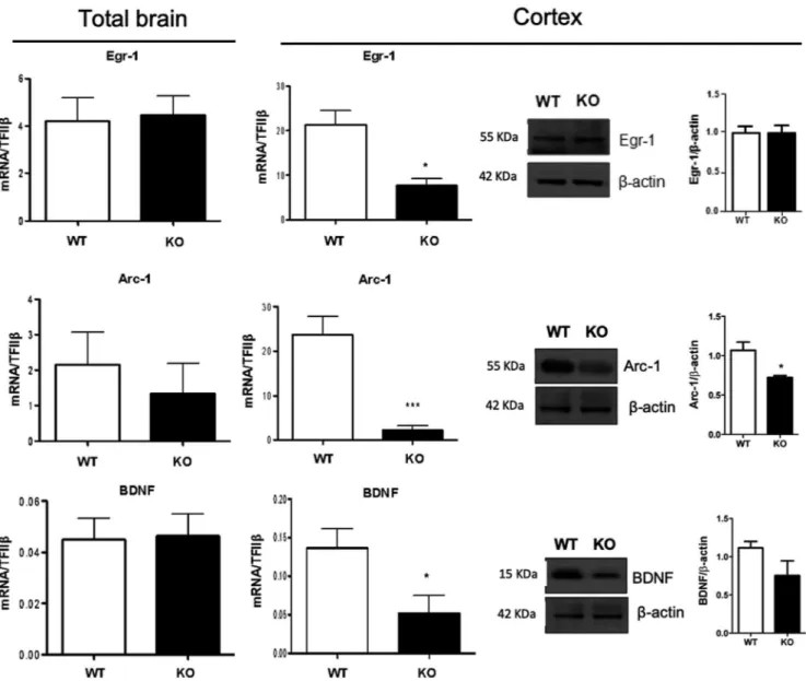
factors that are rapidly and selectively regulated in specific brain regions associated with learning and memory, including Arc-1, Egr-1, and BDNF.33,34 Considering that the expression
of these factors has previously been shown to be regulated by ω-3 polyunsaturated fatty35,36 we measured the gene
expres-sion of Arc-1, Egr-1, and BDNF in the total brain and cerebral cortex. Interestingly, the expression of these neuroplasticity immediate early genes did not show any significant variation in the total brains of KO mice compared to WT (Figure 2). However, when analyzing them in the cerebral cortex, we ob-served that the mRNA expression of Egr1 and its direct target Arc1, as well as of BDNF was significantly down-regulated in KO mice compared to WT (Figure 2). When trying to cor-roborate such findings in the cortex by analyzing their protein levels, we observed that only Arc-1 was significantly reduced
in KO mice compared to WT, whereas no significant change was observed for BDNF and Egr-1 (Figure 2).
3.3
|
DHA deficiency induces
neuroinflammation but has a small impact on
behavioral functions
As already shown by our previous studies where impairment of systemic DHA synthesis affects specific immune cells, such as T-helper cells and macrophages37,38 and given the
important role of DHA in inflammatory responses, we next examined whether the deficient synthesis of this essential fatty acid can modulate the brain inflammatory status. For this purpose, we examined the expression of several markers
FIGURE 2 Synaptic plasticity markers expression is impaired in the cerebral cortex of KO mice. mRNA expression of Egr-1, Arc-1, and BDNF in the total brain and their mRNA and protein in the cortex of WT and KO mice by RT-PCR and western blotting. Data are shown as mean ± SEM of seven animals per group (WT and KO), each in duplicate. *P < .05 and ***P < .001 vs WT by unpaired Student's t test
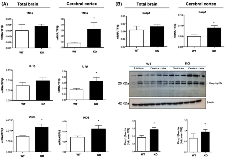
of brain inflammation in total brain and cerebral cortex of KO and WT mice, among which the major pro-inflammatory cy-tokines in the brain (TNF and IL-1β), inducible nitric oxide synthase (iNOS), and caspase-1 (Casp1), all of which play a key role in the onset and progression of neuroinflammatory diseases.39 In particular, the total brains of KO mice showed a
significant increase in iNOS gene expression but no variation in TNF, IL-1β, and Casp1 (Figure 3A,B) compared to WT mice. However, Casp1 protein expression was significantly higher in KO mice compared to WT (Figure 3B). Conversely, when investigating the expression of these pro-inflammatory markers in the cerebral cortex, we observed that mRNAs of both cytokines, as well as iNOS and Casp1, were significantly up-regulated in KO mice. Since Casp1 is usually in its physi-ologically inactive zymogen form and requires cleaving into its p20 subunit to display its pro-inflammatory activity,40 we
also analyzed the protein expression of its biologically active fragment p20 in WT and KO mice and we found that KO mice showed a significant increase in p20-casp1 expression com-pared to WT mice (Figure 3A,B), thus confirming a higher inflammatory status in mice deficient for DHA synthesis.
Since the presence in the brain of inflammatory markers like cytokines and iNOS is linked to glial cells’ activation, we next investigated the effect of endogenous DHA deficiency on astrocytes and microglia functional state and their mor-phology in the cerebral cortex. Although quantitative anal-ysis of cortical GFAP protein level and GFAP positive and reactive astrocytes did not show any significant differences between KO and WT mice (Figure 4D-F), we observed a sig-nificant increase of Iba1 protein level and in the number of Iba1 + cells in the cerebral cortex of KO mice compared to WT (Figure 4A-C), suggesting that DHA-deficient mice dis-played not only an increased number of microglia, but these cells displayed a different morphological state. Interestingly, when closely observing the morphology of Iba1 + microglia, we noticed that the microglia of KO mice showed different morphological features. Thus, we performed Sholl analysis and found that the cerebral cortex of KO mice microglia displayed a higher degree of complexity and ramification compared to those of WT, presenting a larger cell body (in-crease perimeter and cross-sectional area) (Figure 4A, inset) as well as an increased number of intersections and nodes
FIGURE 3 Inflammatory markers expression is impaired in the cerebral cortex of KO mice. A-B, mRNA expression of TNF, IL-1β, iNOS, and Casp1 as well as protein expression of Casp1(p20) in total brain and cortex of WT and KO mice by qRT-PCR and western blotting. Data are shown as mean ± SEM of seven (qRT-PCR) or four (western blotting) animals per group (WT and KO), each in duplicate. *P < .05 vs WT by unpaired Student's t test
FIGURE 4 DHA deficiency alters microglia response without affecting astrogliosis. A, Immunofluorescence staining of microglia (Iba1) in the cerebral cortex of WT and KO mice. Double-labeled and merged confocal images of Iba1 (red) plus NeuroTrace Nissl staining (green) (scale bars = 100 μm). Insets: Single-labeled images of Iba1 staining (scale bars = 10 μm). B, Western blotting and its densitometry of Iba1. Immunoblot images are cropped from different parts of the same gel. C, Histogram of the number of Iba1 positive cells in the cerebral cortex of WT and KO mice. D, Immunofluorescence staining of astrocytes (GFAP) in the cerebral cortex of WT and KO mice. In the lower portion of the sections it is well visible also the corpus callosum (cc). Double-labeled and merged confocal images of GFAP (red) plus NeuroTrace Nissl staining (green) (scale bars = 100 μm). E, Western blotting and its densitometry of GFAP. Immunoblot images are cropped from different parts of the same gel. F, Histogram of the number of GFAP positive cells in the cerebral cortex of WT and KO mice. G-K, Histograms of the Sholl analysis of microglia in the cerebral cortex of WT and KO mice showing the perimeter (G) and the cross-sectional area of the Iba1 + cells (H), the number of intersections (I), and the number of nodes/branch points at the different radii (J). All results are mean ± SEM or representative of six animals per group (WT and KO) *P < .05, **P < .01, and ***P < .001 vs WT by unpaired Student's t test
of their processes (Figure 4G-J), suggesting the different state of microglia in KO mice compared to WT. To show that the reduced expression of genes associated with neural plasticity and the exacerbated inflammation observed in KO mice might have a physiological impact in this mouse model deficient for DHA synthesis, we also performed some behav-ioral analyses. Thus, to examine responses to an unfamiliar environment, Elovl2 WT and KO mice were evaluated in the open field test. Our results show that even if no statistical difference was present in the number of visits to the center zone, the percentage of time spent and the distance covered in the center zone were statistically significant between the two groups of animals (Figure 5A). The same set of animals was also subjected to the NOR test, in which KO animals show a tendency to spend less time, although not significant, at the novel object compared to WT (Figure 5B), suggesting no
deficit in object memory. Next, we also evaluated the spatial learning and memory with the Barnes Maze test in a small group of female mice and, also in this case, no differences be-tween WT and KO were observed in the learning curve and in the amount of time spent in the target quadrant (Figure 5C). These results suggest that even if the deficiency of endoge-nous DHA affects inflammatory and neuroplasticity markers, it does not overall influence behavioral functions.
3.4
|
Neuroplasticity and
neuroinflammatory markers are restored by
dietary DHA supplementation
Given the alteration observed in the cerebral cortex of KO mice in terms of expression of neuroplasticity and
FIGURE 5 DHA deficiency does not alter behavioral functions. A, Individual exploration trajectories showing the change in the exploratory activity of WT and KO mice. Brown lines indicate locomotion (horizontal movements), blue lines indicate rears (vertical movements), and green lines indicate center area and corners. Exploratory activity was calculated as the number of visits to the center zone, percentage of time spent in the center zone, and percentage of distance in the center zone. Data are shown as mean ± SEM of 12 WT and 16 KO mice and analyzed by the unpaired Student's t test. B, Working memory consolidation with two objects (novel and familiar) evaluated as the percentage of time spent at exploring the objects. Data are shown as mean ± SEM of seven animals per group (WT and KO) and analyzed by the unpaired Student's t test. C, Spatial and learning memory evaluated as the ability of animals to seek out for escape holes (time spent in quadrants of target, opposite, negative or positive holes). Data are shown as mean ± SEM of 8 WT and 7 KO and analyzed by the unpaired Student's t test
neuroinflammation markers, we next questioned whether a short-term dietary supplementation with DHA could re-store some of these markers in the cerebral cortex of KO mice. In particular, the reintroduction of DHA in KO mice
significantly increased the mRNA expression levels of Arc-1 and reduced that of IL-1β and Casp1 compared to KO, with IL-1β and Casp1 also showing comparable levels to those of WT mice (Figure 6A). This DHA-induced effect was also
FIGURE 6 Dietary supplementation with DHA restores neuroplasticity and microglia status in the cerebral cortex. A, mRNA expression of Arc-1, IL-1β, and Casp1 in the cortex by qRT-PCR. Data are shown as mean ± SEM of four animals per group (WT, KO, and KO + DHA), each in duplicate. ***P < .001 vs WT and *P < .05 vs KO by one-way ANOVA. B, Protein expression of Arc-1 by western blotting and (C) level of IL-1β by ELISA. Data are shown as mean ± SEM of four animals per group (WT, KO, and KO + DHA), each in duplicate. ***P < .001 vs WT and *P < .05 vs KO by one-way ANOVA followed by Bonferroni post-hoc test. D, Immunofluorescence staining of microglia in the cortex of WT, KO, and DHA-supplemented KO (KO + DHA) mice. Double-labeled and merged confocal images of Iba1 (red) plus NeuroTrace Nissl staining (green) in the cerebral cortex (scale bars = 100 μm). Inset: Single-labeled images of Iba1 staining (scale bars = 10 μm). E, Histogram of the number of Iba1 positive cells in the cerebral cortex. Data are shown as mean ± SEM of six animals per group (WT, KO, and KO + DHA). ***P < .001 and **P < .01 by one-way ANOVA followed by the Bonferroni post-hoc test (F-I). Histograms of the Sholl analysis of microglia in the cerebral cortex of WT, KO, and KO + DHA mice showing the perimeter (F) and the cross-sectional area of the cells (G), the number of intersections (H), and the number of nodes/branch points of intersections at the different radii (I). Results are mean ± SEM or representative of six animals per group (WT, KO, and KO + DHA). **P < .01 and ***P < .001 by one-way ANOVA followed by Bonferroni post-hoc test. J, Schematic representation of DHA deficiency-induced impact on brain plasticity and inflammation markers
corroborated analyzing the protein levels of Arc-1 and IL-1β (Figure 6B-C), with the former showing even higher levels of expression compared to WT (Figure 6B), while the levels of the latter were equal to WT mice (Figure 6C). Similarly, we observed a marked and significant decrease in the num-ber of Iba1 + cells in DHA-supplemented KO mice com-pared to KO mice (Figure 6D-E), showing that the number of Iba1 + cells was comparable to WT mice (Figure 6F-I). Of note, the DHA-deficiency-induced changes in the morphol-ogy of microglia observed in KO mice were also counter-acted upon DHA supplementation, with microglia presenting a morphological state similar to that observed in WT mice (Figure 6D,F-I). These findings were supported by the evi-dence that DHA supplementation significantly restored the brain levels of DHA (Figure S1A), suggesting a key role of DHA in mediating our observed effects. Of note, DHA sup-plementation did not impact on body weight (Figure S1B) or food intake (Figure S1C).
Furthermore, to rule out the possibility of stain infiltrated Iba1 + monocytes/macrophages from the periphery, we also performed double immunofluorescence combining Iba1 with TMEM-119, a more specific marker of resident microg-lia.41 Confocal analysis of Iba1/TMEM-119 confirmed that
the number of microglia TMEM-119 + was significantly higher in KO mice and reduced upon DHA reintroduction, but also demonstrated that both markers perfectly colocalize (Figure S2), thus confirming that the DHA-induced modula-tion of neuroinflammamodula-tion more likely involves only resident microglia.
4
|
DISCUSSION
The beneficial properties of dietary ω-3 polyunsaturated fatty acids (PUFA), particularly of DHA, have been recognized for decades and their metabolic dysfunction has been linked to a range of diseases including inflammatory and neurode-generative disorders.42,43 Indeed, DHA is an important
com-ponent of neural membranes, regulating membrane fluidity, permeability, and viscosity in synaptic membranes as well as playing a key role in modulating neurotransmission and syn-aptic function and plasticity.44 However, although its crucial
function in brain processes is undisputed, the specific role of the endogenous synthesis of DHA in the brain and how this affects neural plasticity and inflammation is still obscure.
Thus, in this study, we thoroughly investigated for the first time whether the endogenous synthesis of DHA can impact on markers of brain plasticity and inflamma-tion. For this purpose, we used mice deficient for Elovl2 (Elovl2−/−), a key enzyme involved in the synthesis of
DHA.45 Our group has previously shown that these mice
are characterized by severely reduced DHA levels in testis, serum, liver, mammary gland and white adipose tissue6,26,44
as well as by a pro-inflammatory status of innate immune cells.25 In the present work, we collected the unprecedented
evidence of acyl-CoA levels in brain by targeted lipidom-ics and we demonstrated that Elovl2-mediated endogenous DHA synthesis plays a role in the expression of several brain plasticity and inflammatory markers. Although, to date, it is unclear why brain phospholipids are specifically enriched in DHA and low in DPA and EPA, the fact that DHA enrichment is conserved across species46,47 suggests
that there is a highly specific requirement for this essential fatty acid in neuronal membranes. Indeed, we found a sig-nificant downregulation of several neural plasticity factors (Arc-1, Egr-1, and BDNF) and a concomitant upregula-tion of key inflammatory markers (TNF, IL-1β, iNOS, and Casp1) in Elovl2−/− mice. Interestingly, variations of such
markers were particularly evident in the cerebral cortex and not in the total brain, confirming the essential role of this fatty acid in the brain region where it is mostly abun-dant and corroborating our previous results demonstrating the unique role of Elovl2 in the formation of DHA in the brain.26 Our data are in line with previous findings
demon-strating that the expression of these markers as well as their related learning and memory functions is modulated by omega-3 fatty acids in the adult brain.36,48,49
However, although decreases in BDNF and Arc-1 ex-pression are commonly linked to the development of long-term cognitive and behavioral alterations11,13,50,51 and their
reduction in the prefrontal cortex are associated with de-pression,50 the mechanism underlying brain DHA
reduc-tions and behavioral alterareduc-tions are still a matter of debate. In addition, considering the substantial evidence regard-ing the importance of DHA durregard-ing brain development and on resulting cognitive functions, further investigations are needed to assess a functional relationship between cognitive/behavioral alterations and DHA deficiency in Elovl2−/− mice.
Of note, it is now well established that the brain has fun-damental interactions with the immune system.52 One of the
most exciting aspects of such neuro-immune communica-tion is that the immune system actively participates in brain functions and synaptic plasticity. In particular glial cells have emerged as active players of synapses, able to shape synapse structure and function.53 Indeed, we observed a significant
upregulation of several genes involved in neuroinflammation such as iNOS, caspase-1, and pro-inflammatory cytokines (IL-1β and TNF) in the cerebral cortex of DHA-deficient mice; such high and sustained pro-inflammatory status is detrimental for both neurons and glial cells, resulting in cell dysfunction or death54,55 as well as in the pathogenesis of
several cognitive disorders.56-58 Of note, several cytokines
have been reported to suppress synaptic plasticity by sup-pressing Arc expression in a BDNF-dependent manner and the latter, together with Egr-1, is a transcriptional regulator
of Arc, thus accounting for our marked changes in Arc-1 at both mRNA and protein level, while Erg-1 and BDNF only show changes in their mRNA but not protein. However, the notion that pro-inflammatory cytokines are only expressed in the brain in response to pathological stimuli has been chal-lenged by emerging data, indicating that certain cytokines can also play an active role in synaptic plasticity in physio-logical condition.59
Yet, the ability of fatty acids to modulate both inflam-mation and synaptic functions is coming to the foreground and alterations of their availability, either due to insuffi-cient dietary consumption or impaired metabolism, might play a role in the pathogenesis of several cognitive disor-ders.38 In this regard, significant differences in the
synap-tic proteome of DHA-adequate and DHA-deficient mouse brains have been reported60 providing a molecular basis for
the significant impact of nutritional DHA on some brain functions such as learning and memory. The evidence that free DHA can enter the brain through the mfsd2a and that this transporter is regulated both in Elovl2 mice and upon DHA supplementation suggests not only that Major Facilitator Superfamily Domain containing 2a (Mfsd2a) plays a compensatory role to balance the DHA levels in the brain of Elovl2 mice but also that exists a dynamic inter-play between DHA synthesis and DHA uptake in the con-trol of systemic levels.
Intriguingly, glial cells play a crucial role in the regula-tion of both inflammatory and brain plasticity processes.61,62
The fact that our findings revealed the alteration of microg-lia, but not of astrocytes, in Elovl2−/− mice, suggests that this
specific resident immune cell of the brain is specifically af-fected by DHA deficiency. In particular, not only did we find that the number of Iba1 + cells in the cerebral cortex is in-creased in DHA deficient mice, but also that its morphology undergoes significant changes. Microglia are dynamic cells whose morphological changes seem to be associated with their functional activities, whereby ramified microglia act as surveying cells, whereas hyper-ramified microglia are char-acterized by increased secretion of pro-inflammatory medi-ators.62 This suggests that microglia might be responsible
for the up-regulation of iNOS, IL-1β, and TNF observed in Elovl2−/− mice. Although our findings do not clarify the
pre-cise cellular source of cytokines, however, our results suggest a temporal correlation between the microglia morphological changes and cytokines production. More studies are required to establish the cellular source of cytokines and, in particu-lar, to demonstrate that their production can be attributed to microglia.
Furthermore, since microglia also play a role in syn-aptic remodeling and neuronal plasticity,61 it is also
pos-sible that such activated inflammatory status is somehow associated with the downregulation of markers of synaptic plasticity in Elovl2−/− mice. This is particularly important
since the brain and the immune systems are engaged in a bidirectional crosstalk and our data suggest that microg-lia might engage in mediating such a complex interaction. Furthermore, the fact that such changes are even influenced by DHA deficiency alone, in absence of any brain insult, suggests that ω-3 essential fatty acid is a master regulator of brain responses and an intriguing interface between “mind” and “body.” However, only a few groups have evaluated the consequences of PUFAs manipulation on microglia in vivo and in cerebral cortex of adult mice after a pathologi-cal event.63,64 Accordingly, the evidence that reintroducing
DHA in the diet of Elovl2−/− mice restored the expression
levels of key neuroinflammatory (IL-1β and Casp-1) and neuroplasticity (Arc-1) markers as well as prevented the microglia DHA deficiency-induced morphologic changes in the cerebral cortex further corroborates our findings. Of note, whether such DHA-induced neuroprotective effects are mediated by this fatty acid alone or by its derived and specialized pro-resolving mediators (SPMs) have yet to be unraveled. The discoveries that several DHA-derived SPMs, such as resolvins or neuroprotectins, play critical roles in modulating microglia responses as well ad in the modulation of reactive species24,65-67 is suggestive of their
possible involvement in controlling neuroplasticity and in representing a link between the immune and the central ner-vous systems. In addition, our findings show that even in the absence of inflammatory stimuli or without the induc-tion of specific diseases, systemic DHA deficiency induces a basal inflammatory activity, which is characterized by an overall upregulation of several inflammatory markers; how-ever, this only has a little impact on behavioral functions. Further experiments to prove whether such DHA-deficient mice are more responsive to inflammatory events or are more susceptible to chronic neuroinflammatory diseases or cognitive disorders are needed. Yet the evidence that a ω-3 deficient diet is associated with exacerbated inflamma-tion and memory deficits in another mice model exposed prenatally to LPS68 suggests that DHA-induced profound
and macroscopic cognitive alterations appear mostly under inflammatory conditions and account for the discrete be-havioral dysfunctions observed here. It is important to add that since the Iba1 microglia marker can also stain macro-phages migrated from the periphery, TMEM-119/Iba1 co-localization excludes the possibility that endogenous DHA synthesis may also impact recruitment and activation of infiltrated cortical monocytes/macrophages.
Furthermore, we cannot exclude also the possibility that our DHA-induced effects on microglia are mediated by a direct action on these cells or are indirectly mediated by influencing other molecules released by astrocytes or even neurons, calling for future mechanistic studies unraveling the cellular and molecular signaling pathways. The unraveling of these issues would also be crucial to understand whether the
changes in markers of brain plasticity and neuroinflammation take place within the same cells and are linked to one another and what are the mechanisms underlying their interaction.
In conclusion, as summarized in Figure 6J, this study has highlighted the important role of the DHA-generating Elovl2 enzyme in modulating brain neuroplasticity and inflamma-tion, with conceivable and potential implications in affecting CNS homeostasis. Indeed, our findings suggest that the enzy-matic machinery that leads to DHA synthesis is involved, at least in part, in the maintenance of brain functions and sup-ports the notion that diets rich in DHA could be sufficient to activate some compensatory pathways that might reduce neuroinflammation.
ACKNOWLEDGMENTS
This work was partly supported by Fondazione Italiana Sclerosi Multipla (FISM grant 2015/R/8) to VC, by the UK Biotechnology and Biological Sciences Research Council (Strategic Program grant BBS/E/C/000I0420) to RPH and JAN and by Swedish Research Council and the Swedish Foundation for Strategic Research (grant IB13-0026) to TH. The behavioral analyses were conducted and analyzed at Karolinska Institutet Mouse Clinic (KIMC; https ://ki.se/ en/km/pheno typing-of-mouse-models-at-ki-mouse-clinic). We thank Michael Gaudry for the English language editing support.
CONFLICTS OF INTEREST
The authors declare no conflict of interest.
AUTHOR CONTRIBUTION
E. Talamonti, M.T. Viscomi, and V. Chiurchiù conceived and designed the study; E. Talamonti, V. Sasso, H. To, R.P. Haslam, K. Pernold, and A. Asadi performed and analyzed the data; J.A. Napier, B. Ulfhake, T. Hessa, A. Jacobsson, V. Chiurchiù, and M.T. Viscomi provided reagents and tools; V. Chiurchiù, M.T. Viscomi, and E. Talamonti wrote the paper.
REFERENCES
1. Salem N, Litman B, Kim HY, Gawrisch K. Mechanisms of ac-tion of docosahexaenoic acid in the nervous system. Lipids. 2001;36:945-959.
2. Luchtman DW, Song C. Cognitive enhancement by omega-3 fatty acids from child-hood to old age: findings from animal and clinical studies. Neuropharmacol. 2013;64:550-565.
3. Guillou H, Zadravec D, Martin PG, Jacobsson A. The key roles of elongases and desaturases in mammalian fatty acid metabolism: Insights from transgenic mice. Prog Lipid Res. 2010;2:186-199. 4. Tvrdik P, Westerberg R, Silve S, et al. Role of a new mammalian
gene family in the biosynthesis of very long chain fatty acids and sphingolipids. J Cell Biol. 2000;3:707-718.
5. Ohno Y, Suto S, Yamanaka M, et al. ELOVL1 production of C24 acyl-CoAs is linked to C24 sphingolipid synthesis. Proc Natl Acad
Sci U S A. 2010;43:18439-18444.
6. Pauter AM. Both maternal and offspring Elovl2 genotypes de-termine systemic DHA levels in perinatal mice. J Lipid Res. 2017;1:111-123.
7. Bazan NG. Lipid signaling in neural plasticity, brain repair, and neuroprotection. Mol Neurobiol. 2005;1:89-103.
8. Wu A, Ying Z, Gomez-Pinilla F. The salutary effects of DHA dietary supplementation on cognition, neuroplasticity, and membrane homeostasis after brain trauma. J Neurotrauma. 2011;10:2113-2122.
9. Kim HY. Neuroprotection by docosahexaenoic acid in brain injury.
Mil Med. 2014;11:106-111.
10. Labrousse VF, Nadjar A, Joffre C, et al. Short-term long chain omega3 diet protects from neuroinflammatory processes and mem-ory impairment in aged mice. PLoS ONE. 2012;5:36861.
11. Moranis A, Delpech JC, De Smedt-Peyrusse V, et al. Long term adequate n-3 polyunsaturated fatty acid diet protects from depres-sive-like behavior but not from working memory disruption and brain cytokine expression in aged mice. Brain Behav Immun. 2012;5:721-731.
12. DeMar JC, Ma K, Bell JM, Igarashi M, Greenstein D, Rapoport SI. One generation of n-3 polyunsaturated fatty acid deprivation increases depression and aggression test scores in rats. J Lipid Res. 2006;1:172-180.
13. Fedorova I, Hussein N, Di Martino C, et al. An n-3 fatty acid deficient diet affects mouse spatial learning in the Barnes circular maze. Prostaglandins Leukot Essent Fatty Acids. 2007;77:269-277.
14. Lafourcade M, Larrieu T, Mato S, et al. Nutritional omega-3 de-ficiency abolishes endocannabinoid-mediated neuronal functions.
Nat Neurosci. 2011;3:345-350.
15. Moriguchi T, Greiner RS, Salem N. Behavioral deficits associated with dietary induction of decreased brain docosahexaenoic acid concentration. J Neurochem. 2000;6:2563-2573.
16. Madore C, Nadjar A, Delpech JC, et al. Nutritional n-3 PUFAs deficiency during perinatal periods alters brain innate immune sys-tem and neuronal plasticity-associated genes. Brain Behav Immun. 2014;41:22-31.
17. Minatohara K, Akiyoshi M, Okuno H. Role of immediate-early genes in synaptic plasticity and neuronal ensembles underlying the memory trace. Front Mol Neurosci. 2015;8:78.
18. Duclot F, Kabbaj M. The role of early growth response 1 (EGR1) in brain plasticity and neuropsychiatric disorders. Front Behav
Neurosci. 2017;11:35.
19. Belkind-Gerson J, Carreón-Rodríguez A, Contreras-Ochoa CO, Estrada-Mondaca S, Parra-Cabrera MS. Fatty acids and neurode-velopment. J Pediatr Gastroenterol Nutr. 2008;47:7-9.
20. Zhang YP, Lou Y, Hu J, Miao R, Ma F. DHA supplementation improves cognitive function via enhancing Aβ-mediated auto-phagy in Chinese elderly with mild cognitive impairment: a ran-domised placebo-controlled trial. J Neurol Neurosurg Psychiatry. 2018;89:382-388.
21. Ciappolino V, Mazzocchi A, Botturi A, et al. The role of docosa-hexaenoic acid (DHA) on cognitive functions in psychiatric disor-ders. Nutrients. 2019;11:7692019.
22. Serhan CN. Pro-resolving lipid mediators are leads for resolution physiology. Nature. 2014;7503:92-101.
23. Serhan CN, Dalli J, Karamnov S, et al. Macrophage proresolving mediator maresin 1 stimulates tissue regeneration and controls pain. FASEB J. 2012;4:1755-1765.
24. Bisicchia E, Sasso V, Catanzaro G, et al. Resolvin D1 halts remote neuroinflammation and improves functional recovery after focal brain damage via ALX/FPR2 receptor-regulated microRNAs. Mol
Neurobiol. 2018;55:6894-6905.
25. Talamonti E, Pauter AM, Asadi A, Fischer AW, Chiurchiù V, Jacobsson A. Impairment of systemic DHA synthesis af-fects macrophage plasticity and polarization: implications for DHA supplementation during inflammation. Cell Mol Life Sci. 2017;15:2815-2826.
26. Zadravec D, Tvrdik P, Guillou H, et al. ELOVL2 controls the level of n-6 28:5 and 30:5 fatty acids in testis, a prerequisite for male fer-tility and sperm maturation in mice. J Lipid Res. 2011;2:245-255. 27. Larson TR, Graham IA. Technical advance: a novel technique for
the sensitive quantification of acyl CoA esters from plant tissues.
Plant J. 2001;1:115-125.
28. Haynes CA, Haynes CA, Allegood JC, et al. Quantitation of fatty acyl-coenzyme As in mammalian cells by liquid chromatogra-phy-electrospray ionization tandem mass spectrometry. J Lipid
Res. 2008;5:1113-1125.
29. Fahlström A, Zeberg H, Ulfhake B. Changes in behaviors of male C57BL/6J mice across adult life span and effects of dietary restric-tion. Age. 2012;34:1435-1452.
30. Attar A. A shortened Barnes maze protocol reveals memory defi-cits at 4-months of age in the triple-transgenic mouse model of Alzheimer's disease. PLoS ONE. 2013;8:80355.
31. Viscomi MT, Florenzano F, Latini L, Amantea D, Bernardi G, Molinari M. Methylprednisolone treatment delays remote cell death after focal brain lesion. Neuroscience. 2008;4:1267-1282. 32. Lo Iacono L, Catale C, Martini A, et al. From traumatic childhood
to cocaine abuse: the critical function of the immune system. Biol
Psychiatry. 2018;84:905-916.
33. Monteggia LM. Elucidating the role of brain-derived neurotrophic factor in the brain. Am J Psychiatry. 2007;12:1790.
34. Shepherd JD, Bear MF. New views of Arc, a master regulator of synaptic plasticity. Nat Neurosci. 2011;3:279-284.
35. Kitajka K, Sinclair AJ, Weisinger RS, et al. Effects of dietary omega-3 polyunsaturated fatty acids on brain gene expression.
Proc Natl Acad Sci U S A. 2004;30:10931-10936.
36. Rao JS, Ertley RN, Lee HJ, et al. n-3 polyunsaturated fatty acid deprivation in rats decreases frontal cortex BDNF via a p38 MAPK-dependent mechanism. Mol Psychiatry. 2007;1:36-46. 37. Chiurchiu V, Leuti A, Maccarrone M. Bioactive lipids and
chronic inflammation: managing the fire within. Front Immunol. 2018;9:38.
38. Chiurchiù V, Leuti A, Dalli J, et al. Proresolving lipid mediators resolvin D1, resolvin D2, and maresin 1 are critical in modulating T cell responses. Sci Transl Med. 2016;353:353ra111.
39. Thornton P, Pinteaux E, Gibson RM, Allan SM, Rothwell NJ. Interleukin-1-induced neurotoxicity is mediated by glia and re-quires caspase activation and free radical release. J Neurochem. 2006;1:258-266.
40. Elliott JM, Rouge L, Wiesmann C, Scheer JM. Crystal structure of procaspase-1 zymogen domain reveals insight into inflammatory caspase autoactivation. J Biol Chem. 2009;10:6546-6553. 41. Bennett ML, Bennett FC, Liddelow SA, et al. New tools for
study-ing microglia in the mouse and human CNS. Proc Natl Acad Sci U
S A. 2016;113:1738-1746.
42. Lorente-Cebrian S, Costa AG, Navas-Carretero S, et al. An update on the role of omega-3 fatty acids on inflammatory and degenera-tive diseases. J Physiol Biochem. 2015;2:341-349.
43. Echeverria F, Valenzuela R, Catalina Hernandez-Rodas M, Valenzuela A. Docosahexaenoic acid (DHA), a fundamental fatty acid for the brain: new dietary sources. Prostaglandins Leukot
Essent Fatty Acids. 2017;124:1-10.
44. Oster T, Pillot T. Docosahexaenoic acid and synaptic protection in Alzheimer's disease mice. Biochim Biophys Acta. 2010;8:791-798. 45. Pauter AM, Olsson P, Asadi A, et al. Elovl2 ablation demonstrates
that systemic DHA is endogenously produced and is essential for lipid homeostasis in mice. J Lipid Res. 2014;4:718-728.
46. Crawford MA, Casperd NM, Sinclair AJ. The long chain metabo-lites of linoleic avid linolenic acids in liver and brain in herbivores and carnivores. Comp Biochem Physiol B. 1976;3:395-401. 47. Farkas T, Kitajka K, Fodor E, et al. Docosahexaenoic
acid-contain-ing phospholipid molecular species in brains of vertebrates. Proc
Natl Acad Sci U S A. 2000;12:6362-6366.
48. Tanaka K, Farooqui AA, Siddiqi NJ, Alhomida AS, Ong WY. Effects of docosahexaenoic acid on neurotransmission. Biomol
Ther. 2012;2:152-157.
49. Brand A, Schonfeld E, Isharel I, Yavin E. Docosahexaenoic ac-id-dependent iron accumulation in oligodendroglia cells pro-tects from hydrogen peroxide-induced damage. J Neurochem. 2008;4:1325-1335.
50. Covington HE, Lobo MK, Maze I, et al. Antidepressant effect of optogenetic stimulation of the medial prefrontal cortex. J Neurosci. 2010;48:16082-16090.
51. Larrieu T, Madore C, Joffre C, Laye S. Nutritional n-3 polyunsat-urated fatty acids deficiency alters cannabinoid receptor signaling pathway in the brain and associated anxiety-like behavior in mice.
J Physiol Biochem. 2012;4:671-681.
52. Prinz M, Priller J. The role of peripheral immune cells in the CNS in steady state and disease. Nat Neurosci. 2017;2:136-144. 53. Mottahedin A, Ardalan M, Chumak T, Riebe I, Ek J, Mallard C.
Effect of neuroinflammation on synaptic organization and function in the developing brain: implications for neurodevelopmental and neurodegenerative disorders. Front Cell Neurosci. 2017;11:190. 54. Mamik MK, Power C. Inflammasomes in neurological
dis-eases: emerging pathogenic and therapeutic concepts. Brain. 2017;9:2273-2285.
55. Kempuraj D, Thangavel R, Selvakumar GP, et al. Brain and pe-ripheral atypical inflammatory mediators potentiate neuroinflam-mation and neurodegeneration. Front Cell Neurosci. 2017;11:216. 56. Khairova RA, Machado-Vieira R, Du J, Manji HK. A potential
role for pro-inflammatory cytokines in regulating synaptic plas-ticity in major depressive disorder. Int J Neuropsychopharmacol. 2009;4:561-578.
57. Wohleb ES. Neuron-microglia interactions in mental health disor-ders: “for better, and for worse”. Front Immunol. 2016;7:544. 58. Solas M, Milagro FI, Ramirez MJ, Martinez JA. Inflammation and
gut-brain axis link obesity to cognitive dysfunction: plausible phar-macological interventions. Curr Opin Pharmacol. 2017;37:87-92. 59. Levin SG, Godukhin OV. Modulating Effect of Cytokines
on Mechanisms of Synaptic Plasticity in the Brain. Biochem. 2017;3:264-274.
60. Sidhu VK, Huang BX, Kim HY. Effects of docosahexaenoic acid on mouse brain synaptic plasma membrane proteome analyzed by mass spectrometry and (16)O/(18)O labeling. J Proteome Res. 2011;12:5472-5480.
61. Paolicelli RC, Bolasco G, Pagani F, et al. Synaptic pruning by microglia is necessary for normal brain development. Science. 2011;6048:1456-1458.
62. Nayak D, Roth TL, McGavern DB. Microglia development and function. Annu Rev Immunol. 2014;32:367-402.
63. Ji A, Diao H, Wang X, et al. n-3 polyunsaturated fatty acids inhibit lipopolysaccharide-induced microglial activation and dopaminer-gic injury in rats. Neurotoxicol. 2012;4:780-788.
64. Pu H, Pu H, Guo Y, et al. Omega-3 polyunsaturated fatty acid sup-plementation improves neurologic recovery and attenuates white matter injury after experimental traumatic brain injury. J Cereb
Blood Flow Metab. 2013;9:1474-1484.
65. Sheets KG, Jun B, Zhou Y, et al. Microglial ramification and redistribution concomitant with the attenuation of choroidal neovascularization by neuroprotectin D1. Mol Vis. 2013;19: 1747-1759.
66. Leuti A, Maccarrone M, Chiurchiù V. Proresolving lipid media-tors: endogenous modulators of oxidative stress. Oxid Med Cell
Longev. 2019;18:8107265.
67. Krashia P, Cordella A, Nobili A, et al. Blunting neuroinflamma-tion with resolvin D1 prevents early pathology in a rat model of Parkinson's disease. Nat Commun. 2019;10:3945.
68. Labrousse VF, Leyrolle Q, Amadieu C, et al. Dietary omega-3 deficiency exacerbates inflammation and reveals spatial memory deficits in mice exposed to lipopolysaccharide during gestation.
Brain Behav Immun. 2018;73:427-440.
SUPPORTING INFORMATION
Additional supporting information may be found online in the Supporting Information section.
How to cite this article: Talamonti E, Sasso V, To H,
et al. Impairment of DHA synthesis alters the expression of neuronal plasticity markers and the brain inflammatory status in mice. The FASEB
Journal. 2020;34:2024–2040. https ://doi.org/10.1096/ fj.20190 1890RR