Synthetic PreImplantation Factor (PIF)
prevents fetal loss by modulating LPS induced
inflammatory response
Nicoletta Di Simone1☯, Fiorella Di Nicuolo1,2☯, Riccardo Marana1,2, Roberta Castellani1, Francesco Ria3, Manuela Veglia1, Giovanni Scambia1, Daniel Surbek4, Eytan Barnea5,6‡, Martin Mueller4,7‡*
1 Department of Obstetrics and Gynecology, UniversitàCattolica Del Sacro Cuore, A. Gemelli Universitary Hospital, Rome, Italy, 2 International Scientific Institute Paolo VI, ISI, UniversitàCattolica Del Sacro Cuore, A. Gemelli Universitary Hospital, Rome, Italy, 3 Institute of General Pathology, UniversitàCattolica Del Sacro Cuore, Rome, Italy, 4 Department of Obstetrics and Gynecology, University Hospital Bern, Bern, Switzerland, 5 The Society for the Investigation of Early Pregnancy (SIEP), Cherry Hill, New Jersey, United States of America, 6 BioIncept LLC, Cherry Hill, New Jersey, United States of America, 7 Department of Obstetrics, Gynecology, and Reproductive Sciences, Yale University School of Medicine, New Haven, Connecticut, United States of America
☯These authors contributed equally to this work. ‡ These authors also contributed equally to this work. *[email protected]
Abstract
Maternal control of inflammation is essential during pregnancy and an exaggerated response is one of the underlying causes of fetal loss. Inflammatory response is mediated by multiple factors and Toll-like receptors (TLRs) are central. Activation of TLRs results in NALP-3 mediated assembly of apoptosis-associated speck-like protein containing a CARD (ASC) and caspase-1 into the inflammasome and production of pro-inflammatory cytokines IL-1βand IL-18. Given that preventing measures are lacking, we investigated PreImplanta-tion Factor (PIF) as therapeutic opPreImplanta-tion as PIF modulates InflammaPreImplanta-tion in pregnancy. Addi-tionally, synthetic PIF (PIF analog) protects against multiple immune disorders. We used a LPS induced murine model of fetal loss and synthetic PIF reduced this fetal loss and increased the embryo weight significantly. We detected increased PIF expression in the pla-centae after LPS insult. The LPS induced serum and placenta cytokines were abolished by synthetic PIF treatment and importantly synthetic PIF modulated key members of inflamma-some complex NALP-3, ASC, and caspase-1 as well. In conclusion our results indicate that synthetic PIF protects against LPS induced fetal loss, likely through modulation of inflamma-tory response especially the inflammasome complex. Given that synthetic PIF is currently tested in autoimmune diseases of non-pregnant subjects (clinicaltrials.gov,NCT02239562), therapeutic approach during pregnancy can be envisioned.
a1111111111 a1111111111 a1111111111 a1111111111 a1111111111 OPEN ACCESS
Citation: Di Simone N, Di Nicuolo F, Marana R,
Castellani R, Ria F, Veglia M, et al. (2017) Synthetic PreImplantation Factor (PIF) prevents fetal loss by modulating LPS induced inflammatory response. PLoS ONE 12(7): e0180642.https://doi.org/ 10.1371/journal.pone.0180642
Editor: Colette Kanellopoulos-Langevin, Xavier
Bichat Medical School, INSERM-CNRS - Universite´ Paris Diderot, FRANCE
Received: October 19, 2016 Accepted: June 19, 2017 Published: July 12, 2017
Copyright:© 2017 Di Simone et al. This is an open access article distributed under the terms of the Creative Commons Attribution License, which permits unrestricted use, distribution, and reproduction in any medium, provided the original author and source are credited.
Data Availability Statement: Data are available
fromhttps://figshare.com/s/ 73ea9df023bbe848e298.
Funding: The work was supported by a research
grant from the Università Cattolica del Sacro Cuore (D1, 2014) and by Istituto Scientifico
Internazionale, Paolo VI Institute, Università Cattolica del Sacro Cuore, Rome, Italy. Dr. Eytan R. Barnea is the (uncompensated) Chief Scientist for BioIncept, LLC. BioIncept, LLC did not have any
Introduction
During pregnancy, allogeneic fetal cells invade the maternal decidua and are protected from the maternal immune system. Controlling the maternal response to inflammation is essential as an exaggerated response is one of the underlying causes of early and even later fetal loss [1]. Fetal loss is associated with multiple causes such as anatomic, genetic, and hematologic disor-ders but immune defects emerged as central players recently. Not surprisingly, proper immune adaptations play a key role in prevention of pregnancy disorders including preeclampsia, fetal growth restriction, and premature birth [2].
One of the essential players of an immune system during pregnancy is a set of pathogen rec-ognition receptors: toll-like receptors (TLRs) in a trophoblast. Once TLRs are activated by pathogen associated molecular patterns (PAMPs), robust activation of NFκ-B and MAP kinase signaling pathways induce up-regulation of associated genes. These genes are mostly pro-inflammatory and NALP–inflammasome is central [3,4]. To date four main types of inflam-masomes have been described including the NALP-3 subtype. Upon activation of NALP-3, apoptosis-associated speck-like protein containing a CARD (ASC) and caspase-1 are assem-bled into the inflammasome [4,5]. This multi-protein complex enables the caspase-1-mediated proteolytic processing of the pro-inflammatory cytokines IL-1β, IL-18 and IL-33, thus generat-ing their respective mature secreted forms. All of these events are necessary durgenerat-ing inflamma-tory response [5].
The lipopolysaccharide (LPS) induced inflammation is a well-documented and frequently used model to study induced fetal loss [6,7]. The bacterial antigen LPS is a PAMP in the extra-cellular milieu. It induces macrophage-derived TNF-α production which in turn activates NK cells and IFN-γ secretion resulting in a positive feed-back [8]. This pathway leads to activa-tion of the uterine and placental endothelium and the release of embryotoxic cytokines [9,10]. We hypothesize that counteracting this adverse environment through targeted therapy would constitute important progress in pregnancy management and fetal loss prevention. A potent candidate in fetal loss prevention is PreImplantation Factor (PIF) as PIF regulates the inflam-matory response during pregnancy [11]. PIF is a 15-amino acid peptide secreted by the embryo and is associated with favorable pregnancy outcome [12–15]. PIF‘s essential role dur-ing pregnancy is supported by the ability to promote embryo development, endometrial recep-tivity, and trophoblast invasion [16–21]. Interestingly, PIF interaction with maternal immune system goes beyond binding to CD14+T regulatory cells as it targets the adaptive arm of immunity (CD3+cells) as well [22]. PIF‘s mode of action is mediated in part by reducing oxi-dative stress and protein misfolding, thereby protecting against embryo toxicity [20,21]. In addition, PIF reduces natural killer cells toxicity by down-regulating the pro-inflammatory CD69 expression [23]. Together, PIF is a unique and pregnancy essential peptide which pos-sesses immune-modulatory and not immune-suppressive properties. Not surprisingly, syn-thetic PIF (PIF analog) protects against multiple immune disorders [24–30] and received a FAST-Track FDA approval for clinical trial in autoimmune diseases of non-pregnant subjects (clinicaltrials.gov, NCT02239562).
Whether synthetic PIF administration could be effective in improving pregnancy-induced pathologiesin vivo is not yet established. In view of its translational potential, we used an intact immune murine model to study the mechanisms of LPS-induced fetal loss and examined syn-thetic PIF’s potential as a treatment.
Materials and methods
Synthetic PIF15(MVRIKPGSANKPSDD) was synthesized by solid-phase peptide synthesis
(Peptide Synthesizer, Applied Biosystems) employing Fmoc (9-fluorenylmethoxycarbonyl)
role in the study design, data collection and analysis, decision to publish, or preparation of the manuscript. The specific role of this author is articulated in the ‘author contributions’ section.
Competing interests: PIF is a proprietary
compound owned by BioIncept, LLC. Dr. Eytan R. Barnea is its (uncompensated) Chief Scientist. The other authors declare no conflict of interest. This does not alter our adherence to PLOS ONE policies on sharing data and materials.
chemistry at Bio-Synthesis, Inc. (Lewisville, TX, USA). Final purification was carried out by reversed-phase HPLC and identity was verified by matrix-assisted laser desorption/ionization time-of-flight mass spectrometry and amino acid analysis at >95% purity. Anti-PIF monoclo-nal antibody against MVRIKPGSANKPSDD was generated in (Genway, SanDiego, CA, USA).
Animals and treatments
Female Swiss mice (7–8 weeks old) were supplied from Centro Ricerche Sperimentali (CEN-RIS), Università Cattolica del S. Cuore, Roma and were paired with adult Swiss mice; the day of appearance of post-coitum vaginal plug was considered as day 0 of gestation. Animals were housed in accordance with Ethics Committee and Veterinary Department guidelines. Accli-matization of animals to the laboratory environment was allowed prior to surgery. Aseptic rodent survival surgery guidelines were followed. Animals received food and water ad libitum and were housed under controlled conditions of light (12h light/12h dark) and temperature (23–25˚C). In preliminary experiments we tested several doses of LPS (from 0.01μg/g to 1.0μg/g; from Escherichia Coli serotype 0111:B4; Sigma-Aldrich, St Louis MA, USA) to delin-eate its ability to induce fetal demise. In our model, we choose the LPS concentration that was able to induce ~ 80% of fetal loss (0.1μg/g).
The experimental protocol included 4 groups (n = 18 pregnant animals each group;Fig 1A). Two groups of pregnant mice were treated with synthetic PIF (1μg/g mouse /day) or with phosphate buffered solution, PBS (200μl, control group) using micro-osmotic pumps from day 0 until day 15 of gestation. Briefly, mice were anesthetized via intraperitoneal injection of Ketamine and Xylazine (ketamine 80–100 mg/kg, xylazine 10–12.5 mg/kg). Once the animal has lost its righting reflex we proceed with surgical preparation of implantation site, pumps were implanted subcutaneously on the back of mice by making a small cut in the mid-scapu-lary region and incision was closed with wound clip. After recovery from anesthesia, mice were monitored for several signs including bleeding, discomfort, or pain. If needed, to alleviate postoperative pain, local anesthesia was used successfully (lidocaine, 4 mg/kg, 0.4 mL/kg of a 1% solution). Notably, the dosage of synthetic PIF was used in multiple animal studies previ-ously [24,25,27,28,30]. Additionally, on day 7 of gestation, each of these two groups was injected, intra-peritoneum, with LPS (0.1μg/g mouse/200 μl PBS) or PBS (200 μl). Thus, fol-lowing 4 groups were investigated (Control, synthetic PIF, LPS, and LPS+synthetic PIF). All mice were sacrificed on day 15 of pregnancy, the uteri were dissected and placentae were har-vested. Briefly, mice were anesthetized via intraperitoneal injection of Ketamine and Xylazine (ketamine 80–100 mg/kg, xylazine 10–12.5 mg/kg) and sacrificed by cervical dislocation. The number of viable and resorbed embryos was recorded. The fetal loss rate was calculated as follows: Loss rate = (number of demised fetuses/number of total fetuses) x 100. The weight of viable fetuses and placentae were recorded and placental/fetal weight ratio calculated. The pla-centae were further used for western blot, immunohistochemistry, and cytokine analysis. The blood samples were centrifuged and the serum was stored at -80˚C.
All procedures followed the requirements of Commission Directive 86/609/EEC concern-ing the protection of animals used for experimental and other scientific purposes. All the experimental procedures were approved by the local ethical committee on preclinical studies [n˚ 5647/14 (A13 D)] Universita‘Cattolica del Sacro Cuore Roma, Italy.
Placental PIF immunofluorescence
Endogenous PIF was detected in placental tissue using immunofluorescence. Briefly, placental samples were fixed in formalin and processed through embedded paraffin for the histological evaluation. Paraffin sections (3μm) were dewaxed in Histosol (Sigma Chemical Co; St Louis,
Fig 1. Experimental setup and fetal outcomes after LPS induced insult and synthetic PIF treatment. (A) Experimental setup: We used 4 experimental groups (n = 18 pregnant animals each group). Control group received PBS (200ul/day from postnatal day 0 until day 15 (P0-15) and 200ul PBS on P7. Synthetic PIF group received synthetic PIF (1ug/g mouse/day) from P0-15 and PBS on P7. LPS group received PBS from P0-15 and LPS (0.1ug/g mouse) on P7. LPS+sPIF group received synthetic PIF and LPS (as above). Fetal outcomes are presented in (B) fetal loss, (C) placental weight, and (D) fetal weight, and (E) Ratio placental/fetal weight.*p<0.05,**p<0.01, and***p<0.001. sPIF: synthetic PreImplantation Factor; LPS: Lipopolysaccharides. Data are mean±SD.
MO) and rehydrated through descending grade of alcohol (95–70%) to distilled water (dH2O). PIF was detected with a Biotin conjugated antibody against PIF, (Biosynthesis Code: AB1473-1) dil. 1:100 and a secondary reagent Alexa Fluor1633 streptavidin (biotin-binding protein) (Thermo Fisher Scientific Code: S-21375, Rockford IL. USA) dil. 1: 200 and counter-stained using hematoxylin and eosin (H&E). Finally, samples were mounted with EMS Shield Mount (Electron Microscopy Sciences Code: 17985–150). All images were obtained with a DM6000 B microscope (Leica Microsystems) at 20x magnification in a blinded fashion.
SDS–PAGE and immunoblotting
Placental tissues were collected (approximately 13 from each pregnant mouse), washed with PBS, minced, and lysed using 1% NP40 in the presence of protease inhibitors (Roche Diagnos-tics, Indianapolis, IN, USA). Protein concentrations were calculated by the BCA assay (Pierce Biotechnology, Rockford, IL, USA). For Western blotting 18–20 placentae were taken ran-domly from each group and 50μg of total lysates were separated by 10% SDS-PAGE electro-phoresis under reducing conditions. After gel electroelectro-phoresis and transfer of proteins to a nitrocellulose membrane, nitrocellulose sheets were blocked at room temperature for 1 h in 5% non-fat dry milk, and incubated overnight at +4˚C with a specific primary antibody (anti-NALP-3, or anti- apoptosis-associated speck-like protein containing a CARD (ASC), 1:200, ThermoFisher Scientific, Rockford, IL, USA). The membranes were washed with PBST and incubated in specific horseradish peroxidase-conjugated IgG diluted 1:2000 in 5% non-fat dried milk in PBST. Bound secondary antibody was detected by chemiluminescence. Bands were analyzed with the use of a Gel Doc 200 Image Analysis System and quantified with the use of Quantity One Quantitation Software (both from BioRad). The level of NALP-3 or ASC was estimated versus the constant level of a 42-kDa protein present in the cytosolic extract (β-actin), which was identified with the use of a mouse monoclonal anti-humanβ-actin antibody (Sigma-Aldrich, St Louis, MO, USA).
ELISA assay
Caspase-1 levels were measured in lysates obtained from placentae (18–20 placentae taken ran-domly from each group) by an enzyme-linked immunoassay (ELISA) according to manufac-turer’s instructions (USCN Life Science Inc. and Cloud-Clone Corp. Houston, TX, USA). Briefly, samples or standard (100μl) were added to each well coated with monoclonal anti-caspase-1 antibody. After 2h of incubation at 37˚C, wells were washed and incubated with a specific enzyme-linked polyclonal antibody, horseradish peroxidase. Then, tetramethyl-benzi-dine substrate solution was added to each well, and the color developed in proportion to the amount of the proteins bound in the initial step. The plate was read on a Titertek Multiscan plus Mk II plate reader (ICN Flow Laboratories, Irvine, CA) measuring the absorbance at wavelengths of 450 nm.
Multiplex bead array analysis
Placental tissues (18–20 placentae taken randomly from each group) were washed with PBS, minced and lysed using 1% NP40 in the presence of protease inhibitors (Roche Diagnostics, Indianapolis, IN, USA). The placental extract supernatant and serum inflammatory cytokines and chemokines (TNF-α, IFN-γ, IL-1b, IL-18, GM-CSF, GRO, Eotaxin, IL-2, IL-4, IL-5, IL-6, IL-9, IL-10, IL12p70, IL-13, IL17a, IL-22, Il-23, IL27, IP10, MCP1, MCP3, MIP1α, MIP2, MIP1β, RANTES) were analyzed using a Multiplex Bead Array System, Procarta1
Immunoas-say Kit (eBioscience). Briefly, 50μl of sample or standard solution were added to 25 μl of the bead mixture in a well on the plate. Serum or placental lysates samples or recombinant
standard were then allowed to bind to their respective primary antibodies on the spheres dur-ing the 2 hours of incubation. After washdur-ing, a mixture of biotinylated secondary antibodies was added to each well and allowed to bind to the captured analyte on the beads. After removal of excess antibodies, streptavidin-RPE (fluorochrome) was added and allowed to bind to the biotin on the secondary antibodies during the third incubation step. The excess streptavidin-RPE was washed away and the fluorescence of the bead (which identifies the immunological marker) and the RPE fluorescence were quantitated. The RPE fluorescence was directly pro-portional to the concentration of each analyte present in the original sample. The analyses of levels of cytokines were made using the Luminex 100 instrument (Luminex Corp., Austin, TX, USA) and STarStation software (V1.1, Applied Cytometry Systems, Sheffield, UK).
Statistical analysis
The results are presented as the mean± standard deviation (SD). The data were analyzed using one-way analysis of variance (ANOVA) followed by a post–hoc test (Bonferroni test). Statisti-cal significance was determined at p<0.05.
Results
Synthetic PIF prevents fetal loss
Given the endogenous PIF pro-pregnancy propertiesin vitro [14], we examined whether syn-thetic PIF can replicate its functionsin vivo as well. We used a well-established model of LPS induced fetal loss (Fig 1A) [6,7]. We decided to use this model because of the known optimal reproductive outcome in these mice. Therefore, changes in fetal survival can be related to synthetic PIF treatment. We administered LPS on day 7 of gestation and analyzed pregnancy outcome on day 15 of gestation (expected time of delivery day 18–22) mimicking early inflam-matory insult during pregnancy (Fig 1A) [7,31]. We detected an increased fetal loss rate in mice treated with LPS compared with mice injected with PBS alone (Fig 1Bcompare red to black bars) and synthetic PIF treatment reduced the LPS induced fetal loss significantly (Fig 1Bcompare green to red bars). Interestingly, PIF reduced fetal loss as compared to control mice as well (Fig 1Bcompare green to black bars). We concluded that in an inflammatory compromised pregnancy synthetic PIF treatment results in increased fetal survival and decided to evaluate fetal changes next.
To determine the PIF‘s effect on the fetuses, we evaluated placental and fetal weights next. Although we did not detect significant differences in placental weight (Fig 1C), synthetic PIF treatment increased the embryo weight compared to LPS treated or control animals (Fig 1D
compare green to black and red bars). In further analysis, we determined the ratio of placental to fetal weight in each group. We detected an increased ratio after LPS insult (Fig 1Ecompare red to black bars) and decreased after synthetic PIF treatment (Fig 1Ecompare green to black and red bars). This observation is quite intriguing as it suggests that although the placental weight did not change the LPS induced fetal loss was due to placental alterations [32]. To fur-ther understand PIF’s effect on the LPS induced insult in the placenta, we tested PIF expression next.
LPS leads to PIF expression in the placenta
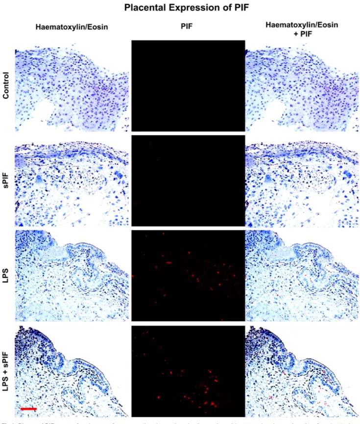
We examined the presence of PIF positive cells by specific anti-PIF monoclonal antibody at day 15 of gestation in the placentae. In line with previous studies [19], we detected minimal to no PIF expression in synthetic PIF or control mice in late gestation (Fig 2: compare two upper panels). Surprisingly, LPS treatment resulted in increased endogenous PIF expression in the
Fig 2. Placental PIF expression. Images of representative placental section (we evaluated 3 consecutive placental sections from 8–10 mice per group) stained using hematoxylin and eosin (left panels) and examined the presence of PIF by specific anti-PIF antibody (red
immunofluorescence: middle panels). Merged images are the right panels. We detected no PIF positive cells in Control and synthetic PIF groups (two upper panels). LPS induced inflammatory insult during pregnancy resulted in PIF expression and additional synthetic PIF treatment did not further increase this expression (two lower panels). sPIF: synthetic PreImplantation Factor; LPS: Lipopolysaccharides. Scale bar: 50μm.
placenta and this expression was not additionally amplified by synthetic PIF administration (Fig 2: compare two lower panels). Notably, PIF expression was noticed predominantly in the cytotrophoblast compartment which is in line with previous reports [15]. We postulate that increased endogenous PIF expression may reflect a protective response to the LPS induced inflammatory insult. Further experiments will address the LPS-induced changes in the pla-centa during embryo development but are beyond the scope of the manuscript. As this endog-enous PIF expression did not result in prevention of fetal loss (Fig 1B) and cytokines play a critical role in prevention of fetal loss, we focused on local (placenta) and global (serum) cyto-kine/chemokine levels next.
PIF attenuates LPS induced inflammatory signature and inflammasome
activation
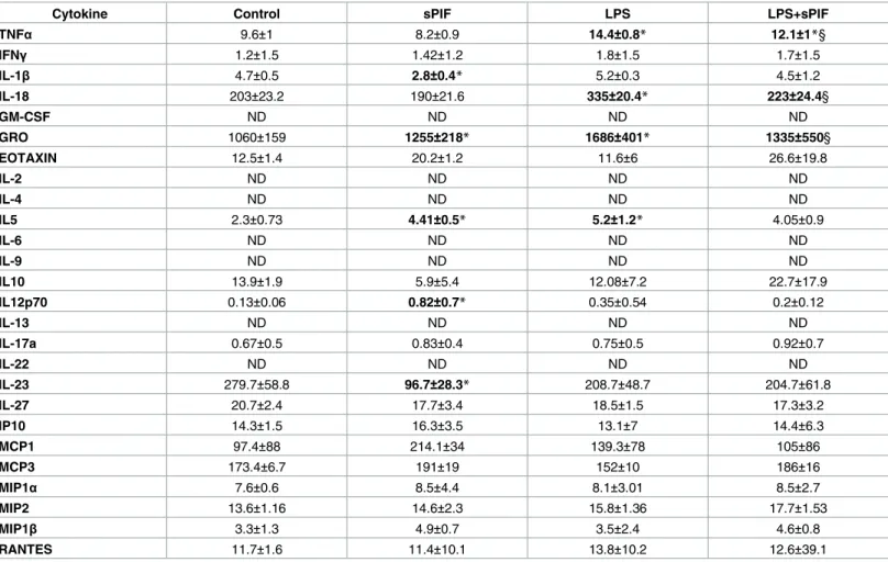
Given that cytokines play a critical role in the inflammatory response and the placenta is a well-defined source of cytokine production, we tested the placental cytokine/chemokine profile first. We used a multiplex bead array assay to detect levels of 22 cytokines/chemokines. As shown inTable 1(placental cytokines), LPS administration resulted in increased levels of Tumor necrosis factor (TNF)-α (prime pro-inflammatory cytokine), IL-18 (an inflammasome-related cytokine), and growth inflammasome-related oncogene (GRO: neutrophil-attractive chemokine) and synthetic PIF prevented these increase significantly. To further test the hypothesis of divergent local (placenta) and systemic (serum) inflammatory control, we tested serum cytokine profile as well (Table 2). Indeed, the global inflammatory response was much stronger than in the pla-centa (Table 2: 14/22 circulating cytokines increased after LPS) and synthetic PIF restored 11 of the 14 LPS induced cytokines/chemokines (Table 2: IFN-γ, 18, GM-CSF, GRO, 4, IL-5, IL-12p70, IL-17a, IL-22, IL-27, and MIP-1β). Notably, we detected changes after synthetic PIF administration independent of LPS insult as well. For example synthetic PIF up-regulated eotaxin, a chemokine which is involved in placental implantation process [33].
We hypothesize that increased endogenous PIF expression in the placenta (Fig 2) results in the divergent inflammatory response at placental and peripheral levels (Tables1and2). How-ever, as in case of progesterone supplementation [34], additional synthetic PIF supplementa-tion is necessary to prevent both the local and peripheral LPS-induced response (Tables1and
2) and therefore provide a protective effect (Fig 1).
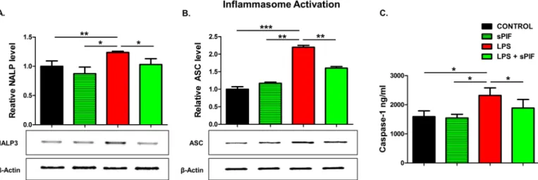
Given that synthetic PIF can reduce major inflammasome signaling cytokines such as IL-1β and IL-18 [35], we decided to test key components of this pathway in the placentae next. Nota-bly, IL-18 contributes to preterm birth in part by modulating TLR4 signaling [36] and TLR4 activation is necessary for PIF effects [27,28]. We focused on NALP-3 and ASC proteins expression in the placenta as these are the key components of inflammasome signaling [4,5]. Notably, this multi-protein complex enables caspase-1-mediated production of pro-inflamma-tory cytokines such as IL-1β and IL-18. We detected increased placental expression of both NALP-3 and ASC proteins in LPS-treated mice (Fig 3A and 3Bcompare red to black bars). In line with our hypothesis, synthetic PIF reduced this activation significantly (Fig 3A and 3B
compare green to black and red bars). We tested downstream inflammasome activation (cas-pase-1) as well. Again, we detected increased levels of caspase-1 in the placenta after LPS expo-sure and the addition of PIF reduced this protein’s levels significantly (Fig 3Ccompare green to red and black bars). Together, we document increased endogenous PIF expression after an LPS insult (Fig 2). Importantly, only application of synthetic PIF results in prevention of increase in cytokine levels after the LPS challenge including the inflammasome pathway. We hypothesize that as in case of progesterone supplementation [34], a combined local and sys-temic inflammatory control is necessary to reduce LPS-induced fetal loss.
Discussion
Our current findings are in line with the notion of PIF‘s trophic and protective action on the embryo, decidua, and trophoblast [12]. Synthetic PIF administration alone decreased fetal loss and improved fetal weight without modulating the inflammasome pathway (Figs1B, 1Dand
3). These favorable effects suggest a beneficial role in optimization of the implantation process. In the current study, we did not detect changes in the number of implanted embryos and plug/ pregnancy rate (S1 Table). However, the current experimental setup is not suitable to answer this specific question as this model does not share features with human early recurrent miscar-riage as the CBA/J x DBA/2 mouse model (mating of CBA/J females (H2k) with DBA/2J males (H2d)) [37–39]. On the other hand, synthetic PIF increased serum levels of eotaxin indepen-dent of the LPS insult (Table 2). Eotaxins regulate extravillous throphoblast function during uterine decidual vessel remodeling [33]. Furthermore, eotaxin has been shown to promote eosinophil adhesion to vascular cell adhesion molecule-1, a major ligand for the integrins [40]. As trophoblast differentiate into an endovascular phenotype a4 integrin is up-regulated [33]
Table 1. Placental inflammatory signature.
Cytokine Control sPIF LPS LPS+sPIF
TNFα 9.6±1 8.2±0.9 14.4±0.8* 12.1±1*§ IFNγ 1.2±1.5 1.42±1.2 1.8±1.5 1.7±1.5 IL-1β 4.7±0.5 2.8±0.4* 5.2±0.3 4.5±1.2 IL-18 203±23.2 190±21.6 335±20.4* 223±24.4§ GM-CSF ND ND ND ND GRO 1060±159 1255±218* 1686±401* 1335±550§ EOTAXIN 12.5±1.4 20.2±1.2 11.6±6 26.6±19.8 IL-2 ND ND ND ND IL-4 ND ND ND ND IL5 2.3±0.73 4.41±0.5* 5.2±1.2* 4.05±0.9 IL-6 ND ND ND ND IL-9 ND ND ND ND IL10 13.9±1.9 5.9±5.4 12.08±7.2 22.7±17.9 IL12p70 0.13±0.06 0.82±0.7* 0.35±0.54 0.2±0.12 IL-13 ND ND ND ND IL-17a 0.67±0.5 0.83±0.4 0.75±0.5 0.92±0.7 IL-22 ND ND ND ND IL-23 279.7±58.8 96.7±28.3* 208.7±48.7 204.7±61.8 IL-27 20.7±2.4 17.7±3.4 18.5±1.5 17.3±3.2 IP10 14.3±1.5 16.3±3.5 13.1±7 14.4±6.3 MCP1 97.4±88 214.1±34 139.3±78 105±86 MCP3 173.4±6.7 191±19 152±10 186±16 MIP1α 7.6±0.6 8.5±4.4 8.1±3.01 8.5±2.7 MIP2 13.6±1.16 14.6±2.3 15.8±1.36 17.7±1.53 MIP1β 3.3±1.3 4.9±0.7 3.5±2.4 4.6±0.8 RANTES 11.7±1.6 11.4±10.1 13.8±10.2 12.6±39.1
Cytokine levels were measured in placental lysates obtained from mice treated with PBS (Control), synthetic PreImplantation Factor (sPIF), Lipopolysaccharides (LPS), and LPS+sPIF by multiplex bead array assay. The results are expressed as ng/ml (Mean±SD). Statistical significance: *P<0.05 vs CTR;
§P<0.05 vs LPS.
Significant changes are marked in bold. https://doi.org/10.1371/journal.pone.0180642.t001
and up-regulation of integrins by PIF during throphoblast invasion was reported previously [15,18]. Thus, current and previous data support the use of synthetic PIF in early recurrent pregnancy loss [20,23,41] and studies are in preparation.
Synthetic PIF‘s effects on placental/fetal weight ratio after LPS insult are of interest as well (Fig 1E). LPS was reported to reduce uterine blood flow to the fetus and increase uterine resis-tance without affecting the placental size thereby leading to fibrin deposits and disseminated intravascular coagulation [42]. Thus, growth retardation is expected and was observed in our study (Fig 1D: compare red to black bars). The role of the inflammasome specifically NALP-3 in LPS induced pathology in placental cells has been previously described [35]. Importantly, we report that the LPS induced activation of placental inflammasome can be prevented by syn-thetic PIF administration (Fig 3). This is supported by the fact that PIF‘s effects dependent on TLR4 signaling [27,28] and TLR4 is the main receptor of inflammasome activation [43]. Fur-thermore, synthetic PIF targets Kv1.3b channel to reduce K+flux which activates NALP-3 expression [29,43]. We hypothesize that synthetic PIF‘s effects on placental caspase-1 levels
Table 2. Serum inflammatory signature.
Cytokine Control sPIF LPS LPS+sPIF
TNFα 6.6±0.4 5.6±1.1 8.6±0.9* 7.7±1.0 IFNγ 3.16±0.2 3.39±1.4 16.6±1.6* 5.92±1.1§ IL-1β 3.4±1 2.5±1.1 5.2±1.3* 4±1.3 IL-18 266±65 150±25* 369±26* 268±64§ GM-CSF 6.4±1 5.3±0.7 11.06±1.6* 8.2±1.5§ GRO 15.26±1.8 18.06±2.2 24.9±2.9* 17.07±1.2§ EOTAXIN 278±61 486±46* 226±37 430±20§ IL-2 ND ND ND ND IL-4 2.53±0.48 1.74±0.9 7.72±0.2* 3.95±0.3*§ IL-5 3.98±0.5 2.66±0.4* 13.7±0.11* 6.8±0.5*§ IL-6 ND ND ND ND IL-9 ND ND ND ND IL-10 7.68±1.1 6.65±1.1 11.69±2.2 8.23±1.3 IL12p70 1.36±0.49 0.94±0.7* 3.14±1.73* 2.07±1.2*§ IL-13 ND ND ND ND IL-17a 5.52±0.06 4±0.7 13.6±0.54* 10.4±0.12* IL-22 35.89±2.1 15.9±27* 47.3±5.6* 36.2±2.6§ IL-23 36.29±8.6 21.6±2.3* 66.9±3.2* 50.9±5.2 IL-27 22.7±1.1 13.4±1.08* 54.5±2.7* 38.7±3.1§ IP10 28.6±11.2 41.5±12.8 22.5±12.7 35.8±14.6 MCP1 47.04±3.3 34.1±2.7* 49.01±1.9 41.8±3.9 MCP3 176.8±71 146.4±35 168.4±58 167.3±93.2 MIP1α 2.6±1 3.08±0.62 3.1±0.96 3.3±1 MIP2 24.7±2.9 24.9±3.4 27.6±4.1 26.3±1.5 MIP1β 3.95±0.3 1.9±0.7* 6.4±0.4* 3.6±0.8§ RANTES 14.2±8.4 22.2±8.5 21.6±7.7 26.5±11.7
Cytokine levels were measured in serum obtained from mice treated with PBS (Control), synthetic PreImplantation Factor (sPIF), Lipopolysaccharides (LPS), and LPS+sPIF by multiplex bead array assay. The results are expressed as ng/ml (Mean±SD). Statistical significance:
*P<0.05 vs CTR; §P<0.05 vs LPS.
Significant changes are marked in bold. https://doi.org/10.1371/journal.pone.0180642.t002
protect against apoptosis and compromise critical oxygen and nutrients transfer to the fetus [44].
Furthermore, the placental/fetal weight ratio relevance for pregnancy outcome was addressed by large population studies previously and an increased ratio was noted in prema-ture birth and fetal demise [32,45,46]. Various pathologic and inflammatory conditions are associated with abnormal placental/birthweight ratios including chronic hypertension, pre-eclampsia, and chorioamnionitis. The present study underscores the fact that the placenta (a primitive organ, supplied by maternal circulation) and the fetus (complex, more evolved with higher oxygen and nutrient demands) are coupled entities. Herein, inflammatory insult affects placental function (but not weight/growth) and still results in reduced fetal weight. We hypothesize that the noted improved fetal-placental exchange is partially due to PIF’s role in promoting implantation [20,23] and/or reducing vascular inflammation [29]. However, it is still not understood how synthetic PIF increases fetal weight despite of LPS insult (Fig 1D). Although hypothetical, the modulation of the H19/insulin-like growth factor 2 (Igf2)
imprinted locus is intriguing. H19 is a long non-coding RNA and placental H19 misregulation is associated with the imprinting disorder such fetal overgrowth disorder (Beckwith–Wiede-mann syndrome) and intrauterine growth retardation (Silver–Russell syndrome) [47]. Nota-bly, we recently reported the close functional interactions of H19 and microRNA let-7 [48,49] and let-7 expression is modulated by synthetic PIF [27]. Thus, synthetic PIF‘s role on H19 modulation in intrauterine growth restriction models is currently investigated but beyond the scope of this manuscript. Notably, women with unexplained infertility and women with endo-metriosis show decreased H19 expression in eutopic endometrium suggesting a direct clinical link [50,51].
Synthetic PIF‘s effect on local and peripheral cytokine level are of interest as well (Tables1
and2). Altered balance of pro- and anti-inflammatory cytokines forms the basis of multiple pregnancy disorders and results in fetal and maternal pathological changes [12,52–56]. For example, IL-18 is significantly elevated at the onset of labor [57] and contributes to preterm birth [36,57]. Preterm birth or placental hypoxia is associated with high concentrations of granulocyte macrophage-colony stimulating factor (GM-CSF) [58,59]. GRO a well-defined neutrophil-attractive chemokine increases in case of chorioamnionitis [60]. Besides maternal morbidities, prenatal LPS insult results in sustained inflammation in fetus and newborn,
Fig 3. Inflammasome pathway analysis (18–20 placental lysates for each group). Representative gel images of (A) relative NALP-3 and (B) relative ASC protein levels in placental tissue lysates. The downstream cytokine caspase-1 levels in lysates were measured using enzyme-linked immunoassay (C). Data are mean±SD.β-Actin was the control.*p<0.05. sPIF: synthetic PreImplantation Factor; LPS: Lipopolysaccharides. https://doi.org/10.1371/journal.pone.0180642.g003
which is associated with an increased risk for adverse outcomes such as brain damage and pul-monary or intestinal complications [61–64]. Given that synthetic PIF prevented the increase of those cytokines/chemokines in the serum (Table 2) and partially in the placenta (Table 1), we envision synthetic PIF treatment in pregnancies at risk as in case of progesterone [52]. This is supported by the fact that synthetic PIF reduces the inflammasome response (Fig 3) and NALP-3 induced IL-18 and IL-1β impact the pathogenesis of preeclampsia, preterm birth, and perinatal brain injury [3,65,66].
The simple notion of TH2overbalance during pregnancy is not specific enough. More
recent insights into immunological operative mechanisms in pregnancy favor the TH1/TH2
cooperation [67]. The dual role of IL-4 is an examples [68]. Synthetic PIF prevented the LPS induced Th2-type cytokine IL-4 in the serum (Table 2). Notably, IL-4–induced natural killer
cells produce higher levels of IFN-γ, IL-10, and GM-CSF while exhibiting high cytotoxicity [68]. Not surprisingly, synthetic PIF prevented the increase of those cytokines in the serum (Table 2) in line with natural killer cells being PIF‘s target [23]. Finally, synthetic PIF modu-lated IL-22, a member of IL-10 family (Table 2) and increased IL-22 levels are an inflammatory marker of placental dysfunction and early pregnancy loss [56,69].
In conclusion, we provide evidence supporting synthetic PIF as a targeted therapy in inflammatory induced pregnancy loss. This study is in line with PIF‘s efficacy previously observed in several preclinical models of immune disorders [24–30]. Additionally, this is the first description of placental expression of PIF after LPS-induced inflammatory insult. Syn-thetic PIF is currently in first-in-human Phase Ib clinical trial (NCT02239562) for adult patients with autoimmune disease. Upon completion the use of synthetic PIF as treatment for perinatal brain injuries is planned [27,28]. Synthetic PIF therapy in pregnancy disorders such as preeclampsia, preterm birth, and early pregnancy loss are in preparation as well [12,19,41].
Supporting information
S1 Table. Detailed outcomes of the performed experiments. Detailed results of the
per-formed experiments and groups (Control, sPIF, LPS, and LPS+sPIF) are summarized includ-ing the number of implanted embryos and plug/pregnancy rate.
(PDF)
Acknowledgments
We thank A. Carter and S. Zinn for editorial assistance.
Author Contributions
Conceptualization: Nicoletta Di Simone, Fiorella Di Nicuolo, Riccardo Marana, Roberta
Cas-tellani, Manuela Veglia, Giovanni Scambia, Daniel Surbek, Martin Mueller.
Data curation: Nicoletta Di Simone, Fiorella Di Nicuolo, Riccardo Marana, Francesco Ria,
Manuela Veglia, Giovanni Scambia, Daniel Surbek, Martin Mueller.
Formal analysis: Nicoletta Di Simone, Fiorella Di Nicuolo, Riccardo Marana, Roberta
Castel-lani, Francesco Ria, Manuela Veglia, Giovanni Scambia, Daniel Surbek, Eytan Barnea, Mar-tin Mueller.
Funding acquisition: Nicoletta Di Simone, Eytan Barnea.
Investigation: Nicoletta Di Simone, Fiorella Di Nicuolo, Francesco Ria, Eytan Barnea, Martin
Methodology: Nicoletta Di Simone, Fiorella Di Nicuolo, Riccardo Marana, Roberta Castellani,
Francesco Ria, Manuela Veglia, Giovanni Scambia, Daniel Surbek, Eytan Barnea, Martin Mueller.
Project administration: Martin Mueller. Resources: Martin Mueller.
Supervision: Daniel Surbek, Eytan Barnea.
Validation: Fiorella Di Nicuolo, Eytan Barnea, Martin Mueller.
Visualization: Nicoletta Di Simone, Fiorella Di Nicuolo, Martin Mueller.
Writing – original draft: Nicoletta Di Simone, Fiorella Di Nicuolo, Roberta Castellani, Eytan
Barnea, Martin Mueller.
Writing – review & editing: Martin Mueller.
References
1. Christiansen OB. Reproductive immunology. Molecular immunology. 2013; 55(1):8–15.https://doi.org/ 10.1016/j.molimm.2012.08.025PMID:23062611.
2. Clark DA. The importance of being a regulatory T cell in pregnancy. J Reprod Immunol. 2016; 116:60– 9.https://doi.org/10.1016/j.jri.2016.04.288PMID:27219894.
3. D’Ippolito S, Tersigni C, Marana R, Di Nicuolo F, Gaglione R, Rossi ED, et al. Inflammosome in the human endometrium: further step in the evaluation of the "maternal side". Fertil Steril. 2016; 105 (1):111–8.e1-4.https://doi.org/10.1016/j.fertnstert.2015.09.027PMID:26474737.
4. Dunne A. Inflammasome activation: from inflammatory disease to infection. Biochemical Society trans-actions. 2011; 39(2):669–73.https://doi.org/10.1042/BST0390669PMID:21428959.
5. Nold-Petry CA, Nold MF, Nielsen JW, Bustamante A, Zepp JA, Storm KA, et al. Increased cytokine pro-duction in interleukin-18 receptor alpha-deficient cells is associated with dysregulation of suppressors of cytokine signaling. J Biol Chem. 2009; 284(38):25900–11.https://doi.org/10.1074/jbc.M109.004184
PMID:19592492;
6. Silver RM, Edwin SS, Umar F, Dudley DJ, Branch DW, Mitchell MD. Bacterial lipopolysaccharide-medi-ated murine fetal death: the role of interleukin-1. Am J Obstet Gynecol. 1997; 176(3):544–9. PMID:
9077604.
7. Robertson SA, Care AS, Skinner RJ. Interleukin 10 regulates inflammatory cytokine synthesis to protect against lipopolysaccharide-induced abortion and fetal growth restriction in mice. Biol Reprod. 2007; 76 (5):738–48.https://doi.org/10.1095/biolreprod.106.056143PMID:17215490.
8. Rossol M, Heine H, Meusch U, Quandt D, Klein C, Sweet MJ, et al. LPS-induced cytokine production in human monocytes and macrophages. Crit Rev Immunol. 2011; 31(5):379–446. PMID:22142165. 9. Clark DA. Immunological factors in pregnancy wastage: fact or fiction. Am J Reprod Immunol. 2008; 59
(4):277–300.https://doi.org/10.1111/j.1600-0897.2008.00580.xPMID:18336385.
10. Moffett-King A. Natural killer cells and pregnancy. Nat Rev Immunol. 2002; 2(9):656–63. Epub 2002/09/ 05.https://doi.org/10.1038/nri886PMID:12209134.
11. Barnea ER. Applying embryo-derived immune tolerance to the treatment of immune disorders. Ann N Y Acad Sci. 2007; 1110:602–18.https://doi.org/10.1196/annals.1423.064PMID:17911476.
12. Barnea ER, Almogi-Hazan O, Or R, Mueller M, Ria F, Weiss L, et al. Immune regulatory and neuropro-tective properties of preimplantation factor: From newborn to adult. Pharmacol Ther. 2015; 156:10–25.
https://doi.org/10.1016/j.pharmthera.2015.10.008PMID:26546485.
13. Stamatkin CW, Roussev RG, Stout M, Absalon-Medina V, Ramu S, Goodman C, et al. PreImplantation Factor (PIF) correlates with early mammalian embryo development-bovine and murine models. Reprod Biol Endocrinol. 2011; 9:63.https://doi.org/10.1186/1477-7827-9-63PMID:21569635;
14. Ramu S, Stamatkin C, Timms L, Ruble M, Roussev RG, Barnea ER. PreImplantation factor (PIF) detec-tion in maternal circuladetec-tion in early pregnancy correlates with live birth (bovine model). Reprod Biol Endocrinol. 2013; 11:105.https://doi.org/10.1186/1477-7827-11-105PMID:24238492;
15. Moindjie H, Santos ED, Loeuillet L, Gronier H, de Mazancourt P, Barnea ER, et al. Preimplantation fac-tor (PIF) promotes human trophoblast invasion. Biol Reprod. 2014; 91(5):118.https://doi.org/10.1095/ biolreprod.114.119156PMID:25232018.
16. Paidas MJ, Krikun G, Huang SJ, Jones R, Romano M, Annunziato J, et al. A genomic and proteomic investigation of the impact of preimplantation factor on human decidual cells. Am J Obstet Gynecol. 2010; 202(5):459.e1–8.https://doi.org/10.1016/j.ajog.2010.03.024PMID:20452489;
17. Duzyj CM, Barnea ER, Li M, Huang SJ, Krikun G, Paidas MJ. Preimplantation factor promotes first tri-mester trophoblast invasion. Am J Obstet Gynecol. 2010; 203(4):402.e1–4.https://doi.org/10.1016/j. ajog.2010.06.060PMID:20708167;
18. Barnea ER, Kirk D, Paidas MJ. Preimplantation factor (PIF) promoting role in embryo implantation: increases endometrial integrin-alpha2beta3, amphiregulin and epiregulin while reducing betacellulin expression via MAPK in decidua. Reprod Biol Endocrinol. 2012; 10:50. https://doi.org/10.1186/1477-7827-10-50PMID:22788113;
19. Barnea ER, Vialard F, Moindjie H, Ornaghi S, Dieudonne MN, Paidas MJ. PreImplantation Factor (PIF*) endogenously prevents preeclampsia: Promotes trophoblast invasion and reduces oxidative stress. J Reprod Immunol. 2016; 114:58–64.https://doi.org/10.1016/j.jri.2015.06.002PMID:26257082. 20. Stamatkin CW, Roussev RG, Stout M, Coulam CB, Triche E, Godke RA, et al. Preimplantation factor
negates embryo toxicity and promotes embryo development in culture. Reprod Biomed Online. 2011; 23(4):517–24.https://doi.org/10.1016/j.rbmo.2011.06.009PMID:21900046.
21. Barnea ER, Lubman DM, Liu YH, Absalon-Medina V, Hayrabedyan S, Todorova K, et al. Insight into PreImplantation Factor (PIF*) mechanism for embryo protection and development: target oxidative stress and protein misfolding (PDI and HSP) through essential RIKP [corrected] binding site. PLoS One. 2014; 9(7):e100263.https://doi.org/10.1371/journal.pone.0100263PMID:24983882; 22. Barnea ER, Kirk D, Ramu S, Rivnay B, Roussev R, Paidas MJ. PreImplantation Factor (PIF)
orches-trates systemic antiinflammatory response by immune cells: effect on peripheral blood mononuclear cells. Am J Obstet Gynecol. 2012; 207(4):313.e1–11.https://doi.org/10.1016/j.ajog.2012.07.017PMID:
23021695.
23. Roussev RG, Dons’koi BV, Stamatkin C, Ramu S, Chernyshov VP, Coulam CB, et al. Preimplantation factor inhibits circulating natural killer cell cytotoxicity and reduces CD69 expression: implications for recurrent pregnancy loss therapy. Reprod Biomed Online. 2013; 26(1):79–87.https://doi.org/10.1016/j. rbmo.2012.09.017PMID:23186554.
24. Weiss L, Bernstein S, Jones R, Amunugama R, Krizman D, Jebailey L, et al. Preimplantation factor (PIF) analog prevents type I diabetes mellitus (TIDM) development by preserving pancreatic function in NOD mice. Endocrine. 2011; 40(1):41–54.https://doi.org/10.1007/s12020-011-9438-5PMID:
21424847.
25. Weiss L, Or R, Jones RC, Amunugama R, JeBailey L, Ramu S, et al. Preimplantation factor (PIF*) reverses neuroinflammation while promoting neural repair in EAE model. J Neurol Sci. 2012; 312(1– 2):146–57.https://doi.org/10.1016/j.jns.2011.07.050PMID:21996270.
26. Azar Y, Shainer R, Almogi-Hazan O, Bringer R, Compton SR, Paidas MJ, et al. Preimplantation factor reduces graft-versus-host disease by regulating immune response and lowering oxidative stress (murine model). Biol Blood Marrow Transplant. 2013; 19(4):519–28.https://doi.org/10.1016/j.bbmt. 2012.12.011PMID:23266739.
27. Mueller M, Zhou J, Yang L, Gao Y, Wu F, Schoeberlein A, et al. PreImplantation factor promotes neuro-protection by targeting microRNA let-7. Proc Natl Acad Sci U S A. 2014; 111(38):13882–7.https://doi. org/10.1073/pnas.1411674111PMID:25205808;
28. Mueller M, Schoeberlein A, Zhou J, Joerger-Messerli M, Oppliger B, Reinhart U, et al. PreImplantation Factor bolsters neuroprotection via modulating Protein Kinase A and Protein Kinase C signaling. Cell death and differentiation. 2015; 22(12):2078–86.https://doi.org/10.1038/cdd.2015.55PMID:25976303; 29. Chen YC, Rivera J, Fitzgerald M, Hausding C, Ying YL, Wang X, et al. PreImplantation factor prevents
atherosclerosis via its immunomodulatory effects without affecting serum lipids. Thromb Haemost. 2016; 115(5):1010–24.https://doi.org/10.1160/TH15-08-0640PMID:26842698.
30. Shainer R, Almogi-Hazan O, Berger A, Hinden L, Mueller M, Brodie C, et al. PreImplantation factor (PIF) therapy provides comprehensive protection against radiation induced pathologies. Oncotarget. 2016.https://doi.org/10.18632/oncotarget.10635PMID:27449294.
31. Su MT, Lin SH, Chen YC. Association of sex hormone receptor gene polymorphisms with recurrent pregnancy loss: a systematic review and meta-analysis. Fertil Steril. 2011; 96(6):1435–44 e1.https:// doi.org/10.1016/j.fertnstert.2011.09.030PMID:22014881.
32. Haavaldsen C, Samuelsen SO, Eskild A. Fetal death and placental weight/birthweight ratio: a popula-tion study. Acta Obstet Gynecol Scand. 2013; 92(5):583–90.https://doi.org/10.1111/aogs.12105PMID:
23398315.
33. Chau SE, Murthi P, Wong MH, Whitley GS, Brennecke SP, Keogh RJ. Control of extravillous tropho-blast function by the eotaxins CCL11, CCL24 and CCL26. Hum Reprod. 2013; 28(6):1497–507.https:// doi.org/10.1093/humrep/det060PMID:23477905.
34. Aisemberg J, Vercelli CA, Bariani MV, Billi SC, Wolfson ML, Franchi AM. Progesterone is essential for protecting against LPS-induced pregnancy loss. LIF as a potential mediator of the anti-inflammatory effect of progesterone. PLoS One. 2013; 8(2):e56161.https://doi.org/10.1371/journal.pone.0056161
PMID:23409146;
35. Pontillo A, Girardelli M, Agostinis C, Masat E, Bulla R, Crovella S. Bacterial LPS differently modulates inflammasome gene expression and IL-1beta secretion in trophoblast cells, decidual stromal cells, and decidual endothelial cells. Reprod Sci. 2013; 20(5):563–6.https://doi.org/10.1177/1933719112459240
PMID:23184659.
36. Li L, Yang J, Jiang Y, Tu J, Schust DJ. Activation of decidual invariant natural killer T cells promotes lipo-polysaccharide-induced preterm birth. Mol Hum Reprod. 2015; 21(4):369–81.https://doi.org/10.1093/ molehr/gav001PMID:25589517.
37. Clark DA, Manuel J, Lee L, Chaouat G, Gorczynski RM, Levy GA. Ecology of danger-dependent cyto-kine-boosted spontaneous abortion in the CBA x DBA/2 mouse model. I. Synergistic effect of LPS and (TNF-alpha + IFN-gamma) on pregnancy loss. Am J Reprod Immunol. 2004; 52(6):370–8.https://doi. org/10.1111/j.1600-0897.2004.00237.xPMID:15663602.
38. Clark DA, Ding JW, Yu G, Levy GA, Gorczynski RM. Fgl2 prothrombinase expression in mouse tropho-blast and decidua triggers abortion but may be countered by OX-2. Mol Hum Reprod. 2001; 7(2):185– 94. PMID:11160845.
39. Redecha P, van Rooijen N, Torry D, Girardi G. Pravastatin prevents miscarriages in mice: role of tissue factor in placental and fetal injury. Blood. 2009; 113(17):4101–9. https://doi.org/10.1182/blood-2008-12-194258PMID:19234141;
40. Woodside DG, Kram RM, Mitchell JS, Belsom T, Billard MJ, McIntyre BW, et al. Contrasting roles for domain 4 of VCAM-1 in the regulation of cell adhesion and soluble VCAM-1 binding to integrin alpha4-beta1. J Immunol. 2006; 176(8):5041–9. PMID:16585601.
41. Goodale LF, Hayrabedran S, Todorova K, Roussev R, Ramu S, Stamatkin C, et al. PreImplantation fac-tor (PIF) protects cultured embryos against oxidative stress: relevance for recurrent pregnancy loss (RPL) therapy. Oncotarget. 2017; 8(20):32419–32.https://doi.org/10.18632/oncotarget.16028PMID:
28423690.
42. Renaud SJ, Cotechini T, Quirt JS, Macdonald-Goodfellow SK, Othman M, Graham CH. Spontaneous pregnancy loss mediated by abnormal maternal inflammation in rats is linked to deficient uteroplacental perfusion. J Immunol. 2011; 186(3):1799–808.https://doi.org/10.4049/jimmunol.1002679PMID:
21187445.
43. Petrilli V, Papin S, Dostert C, Mayor A, Martinon F, Tschopp J. Activation of the NALP3 inflammasome is triggered by low intracellular potassium concentration. Cell death and differentiation. 2007; 14 (9):1583–9.https://doi.org/10.1038/sj.cdd.4402195PMID:17599094.
44. Ilekis JV, Tsilou E, Fisher S, Abrahams VM, Soares MJ, Cross JC, et al. Placental origins of adverse pregnancy outcomes: potential molecular targets: an Executive Workshop Summary of the Eunice Ken-nedy Shriver National Institute of Child Health and Human Development. Am J Obstet Gynecol. 2016; 215(1 Suppl):S1–S46.https://doi.org/10.1016/j.ajog.2016.03.001PMID:26972897;
45. de Jongh BE, Mackley A, Jain N, Locke R, Paul DA. Effects of advanced maternal age and race/ethnic-ity on placental weight and placental weight/birthweight ratio in very low birthweight infants. Matern Child Health J. 2015; 19(7):1553–8.https://doi.org/10.1007/s10995-014-1662-1PMID:25567078. 46. McNamara H, Hutcheon JA, Platt RW, Benjamin A, Kramer MS. Risk factors for high and low placental
weight. Paediatr Perinat Epidemiol. 2014; 28(2):97–105.https://doi.org/10.1111/ppe.12104PMID:
24354883.
47. Hur SK, Freschi A, Ideraabdullah F, Thorvaldsen JL, Luense LJ, Weller AH, et al. Humanized H19/Igf2 locus reveals diverged imprinting mechanism between mouse and human and reflects Silver-Russell syndrome phenotypes. Proc Natl Acad Sci U S A. 2016; 113(39):10938–43. PMID:27621468; 48. Kallen AN, Zhou XB, Xu J, Qiao C, Ma J, Yan L, et al. The imprinted H19 lncRNA antagonizes let-7
micro-RNAs. Mol Cell. 2013; 52(1):101–12.https://doi.org/10.1016/j.molcel.2013.08.027PMID:24055342; 49. Gao Y, Wu F, Zhou J, Yan L, Jurczak MJ, Lee HY, et al. The H19/let-7 double-negative feedback loop
contributes to glucose metabolism in muscle cells. Nucleic Acids Res. 2014; 42(22):13799–811.https:// doi.org/10.1093/nar/gku1160PMID:25399420;
50. Korucuoglu U, Biri AA, Konac E, Alp E, Onen IH, Ilhan MN, et al. Expression of the imprinted IGF2 and H19 genes in the endometrium of cases with unexplained infertility. European journal of obstetrics, gynecology, and reproductive biology. 2010; 149(1):77–81.https://doi.org/10.1016/j.ejogrb.2009.12. 007PMID:20042264.
51. Ghazal S, McKinnon B, Zhou J, Mueller M, Men Y, Yang L, et al. H19 lncRNA alters stromal cell growth via IGF signaling in the endometrium of women with endometriosis. EMBO molecular medicine. 2015; 7 (8):996–1003.https://doi.org/10.15252/emmm.201505245PMID:26089099;
52. Lissauer D, Eldershaw SA, Inman CF, Coomarasamy A, Moss PA, Kilby MD. Progesterone promotes maternal-fetal tolerance by reducing human maternal T-cell polyfunctionality and inducing a specific cytokine profile. Eur J Immunol. 2015; 45(10):2858–72.https://doi.org/10.1002/eji.201445404PMID:
26249148;
53. Nahum R, Brenner O, Zahalka MA, Traub L, Quintana F, Moroz C. Blocking of the placental immune-modulatory ferritin activates Th1 type cytokines and affects placenta development, fetal growth and the pregnancy outcome. Hum Reprod. 2004; 19(3):715–22.https://doi.org/10.1093/humrep/deh099PMID:
14998975.
54. Abumaree MH, Chamley LW, Badri M, El-Muzaini MF. Trophoblast debris modulates the expression of immune proteins in macrophages: a key to maternal tolerance of the fetal allograft? J Reprod Immunol. 2012; 94(2):131–41.https://doi.org/10.1016/j.jri.2012.03.488PMID:22542910.
55. Germain SJ, Sacks GP, Sooranna SR, Sargent IL, Redman CW. Systemic inflammatory priming in nor-mal pregnancy and preeclampsia: the role of circulating syncytiotrophoblast microparticles. J Immunol. 2007; 178(9):5949–56. PMID:17442979.
56. O’Hern Perfetto C, Fan X, Dahl S, Krieg S, Westphal LM, Bunker Lathi R, et al. Expression of interleu-kin-22 in decidua of patients with early pregnancy and unexplained recurrent pregnancy loss. J Assist Reprod Genet. 2015; 32(6):977–84.https://doi.org/10.1007/s10815-015-0481-7PMID:25925347; 57. Ida A, Tsuji Y, Muranaka J, Kanazawa R, Nakata Y, Adachi S, et al. IL-18 in pregnancy; the elevation of
IL-18 in maternal peripheral blood during labour and complicated pregnancies. J Reprod Immunol. 2000; 47(1):65–74. PMID:10779591.
58. Basraon SK, Menon R, Makhlouf M, Longo M, Hankins GD, Saade GR, et al. Can statins reduce the inflammatory response associated with preterm birth in an animal model? Am J Obstet Gynecol. 2012; 207(3):224 e1-7.https://doi.org/10.1016/j.ajog.2012.06.020PMID:22939729.
59. Hayashi M, Hamada Y, Ohkura T. Elevation of granulocyte-macrophage colony-stimulating factor in the placenta and blood in preeclampsia. Am J Obstet Gynecol. 2004; 190(2):456–61.https://doi.org/10. 1016/j.ajog.2003.07.032PMID:14981389.
60. Marvin KW, Keelan JA, Eykholt RL, Sato TA, Mitchell MD. Use of cDNA arrays to generate differential expression profiles for inflammatory genes in human gestational membranes delivered at term and pre-term. Mol Hum Reprod. 2002; 8(4):399–408. PMID:11912289.
61. Kuypers E, Willems MG, Jellema RK, Kemp MW, Newnham JP, Delhaas T, et al. Responses of the spleen to intraamniotic lipopolysaccharide exposure in fetal sheep. Pediatr Res. 2015; 77(1–1):29–35.
https://doi.org/10.1038/pr.2014.152PMID:25285474.
62. Smit AL, Lambermont VA, Stokroos RJ, Anteunis LJ, Chenault MN, Schaefer SM, et al. Intrauterine Lipopolysaccharide-Induced Chorioamnionitis in a Sheep: Does It Affect the Auditory System? Repro-ductive sciences (Thousand Oaks, Calif). 2016; 23(2):257–63.https://doi.org/10.1177/
1933719115602759PMID:26702124.
63. Al-Amin MM, Alam T, Hasan SM, Hasan AT, Quddus AH. Prenatal maternal lipopolysaccharide admin-istration leads to age- and region-specific oxidative stress in the early developmental stage in offspring. Neuroscience. 2016; 318:84–93.https://doi.org/10.1016/j.neuroscience.2016.01.002PMID:26774051. 64. Rose DR, Careaga M, Van de Water J, McAllister K, Bauman MD, Ashwood P. Long-term altered
immune responses following fetal priming in a non-human primate model of maternal immune activa-tion. Brain Behav Immun. 2017; 63:60–70.https://doi.org/10.1016/j.bbi.2016.11.020PMID:27876552; 65. Adams Waldorf KM, Persing D, Novy MJ, Sadowsky DW, Gravett MG. Pretreatment with toll-like
recep-tor 4 antagonist inhibits lipopolysaccharide-induced preterm uterine contractility, cytokines, and prosta-glandins in rhesus monkeys. Reprod Sci. 2008; 15(2):121–7.https://doi.org/10.1177/
1933719107310992PMID:18187405;
66. Xue P, Zheng M, Gong P, Lin C, Zhou J, Li Y, et al. Single administration of ultra-low-dose lipopolysac-charide in rat early pregnancy induces TLR4 activation in the placenta contributing to preeclampsia. PLoS One. 2015; 10(4):e0124001.https://doi.org/10.1371/journal.pone.0124001PMID:25853857; 67. Sykes L, MacIntyre DA, Yap XJ, Teoh TG, Bennett PR. The Th1:th2 dichotomy of pregnancy and
pre-term labour. Mediators Inflamm. 2012; 2012:967629.https://doi.org/10.1155/2012/967629PMID:
22719180;
68. Kiniwa T, Enomoto Y, Terazawa N, Omi A, Miyata N, Ishiwata K, et al. NK cells activated by Interleukin-4 in cooperation with Interleukin-15 exhibit distinctive characteristics. Proc Natl Acad Sci U S A. 2016; 113(36):10139–44.https://doi.org/10.1073/pnas.1600112113PMID:27551096;
69. Bersani I, De Carolis MP, Foell D, Weinhage T, Rossi ED, De Carolis S, et al. Interleukin-22: biomarker of maternal and fetal inflammation? Immunol Res. 2015; 61(1–2):4–10. https://doi.org/10.1007/s12026-014-8568-2PMID:25407645.