University of Sassari
Department of Biomedical Sciences
INTERNATIONAL PHD SCHOOL IN BIOMOLECULAR AND BIOTECHNOLOGICAL SCIENCES
XXVIII Cycle
INVESTIGATION OF THE MOLECULAR MECHANISMS INDUCING VASCULAR DAMAGE IN SYSTEMIC SCLEROSIS Director:
Prof. LEONARDO A. SECHI
Tutor: GIANFRANCO PINTUS Co-tutor: HUYNH DINH CHIEN
PhD thesis of
DUONG THI BICH THUAN
ATTESTATION OF AUTHORSHIP ACKNOWLEDGEMENT ABBREVIATION ABSTRACT 1. INTRODUCTION ... 1 1.1. Systemic sclerosis ... 1
1.2. Oxidative stress and systemic sclerosis ... 15
1.3. Role of endothelial NADPH oxidases in SSc ... 19
1.4. Endothelial-to-Mesenchymal Transition and Systemic Sclerosis ... 22
1.5. Reactive oxygen species and Endothelial-to-Mesenchymal Transition ... 28
2. RESEARCH OBJECTIVES ... 32
3. MATERIALS AND METHODS ... 34
3.1. Ethical approval ... 34
3.2. Patient selection ... 34
3.3. Cells and Chemicals ... 35
3.4. Cell culture ... 35
3.5. Measurement of intracellular ROS ... 35
3.6. Measurement of intracellular ROS with NADPH inhibitor ... 36
3.7. Molecular cloning and lentiviral particles production ... 37
3.8. Determination of NOX2-associated ROS using NOX-specific redox biosensor p47-roGFP 39 3.9. Measurement of collagen promoter activity ... 39
3.10. Cell proliferation assay (BrdU incorporation Assay) ... 40
3.11. Protein extraction ... 41
3.12. Western Blotting ... 41
3.13. Statistical analysis ... 42
4. RESULTS ... 43
4.1. Clinical and serological characteristics of SSc patients. ... 43
4.2. Sera of patients with SSc increased intracellular ROS level in HPMECs ... 45
4.3. DPI reduced intracellular ROS levels induced by sera of SSc patient. ... 46
4.4. NOX2 is not involved in the increase of intracellular ROS levels induced by sera of patients with SSc. ... 47
4.5. Sera of patients with SSc raised COL1A1 promoter activity in HPMECs. ... 49
4.6. Sera of patients with SSc enhanced proliferative capacity of HPMECs. ... 50
4.7. Iloprost significantly reduced intracellular ROS levels in HPMECs ... 51
4.8. Iloprost reduced COL1A1 promoter activity in HPMECs ... 51
4.9. Sera of SSc patients induces Endothelial-to-Mesenchymal transition ... 53
5. DISCUSSION ... 55
CONCLUSION ... 63
ATTESTATION OF AUTHORSHIP
I hereby declare that this submission is my own work and that, to the best of my knowledge and belief, it contains no material previously published or written by another person except that which appears in the citations and acknowledgements. Nor does it contain material, which to a substantial extent I have submitted for the qualification for any other degree of another university or other institution of higher learning.
I cannot express how much I am thankful and grateful to my tutor Prof. Gianfranco
Pintus for all the support and guidance during my PhD Course. Without him, I do not
think I could perfectly complete my studies and thesis. His dedication and keen interest
had been mainly responsible for completing my work. His timely scholarly advice as well
as his scientific approach and effort have motivated and inspired me toward my future
studies and career.
I would like to thank Prof. Huynh Dinh Chien, my co-tutor in Vietnam for his kindness,
nice concern, and assistance throughout the PhD course.
I would like to show my deep sense of thanks to Dr. Roberta Giordo, Dr. Anna Maria
Posadino, and Dr. Annalisa Cossu, who gave timely suggestions and enthusiastic
guidance to enable me to complete my thesis.
I would like to thank Prof. Piero Cappuccinelli, Prof. Bruno Masala, and Prof. Claudia
Crosio for their great advice and support, especially bringing the unforgettable time
during the time I lived in Sassari.
I would like to extend many thanks to our collaborator Dr. Francesco Boin, Johns
Hopkins University School of Medicine, Baltimore, for his contribution to the clinical part
of this study.
I would like to thank other lab members and our other collaborators Dr. GianLuca Erre
from Unit of Complex Rheumatology, University of Sassari, who also contributed to this
work.
Fiamma, for their sincere friendships and great assistance.
I would like to immensely thank my father and all my family members, friends and
colleagues for providing all the support during my entire PhD Studies.
Lastly, it is my privilege to thank Dion Hyde, for his constant encouragement and
invaluable time spending with me throughout my research period.
α-SMA alpha-smooth muscle actin
4-HNE 4-hydroxynonenal, a common byproduct of lipid peroxidation during oxidative stress
ACR American College of Rheumatology
ADAM17/NOTCH Disintergrin and metalloproteinase domain-containing protein 17 involved in the activation of the Notch signaling pathway
AECA Anti-endothelial cell antibodies ALK5 Activin receptor-like kinase 5 c-Abl c-Abl protein kinase
CTGF Connective tissue growth factor dcSSc diffuse cutaneous systemic sclerosis EMT Epithelial-to-Mesenchymal Transition EndMT Endothelial-to-Mesenchymal Transition ERK Extracellular signal-regulated kinases
ET-1 Endothelial-1
EULAR European League Against Rheumatism FSP-1 Fibroblast specific protein-1
GSK-3β Glycogen synthase kinase 3β
HPASMCs Human pulmonary artery smooth muscle cells HPMECs Human pulmonary microvascular endothelial cells
I-EndMT Induced EndMT
IL-1β Interleukin-1β
JAMs Junctional adhesion molecules lcSSc limited cutaneous systemic sclerosis
miRNAs MicroRNAs
MMP Matrix metalloproteinase-1 MVECs Microvascular endothelial cells
NADPH oxidase (NOX) Nicotinamide adenine dinucleotide phosphate oxidase NF-кB Nuclear factor kappa-light-chain-enhancer of activated
B cells
NO Nitric oxide
O2 ˉ Superoxide radical
OH Hydroxyl radical
ONOO- Peroxynitrite
p38MAPK P38 Mitogen-activated protein kinases PAH Pulmonary artery hypertension
PAI-1 Plasmin activator inhibitor 1 PDGF Platelet derived growth factors
PI3K Phosphoinositide 3-kinase
PKC-δ Protein kinase Cδ
PTU Propylthiouracil
Ras A small GTP-binding protein
ROS Reactive oxygen species
SSc Systemic sclerosis
TGF-β Transforming growth factor-β
TIMPs Tissue inhibitors of metalloproteinases TNF-α Tumor necrosis factor-alpha
VE-Cadherin Vascular endothelial cadherin VEGF Vascular endothelial growth factor
ABSTRACT
Purposes - This study was conducted to investigate whether oxidative stress
and Endothelial-to-Mesenchymal Transition (EndMT) may be part of the molecular machinery inducing vascular damage in systemic sclerosis (SSc). The possibility that iloprost, a drug commonly used in SSc therapy, might modulate the above-mentioned biological phenomena was also investigated.
Results - Sera from SSc patients markedly increased ROS levels,
proliferation, and collagen synthesis in human pulmonary microvascular endothelial cells (HPMECs). Interestingly, SSc sera taken after 5 hours of iloprost infusion could attenuate ROS levels and collagen synthesis.
Preliminary results show that SSc sera induced the conversion of ECs into myofibroblasts through decreasing the endothelial marker, von Willebrand Factor, and increasing α-smooth muscle actin, the myofibroblastic marker.
Conclusion - Exposition of HPMECs to pro-oxidant factors present in SSc
sera increased disease-associated physio-pathological phenomena such as intracellular ROS levels and collagen synthesis. Reduction of the above-mentioned phenomena by iloprost suggests a potential antioxidant effect of this drug. Preliminary data demonstrate the presence of an SSc sera-induced EndMT, indicating this phenotypic shift as an important etiological mechanism of SSc-associated vascular damage and a potential therapeutic target to inhibit obliterative vascular disorder and tissue fibrosis.
1. INTRODUCTION
1.1. Systemic sclerosis
Systemic sclerosis (scleroderma, SSc) is a complex multisystem autoimmune disease of unknown etiology characterized by dysregulated immune system, progressive fibrosis of skin and visceral organs in which extensive vascular alterations are prominent and severe [41]. There are two major subgroups in the commonly accepted classification of systemic sclerosis: limited cutaneous systemic sclerosis (lcSSc) and diffuse cutaneous systemic sclerosis (dcSSc). The degree of skin fibrosis, immunological profile, and microvascular dysfunction determines this clinical classification of the disease [51]. In lcSSc, skin fibrosis is restricted to the fingers, distal extremities and face, whereas in dcSSc, the trunk and proximal extremities are also affected. In patients with lcSSc, Raynaud‘s phenomenon is present for several years before fibrosis appears, whereas patients with dcSSc have rapid disease progression with extensive skin changes and early development of visceral organ complications [2]. Autoantibodies are not only predictive value for clinical evaluation and prognosis but also signs to distinguish between lcSSc and dcSSc. lcSSc is commonly associated with centromere-specific antibodies which occur in 50 to 90% of patients, whereas dcSSc is more often associated with topoisomerase I-or RNA polymerase III-specific antibodies [52, 112]. This classification can be useful, but none of the proposed classifications sufficiently reflects the heterogeneity of the clinical manifestations of systemic sclerosis. Some individuals present with hallmark clinical and serological features of systemic sclerosis in the absence of detectable skin involvement; others manifest the overlap of systemic sclerosis with another connective tissue disease, such as lupus erythematosus, rheumatoid arthritis, polymyositis .
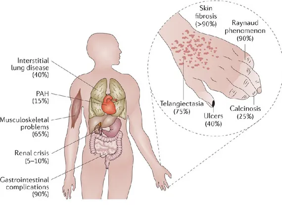
Systemic sclerosis can cause severe dysfunction and failure of almost any internal organ. Major organ involvement leads to decreased survival in SSc. Pulmonary fibrosis and pulmonary arterial hypertension (PAH) cause more than half of all SSc-related deaths. In addition, severe complications of the kidneys, heart, lungs, and gastrointestinal tract generally develop within 3 years of disease onset, particularly in patients with dcSSc [85].
Figure 1.1. Organ complications associated with systemic sclerosis. The uncontrolled
fibrosis and scaring of the skin and internal organs in systemic sclerosis leads to severe and sometimes life-threatening complications. The average frequency of the specific complications is indicated in parentheses. PAH, pulmonary arterial hypertension [2].
1.1.1. Epidemiology
The results of studies of prevalence and incidence estimates of SSc show a substantial variation across geographic regions. Prevalence and incidence of SSc appears to be greater in populations of European ancestry and lower in
Asian groups. Lower estimates of prevalence (<150 per million) and incidence (<10 per million per year) have been recorded in northern Europe and Japan, whereas higher estimates of prevalence (276-443 per million) and incidence (14-21 per million per year) have been reported in southern Europe, North America and Australia [7]. The 2013 revised classification criteria - the American College of Rheumatology (ACR)-European League Against Rheumatism (EULAR) criteria include patients with centromere-specific antibodies and limited cutaneous involvement. As a result, the estimated prevalence of SSc based on the ACR-EULAR classification criteria was much higher than previously published estimates [59].
Like other connective tissue diseases, SSc is sex dependent and occurs more common in women. Female/male ratio ranging from 3:1 to 14:1 and it occurs more frequently in the fourth or fifth decades of life. However, male sex has been consistently shown to be a poor prognostic factor in SSc. In addition, race and ethnic groups are related to distinct phenotypic profiles, and there is a trend towards less favorable outcomes in Africa American patients.
1.1.2. Endothelial Dysfunction in Systemic sclerosis
In SSc, the vasculopathy is one of the earliest pathological events, characterized by endothelial cell activation, and altered vascular tone. The fact that the vascular manifestations consistently precede tissue fibrosis suggests that microvascular endothelial cells (MVECs) are the primary target in this disease. The pathological alterations are accompanied by the presence of proinflammatory cytokines and angiogenic regulatory factors, and the loss of redox control. This complex interaction involves in a number of cells types, particularly the endothelial cells. The vascular endothelium provides a compatible interface to facilitate blood circulation as well as a multifunctional
interface between blood and all internal organs [1]. The vascular endothelium participates in regulating coagulation and fibrinolysis, permeability, vasomotion, and inflammation. The term endothelial dysfunction is most often used to symbolize impairment of endothelium-dependent vasodilation; however, a more expanded definition also includes endothelial interactions with leukocytes, platelets, and regulatory substances [3].
The most prominent clinical vascular dysfunction in SSc is linked to dysregulation of vascular tone leading to vascular spasm and reduced blood flow. An imbalance between the levels of vasoconstrictor and vasodilator mediators is attributed for endothelial dysfunction in SSc. Among these vasodilator agents, nitric oxide (NO), produced by endothelial cells (ECs) is one of the most potent vasodilator. In SSc, the defects in NO production by endothelial nitric oxide synthetase reduce microvascular endothelial cells (MVECs) survival and promote apoptosis. Moreover, endothelin-1 (ET-1), a potent vasoconstrictor peptide originally isolated from ECs is upregulated in SSc sera and tissues. The imbalance between vasodilator and vasoconstrictor signals leaves MVECs vulnerable to apoptosis and promotes an environment of ischemia, hypoxia, and profibrosis.
Hypoxia is a possible trigger that mediates MVECs lesions. Hypoxia followed by reperfusion induces an inflammatory process and oxidative stress that leads to cellular injury. Human MVECs exposing to intermittent hypoxia induced the dysfunction of MVECs, associated with an increase in oxidative stress. Intriguingly, using antioxidants prevents the effect of hypoxia on MVECs.
are formed from preexisting vessels and de novo vessels, respectively. These defects indicate defective role of MVECs differentiation in two processes. Of interest, vascular endothelial growth factor (VEGF) is a very potent angiogenic factor that is overexpressed in skin and sera of SSc patients, but there is evidence suggesting that VEGF receptor may be impaired in the cells based on the observation that MVECs isolated from SSc patients show reduced response to VEGF in vitro. In addition, failed release of bone marrow-derived progenitor cells, which have the potential to initiate vascular repair has been identified in SSc patients. The failure to compensate for vascular injury eventually leads to accumulative loss of capillaries and arterioles, resulting in the well-known vascular complications: painful ulcers on the digits, scleroderma renal crisis occurs in the kidneys, and PAH occurs in the lungs [81].
There appears to be a complex cross talk between MVECs, fibroblasts, and leukocytes, especially in early phases of SSc. It is suggested that junctional adhesion molecules (JAMs) are important elements in this interaction. The levels of JAM-A are unregulated in MVECs especially in early stage SSc, and the soluble JAM-A and JAM-C levels are higher in early stage SSc [80]. This underlines the role of JAMs in activating MVECs, particularly in early stage SSc in which there are more inflammatory infiltrates around the blood vessels.
Finally yet importantly, MVECs may activate fibroblasts and the immune system. As a result, activated fibroblasts precede the overproduction of collagen and other compartments of extracellular matrix (ECM).
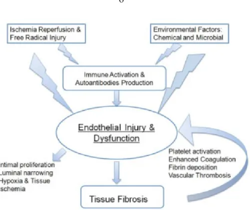
Figure 1.2. Pathogenesis of SSc vasculopathy. Endothelial injury and dysfunction are
initiated by the actions of free radicals or chemical and microbial agents that injure the endothelium, either directly or indirectly. This injury is also initiated by the induction of immune activation and the generation of autoantibodies and activated cellular immunity. The vascular injury activated platelet and coagulation pathways, which results in vascular microthrombosis. The resulting vasculopathy is associated with intimal hyperplasia in the small arterioles, and the ensuing luminal narrowing results in tissue hypoxia and chronic ischemia. Released vascular products, in association with hypoxia and ischemia, collectively contribute to the activation of resident fibroblasts, which in turn perpetuates the vasculopathy by triggering vascular wall fibrosis [81].
1.1.3. Raynaud’s Phenomenon
Raynaud‘s Phenomenon (RP) is the clinical manifestation of cutaneous vessel dysfunction involved thermal regulation of blood flow [124]. This exaggerated respond of the cutaneous circulation to cold exposure results in vasospasm and the characteristic pallor of the skin [39]. Its classical presentation involves the fingers turning white (ischemia), then blue
patients, it is usually the first symptom of disease, and it begins before the onset of clinical signs of tissue fibrosis. RP occurs when the delicate balance between vasodilation and vasoconstriction is disturbed, in favor of vasoconstriction. The presence of RP and dysregulation of cutaneous vascular tone is a predictor of developing SSc. The locations of the most severe skin fibrosis follow the same distribution patterns as typical cutaneous body sites involved in thermoregulation (e.g., the fingers, feet, face, and lower arms), suggesting some causative relationship between the vascular disease and skin fibrosis.
Most research into the changes in vascular function in RP has so far focused on the endothelium. It has been suggested that RP in SSc patients may be triggered by endothelial injury [67] (Figure 1.2). Endothelial activation and/or damage will upset the balance between vasodilation and vasoconstriction leading to underproduction or reduce efficacy of vasodilators and/or overproduction of vasoconstrictors. While the exact mechanism for the initial endothelial injury is unknown, apoptosis induced by infection, immumed-mediated cytoxicity, antiendothelial antibodies, and ischemia-reperfusion injury have all been implicated. In addition to the imbalance of vasoactive factors, including overproduction of the vasoconstrictor ET-1 and underproduction of vasodilator NO and prostacyclin, there is proposing evidence of dysregulation of a variety of neurotransmitters and their receptors that regulate small sensory nerve fibers as sympathetic vasoconstrictor and vasodilator nerves [66]. Decreased release of vasodilatory neuropeptides from sensory nerves and up-regulation of vascular smooth muscle α2C -adrenoreceptors that enhance vasoconstrictive responses to stress or cold stimuli are implicated in the dysfunction of the thermoregulatory vessels leading to RP. Patients with RP who develop SSc exhibit unique characterized
by intimal thickening that can eventually occlude the vessel lumen, thus causing ischemic injury and chronic tissue hypoxia (Figure 1.3). Skin hypoxia has been documented in SSc patients and is a potent stimulus for growth factors that mediate tissue fibrosis [10], suggesting that vascular damage is the primary insult, which then provokes tissue fibrosis.
Figure 1.3. Masson‘s trichrome staining of a digital artery from a patient with SSc (A) and
hematoxylin and eosin staining of a renal artery from a patient of SSc (B). Note of striking fibrotic intimal hyperplasia and the adventitial fibrosis in the digital artery and the onion skin-like intimal thickening composed of smooth muscle cells and increased connective tissue matrix in the renal artery. The intimal hyperplasia results in critical luminal narrowing and even occlusion [81].
1.1.4. Pulmonary arterial hypertension in systemic sclerosis
Pulmonary arterial hypertension, defined by an elevated mean pulmonary arterial pressure (mPAP) greater than 25 mmHg and a pulmonary capillary wedge pressure less than 15 mmHg, is a progressive disorder involving the pulmonary vasculature that leads to right heart failure and ultimately death [82]. PAH is a serious complication of SSc with prevalence approximately 15%. SSc- associated PAH (SSc-PAH) is a leading cause of PAH, presents
around 15-30% in all PAH. SSc-PAH has a dramatic impact on clinical course and overall survival, and remains a major cause of mortality in SSc. In the mechanism of SSc-PAH pathogenesis, inflammation is considered as a pathological hallmark in which inflammatory cells infiltrate in pulmonary perivascular spaces within and around plexiform lesions. On the other hand, the role of growth factors such as TGF-β and VEGF in the development of SSc-PAH is also indicated in the study of Derrett-Smith et al. [27]. Whether this inflammatory process precedes a state of imbalance between vasoactive, growth factors and proliferative mediators leading to abnormal regulation of endothelial and smooth muscle cells as well as fibroblasts, and vascular dysfunction is not elucidated [37].
Endothelial dysfunction seems to play an integral role in mediating the structural changes in the pulmonary vasculature. Disordered endothelial cell proliferation along with concurrent neoangiogenesis, when exuberant, results in the formation of glomeruloid structures known as the plexiform lesions, which are common pathological features of the pulmonary vessels of patients with PAH. Additionally, an altered production of various endothelial vasoactive mediators, such as NO, prostacyclin, ET-1, serotonin, and thromboxane, has been increasingly recognized in patients with pulmonary hypertension. Because most of these mediators affect the growth of the smooth muscle cells, an alteration in their production may facilitate the development of pulmonary vascular hypertrophy and structural remodeling characteristic of pulmonary hypertension. It is conceivable that the beneficial effect of many of the treatments currently available for PAH, such as the use of prostacyclin, NO, and ET antagonists, result at least in part from restoring the balance between these mediators. However, the ultimate cellular and physiological targets of these treatments remain unknown [16].
In addition to the potential consequences of an imbalance in the endothelial production of various mediators, injury to the endothelium may expose the underlying vascular tissue to diverse blood-borne factors that may further promote pathological changes. Endothelial dysfunction may also have adverse consequences on pulmonary vascular hemostasis by altering the production of anticoagulant factors. Recent reports of genetic mutations in the endothelial cells of patients with pulmonary hypertension further underscore the role of these cells in the disease pathogenesis [16].
SSc-PAH shares similar histological features in IPAH, including intimal hyperplasia, medial hypertrophy, adventitial fibrosis, and inflammatory infiltrates. However, there are fewer plexiform lesions, increased intimal fibrosis, more heterogeneity, and a higher prevalence of venoocclusive lesions when compared with IPAH [45].
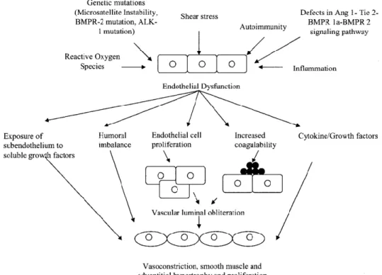
Figure 1.4. Mechanistic role of endothelial dysfunction in pulmonary hypertension and
1.1.5. Collagen deposition
Fibrosis represents the phenotypic expression (typified by skin thickening and interstitial lung fibrosis) of SSc. Patients with the disease have increases in collagen types 1 and 3, with type 1 being the most abundant. Type 1 collagen is encoded by the COL1A1 and COL1A2 genes, which are at least partly controlled by the transcription factor SP1. Increased SP1 binding activity has been recorded in sclerodermic fibroblasts and its activity has shown to be associated with increased gene expression of type 1 collagen in patients with SSc. Gene expression of type 1 collagen is also affected by TGF-β, which indicates a possible synergistic profibrotic interaction between SP1 and the TGF-β pathway via the SMAD3/4 complexes. Reduced amounts of SMAD7 (an inhibitor of collagen gene expression) have been reported in SSc, which suggests that the loss of this inhibitory effect allows TGF-β to stimulate unfettered, excessive accumulation of extracellular matrix.
1.1.6. Therapeutic targets in systemic sclerosis
Conventional treatments for SSc
Among the conventional therapies used for the treatment of SSc (figure 1.5), randomized trials have demonstrated a possible effect for methotrexate and extracorporeal photopheresis in improving skin thickening, as well as a role for cyclophosphamide in stabilizing or even slightly improving interstitial lung disease [24, 69]. Calcium channel blocker mostly contribute to the management of SSc peripheral vascular disease [117], while angiotensin converting enzyme (ACE) inhibitors were proven effective against renal impairment [113]. The prostacyclin analogues significantly improved pulmonary arterial hypertension associated with SSc [4] as well as peripheral vascular disease. The use of D-penicillamine as an antifibrotic agent in SSc is
not supported by the results of a randomized trial that compared high to low doses of this drug [21]. Other medications and therapeutic procedures such as cyclosporine A, sirolimius, tacrolimus, antithymocyte globulin, prazosin, and ketanserin have also been used as a therapy for SSc without achieving solid evidence of benefits [14].
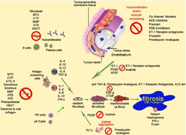
Figure 1.5. Summary of the pathogenetic processes leading to fibrosis in SSc and
mechanism of action of some of the commonly used drugs. PDGF platelet-derived growth factor, ET-1 endothelin-1, IL interleukin, TGF-β transforming growth factor-β, CTGF connective tissue growth factor, bFGF fibroblast growth factor, ECM extracellular matrix,
α-SMA alpha-smooth muscle actin, IFN-γ interferon-γ, MTX methotrexate, CYP
cyclophosphamide, ATG antithymocyte globulin, HSCT hematopoietic stem cell transplantation, IVG intravenous gamma globulin, MMF mycophenolate mofetil, CYC A cyclosporine A, D-pen D-penicilllamine, PDE-5 phosphodiesterase-5, ACE angiotensine converting enzyme, PLTs platelets [14].
Therapeutic target on vascular dysfunction
As stated previously, endothelial damage and vascular dysfunction could be one of the earliest alterations in SSc [107]. The greatest advances have been in the management of vascular complications. Angiotensin converting enzyme (ACE) inhibitors has been accepted as the preferred choice in management of this rapidly fatal complication of SSc. Their mechanism of action has been well known (even in the late 1970s) and thought to be especially appropriate to treat a hypertensive state that was associated with, and driven by, elevated amounts of renin (and thus angiotensin and aldosterone) [19]. A review by Steen and colleagues, based on the Pittsburgh longitudinal database, records that 1-year survival is better than 70% in renal crisis patients treated with ACE inhibitors, but less than 20% in those not treated with ACE inhibitors.
Regarding SSc-associated PAH, some drugs have been approved by US Federal Drug Administration (FDA) due to the improved knowledge of molecular biology. Epoprostenol, treprostinil, and iloprost can supply prostacyclin that the pulmonary vascular endothelium itself no longer supplies in adequate quantities; sildenafil can increase amounts of NO in tissue; and bosetan can inhibit endothelin, which is overproduced in the serum of patients with PAH. These drugs improve the symptoms of PAH as well as the quality of life of patients with SSc-associated PAH. Increasing evidence suggests that the two treatments that have been approved the longest (epoprostenol and bosetan) could improve survival in PAH overall. Several other treatments with good molecular-based rationale have been investigated: inhaled treprostinil (a prostacyclin-like compound), inhaled nitric oxide, and other endothelin inhibitors such as sitaxsentan [46, 83]. Calcium channel blockers (especially nifedipine), intravenous iloprost and epoprostenol, and
nitroglycerin paste (oilment) are effective in lessening RP and, to a lesser extent, in healing digital ulcers. Sildenafil has also shown some effectiveness in a small placebo-controlled trial of patients with secondary RP resistant to multiple therapies [19]. A study conducted by Sfikakis et al. showed small doses of bosentan improve endothelial function without affecting hemodynamic parameters or endothelial activation-related processes, thus supporting a direct, reversible effect of endothelin in SSc-associated vascular injury [110].
Iloprost in vasodilator therapies
The digital arteries of patients with SSc exhibit marked fibrotic intimal hyperplasia and luminal narrowing. Normal vasoconstrictor responses to cold, superimposed on this anatomic obstruction, could cause occlusion of the arterial lumen. Vasodilators that inhibit cold induced vasoconstriction might prevent this vessel closure. Alternatively, vasodilator therapies might be predicted to be minimally effective in the setting of fixed vascular obstruction [29].
Iloprost is a stable analogue of prostacyclin that is associated with a longer duration of vasodilatation [38]. It is widely used for the treatment of vascular dysfunction in systemic sclerosis due to its well-known vasodilator and anti-aggregant effect [36].
According to the recommendations of the European League against Rheumatism (EULAR) for the treatment of systemic sclerosis, iloprost is effective in reducing the frequency and severity of SSc-RP. Iloprost, given intravenously (0.5–3 ng/kg per minute for 3–5 consecutive days sequentially) or orally (50–150 mg twice a day) significantly reduced the frequency of ischaemic attacks, and improved the RP severity score in comparison with
placebo. Oral prostanoids seem to be generally less effective than intravenous iloprost in the treatment of SSc-RP, although some beneficial effects could be seen with higher doses. Intravenous prostanoids (particularly intravenous iloprost) are efficacious in healing digital ulcers in patients with SSc. Intravenous prostanoids (in particular iloprost) should be considered in the treatment of active digital ulcers in patients with SSc [71].
Iloprost may promote repair of damaged endothelium, which could explain vascular effects and clinical improvement weeks after [29]. Dole et al. showed that iloprost can improve healing of cutaneous lesions including ischemic ulcers in patients with SSc. The ischemic lesions occur because of a vasculopathy characterized histologically by abnormal vascular endothelium and intimal thickening, and clinically by vasopasm. The benefit of iloprost on tissue healing may in part be explained by the potential capacity of prostacyclin to inhibit platelet activation and leukocyte adherence to the endothelium thus reducing counteracting defective endothelial function and tissue injury [29].
1.2. Oxidative stress and systemic sclerosis
Reactive oxygen species (ROS) is an expanded term from free radicals which have been defined as species capable of independent existence that contains one or more unpaired electrons in atomic or molecular orbitals [91]. These unpaired electrons make free radicals highly reactive in which superoxide radical (O2 ˉ), hydrogen peroxide (H2O2), hydroxyl radical ( OH), hypochlorous acid (HOCl) and peroxynitrite (ONOO-) are common oxidative molecules in the ROS family [87]. With excessive levels, ROS are addressed oxidative stress and cause cellular damage and cell transformation.
Several sources of ROS mentioned to contribute to oxidative stress in fibrotic processes of SSc. Ischemia-reperfusion is a main cause leading to tissue
damage. In addition, the interaction of cytokines and growth factors with their receptors can induce ROS in SSc. Among those, TGF-β is a pivotal profibrotic cytokine, which expressed the key role in pathogenesis of SSc. Some other significant factors involved are platelet derived growth factors (PDGF), vascular endothelial growth factor (VEGF) [12], connective tissue growth factor (CTGF), angiotensin II, interleukin 3, interleukin 6, tumor necrosis factor-alpha (TNF-α), nerve growth factor, fibroblast growth factor. Angiotensin II, PDGF and TGF-β1 can interact with members of the nicotinamide adenine dinucleotide phosphate oxidase (NOX) family in vascular smooth muscle cells, cardiac, lung and skin fibroblasts [42]. The increased level of superoxide from fibroblasts and monocytes also contributes to abundant sources of ROS in SSc [30].
RP occurring in almost all SSc patients, approximately more than 90% [58], presents the oxidative pathway beside the nonoxidative pathway in pathology of this phenomenon associated with SSc [51]. There is evidence showing induced effects on SSc-associated Raynaud‘s phenomenon. The level of 8-isoprostane, a reliable biomarker of oxidative stress and antioxidant deficiency, has been shown as a valid measure of lipid peroxidation in vivo [89]. It is correlated with the extent of vascular lesion in RP and the severity of fibrosis in SSc patients [94, 114]. Free radical nitric oxide (NO) released by endothelial cells is considered as a beauty in the protective role in controlling vasculature and regulating blood pressure as well as acting as antithrombotic and cytoprotective agent. However, in RP, during reperfusion, free radials and NO produced overwhelmingly lead to peroxynitrate which precedes oxidative vascular damage and endothelial apoptosis, therefore NO is considered as a beast [17, 51].
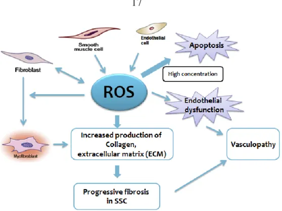
Figure 1.6. Schematic diagram of ROS-induced fibrotic process in SSc.
Abundant studies have shown the relevance of ROS in pathogenesis of SSc in
vitro and in vivo [57, 94, 105, 109]. Oxidative stress is considered as one of
the main background mechanisms contributing to the progress and destruction of this disease, concomitantly clinical manifestations regarding to SSc [51]. Skin and visceral fibroblasts spontaneously produce large amounts of ROS that initiate collagen synthesis [51]. Recently, a preliminary study of Boin et al showed substantially increased intracellular ROS levels in human pulmonary artery smooth muscle cells (HPASMCs) treated by sera of SSc patients with pulmonary artery hypertension (PAH). Employing NOX2ds-tat (gp91ds-tat), a specific inhibitor of NOX2, can effectively reduce PAH-SSc sera -induced ROS, implicating the involvement in the mechanism of increasing ROS of SSc. Exposure of HPASMCs to SSc-PAH sera also resulted in progressive time-related increase of the Collagen promoter activity. Similarly, this effect was inhibited by NOX2ds-tat treatment, suggesting the collagen synthesis activation in HPAMSCs may be driven by
SSc-related PAH sera through NADPH oxidase-dependent ROS generation. Taken together, NADPH oxidase-derived ROS mediates the activation of collagen synthesis [13]. In vivo studies, exploiting BALB/c mice, the SSc murine model injected HOCl every day for 6 weeks induced chronic oxidative stress. As a result, this HOCl-induced oxidative stress operated cutaneous and lung fibrosis in SSc mice. Furthermore, HOCl-treated mice also overexpressed α smooth muscle actin (α-SMA), a biological marker characterized by activated myofibroblasts. These processes occurred through ADAM17/Notch pathway or Ras-ERK pathway or guanosine triphosphate (GTPase) intracellular signaling [5, 6, 68]. Blocking the activation of the Ras-ERK pathway by propylthiouracil (PTU) or simvastatin can prevent the pulmonary fibrosis and cutaneous fibrosis in oxidant-stress animal model of SSc [5, 6].
Some studies reported that anomalous overproduction of ROS activates fibroblasts linking to the increased expression of stimulatory serum autoantibodies to the PDGFR in SSc [8]. While prooxidants may cause the surging of autoantibodies, other studies showed the uncorrelated relation between autoantibodies in endothelial cells and fibroblasts and serum-induced ROS or cell proliferation. Nonetheless, it is undeniable the role of autoantibodies in the vast damage in SSc in which anti-endothelial cell antibodies (AECA) were closely correlated with pulmonary fibrosis with SSc [51, 61]. Oxidative stress may either directly activate ROS-induced differentiation of fibroblasts into myofibroblasts [68] or disrupt the balance between protease and protease inhibitors or both. With the latter mechanism, on the one hand, TGF-β upregulates the expression of extracellular matrix proteins including collagens, on the other hand, TGF-β suppresses protein
plasmin activator inhibitor 1 (PAI-1) and tissue inhibitors of metalloproteinases (TIMPs) [77]. This protease-antiprotease imbalance is likely to be a critical mechanism in the development of SSc lung fibrosis [43].
1.3. Role of endothelial NADPH oxidases in SSc
NOXs are the only enzymes with the primary function of generating ROS [31, 73]. Other enzymes (cyclooxygenases, cytochrome P450, enzymes of the mitochondrial electron transport chain) can produce ROS, but only as a byproduct of their normal function. Of note, these latter enzymes only become ROS-generators in the presence of an external source of ROS (such as NOXs) and can thus be considered as secondary sources of ROS [32]. NOXs catalyze the transfer of electrons from cytosolic NADPH to molecular O2 via their membrane-bound catalytic NOX or DUOX subunit to generate the ROS, superoxide or H2O2. NOX-derived ROS may promote ROS formation from other (normally dormant) enzyme sources, setting up a vicious cycle that further exacerbates vascular oxidative stress [78, 86, 92]. Accumulative evidence highlights the involvement of NADPH oxidase-dependent redox signaling in the profibrotic responses mediated by TGF-β [104].
Seven isoforms of NOX have been described in mammals, each distinguished mainly by the identity of its catalytic subunit. They are the ‗NOXs‘: NOX1-, NOX2- [formerly gp91phox; also known as CYBB (cytochrome b245, b polypeptide)], NOX3-, NOX4-, and NOX5-containing oxidases; and the ‗DUOXs‘ (‗dual oxidases‘): DUOX1- and DUOX2-containing enzymes. Four of the NOXs – NOX1, NOX2, NOX4, and NOX5 – are expressed in endothelial cells.
Of the four endothelial NOXs, three (NOX1, NOX2, and NOX4) are known to exist as multimeric protein complexes, whereby electron transfer occurs through the ‗NOX‘ subunit in association with different combinations of regulatory subunits. Figure 1.7 illustrates the molecular structures of the endothelial NOXs.
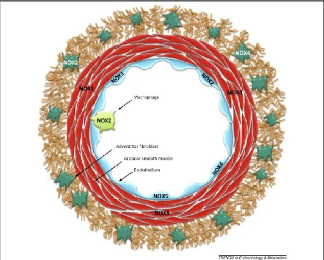
Figure 1.7. NADPH oxidase (NOX) isoform expression in the various cell types of the
blood vessel wall. Four NOX isoforms are expressed in the vascular wall, including NOX1 (in endothelial cells and vascular smooth-muscle cells, VSMCs), NOX2 (in endothelial cells, adventitial fibroblasts, and leukocytes such as monocytes, macrophages, and platelets), NOX4 (in endothelial cells, VSMCs, and adventitial fibroblasts), and NOX5 (in endothelial and VSMCs – not expressed in rodents) [32].
NOX2 was the first NOX isoform to be identified in endothelial cells and, as shall be discussed below, is likely to be the most important in the context of vascular pathology.
NOX4 is the most highly expressed NOX homolog in endothelial cells. It also requires p22phox, but apparently does not associate with Rac1 or any of the cytosolic organizer or activator proteins needed for activation of NOX1 and NOX2 [32]. Current evidence suggests that Nox4 may have a pivotal role in mediating TGF-β-induced profibrotic responses [104]. TGF-β increased Nox4 gene expression without effects on Nox1, Nox2, or Nox5 in human cardiac fibroblasts [22]. Treatment with siRNA against Nox4 suppressed expression of TGF-β target genes including fibronectin, collagen I, α-smooth muscle actin (α-SMA, a marker of myofibroblast), and connective tissue growth factor, indicating that Nox4 was involved in TGF-β-induced differentiation of cardiac fibroblasts to myofibroblasts [22]. In renal tubular epithelial cells, Rhyu et al. showed that inhibition of NADPH oxidase activity with the non-selective Nox inhibitor DPI suppressed EMT and matrix protein production [100].
NOX5 appears to be the only endothelial NOX that can produce ROS in the absence of other ‗phox‘ or Rac subunits. NOX5 is also unique among NOX isoforms in that it contains an N-terminal calmodulin-like domain with four binding sites for Ca2+ (EF hands). Thus, endothelial NOX5 activity can be directly modulated by changes in intracellular [Ca2+] [9].
Figure 1.8. Molecular composition of the endothelial NADPH oxidases (‗NOXs‘). All four
endothelial NOX isoforms are comprised of a catalytic ‗NOX‘ subunit, which may reside in the nuclear, endoplasmic reticulum (ER), or plasma membranes. For NOX1, 2, and 4, additional subunits are required for full enzyme activity. By contrast, NOX5 is a ―stand alone‖ protein whose activity is regulated by Ca2+ binding. The topology of the NOXs is such that NADPH binds to the cytoplasmic domains of the proteins with reactive oxygen species (ROS) generation always occurring on the opposite side of the membrane (i.e., within the nucleus, ER, or the extracellular compartment). It is also noteworthy that whereas NOX1, 2, and 5 generate superoxide, NOX4 appears to generate hydrogen peroxide [32].
1.4. Endothelial-to-Mesenchymal Transition and Systemic Sclerosis
Pathogenesis of systemic sclerosis constitutes numerous complex mechanisms associated with (i) microvascular fibroproliferative lesions, (ii) innate and adaptive immune system abnormalities and uncontrolled accumulation of collagen, (iii) other extracellular matrix compartments produced by
fibroblasts and activated myofibroblasts in skin and various internal organ [64, 95]. In a non-pathological condition, myofibroblasts usually disappear through apoptosis and through transition to a quiescent/senescent state at the later stages of wound healing. However, the persistence of myofibroblasts under an activated phenotype in EndMT contributes to progressive fibrogenesis [90, 111]. The consequence of this process is causing interstitial and perivascular fibrosis in the lungs, heart, kidneys and other organs responsible for the high mortality of SSc patients [11]. The origin of activated myofibroblasts in SSc has been demonstrated from various sources including pericytes and smooth muscle cells (SMCs) from vessel walls, resident fibroblasts, bone marrow-derived fibroblasts [60, 64, 126]. With large amounts of studies, Epithelial-to-Mesenchymal Transition is a well-known source of activated myofibroblasts by which epithelial cells transforms into myofibroblasts. Although it is not extensively studied as EMT, more recent research has been conducted to investigate the mediators and signaling pathways in mechanisms of differentiation of endothelial cells to mesenchymal transition. EndMT is considered as a special form of EMT, vascular endothelial cells share several similar characteristics and molecular mechanisms with epithelial cells to generate fibroblasts and myofibroblasts [74, 99]. EndMT contributes to fibrotic pathogenesis of cardiac fibrosis, pulmonary fibrosis, renal fibrosis, idiopathic portal hypertension, liver fibrosis, and obviously systemic sclerosis [54, 64, 106, 115, 127].
Figure 1.9. Multiple origins of myofibroblasts [126].
Under the conversion of endothelium into mesenchyme, endothelial cells undergo morphological alterations and loss of cell-surface markers. In cell culture, endothelium acquires mesenchymal, fibroblast-like properties such as spindle-shaped morphology, migratory capacity, invasiveness, and enhanced resistance of apoptosis [60, 120]. During EndMT process, the structure of vessel-lining is disrupted due to resident endothelial cells disaggregating from the organized layer of cells on vessel walls and invading the surrounding tissue [76, 97, 120]. Cell-surface markers like vascular endothelial cadherin (VE-Cadherin), CD31 (platelet endothelial cell adhesion molecule-1, PECAM 1) or von Willebrand factor (vWF) are gradually replaced by markers characterized for mesenchyme such as fibroblast specific protein-1 (FSP-1), α-SMA, vimentin and type I and type III collagen [54, 97].
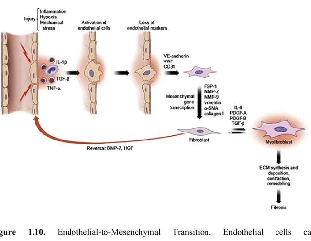
Figure 1.10. Endothelial-to-Mesenchymal Transition. Endothelial cells can
transdifferentiate into fibroblasts under the influence of several factors. This transition is accompanied by the progressive loss of typical endothelial cell markers (VE-cadherin, vWF, CD31) and the transcription of typical mesenchymal cell markers (FSP-1, MMP-2, MMP-9, vimentin, α-SMA, collagen I). This process can be reverted by the administration of BMP-7 or HGF. Under the effect of variety of mediators, fibroblasts differentiate into myofibroblasts which leads to ECM and collagen synthesis [102].
Studies demonstrated the presence of transitional EndMT cells in the pulmonary vasculature of patients with SSc-PAH. The expression of cell surface markers specific for endothelial cells in mesenchymal/fibroblastic cells or vice versa from SSc-PAH patients [48, 97]. Treatment of IL-1β, TNF-α and TGF-β in combination abolished cobblestone structural characteristics and enhanced spindle-like appearance on pulmonary artery endothelial cells. Induced endothelial-mesenchymal transition (I-EndMT) cells exhibit an elevated secretion of proinflammatory proteins and collagen type I. In addition, the presence of I-EndMT cells in cellular barriers leads to substantially
increased paracellular and transcellular permeability which is considered as one of the earliest signs of vascular dysfunction in SSc [43, 48].
The mechanism of EndMT in pathogenesis of SSc vasculopathy is postulated to be induced by synergistic and complex activation of a large variety of cell types exposing to locally various biological mediators, such as inflammatory cytokines, growth factors [54, 95]. However, the precise molecular mechanisms of EndMT process mediated by those mediators and signaling pathways have not been entirely illuminated. Among these mediators, TGF-β has been implicated as a key role in the initiation of EndMT by numerous evidence [23, 49, 64, 90, 97, 120].
TGF-β can either solely activate endothelial cells or synergize with TNF-α and IL-1β to induce EndMT. Maleszewska et al (2013) demonstrated that the treatment with either TGF-β2 or IL-1β resulted in alterations of endothelial cells morphology, followed by a spindle-shaped phenotype. Nevertheless, under a co-stimulation condition the morphological changes were more explicit than in the case of single-factor stimulation [79]. Similarly, Good et al (2015) showed evidence that a combination of TGF-β with IL-1β and TNF-α stimulated the induction of EndMT in pulmonary artery endothelial cells. Notably, the withdrawal of IL-1β, TNF-α, and TGF-β after 6 days failed to convert induced EndMT (I-EndMT) cells to normal structural characteristic. It indicated that phenotypic change was permanent and was not reversible [48, 99]. This observation was in agreement with previous studies that EndMT would be consistent in the cells exposed to activated Ras and TGF-β treatment [53]. However, other reports showed that some anti-fibrotic mediators could dedifferentiate established myofibroblasts. This raises enormous hope for regenerative medicine in the reversal of the EndMT
process, which provides a new intriguing therapeutic target for fibrotic diseases [44, 126].
There are some signaling pathways involved in EndMT in which TGF-β plays a critical role. Both Smad-dependent and Smad-independent pathways are associated in intracellular cascades activated by TGF-β during endothelial-mesenchymal transition. Smad proteins have been shown to bind directly to the promoter of the Snail gene to regulate its transcription [23] . Snail-1, a transcriptional repressor, is crucial for TGF-β-induced mesenchymal transdifferentiation of embryonic stem cell-derived ECs. Snail-1 is a zinc-finger transcription factor that forms a complex with Smad3/Smad4. The active Smad3/Smad4/Snail-1 complex results in potent inhibition of the expression of E-cadherin by directly integrating into specific sequences within the gene promoter and blocking its transcription. In addition to inhibition of E-cadherin, Snail-1 precedes transcriptional events that lead to the expression of a mesenchymal-cell-specific phenotype [64]. In the Smad-independent pathway of TGF-β signaling, there is an involvement of important kinases such as c-Abl protein kinase (c-Abl), protein kinase Cδ (PKC-δ), and glycogen synthase kinase 3β (GSK-3β). GSK-3β is a crucial factor, whose phosphorylation will cause its inhibition and induce the strong up-regulation and subsequent translocation of Snail-1 into the nuclear. In the end, this process increases the expression of mesenchymal cell-specific marker such as α-SMA, type I collagen while reduce the expression of VE-cadherin characterized for endothelial cells. Using inhibitors of c-Abl and PKC-δ, imatinib and rottlerin respectively can inhibit the phosphorylation of GSK-3β, hence; abolish the transdifferentiation of ECs into myofibroblasts. This intervention could be a potential therapeutic target to obliterate the acquisition of the myofibroblastic phenotype through EndMT process [75].
TGF-β2 signals through MEK, PI3K, and p38MAPK pathways are also essential for ECs undergoing EndMT transition [84]. In addition, regulation of EndMT by Wnt [123] and NOTCH signaling [93], Caveolin-1 signaling [25, 26] is indicated to precede endothelial-mesenchymal transition regarding to TGF-β activity.
Other cytokines and growth factors might also play important roles in endothelial-mesenchymal transition such as PDGF, VEGF, endothelial-1 (ET-1), CTGF, MicroRNAs (miRNAs) [64].
1.5. Reactive oxygen species and Endothelial-to-Mesenchymal Transition
With numerous profound studies of the effects of oxidative stress on endothelial dysfunction, ROS have been highlighted in the process of epithelial-mesenchymal conversion [88]. Previous reports have demonstrated that EMT is closely relevant to ROS [74]. Recently, it has been reported that oxidative stress induces the EMT in renal tubular epithelial cells, human epithelial keratinocytes, and lung epithelial cells [28, 40, 101]. An emerging issue is whether there is a similar tendency towards EndMT.
1.5.1. ROS activate/mediate TGF-β-dependent signaling pathways in EndMT.
β is a multifunctional protein including three isoforms (β1, TGF-β2, and TGF-β3) which accounts for the regulation of cell proliferation, differentiation, apoptosis, adhesion, and migration. It therefore shows profound systemic effects on physiological functions [63, 77]. TGF-β stimulates the generation of ROS in various types of cells while ROS activate TGF-β and mediate effects of TGF-β. TGF-β elevates ROS production via NOX4, mitochondria, and microsomes. On the other hand, ROS induce TGF-β gene expression and activate TGF-TGF-β through oxidizing latency association
protein (LAP) or activating MMPs, which in turn release LAP [77]. It is well known that TGF-β is synthesized as a non-active pro-form combining with latency association protein (LAP) to create a latent complex. ROS oxidizing Redox center of LAP can trigger a conformational change resulting to the release of TGF-β1 [65]. Hence, under the stimulation of ROS, TGF-β is activated and increasingly expressed.
It is well documented that TGF-β induced EndMT process in cardiac, pulmonary fibrosis, renal, intestinal and skin fibrosis [20, 53, 54, 103, 127] whereas NADPH oxidases and ROS molecules may have been indicated to play a pivotal role in mediating TGF-β induced fibrotic responses via Smad2/Smad 3 activation [63]. Of note, NOX4-dependent generation of H2O2 is required for TGF-β1-induced myofibroblasts differentiation and elevated ECM production [55, 62].
A recent study of Montorfano group used human umbilical vein endothelial cells (HUVECs) to investigate the role of ROS as well as the underlying mechanism in the conversion of ECs into myofibroblasts. They demonstrated that oxidative stress is a crucial factor in generating the phenotypic conversion of ECs into activated myofibroblasts with an EndMT-like mechanism via a TGF-β1 and TGF-β2-dependent pathway. In addition, this study showed that ROS-induced oxidative stress acted on the conversion through the ALK5/Smad3/NF-кB intracellular pathway. The main point of this study supporting the hypothesis that ROS, TGF-β and EndMT is in an interactional relation is H2O2 induced the expression and secretion of TGF-β1 and TGF-β2, which has been demonstrated to induce the conversion of ECs into myofibroblasts [87]. Another recent study provided evidence that lipopolysaccharide-induced ROS could lead to an EndMT-like process through an ALK5 activity-dependent mechanism [33]. In line with this
observation, Toshio group (2015) also showed novel evidence that endotoxaemia-derived oxidative stress might be one of the promoters of TGF-β-mediated EndMT in pulmonary vascular endothelial cells [118]. This evidence was strongly supported with the newer study by the same authors that the expression of TGF-β1 and TGF-β2 is crucial for the development of the endotoxin-induced endothelial fibrosis mechanism. This increase of endotoxin-induced TGF-β1 and TGF-β2 expression required the activation of NOX and the subsequent generation of ROS [34]. Taken together, oxidative stress is likely a link to mediate the EndMT process induced by TGF-β.
1.5.2. Oxidative stress and EndMT in SSc: is there a link?
Despite the fact that the role of ROS has been well defined in many interstitial organ fibrosis as well as strong there being strong evidences displaying the close relationships between ROS and SSc, EndMT and SSc (stated above) via numerous relevant papers, few studies have conducted experimental research indicating the effects of oxidative stress on EndMT process in SSc. A study conducted by Hao Xu group showed chronic oxidative stress mediates Endothelial-to-Mesenchymal Transition in a murine model of SSc [125]. They isolated microfibrils from the skin of tight skin (Tsk+/-) mice, which showed abnormal big fibrillin-1. EC cultures on this abnormal extracellular matrix leaded to the alterations of morphology and function of ECs, and increased 4-HNE, a well-known fission product of polyunsaturated fatty acid oxidation. There was a transdifferentiation from ECs to mesenchymal cells with the increased presence of FSP-1 and Twist (a transcription factor implicated the endothelial cell to fibroblast transition) and a decrease of VE-cadherin. Beside, abnormal big fibrillin expression was associated with oxidative stress (reduced nitric-oxide-to-superoxide anion ratio). This
D-4F, a peptide binding with high affinity to oxidized lipids attenuated or abolished EndMT. This indicated that the oxidized lipids might play a central role in the mechanisms by which other chronic states of oxidative stress promotes endothelial mesenchymal transition [125]. In this paper, the authors did not mention TGF-β, which has been well documented to be a crucial role in the initiation of EndMT during various diseases [64]. Of note, ECM including fibrillin functions as a reservoir for TGF-β and other growth factors to control mesenchymal differentiation [121]. Whether there is an interaction between TGF-β and oxidative stress to precede EndMT in this case is an intriguing question, which requires more investigation to figure out.
2. RESEARCH OBJECTIVES
Systemic sclerosis (scleroderma, SSc) is a complex multisystem autoimmune disease characterized by dysregulated immune system, progressive fibrosis of skin and visceral organs in which extensive vascular alterations are prominent and severe [41].
Despite of the enormous scientific effort in studying etiology and pathogenesis of scleroderma, an overall picture of its etiopathogenic mechanisms remains obscure. Oxidative stress-mediated vascular dysfunction is one of the important features of the detrimental vascular changes in SSc patients. Although the presence of a large excess of ROS has been supported by numerous studies, which documented the aberrant redox state and demonstrated the involvement of ROS in excessive extracellular matrix deposition, the implication of molecular machinery is still unclear [42]. Many clinical trials with antioxidants have been undertaken to inhibit the progression of SSc.
Iloprost, a stable prostacyclin analogue with vasodilating, anti-platelet, cytoprotective and immunomodulating properties, has been found to be an efficacious alternative to nifedipine for the treatment of SSc [108]. Some studies reported that iloprost infusion reduces oxidative stress and improves fibrosis in SSc patients [36, 108, 116]. However, the mechanism underlying this beneficial effect remains unelucidated.
Aside from Epithelial-to-Mesenchymal Transition (EMT), which has been extensively shown to occur in a variety of fibrotic diseases including SSc [18, 54, 70, 115], a potential role of Endothelial-to-Mesenchymal Transition (EndMT) in scleroderma has been indicated in some reports. EndMT may trigger the enhanced fibroproliferative vasculopathy and is considered as a
novel mechanism for the generation of activated myofibroblasts in SSc [64]. Consonant to what reported above, ROS mediate TGF-β-induced differentiation of myofibroblasts, which accounts for the increased production of fibrillar type I and type III collagen and other extracellular matrix (ECM) proteins, and triggers the expression of α-smooth muscle actin (α-SMA), a process strongly correlated EndMT [22, 64].
The research question of this study was to understand whether oxidative stress and EndMT could be part of the molecular machinery inducing vascular damage in SSc patients.
Therefore, we have been conducting this study with the following intents: 1. To investigate whether SSc sera can induce oxidative stress and collagen synthesis in HPMECs.
2. To examine the effect of intravenous iloprost infusion on SSc sera and how this alteration affects in HPMECs.
3. To investigate whether SSc sera can induce Endothelial-to-Mesenchymal Transition in HPMECs.
3. MATERIALS AND METHODS
3.1. Ethical approval
The study was approved by the Ethics Committee, from both the University of Sassari and the John Hopkins University, Baltimore (USA), and all patients enrolled were informed consent.
3.2. Patient selection
Serum samples were collected from patients with SSc at the Johns Hopkins Scleroderma Center, Division of Rheumatology, Johns Hopkins University, Baltimore, and at the Unit of Complex Rheumatology, Sassari. In Baltimore, nineteen patients were enrolled in the study.In Sassari, sixteen patients were enrolled in the study. The clinical and serological characteristics of these SSc patients in Baltimore and Sassari are summarized in table 4.1 and table 4.2, respectively.
All the patients with SSc had to fulfill the American College of Rheumatology (formerly, the American Rheumatism Association)-SSc classification criteria. Using these guidelines, patients with skin sclerosis limited to hands, forearms, legs below the knee, and face were defined as having limited scleroderma. Those with more extensive skin disease spreading proximal to elbows or knees or involving the trunk were defined as having diffuse scleroderma. The onset of the disease was defined as the time of the first non-Raynaud phenomenon.
The SSc patients from Sassari underwent a monthly intravenous administration by an infusion pump of the prostacyclin analogue iloprost, at the greatest tolerated dose (0.2 ng/kg/min, mean dose 1.3 ng/kg/min), for 5-6 hours/day. Blood samples were taken at two different time points. For each
patient, blood samples were taken at baseline and 5 hours after the cycle of iloprost infusion. Samples were stored at -800C until being assayed.
Previous medications were maintained during the course of the study; no other drugs were started during the study period.
Healthy donors were matched for gender, race, and smoking status.
3.3. Cells and Chemicals
Human pulmonary microvascular endothelial cells (HPMECs) were supplied by Innoprot, S.L, Spain.
Primary antibodies Rabbit polyclonal IgG anti-von Willebrand (Santa Cruz Biotechnology, INC); Goat Polyclonal Anti-Actin Alpha 2 Smooth Muscle Antibody (Novus Biologicals); β-actin antibody from Sigma-Aldrich (Saint-Louis, MO, USA); the HRP-conjugated secondary antibodies were from Santa Cruz Biotechnology, INC and Pierce Biotechnology-Thermo Scientific (Rockford, USA).
3.4. Cell culture
Human pulmonary microvascular endothelial cells (HPMECs) were cultured in endothelial cell medium (ECM) (Innoprot, S.L, Spain) supplemented with 5% of Fetal Bovine Serum (FBS), 1% of Endothelial Cell Growth Supplement (ECGS), and 1% of Penicillin/Streptomycin solution (P/S solution). The cells were maintained at 370C and 5% CO2 in a humid environment.
3.5. Measurement of intracellular ROS
Intracellular ROS levels were determined by using the ROS molecular probe 2‘, 7‘-dichlorodihydrofluorescein diacetate (H2-DCFDA) (Molecular Probe, Eugene, OP) as previously described with minor modification. Within the cell, esterases cleave the acetate groups on H2-DCFDA, thus trapping the
reduced form of the probe (H2-DCF). Intracellular ROS oxidize H2DCF, yielding the fluorescent product, DCF.
HPMECs (105 cells per well) were seeded in black 96-well plates (Costar, Corning, Inc, NY) and incubated with their respective growth medium alone for 24 hours at 370C with an atmosphere of 5% CO2/95% air. Culture media were then removed and cells were pre-incubated for 15 minutes with Earle‘s balanced salt solution (Sigma, MO, USA) containing 10 µM H2-DCFDA, and then washed with PBS. After 15 minutes, cells were treated with sera (10% V/V) of SSc and HD. Fluorescence was measured by using a Tecan GENios plus microplate reader (Tecan, Mannedorf) in a light protected condition for 5 hours. Excitation and emission wavelengths used for fluorescence quantification were 485 nm and 535 nm respectively.
3.6. Measurement of intracellular ROS with NADPH inhibitors
HPMECs (105 cells per well) were seeded in black 96-well plates (Costar, Corning, Inc, NY) and incubated with their respective growth medium alone for 24 hours at 370C with an atmosphere of 5% CO2/95% air. Cells were pre-treated with a broad NOX inhibitor, diphenylene iodonium (DPI) 5µM for one hour or with a specific NOX2 inhibitor, gp91ds-tat 5µM for one hour. Culture media were then removed and cells were pre-incubated for 15 minutes with Earle‘s balanced salt solution (Sigma, MO, USA) containing 10 µM H2-DCFDA, and then washed with PBS. Subsequently, cells were treated with sera (10% V/V) of SSc and HD. Fluorescence was measured by using a Tecan GENios plus microplate reader (Tecan, Männedorf) in a light protected condition for 5 hours. Excitation and emission wavelengths used for fluorescence quantification were 485 nm and 535 nm respectively.
3.7. Molecular cloning and production of lentiviral particles
In this study we have used p47-roGFP, a redox biosensor that allows for noninvasive quantitative imaging of NADPH oxidase activity and pCOL1A1LV-tGFP, a human α(1) procollagen promoter to evaluate the activity of collagen synthesis in cells.
3.7.1. Cloning of the p47-roGFP biosensor construct
The coding sequences of human p47phox and roGFP2 were amplified by PCR from plasmid pcDNA3.1-p47-roGFP (kindly provided from Prof. George G. Rodney). XbaI and SalI restriction sites (underlined in primer sequence) were added to the forward (5‘CGCTCTAGAATGGGGGACACCTTCATCCGT-3‘) and reverse (5‘AGTCGACACTTACTTGTACAGCTCGTCCATG-3‘) primers, respectively. The PCR product was purified and cloned into the lentivector p156CMVGFP (kindly provided from Prof. Franco Galimi) using XbaI and Sal I restriction sites. The final construct has been checked by sequentiation.
Figure 3.1. Construction of the Nox biosensor p47-roGFP. 3.7.2. Cloning of Human A(I) procollagen gene promoter
Human α(I) procollagen promoter (from -804 to +42) was cloned into a pJ241 plasmid using SalI and PmeI restriction sites. Then, the insert with Human α(I) procollagen promoter (from -804 to +42) was cut with SalI and PmeI restriction enzymes and subcloned into a lentivector containing tGFP protein. The final viral construct has been checked by sequentiation.
Figure 3.2. Human α(I) procollagen gene promoter-Lentivector-tGFP.. 3.7.3. HPMEC transduction
Day0: In the T25 flasks, seed the HPMECs in Endothelial Cell Medium
(ECM) (Innoprot, S.L, Spain) supplemented with 5% of FBS, 1% of ECGS, and 1% of P/S solution at appropriate density and incubate overnight (50%-75% confluent).
Day1: Remove the culture medium; add 1.5 mL of ECM complete medium to
cover the surface of cell culture. Thaw the lentiviral stock at room temperature. Add appropriate amount of virus stock. Return cells to 37ºC/CO2 incubator.
Day2: The day after, add ECM complete medium to reach 5 mL of volume. Day3-4: At around 48-72h (depend on the protein) after transduction, check
3.8. Determination of NOX2-associated ROS using NOX-specific redox biosensor p47-roGFP
Determination of NOX2-associated ROS was investigated by using the NOX-specific redox biosensor p47-redox-sensing green fluorescent protein (p47-roGFP). P47-roGFP, which was kindly provided from Prof. George G. Rodney, is a fusion of p47phox, a subunit of NADPH oxidase 2 and roGFP2. P47-roGFP has two fluorescence excitation maxima at 400 (oxidized form) and 485 nm (reduced form) and display rapid and reversible ratiometric changes in fluorescence in response to changes in ambient redox potential. The ratios of fluorescence from excitation at 400 and 485 nm indicate the extent of oxidation and thus the redox potential while canceling out the amount of indicator and the absolute optical sensitivity. Fluorescence measurements were performed in black 96-well plates (Costar, Corning, Inc, NY) on a fluorescence microplate reader GENios plus (Tecan, Männedorf, CH). Cells were excited by using 400 and 485 nm filters and fluorescence values were measured using 535 nm emission filter. These values were averaged and subtracted from the fluorescence values of roGFP.
HPMECs transduced with p47-roGFP lentiviral particles (105 cells/mL) were seeded in black 96-well microplate (Costar, Corning, Inc, NY). Sub-confluent cells then cultured in basal medium containing 10% (V/V) of sera from SSc and HD subjects. Variations of redox status were kinetically followed for 5 hours and values at 2 hours were used for comparison.
3.9. Measurement of collagen promoter activity
Collagen promoter activity was investigated by employing pCOL1A1-LV-tGFP (Innoprot, S.L, Spain), a green fluorescence protein-based lentiviral vector transduced into the cells. This gene is driven by the human COL1A1


![Figure 1.9. Multiple origins of myofibroblasts [126].](https://thumb-eu.123doks.com/thumbv2/123dokorg/8344455.133184/33.892.168.749.131.549/figure-multiple-origins-of-myofibroblasts.webp)