Scuola Dottorale in Biologia
Sezione “Biologia applicata alla salute dell’uomo”
Ciclo di Dottorato XXVIII
Virtual screening for protein function annotation and drug
discovery
Candidato: Docente guida:
Dott.ssa Elena Di Muzio Prof. Fabio Polticelli
Coordinatore:
Prof. Paolo Visca
SUMMARY
1.
INTRODUCTION ... 1
1.1 Protein-ligand interactions and drug discovery ... 1
1.2 Virtual screening ... 4
1.2.1 Molecular docking ... 5
1.2.2 Virtual screening libraries ... 8
2.
AIM OF THE WORK ... 10
3.
METHODS ... 11
3.1 The Protein Data Bank ... 11
3.2 AutoDock Vina ... 12 3.3 DrugBank ... 15 3.4 Molconverter ... 16 3.5 UCSF Chimera ... 16
4.
RESULTS ... 17
4.1 Overview ..…. ... 174.2 Docking of imatinib to HSA and HSA-heme-Fe(III) ... 17
4.2.1 Docking analyses of imatinib binding to ligand-free HSA ... 19
4.2.2 Docking analyses of imatinib binding to HSA-heme-Fe(III) ... 20
4.2.3 Experimental data of imatinib binding to HSA and HSA-heme-Fe(III) ... 21
4.3 Docking of all-trans-RA and all-trans-ROL to HSA ... 22
4.3.1 Docking analysis of retinoids binding to FA-free HSA ... 23
4.3.2 Docking analysis of retinoids binding to FA-bound HSA ... 24
4.3.3 Experimental data of all-trans-RA and all-trans-ROL binding to HSA ... 26
4.4 DockingApp ... 27
4.4.1 Docking files preparation ... 28
4.4.2 Search space setting ... 31
4.4.3 FDA-approved compounds preparation ... 31
4.4.4 Graphical User Interface ... 33
4.4.5 Communication between Python scripts and GUI ... 37
4.5 Application of the procedure to Pseudomonas Aeruginosa targets ... 38
4.5.1 PqsR ... 39
4.5.2 PqsE ... 41
4.5.3 LasR ... 43
4.5.4 WspR …... ... 44
5.
DISCUSSION ... 48
6.
CONCLUSIONS ... 51
7.
REFERENCES ... 52
SUPPLEMENTARY MATERIALS
Reprints of papers published
TITOLO DELLA TESI:
Screening virtuale finalizzato all’annotazione funzionale di proteine e alla scoperta di farmaci
RIASSUNTO
Il docking molecolare è una procedura computazionale che tenta di predire in maniera efficiente la modalità di legame non covalente tra due macromolecole o, più frequentemente, tra una macromolecola (recettore) e una piccola molecola (ligando), partendo dalle rispettive strutture non complessate. Questo approccio è ampiamente usato nello screening virtuale, anche detto
screening in silico, divenuto sempre più popolare nella ricerca farmaceutica per
l'identificazione di possibili candidati, i cosiddetti lead compounds, o composti guida. L'obiettivo di base dello screening virtuale è la riduzione del vasto spazio chimico di piccole molecole organiche a un numero gestibile di composti che hanno la più alta probabilità di condurre all’identificazione di un candidato farmaco. Lo screening virtuale applicato alla scoperta di nuovi farmaci prevede l’uso del docking molecolare, quindi il posizionamento computazionale dei composti nel sito di interesse di una proteina, l’assegnazione di un punteggio e la classificazione dei composti in base al punteggio assegnato. In questo contesto, le banche dati di composti chimici e drug-like sono molto utili perché contengono, nella maggior parte dei casi, i composti in formati pronti per essere utilizzati nella procedura di docking (struttura tridimensionale).
Il presente lavoro è stato finalizzato allo studio del protocollo di docking, ed in particolare del docking proteina-ligando, e alla sua applicazione a sistemi biologici rilevanti da un punto di vista biomedico, come ad esempio l’albumina sierica umana (HSA), con lo scopo di fornire evidenze a supporto degli studi sperimentali. A tal fine, è stato utilizzato AutoDock Vina, un programma open-source per l’identificazione di potenziali farmaci, per il docking molecolare e per lo screening virtuale, che offre capacità multi-core (ossia la possibilità di utilizzare contemporaneamente tutti i processori presenti nella macchina utilizzata), prestazioni elevate e una relativa facilità d'uso. Sostanzialmente sono state eseguite simulazioni di docking sull’albumina per investigare l’interazione tra questa proteina e alcuni ligandi, nello specifico il farmaco antileucemico imatinib, l’acido retinoico e il retinolo. I risultati ottenuti con queste simulazioni sono in accordo con i dati sperimentali e inoltre hanno permesso di identificare il sito preferenziale di legame dei composti oggetto di studio e di analizzare l’interazione a livello atomico.
Simultaneamente alle analisi di docking, è stata sviluppata una piattaforma di docking e
bersagli proteici di Pseudomonas aeruginosa per l'identificazione di nuovi farmaci con potenziale attività antibatterica. E’ noto infatti che la resistenza batterica è una minaccia crescente e ancora pochi nuovi antibiotici attivi contro i batteri multiresistenti sono stati identificati. La combinazione di meccanismi di resistenza batterici e l'uso irrazionale e sconsiderato di antibiotici ha portato ad una situazione allarmante in cui alcune infezioni non hanno alcuna cura. In questo scenario, identificare un nuovo bersaglio batterico, oppure identificare nuovi composti in grado di ridurre o inibire la virulenza batterica, può tradursi in risultati significativi.
Dato l'elevato numero di composti da esaminare (1466 composti approvati dalla Food and Drug
Administration (FDA) per uso farmacologico nell’uomo, disponibili nella banca dati
DrugBank), utilizzando il linguaggio di programmazione Python, è stata sviluppata una procedura automatizzata che permette di calcolare automaticamente i parametri di input per la successiva fase di docking, di classificare i composti secondo la rispettiva energia di legame predetta e di visualizzare il relativo sito di legame sulla proteina di interesse. Tale procedura è stata incorporata in un’applicazione di facile utilizzo denominata "DockingApp". DockingApp permette di effettuare le suddette analisi anche ad utenti privi di alcuna esperienza in bioinformatica. Infatti l’applicazione è dotata di un'interfaccia grafica (graphic user interface o GUI) per guidare l'utente attraverso tutti i passaggi necessari per eseguire le simulazioni. In particolare, l’input dell’applicazione è una proteina con funzione non nota oppure un obiettivo (target) di interesse farmacologico. L'utente deve solamente indicare il file relativo alla struttura tridimensionale del target (in formato PDB); l'input viene poi elaborato per ottenere una struttura con tutte le informazioni necessarie per l'algoritmo di docking (atomi di idrogeno, cariche atomiche). Il nucleo dell'algoritmo è una simulazione automatica di docking proteina-ligando o di virtual screening contro una libreria di composti, nello specifico contro il sottoinsieme di composti presenti in DrugBank approvati dalla FDA, che viene fornito all’utente nella cartella di download e che viene impostato direttamente come database di input nell’interfaccia grafica. Tuttavia, l'utente può personalizzare la libreria, o utilizzarne una propria, indicando semplicemente la cartella relativa nella GUI. L'output è un elenco (memorizzato anche in un file di testo) contenente i migliori risultati derivanti dallo screening, sulla base delle affinità di legame calcolate. Questo file contiene anche alcuni dettagli, come i residui del recettore in contatto con il ligando a soglie di distanza diverse e informazioni circa i composti analizzati. Ovviamente, in questo file vengono memorizzati anche i complessi tra la proteina input e i migliori composti con le rispettive coordinate.
Per eseguire l'applicazione l'utente deve installare MGLtools (ADT) e Autodock Vina. In particolare, MGLtools è utile nella preparazione di recettore e ligando. Infatti i relativi file PDB devono essere convertiti in formato PDBQT, necessario per eseguire una simulazione di docking con Vina. Attraverso l'interfaccia grafica utente ADT è possibile realizzare questa conversione, nonché impostare lo spazio di ricerca desiderata. Tuttavia, per un utente senza (o con limitate) competenze bioinformatiche questi passaggi potrebbero risultare impegnativi. Per superare questa limitazione in questo lavoro sono stati utilizzati alcuni scripts Python forniti dagli sviluppatori di Vina, che consentono la preparazione automatica dei file PDBQT. Per quanto riguarda la definizione dello spazio di ricerca (griglia), cioè della porzione del recettore in cui l’algoritmo di docking cerca le migliori conformazioni del ligando, è stata sviluppata una funzione specifica che consente la regolazione automatica di tale spazio, sia locale, cioè relativo ad un numero limitato di residui, che esteso, per coprire l'intera struttura del recettore.
Sebbene alcuni test siano attualmente in corso, la versione di screening virtuale del programma è stata già testata su 5 bersagli proteici di Pseudomonas aeruginosa che sono coinvolti nella virulenza batterica o sono essenziali per la crescita batterica (PqsE, PqsR, LasR, WspR, MurG), con l’intento di individuare composti approvati dalla FDA che potrebbero avere attività antibatterica. I risultati ottenuti sono ancora in fase di analisi ma sono promettenti perché nella maggior parte dei casi le migliori soluzioni, in termini di energia di legame, relative a ciascun composto analizzato, sono posizionate nel sito di legame del bersaglio con un’affinità di legame migliore se confrontata con il ligando co-cristallizzato.
Riassumendo, con questa applicazione si possono raggiungere due obiettivi: in primo luogo, a partire dalle informazioni derivate dai risultati di docking si può cercare di predire la funzione delle proteine. Ad esempio, per enzimi a funzione non nota, la predizione di probabili substrati basata sulla complementarità strutturale diviene interessante quando il target non ha correlazione strutturale con ortologhi a funzione nota, rendendo tale predizione non realizzabile altrimenti. Da questo punto di vista, l'identificazione di alcuni composti che si legano al bersaglio con la più alta probabilità, cioè l'identificazione di potenziali candidati substrati/inibitori, potrebbe portare all’annotazione della funzione proteica.
In secondo luogo, e sicuramente scopo centrale del presente lavoro, l’applicazione dello
screening virtuale implementato in questa procedura può condurre alla scoperta e/o al
riposizionamento (repurposing) di farmaci, attività che si propone di individuare nuove indicazioni terapeutiche per farmaci già esistenti. Infatti, lo screening automatizzato contro una libreria di composti approvati dalla FDA riduce il costo dello screening sperimentale iniziale e accelera la scoperta di composti guida.
ABSTRACT
Molecular docking is a computational procedure that attempts to efficiently predict non-covalent binding of macromolecules or, more frequently, of a macromolecule (receptor) and a small molecule (ligand), starting with their unbound structures. This approach is widely used in virtual screening, an in silico procedure that has become increasingly popular in the pharmaceutical research for lead identification. The basic goal of the virtual screening is the reduction of the massive virtual chemical space of small organic molecules, to be screened against a specific target protein, to a manageable number of compounds that have the highest chance to lead to a drug candidate. Virtual screening applied to the discovery of new drugs involves docking, computational fitting of structures of compounds to the active site of a protein and scoring and ranking of each compound. In this context, databases of chemical and drug-like compounds are very helpful because, in most cases, contain many compounds in ready-to-dock formats (three-dimensional structures).
The present work was aimed at studying molecular docking protocols, and specifically protein-ligand docking, and its application to biological systems of biomedical relevance, such as human serum albumin (HSA), to provide complementary and supporting evidence to experimental studies. For this purpose was used the program AutoDock Vina, an open-source program for drug discovery, molecular docking and virtual screening, offering multi-core capability (or, in other words, the possibility to use at the same time all the processors of the computer used), high performance, enhanced accuracy, and ease of use. Docking simulations on HSA were carried out to investigate the interaction of this protein with the drug imatinib and with retinoids. Results obtained in these simulations are in agreement with available experimental data and allowed both to identify the preferential binding sites of the ligand/drug, and to investigate the interaction at atomic level.
In addition, a user-friendly procedure was developed, which allows to perform automatic molecular docking simulations and virtual screening analyses. This method was tested on experimental structures of potential Pseudomonas aeruginosa protein targets for the identification of novel lead compounds with potential antibacterial activity. In fact, bacterial resistance is a growing threat and, at present, very few new antibiotics active against multi-resistant bacteria are available. A combination of falling profits, regulatory mechanisms and irrational and injudicious use of antibiotics has led to an alarming situation where some infections have no cure. Finding novel targets, possibly with no cross-resistance, and/or
identification of novel compounds that could reduce or inhibit bacterial virulence, has elements of uncertainty but successful outcomes may translate into significant results.
Given the huge number of compounds to be screened (1466 FDA-approved compounds from DrugBank), an automated procedure for docking studies was developed, using the Python programming language, which allows to calculate the input parameters to perform the docking phase, to rank the compounds according to their predicted binding energy and to visualize the predicted drug binding site on the protein of interest. This procedure has been incorporated in an application named “DockingApp”. DockingApp allows to perform this kind of analyses also to users without any expertise in bioinformatics. In fact, a Java graphical user interface (GUI) was developed to guide the user through all the steps required to perform the docking simulations and analyze the results. In particular, the input of the virtual screening/docking application is a protein with still unknown function or a target of interest from a pharmaceutical viewpoint. The user needs only the three-dimensional structure of the target (i.e. the PDB file); the input is then processed to obtain a structure with all the information needed by the docking algorithm (hydrogen atoms, atomic charges). The core of the algorithm is an automatic protein-ligand docking procedure or a virtual screening analysis against a library of compounds, in particular the subset of FDA-approved compounds retrieved from DrugBank, provided in the download folder. In addition, the user can customize the library, or use his own, simply indicating the relative folder in the GUI. The output is a list (stored in a text file) containing the best hits resulting from the screening, based on the binding affinity calculated. This file also contains some details, such as residues in contact with the ligand at different distance thresholds and details about the compounds. Obviously, in the output are stored also the complexes between the input protein and the best hit compounds with their respective atomic coordinates. To run the application the user must install MGLtools (ADT) and download Autodock Vina. In particular, MGLtools is quite useful in receptor and ligand preparation. In fact, PDB files of the receptor and the ligand must be converted in PDBQT file format, required by Vina to run docking simulations. Through the ADT graphical user interface, it is possible to obtain this conversion as well as to set the desired search space; however, for a user without any (or low) expertise in bioinformatics these steps could be difficult. To overcome this limitation, some Python scripts provided by Vina developers were used in this work, allowing the automatic preparation of PDBQT files. Regarding the search space (docking grid), that is the portion of the receptor that Vina will explore to place the ligand, a specific function was developed to allow to automatically set the search space, either local, relative to a limited number of residues, or extended to cover the entire structure of the receptor.
Although tests are still in progress, the virtual screening version of the application was already tested on five protein targets from Pseudomonas aeruginosa that are involved in bacterial virulence or are essential for bacterial growth (PqsE, PqsR, LasR, WspR, MurG), with the aim to identify FDA-approved compounds with potential antibacterial activity.
The results obtained are still under investigation but they are promising because in most cases the best poses of each compound are placed in the target's binding site with a better affinity if compared with the co-crystallized ligand.
Summarizing, with this application it should be possible to achieve two objectives.
First, starting from the information derived from docking results it may be possible to predict protein function. For enzymes of unknown function, substrate prediction based on structural complementarity becomes attractive when the target enzyme has little relationship to orthologues of known activity, making inference unreliable. From this viewpoint, the identification of few compounds that bind the target with the highest probability, that is the identification of potential substrate/inhibitor candidates, could lead to protein function annotation.
Then, drug discovery and/or repositioning, that is the main goal of the present project, could be achieved. In fact, the automated virtual screening against a library of FDA-approved compounds reduces the cost of the initial experimental screening and accelerates lead compounds discovery. Moreover, this analysis can lead to drug repositioning, which aims to identify new therapeutic indications for existing drugs.
- 1 -
1. INTRODUCTION
1.1 Protein-ligand interactions and drug discovery
Molecular recognition and binding performed by proteins are the background of all biochemical processes in a living cell. In particular, the identification of interactions between drugs and target proteins is a key area in drug discovery. Such interactions with ligands can modulate the function of many classes of pharmaceutically useful protein targets (Yamanishi et al., 2008). Enzyme-substrate, drug-protein, drug-nucleic acid, protein-nucleic acid, and protein-protein interactions play important roles in many biological processes such as signal transduction, cell regulation, and macromolecular complexes assembly. Therefore, determination of the binding mode and affinity between the interacting molecules is crucial to understand the interaction mechanisms and to design therapeutic interventions (Huang and Zou, 2010).
In recent years, the genome-wide detection of compound-protein interactions has become a key issue in chemical genomics research, and gained great importance in drug design. Indeed, it could lead to the successful identifications of new drug leads and therapeutic targets for known diseases. Although biological assays are developing rapidly and becoming more accessible, experimental determination of compound–protein interactions or potential drug–target interactions remains very challenging. For these reasons, effective in silico prediction methods need to be developed, with the aim to reduce the time of the initial steps of the drug design process (Figure 1.1).
In silico pharmacology (also known as computational therapeutics or computational
pharmacology) includes the development of techniques for using software to retrieve, analyze and integrate biological and medical data from many diverse sources. More specifically, it defines the use of this information in the creation of computational models or simulations that can be used to make predictions, suggest hypotheses, and ultimately provide discoveries or advances in medicine and therapeutics (Ekins et al., 2007).
In a drug discovery process, computer-aided drug discovery/design (CADD) is usually used for the following three major purposes. 1) Filter large compound libraries into smaller sets of predicted active compounds that can be tested experimentally. 2) Guide the optimization of lead compounds, whether to increase their affinity or optimize drug metabolism and pharmacokinetics (DMPK). 3) Design novel compounds, either by "growing" starting
- 2 -
molecules one functional group at a time or by piecing together fragments into novel chemotypes. (Figure 1.2) (Aparoy et al., 2012, Sliwoski et al., 2014)
Figure 1.1: Drug design process. Bioinformatics tools are used to reduce the time of the initial steps of the drug design process. The results obtained can provide complementary and supporting evidence to experimental studies.
.
Figure 1.2: CADD in drug discovery/design pipeline. A therapeutic target is identified against which a drug has to be developed. Depending on the availability of structure information, a structure-based approach or a ligand-based approach is used (Sliwoski et al., 2014).
Computer-aided drug discovery has already been used in the discovery of compounds that passed clinical trials and became novel therapeutics in the treatment of a variety of diseases.
- 3 -
Some examples of approved drugs, whose discovery is in large part due to computer-aided drug discovery methods, include: carbonic anhydrase inhibitor dorzolamide, approved in 1995 (Vijayakrishnan, 2009); the angiotensin-converting enzyme (ACE) inhibitor captopril, approved in 1981 as an antihypertensive drug (Talele et al., 2010); the three therapeutics for the treatment of human immunodeficiency virus (HIV) saquinavir (approved in 1995), ritonavir, and indinavir (both approved in 1996) (Van Drie, 2007).
Computer-aided drug discovery methods are broadly classified as either structure-based or ligand-based methods. Structure-based methods are in principle analogous to high-throughput screening (HTS) in that both the target and the ligand structure information is mandatory. Structure-based approaches include ligand docking, pharmacophore, and ligand design methods. Ligand-based methods use only ligand information for predicting activity depending on its similarity/dissimilarity to previously known active ligands (Figure 1.3) (Aparoy et al., 2012, Sliwoski et al., 2014).
Figure 1.3: Basic principles and types of drug design. The term SBDD indicates various approaches wherein the structural knowledge of the drug target is exploited and LBDD indicates the strategies wherein the information from existing ligands of a drug target is utilized. QSAR, Quantitative Structure Activity Relationship; CoMFA, Comparative Molecular Field Analysis; CoMSIA, Comparative Molecular Similarity Index Analysis (Aparoy et
- 4 -
1.2 Virtual screening
Virtual screening is one of the commonly used approaches in the lead identification step and is seen as a complementary approach to experimental high throughput screening to improve the speed and efficiency of the drug discovery and development process. Virtual screening is generally regarded as the top computer-aided drug discovery tool to screen large libraries of chemical structures and reduce them to a key set of likely drug candidates for a specific protein target. It falls into the category of structure-based methods and involves molecular docking (a process to predict the binding mode) of each ligand to the binding site of the target, and scoring (a process to predict the binding affinity). The compounds in the databases screened are ranked to select and experimentally test a small subset of them for biological activity on a given receptor (Aparoy et al., 2012). A typical workflow of a receptor-based virtual screening is illustrated in Figure 1.4.
Figure 1.4: General workflow of a receptor-based virtual screening. The typical workflow consists of a preparation phase for the database and the target, followed by a molecular docking phase, and concluded with the post-processing and compounds selection phases (Cerqueira et al., 2015).
- 5 -
1.2.1 Molecular docking
Molecular docking is a computational procedure that tries to predict non-covalent binding of macromolecules or, more frequently, of a macromolecule (receptor) and a small molecule (ligand) efficiently, starting with their unbound structures. The goal is to predict the bound conformations and the binding affinity (Trott and Olson, 2009). Pioneered during the early 1980s by Kuntz and colleagues (Kuntz et al., 1982), it remains a field of vigorous research, having become a useful tool in drug discovery efforts, and a primary component in many drug discovery programs (Sousa et al., 2006). Some of the most widely used docking programs in high-throughput screening, listed in Figure 1.5, are DOCK (Ewing et al., 2001), FlexX (Rarey
et al, 1996), GOLD (Jones et al., 1997), Glide (Halgren et al., 2004), and AutoDock (Morris et al., 2009) (Table 1.1). These are among the most cited protein-ligand docking programs, as
shown in Figure 1.6 (Sousa et al., 2015).
- 6 -
Figure 1.6: Evolution of the number of citations per year for the seven most cited protein-ligand docking
programs over the period 2001-2011 (Sousa et al., 2015).
An important type of molecular docking is protein-ligand docking because of its biomedical applications in modern structure-based drug design. In fact, the prediction of binding of small molecules to proteins is of particular practical importance because it is used to screen virtual libraries of drug-like molecules to obtain leads for further drug development.
Generally speaking, molecular docking comprises the process of generating a model of a complex based on the known three-dimensional structures of its components, free or complexed with other species. In terms of protein–ligand docking methods, the docking problem can be rationalized as the search for the precise ligand conformations and orientations (commonly referred as posing) within a given targeted protein when the structure of the protein is known or can be predicted. The binding affinity prediction problem addresses the question of how well the ligands bind to the protein (scoring) (Sousa et al., 2006).
The success of a docking algorithm in predicting a ligand binding pose is normally measured in terms of the root-mean-square deviation (RMSD) between the experimentally observed heavy-atom positions of the ligands and the one(s) predicted by the algorithm.
- 7 - where
and the summation is over all N heavy atoms in structure a, the mininum is over all atoms in structure b with the same element type as atom i in structure a.
Docking methods can be classified as rigid-body docking and flexible docking applications depending on the degree to which they consider ligand and protein flexibility during the docking process (Halperin et al., 2002). Rigid body docking methods consider only static geometric/physicochemical complementarities between the ligand and the target and ignore flexibility and induced-fit binding models. More advanced algorithms consider several possible conformations of the ligand or the receptor, or both at the same time according to the conformational selection paradigm (Changeux and Edelstein, 2011). Rigid docking simulations are generally preferred when time is critical, i.e. when a large number of compounds are to be docked during an initial high-throughput screening. However, flexible docking methods are still needed for refinement and optimization of the poses obtained from an initial rigid docking procedure. With the evolution of computational resources and efficiency, flexible docking methods are becoming increasingly used (Sliwoski et al., 2014).
The flexibility of the system in a docking protocol remains a major challenge in the search for the pose that best matches the receptor configuration: indeed the number of degrees of freedom included in the conformational search is a central aspect that determines the searching efficiency (Muegge and Rarey, 2001). In a real biological system, the system would include at least the ligand, the macromolecular receptor, and the solvent molecules. Because of the huge number of degrees of freedom associated with the solvent molecules, they are normally excluded from the simulation, or in special cases implicitly considered in the scoring functions as a way to address the solvent effect (Sousa et al., 2006).
The other critical aspect of every docking protocol is the evaluation and ranking of the ligand conformations predicted based on the search algorithm. Being capable of generating the right conformation is not enough; it is also necessary to be able to recognize it. An effective scoring function, which can be seen as an attempt to approximate the standard chemical potentials of the system, should enable the distinction between the true binding modes and all the other alternative modes explored, or between active and random compounds. However, a very rigorous scoring function would be computationally too expensive, rendering the analysis of several binding modes unfeasible. Hence, a number of assumptions and simplifications have to
- 8 -
be used to reduce the complexity of the scoring functions, with a natural cost in terms of accuracy (Sousa et al., 2006). The scoring functions normally employed in protein–ligand docking can be divided into three major classes. Force field-based scoring functions, which take into account the interaction energy between the receptor and the ligand through a combination of Van der Waals and electrostatic energy terms (Kramer et al., 1999). Empirical scoring functions, which take advantage of experimentally determined binding energies (Wang et al., 2002). Knowledge-based scoring functions, which focus on information statistically derived from large datasets, and thus rely on existing well-determined structures (Gohlke et al., 2000). The imperfections in the scoring function continue to be the major limiting factor of the docking procedure, in terms of speed and accuracy. In fact, a number of physical phenomena known to be determinant in molecular recognition are completely neglected or at least not fully accounted for; for example entropic effects, solvation, and long-range non-bonded interactions (Kitchen
et al., 2004).
1.2.2 Virtual screening libraries
Before starting a virtual screening analysis, it is necessary to collect all the structures to be tested. If the search space is very limited, and it is known which kind of molecules is likely to bind the target, it is possible to draw our own structures and readily start the docking process. However, for most campaigns this is not the case. Usually, before docking, it is necessary to build a library of potential ligands which can contain thousands of structures that have to be tested.
In recent years, several databases of chemical structures have been developed, and are easily accessible, which not only store the structure of these molecules, but also many chemical and biologically relevant information (Cerqueira et al., 2015, Sliwoski et al., 2014). Some widely used databases are listed in Figure 1.7.
Figure 1.7: Widely used chemical compounds libraries. Information about the classes of compounds they host and the size of the repositories are reported (Sliwoski et al., 2014).
- 9 -
Over the past decade, in order to accelerate the drug development process with reduced risk of failure and relatively lower costs, pharmaceutical companies have adopted drug repositioning as an alternative. This strategy involves exploration of drugs that have already been approved for treatment of other diseases and/or whose targets have already been discovered. Existing collections, such as DrugBank (see Figure 1.7), provide access to most FDA-approved and marketed drugs. This approach has the potential to lower the risk of drug development and offers shorter drug development times and lower investment costs than traditional drug discovery methods (Baek et al., 2015, Padhy and Gupta, 2011).
- 10 -
2. AIM OF THE WORK
The present work was aimed at studying and applying protein-ligand docking protocol to biological systems highly relevant from a biomedical point of view, to provide complementary and supporting evidence to experimental studies. Using experimentally characterized macromolecular systems, a tuning of the algorithm parameters and a validation of the scoring system were carried out, by changing the default running parameters, the size of the search space, and the conformational flexibility of the receptor side-chains.
Another objective of this work was that of developing a user-friendly procedure which would allow users without any expertise in bioinformatics to perform molecular docking simulations and virtual screening analyses.
This automated virtual screening/docking platform, which falls in the category of structure-based methods, was tested on experimental structures of Pseudomonas aeruginosa protein targets for the identification of novel lead compounds with potential interest from a pharmaceutical viewpoint. More in general, this procedure was aimed at achieving two primary objectives. First, starting from the information derived from docking results, make it possible to predict the function of an uncharacterized protein. In fact, for enzymes of unknown function, substrate prediction based on structural complementarity becomes attractive when the target enzyme has little relationship to orthologues of known activity, making inferences unreliable. From this viewpoint, the identification of few compounds that bind the target with the highest probability, that is the identification of potential substrate/inhibitor candidates, could lead to protein function annotation.
Second, facilitate drug discovery and/or repositioning activities, that is the main goal of the present project. In fact, the automated screening against a library of FDA-approved compounds would reduce the cost of the initial experimental screening and accelerate lead compounds discovery.
- 11 -
3. METHODS
3.1 The Protein Data Bank
The Protein Data Bank (PDB) (Berman et al., 2000) is an archive for biological macromolecular structures such as proteins and nucleic acids. The data contained in the archive include atomic coordinates, crystallographic structure factors and NMR experimental data. In addition to coordinates, each entry also includes the names of the molecules, primary and secondary structure information, sequence database references, ligand and biological assembly information, details about data collection and structure solution, and bibliographic citations. The primary information stored in the PDB archive consists of coordinate files for biological molecules. These files list the atoms in each protein, and their spatial coordinates. A typical PDB formatted file includes a large "header" section of text that summarizes the information on the protein, followed by the sequence and a list of the atoms and their coordinates. ATOMs and HETATMs are the entries in which each atom is stored. The ATOM keyword is used to identify proteins or nucleic acid atoms, and the keyword HETATM is used to identify atoms of small molecules, metal ions or water molecules. Following this keyword, there is a list of the information about the atom, including its name, its number in the file, the name and number of the residue it belongs to, one letter to specify the chain (in oligomeric proteins), its x, y, and z coordinates, and occupancy and temperature factor values. In addition, another information provided in a PDB file that allows to understand the quality of a protein structure, is the resolution. The resolution is a measure of the level of detail present in the diffraction pattern and the level of detail that will be seen when the electron density map is calculated. High-resolution structures, with High-resolution values around 1.0 Å, are of high quality and the position of every atom in the electron density map is determined precisely.
It should be noticed that X-ray crystallography cannot resolve hydrogen atoms in most protein crystals, thus in PDB files, hydrogen atoms are generally absent. If required (e.g. in docking simulations) hydrogens are added by modeling. On the other hand, hydrogen atoms are always present in PDB files resulting from NMR analysis, and usually present in theoretical models.
- 12 -
3.2. AutoDock Vina
AutoDock Vina (also referred to as Vina; Trott and Olson, 2009) is the program for protein-ligand docking and virtual screening used in this work. Its multi-core capability, high performance and enhanced accuracy, ease of use and free-availability have contributed to its extremely fast dissemination through the docking community (Sousa et al., 2013).
Vina’s scoring function combines some advantages of knowledge-based potentials and empirical scoring functions: it extracts empirical information from both the conformational preferences of the receptor-ligand complexes and the experimental affinity measurements from the PDBbind data set (Wang et al., 2004). The optimization algorithm, used to find the local minimum of the scoring function, is the Broyden-Fletcher-Goldfarb-Shanno (BFGS) method, which is an efficient quasi-Newtonian method (Nocedal and Wright, 1999).
The number of steps in a run is determined adaptively, according to the apparent complexity of the problem, and several runs starting from random conformations are performed. The number of runs is determined by the “exhaustiveness” parameter, that is the accuracy of the global search, roughly proportional to the execution time. In Vina, these runs can be performed concurrently, using multithreading. This allows to take advantage of the shared-memory hardware parallelism, such as the now ubiquitous multicore CPUs.
The optimization algorithm maintains a set of diverse significant minima found that are then combined from the separate runs and used during the structure refinement and clustering stage. The performance of Vina has been compared to that of AutoDock 4.0.1 (Morris et al., 2009) on a set of 190 protein-ligand complexes that had been used as a training set for the AutoDock scoring function (Morris et al., 1998). In this set, the receptors are treated as rigid, and the ligands are treated as flexible molecules with the number of active rotatable bonds ranging from 0 to 32.
Figure 3.1: The fraction of the 190 test complexes for which RMSD< 2Å was achieved by AutoDock and
Vina. In about 80% of cases, Vina was able to place the ligand in a similar position with respect to experimentally
- 13 -
The RMSD cutoff of 2Å between the experimentally observed ligand position on the receptor and conformation and the one(s) predicted by the algorithm, is often used as a criterion of the correct bound structure prediction (Bursulaya et al., 2003). Using the same cutoff value, the fractions of accurate predictions for AutoDock and Vina are reported in Figure 3.1. From the figure it is easy to see the increase in precision of Vina with respect to AutoDock.
Additionally and independently, AutoDock Vina has been tested against a virtual screening benchmark called the Directory of Useful Decoys (Huang et al., 2006) by the Watowich group and was found to be "a strong competitor against the other programs (i.e. GLIDE HTVS, ICM, DOCK, FlexX), and at the top of the pack in many cases".
See https://drugdiscovery.tacc.utexas.edu/#/faq for details.
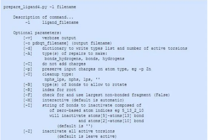
Vina's design philosophy is not to require the user to understand its implementation details, tweak obscure search parameters, cluster results or know advanced algebra. All that is required is the structures of the molecules to be docked (i.e. receptor and ligand) and the specification of the search space including the putative binding site. Vina requires the ligand file to be written in PDBQT format. PDBQT format is very similar to PDB format but it includes partial charges ('Q') and AutoDock 4 (AD4) atom types ('T'). There is one line for each atom in the ligand, plus special keywords indicating which bonds, if any, are required to be rotatable during the docking experiment.
Preparing the receptor and ligand involves ensuring that their atoms are assigned the correct AutoDock atom types, adding Gasteiger charges, merging non-polar hydrogens, detecting aromatic carbons if any, and setting up the 'torsion tree' (only for the ligand). For most atoms, the AD4 atom type is the same as its element; the exceptions are "OA", "NA", "SA" for hydrogen-bond acceptor O, N and S atoms; "HD" for hydrogen-bond donor H atoms; "N" for non-hydrogen bonding nitrogens, and "A" for carbon atoms in aromatic rings.
In particular, starting from the PDB file of the receptor, the conversion to PDBQT file involves: - the deletion of the co-crystallized ligand, if any (HETATM field in PDB file);
- the deletion of the water molecules; - the addition of polar hydrogen atoms; - the addition of partial charges.
If the user wants to allow the flexibility of some residues in the receptor structure, a third file has to be generated, with the information about rotatable bonds in the side chain of each residue. In addition, the steps required to prepare the ligand PDBQT file are:
- the addition of polar hydrogen atoms; - the addition of partial charges;
- 14 -
- the detection of all active torsions within the ligand structure.
In general, the more rotatable bonds in the ligand, the more difficult it will be to find good binding modes in repeated docking experiments. Once the above mentioned steps are concluded, it is necessary to set the search space (i.e. the docking grid), that is the portion of the receptor in which Vina will search for the best orientation and conformation of the ligand. The smaller the search space, the easier it is for the docking algorithm to explore it. On the other hand, it will not explore ligand and flexible side chain atom positions outside the search space. Search spaces bigger than 30 x 30 x 30 (27,000) Å3 should be avoided, unless the "exhaustiveness" parameter is also increased.
These results can be achieved by using AutoDock Tools (ADT, also referred as MGLTools), an auxiliary software developed at the Molecular Graphics Laboratory (MGL) of The Scripps Research Institute for visualization and analysis of molecular structures, for preparing the files, choosing the search space, and viewing the results (Morris et al., 2009). In Figure 3.2, is reported a snapshot of the ADT interface, by which the user can set the search space extension through the specification of its length along the X, Y and Z-axes and of its center (the “grid box” center). All these parameters are then reported in a configuration file (Figure 3.3) which is needed to run the simulation.
The main output of the program is the binding energy value (ΔG): based on this calculated value, the poses obtained for the ligand are ranked and reported in the output file of the program. This file can then be analyzed with many molecular graphics programs, to visualize the preferential binding sites of the ligand and to analyse the interactions at the atomic level.
Figure 3.2: ADT interface with the window to setup the search space. Through this interface it is possible to prepare PDBQT files, choose the search space and visualize the results.
- 15 -
Figure 3.3: Example of Vina’s configuration file. Here are contained all the information needed by Vina to run the docking simulation.
3.3 DrugBank
The DrugBank database (Wishart et al., 2006) is a unique bioinformatics and cheminformatics resource that combines detailed drug (i.e. chemical, pharmacological and pharmaceutical) data with comprehensive drug target (i.e. sequence, structure, and pathway) information. The database contains 8312 drug entries including 2036 FDA-approved small molecule drugs, 233 FDA-approved biotech (protein/peptide) drugs, 93 nutraceuticals and over 6000 experimental drugs. Additionally, 4317 non-redundant protein (i.e. drug target/enzyme/transporter/carrier) sequences are linked to these drug entries. Each DrugCard entry contains more than 200 data fields with half of the information concerning drug/chemical data and the other half related to drug target or protein data (Table 3.1).
Each set of compounds (approved, experimental, nutraceutical, illicit, withdrawn and
investigational) is freely reachable at the website (http://www.drugbank.ca/) as SDF (Structure Data Format) file, that is a chemical file format to represent multiple chemical structure records, delimited by lines consisting of four dollar signs ($$$$), and associated data fields. Obviously, SDF files contain also the coordinates of each compound. In this work, the subset of FDA-approved compounds was used. In fact, a drug that is approved is said to be safe and effective when used as directed. Therefore, the screening against this subset should reduce the risk of low quality hits and, more important, if in the initial screening a drug displays a sufficient affinity for the target, it could be immediately tested in patients.
- 16 -
Table 3.1: Summary of the data fields or data types found in each DrugCard. A more complete listing is provided on the DrugBank home page (Wishart et al., 2006).
3.4 Molconverter
Molconverter is a command line program in Marvin Beans and JChem Suites (http://www.chemaxon.com/products/marvin/molconverter/) that converts between various coordinates file types. Operating in batch mode users can specify various documents, molecules files, graphic and compression/encoding formats. When handling chemical structures users can specify how to handle chemical features relevant to molecule file formats.
In this work, this program was used to convert a two-dimensional SDF file from DrugBank to a three-dimensional PDB file (low energy conformer).
3.5 UCFS Chimera
UCSF Chimera (also referred simply to as Chimera) is a molecular graphics program developed by the Resource for Biocomputing, Visualization, and Informatics at the University of California, San Francisco (Pettersen et al., 2004). In this work, molecular graphics and analyses of docking solutions were performed with this program, including receptor-ligand complex visualization and detailed atomic study of the relative interactions.
- 17 -
4. RESULTS
4.1 Overview
Results obtained in this work are divided into two sections: first, docking analyses on human serum albumin (HSA) are reported, carried out to investigate the interaction of this protein with the drug imatinib and with retinoids. Experimental data relative to these macromolecular systems were provided by Professor Paolo Ascenzi from the Biochemistry Lab of the Department of Science, University of Roma Tre. Data relative to these analyses were published in two papers (Di Muzio et al., 2014, Di Muzio et al., 2015) whose reprints can be found in Supplementary Material. Subsequently, a user-friendly procedure implemented within the present Ph.D. project is described, which allows to perform molecular docking simulations and virtual screening analyses also to users without any expertise in bioinformatics. Although results obtained are still under analysis, the virtual screening version of the program was tested on five targets from Pseudomonas aeruginosa that are involved in bacterial virulence or are essential for bacterial growth with the aim to identify those FDA-approved compounds that could display antibacterial activity.
4.2 Docking of imatinib to HSA and HSA-heme-Fe(III)
Human serum albumin (HSA), the most abundant protein in plasma, is a monomeric multidomain macromolecule, representing the main determinant of plasma oncotic pressure and the main modulator of fluid distribution between body compartments. HSA displays an extraordinary ligand binding capacity, representing a depot and carrier for many endogenous and exogenous compounds. Indeed, HSA represents the main carrier for fatty acids (FA), being able to bind up to nine equivalents of long chain FAs, which represent the primary physiological ligands at multiple binding sites (i.e., the FA1–FA9 sites). Moreover, HSA affects pharmacokinetics of many drugs, provides the metabolic modification of some ligands, renders potential toxins harmless, accounts for most of the anti-oxidant capacity of human plasma, and displays (pseudo-)enzymatic properties (Fanali et al., 2012).
Imatinib is a potent and selective kinase inhibitor approved in the treatment of chronic myelogenous leukemia (CML) and metastatic gastrointestinal stromal tumor (GIST). Imatinib acts by blocking the tyrosine kinase activity of key proteins involved in the pathogenesis of
- 18 -
CML and GIST (Hartmann et al., 2009). Imatinib is approximately 95% bound to plasma proteins, a1-acid glycoprotein (AGP) being the primary carrier. Although the plasma level of AGP is markedly elevated in malignant diseases, in contrast to the decline of HSA level (Gupta and Lis, 2010), half of the normal plasma levels of AGP have been found in patients with hepatic cirrhosis and hepatitis (Kremer et al., 1988); therefore, in these pathological states, HSA may serve as a secondary carrier for imatinib.
Docking simulations of imatinib binding to HSA were performed using the crystal structures of the ligand-free HSA (PDB ID: 1AO6) (Sugio et al., 1999) and of HSA-heme-Fe(III) (PDB ID: 1N5U) (Wardell et al., 2002).
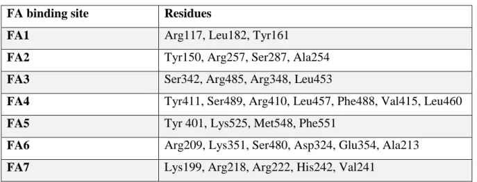
Simulations were carried out using the docking program Autodock Vina with a search space (docking grid) that included the whole protein, in order to carry out “blind” predictions of the imatinib binding site(s). Additional simulations were also carried out restricting the search space only to the FA2, FA6 and FA7 sites. The grid spacing was set to 1 Å per grid unit and the exhaustiveness parameter was increased from the default value of 8 to 24 as suggested by Vina developers for grid sizes larger than 27,000 Å3, which is the case for HSA simulations. Initially the number of solutions (docking poses) generated by Vina was set to the default value of 9, but simulations were also carried out with a number of solutions of 20. The simulations were carried out both by keeping all protein residues rigid and by allowing flexibility of the residues building up the walls of the sites from FA1 to FA7 (Fanali et al., 2012, Fanali et al., 2012a). Dihedral angles involving single bonds of the flexible residues are, by default, varied by a 50 degrees increment during Vina execution. Residues for which flexibility was allowed are reported in Table 4.1. Rotatable bonds of the imatinib lowest energy resonance structure were kept flexible in all the simulations, as suggested by Vina developers (see http://vina.scripps.edu/tutorial.html).
Table 4.1: Residues for which flexibility was allowed in docking simulations of imatinib binding to HSA and
HSA-heme-Fe(III).
FA binding site Residues
FA1 Arg117, Leu182, Tyr161
FA2 Tyr150, Arg257, Ser287, Ala254
FA3 Ser342, Arg485, Arg348, Leu453
FA4 Tyr411, Ser489, Arg410, Leu457, Phe488, Val415, Leu460
FA5 Tyr 401, Lys525, Met548, Phe551
FA6 Arg209, Lys351, Ser480, Asp324, Glu354, Ala213
- 19 -
4.2.1 Docking analyses of imatinib binding to ligand-free HSA
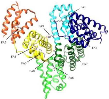
Docking simulations of imatinib binding to HSA, with the search space extended to the whole protein (Figure 4.1), indicated the preferential binding of the drug to the FA1 and FA7 sites with similar binding energy. The number of complexes observed in FA1 and FA7 in docking simulations with a maximum of 9 poses was 4 and 2, respectively. By increasing to 20 the number of possible poses, 9 and 3 complexes were observed in FA1 and FA7, respectively. The highest ranking complexes of imatinib in FA1 and FA7 are shown in Figure 4.2. In simulations carried out restricting the docking search space only to FA2, FA6 and FA7 sites, imatinib was found to bind with the best binding energies and in the majority of the poses (7 out of 9) in FA7 rather than in FA6 site (2 out of 9), while binding in FA2 was not observed. These results suggest that in ligand free-HSA imatinib binds preferentially to the FA1 and FA7 sites.
Figure 4.1: Ribbon representation of the three-dimensional structure of HSA. The subdomains of HSA are rendered with different colors (domain IA, in blue; domain IB, in cyan; domain IIA, in dark green; domain IIB, in light green; domain IIIA, in yellow; and domain IIIB, in red). The FA1–FA9 sites are occupied by capric acid (in ball-and-stick representation). Atomic coordinates were taken from the PDB entry 1E7E (Bhattacharya et al., 2000). The FA binding sites are numbered according to literature (Bhattacharya et al., 2000).
- 20 -
Figure 4.2: Atomic details of the highest ranking complexes obtained by docking simulations between
imatinib and ligand-free HSA. (A) Imatinib bound to FA1. (B) Imatinib bound to FA7. Atomic coordinates were
taken from the PDB entry 1AO6 (Sugio et al., 1999).
4.2.2 Docking analyses of imatinib binding to HSA-heme-Fe(III)
Docking simulations of imatinib binding to HSA-heme-Fe(III), with the search space extended to the whole protein, indicated that the most likely binding sites are FA2 and FA7, binding energy values being of the same order of magnitude. When the maximum number of docking poses was set to 9, the number of complexes observed in FA2 and FA7 was 2 and 6, respectively. Setting the maximum number of docking poses to 20 resulted in 1 and 9 complexes in FA6 and FA7, respectively. When the search space was restricted only to FA2 and FA7 sites, 1 complex was observed in FA2 and 8 in FA7. These results suggest that in HSA-heme-Fe(III), imatinib binds preferentially to the FA2 and FA7 sites as illustrated in Figure 4.3.
- 21 -
Figure 4.3: Atomic details of the highest ranking complexes obtained by docking simulations between
imatinib and HSA-heme-Fe(III). (A) Imatinib bound to FA7. (B) Imatinib bound to FA2. Atomic coordinates
were taken from the PDB entry 1N5U (Wardell et al., 2002).
4.2.3 Experimental data of imatinib binding to HSA and HSA-heme-Fe(III)
Experimental analyses were carried out to investigate thermodynamics of imatinib binding to full-length HSA and its recombinant Asp1-Glu382 truncated form (containing only the FA1, FA2, FA6, and FA7 binding sites; trHSA), in the absence and presence of ferric heme (heme-Fe(III)). Imatinib binding to HSA was followed by competitive inhibition of dansyl-arginine and dansyl-sarcosine association, whereas imatinib binding to trHSA was followed by competitive inhibition of dansyl-arginine association. Competitive inhibition of dansyl-arginine and dansyl-sarcosine association reflects ligand binding to the FA7 site and the FA3–FA4 cleft, respectively. As resulted from these analyses, imatinib affects only dansyl-arginine binding to HSA and trHSA, thus suggesting that imatinib binds selectively to the FA7 site of HSA, which accommodates dansyl-arginine, but not to the FA3–FA4 cleft that binds dansyl-sarcosine. Moreover, from the experimental studies resulted that imatinib affects HSA-heme-Fe(III) and trHSA-heme-Fe(III) reactivity, i.e. peroxynitrite isomerization. Even if, at present, the available data do not allow to identify unambiguously the low affinity imatinib binding site(s) modulating the HSA-heme-Fe(III) reactivity, possible candidates are the FA2 and/or FA6 sites. This is because no single chromophore is available to probe both sites at the same time. However, docking simulations (Figure 4.3, panel B) suggest that FA2 is the most probable imatinib low affinity binding site. For further details on the experimental characterization of imatinib binding to HSA, see the reprint of the paper Di Muzio et al. (2014) in the Supplementary Materials.
- 22 -
4.3 Docking of all-trans-RA and all-trans-ROL to HSA
Dietary vitamin A and its natural and synthetic analogs (named retinoids) are naturally-occurring, fat-soluble, unsaturated isoprenoids present in all living organisms. Retinoids mainly influence cell growth, differentiation and death, the deregulation of retinoid signaling pathways being linked to tumorigenesis. For these reasons, they soon emerged as potential therapeutic agents for several diseases, including cancer (di Masi et al., 2015). Vitamin A, also named
all-trans-retinol (all-trans-ROL), is converted into two classes of biologically active retinoids, i.e.
11-cis-retinoids and acidic retinoids. Among acidic retinoids, all-trans-retinoic acid (all-trans-RA) and 9-cis-retinoic acid (9-cis-(all-trans-RA) represent the main metabolic products.
RA, ROL, and all-trans-retinal bind to specific and aspecific proteins, which solubilize, protect, and detoxify retinoids in the extracellular environment (di Masi et al., 2015, Folli et al., 2010, O'Byrne and Blaner, 2013). Among the extracellular retinoid-binding proteins (RBP), the epididymal retinoid-binding protein (ERBP), the interphotoreceptor matrix retinoid-binding protein (IRBP), and the retinoid binding protein 4 (RBP4) play a central role in ROL transport, whereas lipocalin-type prostaglandin D synthase (also known as β-trace) and HSA transport preferentially all-trans-RA (Kuruvilla et al., 1991, Zahn et al., 1993, O'Byrne and Blaner, 2013). This suggests that RBPs may function as alterative carriers of ROL and RA in health and disease. Indeed, HSA may act as a secondary carrier in human diseases associated with reduced levels of IRBP (e.g., in the early stages of diabetic retinopathy) (Garcia-Ramírez et al., 2009) or RBP4 (e.g., in prolonged dietary vitamin A deficiency, in migraine, in HIV, in diabetic patients treated with anti-TNFa-therapy, and in end-stage renal disease after kidney transplantation) (Zhang et al., 2014, Kotzé et al., 2015).
Docking simulations of all-trans-RA and all-trans-ROL binding to HSA were performed using the crystal structure of ligand-free HSA (PDB ID: 1AO6) (Sugio et al., 1999), and of FA-bound HSA (PDB ID: 3SQJ) (He et al., 2011). All-trans-RA and all-trans-ROL three-dimensional structures were obtained respectively from the crystal structures of the all-trans-RA-bound ligand binding domain of RAR-γ (PDB ID: 2LBD) (Renaud et al., 1995) and of the all-trans-ROL-bound retinol-binding protein (PDB ID: 1KT6) (Calderone et al., 2003).
Simulations were carried out using Vina with a search space (docking grid) that included the whole protein, in order to carry out “blind” predictions of the all-trans-RA and all-trans-ROL binding sites. Additional simulations were also carried out restricting the search space only to the FA1 site, in order to obtain more accurate details on all-trans-RA and all-trans-ROL interaction with HSA.
- 23 -
The simulations were carried out both by keeping all protein residues rigid and by allowing flexibility only of the residues building up the walls of the FA sites (FA1 to FA9). Residues for which flexibility was allowed are those reported in Table 4.1. In addition, flexibility was allowed for residues Lys195, Asp451, Ser454 belonging to the FA8 site, and for residues Asp187, Lys432 belonging to the FA9 site. Rotatable bonds of the all-trans-RA ad ROL lowest energy resonance structures were kept flexible in all the simulations.
4.3.1 Docking analysis of retinoids binding to FA-free HSA
Docking simulations of all-trans-RA and all-trans-ROL binding to HSA, with the search space extended to the whole protein, indicated the preferential binding of both compounds to the FA1 site, in which retinoids bind in the vicinity of the FA1 site residue Tyr161. Interestingly, this residue coordinates the iron ion in the HSA-heme-Fe(III) derivative (Wardell et al., 2002 ). An overall view of the nine lowest energy retinoids-HSA complexes and atomic details of the lowest energy complex obtained for both compounds are shown in Figure 4.4. The number of complexes observed in FA1 in docking simulations with a maximum of 9 poses was 5 for
RA and 6 for ROL. The binding affinity of the best all-RA and
all-trans-ROL poses was −8.1 kcal/mol in both cases. In other poses of this simulation, all-trans-RA was located in the FA6 site (3 poses) and in the FA9 site (1 pose), while all-trans-ROL in the FA9 site (3 poses). The simulation with the search space reduced to the FA1 site provided more reliable details of the interaction: in this case, the binding affinity of RA and all-trans-ROL for the FA1 site of HSA was −8.8 kcal/mol and −8.0 kcal/mol, respectively. The highest ranking complexes of all-trans-RA and all-trans-ROL bound to the FA1 site of HSA are shown in Figure 4.4 (panels C and D).
- 24 -
Figure 4.4: Schematic representation of all-trans-RA and all-trans-ROL complexes with FA-free HSA as
obtained by docking simulations. Overall view of the nine lowest energy all-trans-RA-HSA complexes (panel
A). Overall view of the nine lowest energy all-trans-ROL-HSA complexes (panel B). Atomic details of the lowest energy all-trans-RA-HSA complex (panel C). Atomic details of the lowest energy all-trans-ROL-HSA complex (panel D). Both all-trans-RA and all-trans-ROL bind in the vicinity of the FA1 site residue Tyr161, which coordinates the iron in HSA-heme-Fe(III) derivative.
4.3.2 Docking analysis of retinoids binding to FA-bound HSA
Docking simulations of all-trans-RA and all-trans-ROL binding to FA-bound HSA, with the search space extended to the whole protein, indicated the binding of RA and all-trans-ROL in three different sites. In Figure 4.5 is shown the overall view of the complexes obtained. It should be noted that in the FA-HSA structure used in this simulations, the FA1 to FA7 sites are occupied by myristic acid (He et al., 2011).
In the first-ranking solution both ligands were placed in the FA8 site, located at the base of the crevice between subdomains IA-IB-IIA on one side and subdomains IIB-IIIA-IIIB on the other side. The FA8 site is relevant for ligand recognition only when the FA1 to FA7 sites of HSA
- 25 -
are occupied by long-chain FAs such as myristic acid (Fanali et al., 2012, Bhattacharya et al., 2000).
The binding affinity of the best all-trans-RA and all-trans-ROL poses in the FA8 site was −8.5 kcal/mol and −8.6 kcal/mol, respectively. In other poses, all-trans-RA and all-trans-ROL bind near the FA1 site occupied by myristate with an apparent free energy of −8.4 kcal/mol and −8.3 kcal/mol, respectively.
Figure 4.5: Schematic view of the all-trans-RA and all-trans-ROL interaction with FA-bound HSA as
obtained by docking simulations. Overall view of the nine lowest energy all-trans-RA-HSA complexes (panel
A). Overall view of the nine lowest energy all-trans-ROL-HSA complexes (panel B). Myristate molecules occupying the FA1 to FA7 sites (PDB ID: 3SQJ, He et al., 2011) are shown in ball-and-stick representation and colored in orange.
The binding affinity of the best all-trans-RA and all-trans-ROL poses in the FA8 site was −8.5 kcal/mol and −8.6 kcal/mol, respectively. In other poses, all-trans-RA and all-trans-ROL bind near the FA1 site occupied by myristate with an apparent free energy of −8.4 kcal/mol and −8.3 kcal/mol, respectively. Similar co-binding of aromatic drugs or drug-like molecules such as indomethacin and triiodobenzoic acid in the FA1 site was already reported (Curry et al., 1998, Ghuman et al., 2005) . Lastly, all-trans-RA and all-trans-ROL bind to the FA9 site of HSA, located in the upper region of the cleft built up by subdomains IA-IB-IIA on one side and subdomains IIB-IIIA-IIIB on the other side. Of note, the FA9 site is an additional binding pocket of HSA relevant for ligand recognition only when long-chain FAs, such as myristic acid, occupy the FA1 to FA7 sites of HSA (Bhattacharya et al., 2000). All-trans-RA and all-trans-ROL bind to the FA9 site of HSA in a fashion similar to that observed for thyroxine recognition when long-chain FAs, such as myristic acid, occupy the FA1 to FA7 sites (Petitpas et al., 2003).
- 26 -
The binding affinity of the best RA and ROL poses in this cleft was −8.0 kcal/mol and −7.6 kcal/mol.
Summarizing, docking analyses here reported allowed both to identify the preferential binding sites of the ligand(s)/drug, and to investigate the interaction at atomic level. Moreover, results obtained are in agreement with experimental data simultaneously obtained (see below; Di Muzio et al., 2014, Di Muzio et al., 2015).
4.3.3 Experimental data of all-trans-RA and all-trans-ROL binding to HSA
Thermodynamics of all-trans-RA and all-trans-ROL (i.e., retinoid) binding to the FA1 site, the FA3-FA4 cleft, and the FA7 site of HSA was followed by competitive inhibition of heme-Fe(III), dansylsarcosine, and dansyl-arginine association, respectively. Functional studies demonstrated that, in the absence of FAs, all-trans-RA and all-trans-ROL bind to the FA1 site of HSA, impairing competitively heme-Fe(III) association. On the other hand, all-trans-RA and all-trans-ROL do not bind to the FA3-FA4 cleft and the FA7 site, respectively. In fact, retinoids do not affect the association of dansyl-sarcosine and dansyl-arginine with HSA. Noteworthy, the FA7 site, the FA3-FA4 cleft, and the FA1 pocket (located in subdomains IIA, IIIA, and IB, respectively) represent the first, the second, and the third major ligand binding region of HSA, respectively. These data are in full agreement with the docking studies and thus the complexes obtained by docking simulations results can be used to infer the nature of the atomic interactions leading to retinoids binding to HSA. For further details on the experimental characterization of retinoids binding to HSA, see the reprint of the paper Di Muzio et al. (2016) in the Supplementary Materials.
- 27 -
4.4 DockingApp
“DockingApp” is the name given to the procedure implemented in Python programming language, which allows to perform molecular docking simulations and virtual screening analyses also to users without any expertise in bioinformatics. In Figure 4.6 is reported the “splash screen” of the application GUI.
Figure 4.6: Splash screen of the DockingApp application. It will be soon available at our lab website whose URL is reported in the figure.
Figure 4.7: Algorithm overview. With DockingApp it is possible to carry out both docking and virtual screening analyses.