A
A
l
l
m
m
a
a
M
M
a
a
t
t
e
e
r
r
S
S
t
t
u
u
d
d
i
i
o
o
r
r
u
u
m
m
–
–
U
U
n
n
i
i
v
v
e
e
r
r
s
s
i
i
t
t
à
à
d
d
i
i
B
B
o
o
l
l
o
o
g
g
n
n
a
a
DOTTORATO DI RICERCA
Scienze Chimiche
Ciclo XXI
Settore/i scientifico disciplinari di afferenza: Settore Disciplinare: CHIM/02
Coordinatore: Chiar.mo Prof. Giuliano Longoni
TITOLO TESI
Theoretical insight into the properties of light
induced events of photochromic systems and
rhodopsin proteins.
Presentata da: Tomasello Gaia
Coordinatore Dottorato Relatore
Chiar.mo Prof. Giuliano Longoni Chiar.mo Prof. Giorgio Orlandi
Contents:
Chapter 1:Introduction
•
1.1.Preface
•
1.2 Inside the photochemicalprocesses
•
1.3 Photochromic compounds: fulgides and stilbenoid systems.
•
1.4 Rhodopsin photochemistry.
References
Chapter 2:
Quantum Mechanics/Molecular Methods
•
2.1
The Schrödinger Equation and the Born-Oppenheimer
Approximation
•
2.2 Molecular Orbitals and Valence Bond theories.
•
2.3 Basis Sets: The LCAO Approximation
•
2.4 Hartree-Fock Theory
•
2.5 Configuration Interaction (CI)
•
2.6 MC-SCF: Multiconfigurational Self-Consistent-Field
•
2. 7 CASSCF and RASSCF
2.7.1 Selecting the active space•
2.8 Multiconfigurational Second Order Perturbation Theory
Method: CASPT2
•
2.9 MultiState CASPT2
•
2.10 The evaluation of bond order in molecules
•
2.11 QMMM methods
References:
Chapter 3: Potential Energy Surfa
ce
•
3.1 General Concepts
•
3.2 Critical Points Determination
•
3.3 Energy minimization
•
3.4 The use of derivatives in minimizations
•
3.5 Newton-Raphson method
•
3.6 The Steepest Descent and the Quasi-Newton Methods
•
3.7 Vibrational normal modes analysis
•
3.9 Methods based on local information
•
3.10 Minimum Energy Path determination
•
Intrinsic Reaction coordinate method
•
3.11 Methods based on interpolation between reactant and
product
•
3.12 Conical Intersections: no crossing roles
•
3.13 The Physical chemistry of conical intersections
•
3.14 Conical Intersection Topology
•
3.15 Conical Intersection Optimization
References
Chapter 4: Conceptual aspects of photochemistry.
•
4.1 What happens when a molecule absorbs light?
•
4.2 Adiabatic and non-adiabatic processes?
•
4.3 The “Physical Chemistry” of Non-Radiative Decay
•
4. 4 Ultrafast processes
•
References
Chapter 5
: Fulgide Photochromism.
•
5.1 Introduction
•
5.2 Computational method
•
5.3 Results and discussion
5.3.1Vertical excitations: the open-ring isomer E 5.3.2 The open→ close ring reaction
5.3.3The close-ring isomer and the open ring reaction
•
5.4 Interpretation of the results
•
5.5 Conclusions
References
Chapter 6
: Stilbene photo-physical properties
•
6.1 Introduction
•
6.2 Computational strategies adopted and consequent results
6
.2.1Appling CASSCF//CASPT2 methods: Vertical excitations. 6.2.2 Appling CASSCF//CASPT2 and CC2 methods: trans* relaxation. 6.2.3 Appling CASSCF//CASPT2 and CC2 methods: twisted region. 6.2.4 Appling CASSCF//CASPT2 and CC2 methods: cis* relaxation
•
6.3 Appling RASSCF method
•
6.4Conclusion
References
•
Chapter 7
: Electrostatic control of the
photoisomerization efficiency in Rhodopsin
•7.1. Introduction
•
7.2. Computational Details
•
7.3 Result and Discussio
n
7.3.1 The Glu181 issue: neutral or protonated?
7.3.2 The counterion and the other polar/charged residues: counterion quenching by the protein pocket.
•
7.4 Mutations and Vision Deficiencies
7.4.1 Rh related vision deficiencies.
7.4.2 Color pigments related pathologies.
•
7.5. Conclusions
References
•
Chapter 8: Steric control of photoisomerization
efficiency and spectroscopic properties in
Rhodopsin
•
8.1 Introduction
•
8.2 Computational Methods
•
8.3 Results and Discussion
8.3.1Steric catalysis8.3.2 The trap S2 state
•
Transient absorption along the S
1photoisomerization
•
Conclusions
References
•
Chapter 9: Conclusions
References
-CHAPTER
1-Introduction
1.1 Preface
The light-material interaction is one of the most interesting natural phenomena, being responsible of the existence of life on the earth, and therefore originating photo-biological processes of primary importance. The absorption of a photon transfers the system from the ground state to an electronically excited state. The electronic distribution and nuclear configuration of a photochemically activated molecule generally differ substantially from those of a thermally activated molecule so that the excited state is actually an electronic isomer of the corresponding ground-state system. Further the photochemical and photophysical properties are typical of the photo-excited molecule differing from those shown on the ground state, leading the system towards new processes : photochemistry is the science that study and rationalize such processes. The aim of this introduction is o give the basic information about the photochemical concepts common interest to some of the problems. It is necessary at this point to emphasize how there are some molecular properties not really available to investigate and rationalize from an experimental point of view. Computational photochemistry arises from the necessity of associating accurate computational methodologies of simulation to advanced experimental techniques in order to clarify several aspects
of photochemistry experimentally inaccessible. The modern strategies of calculations and the even more sophisticated computations techniques is nowadays possible to explore and characterize widely the topology of the potential energy surface of a chromophore excited states, and thus to properly rationalize (explain) its photoreactivity.
1.2 Inside the photochemical processes.
The purpose of this thesis is to investigate, through high-level computations, some of the interesting photo-biochemical processes involve conjugated π-systems, such as those occurring in photochromic systems2 of retinal chromophores3. The goal is to
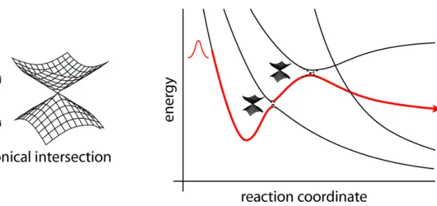
control and maximize photochromic efficiency and to increase our general knowledge of the “primary events” which control the final outcome of the absorption of a photon. The computations make use of a recently implemented novel technique allowing to locate and follow all the steepest descent paths departing from the Frank-Condon point or real crossing point. Once the system is promoted to the excited state decay back to the ground state can occur by fluorescence, or by internal conversion via a radiationless process. For many years the mechanism of decay without emission of radiation was not well understood. It was thought to occur at a minimum on the excited state, in a region of the potential where the ground and excited state surfaces become close at an avoided crossing. Such processes are slow, however, and fail to explain how systems could decay on a femtosecond timescale. More recently, it has been recognized that a true crossing of potential surfaces can take place, creating a new topological feature called a conical intersection (CI)4.
The intersection acts like a funnel linking the excited and ground state surfaces,
providing a fully-efficient decay path back to the ground state products. There need not be a transition state separating the Frank-Condon region and CI, but if one is present it will control the conditions under which the radiationless route is accessible. The idea that a conical intersection could play a key role in the decay mechanism for excited state reactions was proposed by Teller5 more than 30 years ago. The literature now contains
many theoretical studies of organic systems which find low-lying surface crossings. The photochemical events investigated in this thesis are mainly characterize from an ultrafast deactivation and observing ultra-fast radiationless decay is the main indication of a mechanism involving a CI6. Modern experimental techniques have enabled
researchers to verify the existence of conical intersections. In particular laser spectroscopy can now be performed at a sub-picosecond level, giving reliable data for excited state lifetimes, decay rates, and energy thresholds for these reactions. Thus experimental evidences are interpreted and rationalized on the basis topological characteristic individuated by calculations. For example, if the radiationless decay is thermally driven, and it is ‘switched-on’ at a certain temperature, this suggests that a transition state lies on the excited state path leading to the intersection. Quantum yield measurements provide information about the product
distribution, and hence the possible ground state paths accessible from the CI.
Thus computational approach may model the photochemistry of several systems explaining the origin of them photochemical properties.
1.3 Photochromic compounds: fulgides and stilbenoid systems.
Photochromism7 is a reversible transformation of a chemical species induced in one or
both directions by absorption of electromagnetic radiation between two forms, A and B, having different absorption spectra (fig.1.1). Although this phenomenom is largely investigated only in the past 30 years, in reality it origins in 1800. More precisely, on 1870 Fritzche8 the bleaching of an orange-colored solution of tetracene in the daylight
but limited until the 1940–1960 period, which saw an increase of mechanistic and synthetic studies, particularly in the research groups of Hirshberg an Fischer9 in Israel.
In 1950, Hirshberg suggested the term “photochromism”, from the Greek words phos (light) and chroma (color), to describe the phenomenon. This is the name used today. However, it is not limited to colored compounds; it applies to systems absorbing from the far UV to the IR, and to very rapid or very slow reactions.
Abs
λ\ nm
300 400 500 600A
B
A
h
ν1B
h
ν2/
∆Figure1.1 Schematic example of a reversible transformation of a single chemical species between two
states, the absorption spectra of which are clearly different, the transition in at least one direction being induced by electromagnetic radiation
Photochromism expanded during the 1960s in parallel with the development of physical methods (IR, NMR, X-ray, UV, time-resolved and flash spectroscopy) and organic synthesis. Photochromic glasses became available at that period and further stimulated research. Applications, such as the photochromic micro image (PCMI) process12, which
showed the possibility of reducing the a huge number of pages to about 6 cm2, attracted
considerable interest. However, it appeared that the photodegradation of the known families of organic photochromic systems limited their potential for applications 13. A
revival of activity started in the 1980s, essentially because of the development of fatigue-resistant spirooxazine and chromene derivatives. They triggered the fabrication and commercial application of photochromic ophthalmic lenses. Since then, other commercial systems have been developed, and new photochromic systems have been discovered and explored. Examples of potential applications utilizing the physical or chemical changes that accompany the observed shift of the absorption maxima include optoelectronic system,photoswitching transport through membrane, optical information storage, photochemical switchable enzymatic systems, nonlinear optical devices. The main classes of photochromic compounds , organic compounds and biological receptors are in this introduction briefly described, since the subjects treated in this thesis belong these two classes. Among the organic compounds we have investigated the excellent photochromism of fulgides14, and the complex photochromism of stilbene15.
At the microscopic level, photochromism originates from a chemical transformation triggered by a photon absorption, such as ring-closure or ring-opening, and the widest and the most important group of photochromic system is that based on pericyclic reactions. Of the many pericyclic reactions, electrocyclizations have proved to be especially suitable as a basis for photochromism. There are mainly two photochromic species, called ‘one way’ or ‘two way’ 16. In this class of compounds, the photochromic
transformation arises from different photochemical process (hydrogen shift, electrociclyzation, cis-trans or E/Z isomerization). Therefore if compounds like fulgides or diarylethenes are ‘two way’ photochromic systems ,being photochemically converted both to the open and the closed form when irradiated at suitable different wavelengths, compounds like the dihydroazulene couple or dihydronaphtalene17 couple (fig1.2) are
examples of ‘one way’ photochromism since the back reaction can occur only thermally. Of the many pericyclic reactions, electrocyclizations have proved to be especially suitable as a basis for photochromism. The 4n+2 systems whose photochromism results from the electrocyclic 1,3,5-hexatriene/1,3-cyclohexadiene interconversion18 comprise a very large number of species. The fulgides belong to the
second mentioned class of photochromic compounds for which the transformation is largely driven photochemically and consequently the thermal reversal or decoloration of the colored species is forbidden (for this reason they are classified as “two way” photochromic systems). For their features and favorable photochemical properties, fulgides are photochromic systems particularly investigated for the potential industrial applications as optical devices, molecular switches, data storage. The discovery of fulgide systems has to be collocated around one century ago and is due to Hans Stobbe19, who observed for the first time triphenyl-fulgide changing color from
yellow-orange to brown, under light exposition, while turning back to the original color under the dark light.
Figure 1.3 There are reported examples of organic compounds belonging to two main species of
photochromic systems. On one hand, dihydroazulene and dihydronaphtalene derivatives belong to “one way ” group, where the back reaction can occur only thermally, and thus whose photochromism is
CN CN CN CN DHA VHF hν
∆
X X X X CHD HT hν hν’ NC CN Ph Ph NC CN Ph Ph hν ∆ o Me O O O Me Me Me Me o O O O Me Me Me Me Me hν hν’ E C BDQM DHN-1 ‘ONE WAY’ ‘TWO WAY’defined also of type T. On the other hand diarylethenes or fulgides belong to the “two way” group, where the back reactions occur photochemically, and whose photochromism is defined also of type P.
Initially Stobbe supposed that the origin of these special properties was correlated with some kind of crystal effect; nevertheless he changed idea while he noticed how such similar properties characterized also similar compounds. The denomination “fulgide”, arising from latin language and meaning twinkle has to be attributed to the same Stobbe20, the first one observing the particular of some crystal forms. Among the
several species of fulgides, featured by the different chemical structure (from which photochromic properties arise), the most interesting species are those called “aromatic fulgides” or “heterocyclic-fulgides. The aromatic fulgides are distinguished depending on the kind of system condensed on the ciclohexadiene-succinic-anidrid moiety, whose photochromism results from the electrocyclic 1,3,5-hexatriene/1,3-cyclohexadiene interconversion: the experimental data are mainly concentrated on the furyl-classes. In particular compounds as indolylfulgide, furylfulgide, because of its high efficiency as potential photochromic systems, can be used as switch controls or aberchrome molecules. Remarkable, photochromic actinometers can be used repeatedly, thus obviating the need for a fresh sample for each measurement. Some thermally stable photochromic compounds, such as the following, fulfill this requirement. The fulgide we have investigated in this thesis, is suitable for being used as chemical actinometer, since the absorption wavelenght are within the range of coloration (310–370 nm) or decoloration (435–545 nm) domain. Organic compounds as azobenzene and stilbene may be used in the field of chemical actinometry or photoresponsive materials. Reversible photoinduced changes of physical and chemical properties can be transferred to the micro-environment by a photochromic molecule incorporated in the system. Some examples are given from chiroptical molecular switches, where photoisomerization of photochromic units (azobenzene, spyropirans) in poly (α-amino acids) is able to trigger a random coil to α-helix transition. This primary photochemical event occurring in the side chains is amplified and transduced by the structural variations of the macromolecular main chains. The latter are accompanied by large and reversible variations of optical activity21. Because of their reversibility, these systems
can act as “chiroptical molecular switches”22 In this thesis stilbene photochemistry, has
been extensively studied using accurate computational strategies with the aim of clarify the mechanism behind the radiationless deactivation to the ground state. Stilbenes photochromism is due to cis-trans isomerization, and it represents one of the most interesting challenge in spectroscopy. Stilbene and stilbenoid compounds are indeed frequently textbook examples of photochemical cis-trans isomerization.
Their reactivity has an important role as model compound of biological phototropic systems. They, also serve as building blocks for organic materials, whose properties could be used in optical and electro-optical applications such as optical data storage, laser dyes, nonlinear optics, or photochemically, cross-linked polymers. Stilbene photochemistry has always been particularly difficult to rationalize: when rotate to twisted geometry stilbene arrives at a minimum of potential energy, called the “phantom state”, from which it decays to the ground state through nonadiabatic transitions. Understanding the nature of this “phantom state” may permit to amply the knowledge of photochemical properties of more extended system, as stiff-stilbene, or the so called Feringa molecular motors. Concerning to this, it is necessary, at this stage to briefly described one of the most interesting application in this field. One of the main current scientific challenges is the bottom-up construction of systems that represent nanosize analogues of switches, devices and motors. The control of chirality in a molecular switching system allows inter-conversion between molecules of opposite helicity using different wavelengths of light. Such bistable chiral switches are of potential use in optical data storage and processing at the molecular level. The control of molecular chirality is even more subtle in the case of molecular motor systems. The exquisite control of chirality using light as an energy source has resulted in a controlled, repetitive 360° unidirectional rotation in two generations of molecular motor systems. It is hard to imagine daily the life without motors. Controlled rotary and linear motor is at the heart of the variety of machinery our society depend on. For most individual it goes unnoticed that at any instant numerous molecular motors are also engaged in pour body to control such delicate processes catalyst, transport and muscle movement. The fascinating molecular motors discovered in biological systems such as ATP- ase rotary and muscle linear motors offer a great source of inspiration to design synthetic motor systems in which controlled translational or rotary motion can be accomplished. In designing molecular motors one has to take in consideration that, compared to ordinary motors, where energy input induces motion, in molecular motors input has also to restrain motion. Rotary motion around a single carbon-carbon bond in alkane for instance is extremely fast with an energy barrier of only 20 kj/mol and there is not control over the directionality of the rotary motion. The directionality may be established with a double bond carbon-carbon, which is broken upon light irradiation leading the system isomerization on excited state following a precise direction. The problem of mutual random motion of molecules has recently emphasized in discussion or ratchet-type molecular motors and nano-machines. In synthesis approaches towards artificial machinery a variety of molecular or supra-molecular systems have been designed in which an external chemical, electrochemical or photochemical stimulus induces a switching process or movement within the molecule or triggers a change in shape or assembly of molecules.
1.4 Rhodopsin photochemistry.
Photochemical processes are not only interesting for technological and industrial applications, but many biological systems are photochromic, with the photo-induced reactions of fundamental interest for understanding biochemical mechanism. Usually the overall reactivity of the protein, is due to the non-proteic moiety of the entire molecule, which is the chromophore, responsible of light absorbing and of the
photochemical process featuring the protein function.
Many intriguing photobiochemical processes involve conjugated π-systems. Examples are the photoisomerization of the retinal chromophores, which trigger the conformational changes underlying the activity of rhodopsin proteins21, and the set of
photochemical reactions characterizing the bio-genesis of vitamin D22. The theoretical
chemist who wants to perform an accurate computational study of these reactions, would face the unovercoming problem due to the size of his target. Then, to apply accurate computational methods, it is necessary to reduce the dimension of the molecule, by selecting a model-system with the same chemical reactivity but a smaller size. It is usually a good approximation to limit our study only on the polyenic moiety of the system, that in the case of rhodopsin proteins is retinal chromophore, covalently linked to the protein via a lysine through a protonated Shiff base. Among the several proteins, retinal chromophore belong mainly to two different biological photo-receptors: bacteriorhodopsin, a light-activated proton pump that supports phototrophic growth in the oxygen-deficient conditions and rhodopsin, of crucial interest in biological photochemistry, since the reactivity is at basis of mechanism of vision process.
The production of a neural image of the light converging upon an animal begins with the focusing of that light onto a sheet of receptors. Figure depicts a generalized bird eye. In terrestrial animals most of the focusing power of the eye is the air/cornea interface. light impinging upon the eye from particular directions is redirected to particular locations on the retina, the tissue lining the inside of the eye. Within the retina are photoreceptors, cells specialized for transducing relative light intensities into neurochemical signals that can be passed along to the central nervous system.
Figure 1.4 A vertebrate eye.
Although there is considerable diversity among the eyes of various vertebrates, they all share some common features. In particular they have optical elements such as the cornea and lens which focus light in order to form an image on the retina.
At the basis of phototransduction mechanism there are the photoreceptors outer segment, where the light is absorbed and transduced in vertebrates. Embedded in the disk membranes are large numbers of photopigment molecules. Each photopigment consists of a chromophore covalently bound to a protein called an opsin. Phototransduction processes described here are the same in all animals with eyes. Opsin does not absorb visible light, but when it is bonded with 11-cis-retinal to form rhodopsin, which has a very broad absorption band in the visible region of the spectrum. The peak of the absorption is around 500 nm, which matches the output of the sun closely. Upon absorption of a photon of light in the visible range, cis-retinal can isomerize to all-trans-retinal. The shape of the molecule changes as a result of this isomerization.
Figure 1.5 There is represented a scheme summarizing the phototransduction process. The diagram in the
center schematically depicts how a photopigment is embedded within a disk membrane of a photoreceptor outer segment. The filled yellow circle is a lysine to which is attached the chromophore retinal. The absorption of a photon may cause the chromophore to convert from the 11-cis form to the all trans form as shown by the chemical reaction. This conformational change in the chromophore causes a change in the shape of the opsin molecule. The opsin's change in shape converts it from an inactive to an activated enzyme, and thus is light absorption converted to a biochemical signal in a photoreceptor.
Rhodopsin is the only photoreceptor protein (a visual pigment) in the outer segment of rod visual cell responsible for twilight vision. It has been extensively studied more than any other visual pigments such as cone pigments responsible for color vision because of relative ease of preparation and abundance. Rhodopsin has 11-cis retinal as its chromophore, which is embedded inside a single peptide transmembrane protein called opsin. The role of rhodopsin in the signal transduction cascade of vision is to activate transducin, a heterotrimeric G-protein, upon absorption of light23. Therefore, a central
question in rhodopsin is how light energy is used to change protein structure through photochemical reaction of the retinal chromophore. Rhodopsin (opsin), a member of G-protein coupled receptor family, is composed of 7-transmembrane helices. The 11-cis retinal forms the Schiff base linkage (see fig.1.6) with a lysine residue of the 7th helix (Lys296 in the case of bovine rhodopsin), and the Schiff base is protonated, which is stabilized by a negatively charged carboxylate (Glu113 in the case of bovine rhodopsin)24. The beta-ionone ring of the retinal is coupled with hydrophobic region of
opsin through hydrophobic interactions26. Thus, the retinal chromophore is fixed by
three kinds of chemical bonds in the retinal binding pocket of rhodopsin. Absorption of a photon by the chromophore induces a primary photoreaction, followed by conformational changes of protein, and eventually activates transducin. This is called the “bleaching process” because rhodopsin loses its color. Several intermediate states are formed during the bleaching process, which have been identified by visible spectroscopy (fig. 1.7).
Figure.1.6 The
chromophore retinal
covalently bound to lysine 296 through a protonated Schiff base.
Among the bleaching processes of rhodopsin, we define the “primary process” as the event of formation of bathorhodopsin (fig. 1.7). Bathorhodopsin is formed and stable in the picosecond time domain. It is also stable at low temperature (<-140°C) . Unlike bathorhodopsin, photorhodopsin cannot be stabilized at low temperature, as well as the excited state of rhodopsin. There has been a fundamental question on the nature of the primary photoprocess in rhodopsin: What is the primary reaction in vision? To address this question, the primary process of rhodopsin has been extensively studied from both computational and experimental points of view.
Figure 1.7 Photobleaching process of bovine rhodopsin. The primary process is shown in the frame,
which involves excited state of rhodopsin, photorhodopsin, and bathorhodopsin. Metarhodopsin-II that activates transducin is underlined.
Recently, bovine rhodopsin was crystallized by Okada et al.27, and its three-dimensional
structure was determined with 2.8 Å resolution . These accomplishments promise better understanding of the primary reaction mechanism in rhodopsin, and existing data by various techniques have to be reexamined on the basis of the structural background. Because of the limited space, we would like to make brief comments in the following. Extensive studies by means of ultrafast spectroscopy of rhodopsin have provided an answer to the question relative to the primary event mechanim. Femtosecond spectroscopy of rhodopsin eventually captured the excited state of rhodopsin, and as the consequence, we know that unique photochemistry takes place in our eyes. Namely, the cis-trans isomerization in vision is a coherent reaction through the barrierless potential surface, in which formation of photorhodopsin occurs in 200 fs. Such amazingly fast reaction is facilitated in the protein environment, and vibrational analysis of primary intermediates, such as resonance Raman and infrared spectroscopies, have provided insight into the isomerization mechanism from structural background. The atomic structure of bovine rhodopsin has further encouraged detailed understanding of the mechanism.
Remarkable progress has taken place during the past 10 years, and this progress is shown in this article. What will be the progress in the next 10 years? Regarding the dynamics, we still do not know the origin of the nonreactive pathways. On the other hand, we have started to study the role of the protein environment during the retinal isomerization from the structural background. Further efforts by means of spectroscopy, diffraction, and theory will provide us with new insights into the structure and dynamics of photoisomerization in rhodopsin.
References:
1) Nicholas J.Turro ‘Modern Molecular Photochemistry’, Yoshizawa, T.; Kuwata, O. In CRC Handbook of Organic Photochemistry and Photobiology;
2) 1. Irie,M. Chem.Rev. 2000, 100, 1685. Bouas-Laurent,H. Bouas-Laurent,H. ; Durr,H. Pure Appl. Chem. 2001, 73, 639
3) a)Kandori, H.; Shichida, Y.; Yoshizawa, T. Biochemistry-Moscow 2001, 66, 1197.Needleman, R. b)Bacteriorhodopsin and Rhodopsin. In CRC Handbook of
Organic Photochemistry and Photobiology; ed.; Horspool, W. M., Song, P.-S.,
Eds.; CRC Press: Boca Raton, FL, 1995; Vol. pp 1508-1515.
4) Herzberg, G. & Longuet-Higgins ,H.C. Trans. Faraday Soc. ,1936,35, 77. Bernardi, F.; Olivucci, M. & Robb ,M.A. Chem. Soc. Rev. ,1996,25, 321-328. 5) Teller, E. J. Phys. Chem. ,1937,41, 109.
6) Zewail, A. H. Femtochemistry: Ultrafast Dynamics of the Chemical Bond; World Scientific: Singapore 1994; Vols. I and II.
7) 1. Irie,M. Chem.Rev. 2000, 100, 1685. Bouas-Laurent,H. Bouas-Laurent,H. ; Durr,H. Pure Appl. Chem. 2001, 73, 639
8) Fritzsche.,J. Compt. Rend. Acad. Sci., Paris 1867, 69, 1035. 9) Fisher, E. Hirshberg,Y. J.Chem.Soc.1952, 4522.
11) 1. Yoshizawa, T.; Kuwata, O. In CRC Handbook of Organic Photochemistry and Photobiology; Horspool, W. M., Song, P.-S., Eds.; CRC Press: Boca Raton, FL, 1995; pp 1493-1499.
12) 6. Photochromism, G. H. Brown (Ed.), Wiley-Intersciences, New York 1971. 13) 7. R. C. Bertelson. Mol. Cryst. Liq. Cryst. 246, 1 (1994)
14) Heller, H. G.; Szewczyk, M. J. J. Chem. Soc. Perkin Trans.1 1974, 12, 1487. 15) (1).a)Syage,J.A; FelkerP.M;Zewail A,H. Chem Phys 1981,11,4685;b)Syage
,J.A;
Lambert,W.R.Felker,P.M.;Zewail,A.H.Hochstrasser,R.M.Chem.PhysLett.1982, 88,266.c)Felker,P.M.Zewail,J.Phys.Chem.,1985,89,5402.d) Orlandi,G.
Siebrand W.Chem.Phys.Lett 1975 30,352
16) 1. Irie,M. Chem.Rev. 2000, 100, 1685. Bouas-Laurent,H. Bouas-Laurent,H. ; Durr,H. Pure Appl. Chem. 2001, 73, 639
17) Gaia Tomasello, Francois Ogliaro, Michael J. Bearpark, Michael A. Robb, and Marco Garavelli J. Phys. Chem. A; 2008; 112(41) pp 10096 - 10107;
18) Celani, P.; Bernardi, F.; Robb, M. A.; Olivucci, M. J. Phys. Chem. 1996, 100, 19364-19366.
19) Stobbe, H. Die Fulgide, Annalen,380,1,1911.
20) Stobbe,H.Phototropiererscheinugen bei Fulgiden und Anderen Stoffen, Annalen,359,1,1908.
21) A. Fissi, O. Pieroni, G. Ruggeri, F. Ciardelli. Macromol. 28, 302 (1995) and references therein. B. L. Feringa, W. F. Jager, B. de Lange. J. Chem. Soc., Chem. Commun. 288 (1993).
22) Yoshizawa, T.; Kuwata, O. In CRC Handbook of Organic Photochemistry and Photobiology; Horspool, W. M., Song, P.-S., Eds.; CRC Press: Boca Raton, FL, 1995; pp 1493-1499
23) (a) Jacobs, H. J. C.; Havinga, E. Photochemistry of Vitamin D and Its Isomers and of Simple Trienes. In Advances in Photochemistry; Pitts, J. N., Jr.,
Hammond, G. S.; Gollnick, K. Eds.; John Wiley & Sons: New York, 1979; Vol. 11, pp 305-373; (b) Dauben, W. G.; McInnis, E. L.; Mincho, D. M.
Photochemical rearrangements in trienes. In Rearrangements in ground and excited states; De Mayo, P. Ed.; Academic Press: London, 1980; Vol. 3, pp 91-129.
24) a)Khorana, H. G. (1992) J. Biol. Chem., 267, 1 4.b). Hofmann, K. P., and Helmreich, E. J. M. (1996) Biochim.Biophys. Acta, 1286, 285 322.
25) a)Sakmar, T. P. (1998) Prog. Nucleic Acid Res. Mol. Biol., 59,1 33.b) Shichida, Y., and Imai, H. (1998) CMLS, Cell. Mol. Life Sci., 54, 1299 1315.
26) Matsumoto, H., and Yoshizawa, T. (1975) Nature, 258, 523-526.
27) Okada, T., Trong, I. L., Fox, B. A., Behnke, C. A., Stenkamp, R. E., and Palczewski, K. (2000) J. Struct. Biol., 130, 73-80.
(THEORETICAL BACKGROUND)
-CHAPTER
2-Quantum Mechanics/Molecular Methods
When studying photochemical systems one encounters several fundamental problems:the detailed processes as the bond-breaking / bond-making process, the cis-trans photo-isomerization, which occur in these reactions must be modeled using some form of quantum-mechanical potential surface. Furthermore for some of the systems analyzed in this thesis, environment influence of the protein must be considered together with the chromophore reaction. Recently the strategy of QMMM method is one of the most adopted. In the next chapter it will be briefly described in what this method consists. For the moment it is sufficient to emphasize how through this strategy is possible to treat larger molecule, as entire proteins or enzyme, by studying separately the small part of system (of limited number of atoms) responsible of the reaction with a quantum mechanical potential, and the surrounding protein with molecular mechanics potential.
The next section briefly describes the QM methods that have been adopted to investigate the electronic properties of system responsible of the reaction (as a chromophore in the proteic environment); through the potential energy surface of ground state and the excited state ( computed by solving the Schroedinger equation) is possible to map the reaction path of the molecule after photon absorption and the following relaxation channels on the ground state populated after the decay. The remainder of the thesis documents applications of the method to some of important photochemical reactions.
2.1
The Schrödinger Equation and the Born-Oppenheimer Approximation
The development of quantum mechanics1 helped to consolidate the theoretical
foundations of the experimental chemical sciences. The total energy of a system is given by the non-relativistic, time-independent, ignoring the spin-orbit coupling, Schredinger equation: ) , ( ) , ( ˆ R r E R r HΨ = Ψ (2.1) In this equation Ψ is the wave function, R and r denote respectively the relative positions of the nuclei and of the electrons within the molecule, and the Hamiltonian
H
ˆ
operator can be decomposed in 5 terms: the kinetic energy of the electrons
( ˆ
T
e)
and nuclei( ˆ
T
N)
, the attraction of the electrons to the nuclei( ˆ
V
e N)
, and the interelectronic( ˆ
V
e)
and internuclear repulsion( ˆ
V
N)
:∑
∑ ∑
∑
∑
∑
< < + + − − ∇ − = l k kl l k i k ik i j ij k k k i i e r Z Z e r e r Z e m m H 2 2 2 2 2 2 2 2 ˆ (2.2)ˆ
H
=
T
ˆ
e+
T
ˆ
N+
V
ˆ
eN+
V
ˆ
e+
V
ˆ
N (2.3)where i and j run over electrons, k and l run over nuclei. The energy and many properties of the system can be obtained by solving the Schrödinger equation for Ψ, subject to the appropriate boundary conditions. Many different wavefunctions are solutions to it, corresponding to different stationary states of the system. If the potential energy V is not a function of time, the Schrödinger equation can be simplified using the mathematical technique known as separation of variables. For the problems we are interested on, this separation is valid, and we focus entirely on the familiar time-independent Schrödinger equation. Under typical physical conditions, the nuclei of molecular systems are moving much more slowly than the electrons. Therefore the electronic ‘relaxation’ with respect to nuclear motion is considered instantaneous and it becomes possible to decouple these two motions, and compute electronic energies for
fixed nuclear positions. This assumption is at the base of the adiabatic Born-Oppenheimer approximation2, and within this approximation it will be defined the
concept of Potential Energy Surface (PES). Rather than attempting to solve the Schrödinger equation in the space of all the variables, the Born-Oppenheimer approximation may be formulated by writing down the (time-independent) Schrödinger equation for only the electrons in the field of fixed nuclei, using the electronic Hamiltonian instead of the full Hamiltonian:
( )
r R E( ) ( )
R r RHˆelecΨelec , = eff Ψelec , (2.4)
In this formulation Hˆelecis the electronic Hamiltonian, differing from the full, only for neglecting the kinetic energy term of the nuclei. The eigenvectors of the equation (2.4) are the adiabatic electronic wavefunctions elec
( )
r R ,
Ψ , where the electronic coordinates
)
(r being independents from the velocity of the nuclei, vary parametrically with nuclear
configuration (R). Consequently, we can write the total wavefunction Ψ ( rR, ) as the product of the electronic wavefunction elec
( )
r R ,
Ψ and a nuclear wavefunction nucl
( )
R Ψ : ) , ( rR Ψ = elec
( )
r R nucl( )
R ⋅ΨΨ , . Solving the equation (2.4) for a particular nuclear geometry gives, in general, an infinite set of eigenvalues Eeff. Moving in the
configuration space R then maps out what is called the potential energy surface (PES) for each electronic state.The latter is one of the most important concepts in computational chemistry. The lowest energy solution gives the ground state surface.
The other roots generate the excited state surfaces, although we might only be concerned with the lowest of these.
In most situations, the B-O approximation is reliable, but for one stage of a photochemical reaction it fails. When two potential surfaces are nearly degenerate, and the nuclear velocity is non-zero, nuclear and electronic motion become coupled, and the molecule no longer feels the effect of a single electronic state. The idea of a surface hop is then used as a way of dealing with this phenomenon in a semi-classical way, whilst still retaining the classical trajectory model.
The main task of theoretical studies of electronic structures is to solve, at least approximately, the Schrödinger equation for the electronic wavefunction and hence find the effective nuclear potential function Eeff(R). The potential surface Eeff(R) is
fundamental to the quantitative description of chemical structures and reactivity, as we will see in Chapter 3.
2.2 Molecular Orbitals and Valence Bond theories.
We can only solve analytically the Schoedinger equation for the simplest systems, like the hydrogen atom or molecular hydrogen cation, H2+(1-electronic systems). The
problem with the systems with more electrons is due to the interelectronic repulsion energy
∑
<j i rij
e2
; this term in the Hamiltonian does not permit the variables separation for each electron, trough which it is possible to solve the Scroedinger equation. Since it is not possible to solve the Schroedinger equation for systems with two and more electrons, approximations have to be introduced. The procedure to obtain an energy and a wavefunction as an approximate solution of the Schrödinger equation should be completely specified in terms of nuclear positions and the number and spins of electrons in the molecule.
The first approximation we will consider arises from the interpretation of |Ψ|2 as a probability density for the electrons within the system. Molecular Orbital (MO) theory is an approach to molecular quantum mechanics which uses one-electron functions (or orbitals) to approximate the full wavefunction Ψ . The square of the wavefunction,
Ψ2 (or |Ψ|2 if Ψ is complex), is interpreted as a measure of the probability density for the particles described. Therefore we require Ψ tobe normalized: if we integrate over all space, the probability should be unity (the probability of finding all the particles anywhere in space is unity). Secondly, according with Pauli’s principle3, Ψ must also be
antisymmetric, meaning that it must change sign when two identical particles (such as electrons) are interchanged. For an electronic wavefunction, antisymmetry is a physical requirements following from the fact that the electrons are fermions.† More specifically,
this requirement means that any valid wavefunction must satisfy the following condition:
Ψ(r1,...,ri,...,rj,...,rn) = - Ψ(r1,...,rj,...,ri,...,rn) (2.5)
† Fermions are particles that have the properties of antisymmetry and half-integral spin quantum number,
The orbital description of atoms and molecules electronic structures is probably the most powerful unifying tool to provide such understanding. There are two main theories, which use the orbitals description: Molecular Orbital (MO) theory and Valence Bond theory(VB).
Molecular orbital (MO) theory is an approach to molecular quantum mechanics which uses one-electron functions (or orbitals) to approximate the full wavefunction
Ψ.This approximate treatment of electron distribution and motion assigns individual electrons to one-electron functions termed spin-orbitals. These comprise a product of a spatial function, termed molecular orbital, φ1(x,y,z), φ2(x,y,z), φ3(x,y,z),...., and either
α or β spin function (describing the spin component of the electron). The spin-orbitals are allowed complete freedom to spread throughout the molecule, their exact form being determined variationally to minimize the total energy. The variational method, yield a total energy that is an upper bound to that which would result from exact solution of the full Schrödinger equation.
For simplification reasons molecular orbitals are chosen to be orthogonal to each other and normalized (normalization correspond to the physical requirement that the probability of finding the electron anywhere in space is unity):
φi *
∫
φidτ = 1 (2.6) φi *∫
φ jdτ = 0 , i≠jIn the simplest version of the theory, as in the restricted Hartree-Fock (RHF)4 method,
a single assignment of electrons to orbitals is made: they are ordered by energy and the electrons are used to populate starting from the lowest one, featuring the so called
electron configuration. The orbitals are then combined together to form a suitable many-electron wavefunction Ψ which is the simplest MO approximation to the Schrödinger equation. The simplest possible way of making Ψ for the description of an n-electron molecular system would be in the form of a simple product of spin orbitals (the so called Hartree product). Since the electronic wave function has to satisfy the Pauli’s exclusion principle, it must be antisymmetric. To ensure this condition the wave function is written as a Slater determinant:
Ψ
(
x
1, x
2, . . . , x
N)
=
Φ
1( )
x
1Φ
2( )
x
1L
Φ
N( )
x
1Φ
1( )
x
2Φ
2( )
x
2L
Φ
N( )
x
2M
M
O
M
Φ
1( )
x
NΦ
2( )
x
NL
Φ
N( )
x
N (2.7)Trough this formulation the property of antisymmetry is ensured ; in fact swapping two rows of the determinant corresponds to swapping two electrons, having the effect of changing the determinant sign. Furthermore according with the Pauli3 exclusion
principle,: it is not possible for a molecular orbital φi to be occupied by two electrons of the same spin. This is also guaranteed by determinantal formalism because the determinantal wavefunction vanishes if two columns are identical. Hence orbitals may be classified as doubly occupied, singly occupied or empty. Most molecules have an
even number of electrons and their ground states may be represented by closed-shell wavefunction with orbitals either doubly occupied or empty. The open-shell wavefunction represents an electronic configuration with the last orbitals partially occupied, as mono-occupied.
The MO approach is particularly useful for describing excited states and is quite popular in spectroscopy, because an electronic transition can be visualized like an electronic excitation from the molecular orbital
Φ
j to theΦ
j+1. In VB theory one starts with the occupied atomic orbitals of the atoms and constructs a many-electrons wave function to describe bonding directly in terms of these atomic orbitals. VB theory is most useful for describing reactions, bond dissociation and spin recoupling, because the many-electrons states are built into VB. Important chemical concepts such resonance are based on VB concepts. Indeed while the molecular orbitals theory uses delocalized orbitals, the valence bond theory uses localized orbital to built the state wave function. For instance the benzene wave function is described by the six resonance structures (each combination of Slater determinants); two of them are:However, VB is considered less accurate and less reliable than MO and it is much less obvious how to describe excited states.
2.3 Basis Sets: The LCAO Approximation
In MO theory one considers combinations of the atomic orbitals to generate molecular orbitals: thus the electronic state wave function is constructed using the molecular orbitals. In reality a further approximation involves expressing the molecular orbitals φi as linear combination of a pre-defined finite set of N one-electron functions known as basis functions. φ i = cµi µ =1 N
∑
χµ (2.8) where the coefficients cµiare known as the molecular orbital expansion coefficients.The basis functions (also chosen to be normalized) constitute the basis set. These basis functions (which are defined in the specification of the model) are usually centered on the atomic nuclei and so bear some resemblance to atomic orbitals. If the basis functions are the atomic orbitals for the atoms making up the molecule, the previous equation is often described as the linear combination of atomic orbitals (LCAO) approximation, and is frequently used in qualitative descriptions of electronic structure. However, the actual mathematical treatment is more general than this, and any set of appropriately defined functions may be used.
To provide a basis set that is well defined for any nuclear configuration and therefore useful for theoretical model, it is convenient to define a particular set of basis functions associated with each nucleus, depending only on the charge on that nucleus. Such functions may have the symmetry properties of atomic orbitals, and may be classified as
received widespread use: Slater-type functions5 and gaussian-type functions 6. The
former provide a reasonable representations to atomic orbitals but are not well suited to numerical work, and their use in practical molecular orbital computations has been limited. The latter are less satisfactory as representation of atomic orbitals (particularly because they do not have a cusp at the origin), nevertheless they have the important advantage that all integrals in the computations can be evaluated explicitly (analytically) without recourse to numerical integration.
Actually, linear combination of gaussian functions are used to define the basis functions (for example an s-type basis function χ µ can be expanded in terms of s-type
gaussians gs):
χ µ = dµs
s
∑
gs(2.9)
where the d coefficients (called contraction coefficients) are fixed constants within a given basis set. Basis functions of this type are called contracted gaussians, the individual g being termed primitive gaussians.
All of the construction result in the following expansion for molecular orbitals:
φ i = cµi µ =1 N
∑
dµm m∑
gm (2.10) where m represents the generic angular type (s, p, d, f,...).Given the basis set, the unknown coefficients cµi are determined so that the total
electronic energy calculated from the many-electron wavefunction is minimized and, according to the variational theorem, is as close as possible (an upper bound) to the energy corresponding to the exact solution of the Schrödinger equation. The Self Consistent Field (SCF) procedure is the iterative methods through which expansion coefficients cµi are optimized to gain the minimum value of the energy (a threshold in
the energy gap between consecutive iterations is chosen, under which the procedure is considered “convergent”). This energy and the corresponding wavefunction represent the best that can be done within the chosen approximation, that is, the best given the constrains imposed by: (a) the use of a limited (not infinite) basis set, and (b) the constraint in the wavefunction extension (for the RHF methods being the use of a single-determinant wavefunction).
2.4 Hartree-Fock Theory
Molecular orbitals for a system can be obtained by solving Hartree-Fock Self Consistent Field (SCF) equations:
ˆ
F
Ψ=
E
Ψ
(2.11)ˆ
F
= −
1
2
∇
i 2−
Z
kr
ij k=1 M∑
+
V
i{ }
j
(2.12)By comparison with the (2.12) it is evident that the Fock operator does not include the simultaneous inter-electronic repulsion. The final term represents a potential interaction with all of the other electrons occupying orbitals {j}(each electron fells the average field of all other electrons in the molecule) and may be computed as ;
{ }
∑ ∫
≠ = i j ij j i dr r j V ρ (2.13)ρ
j= Ψ
j 2 (2.14)where ρj is the charge (probability) density associated with electron j.
Thus it is apparent how the Hartree-Fock operator is a function of its own solutions, and so why it is not possible to determine directly the eigenfunctions Ψ .To finesse this problem, Hartree proposed an iterative “self consistent field “ method, in which one itguesses the wave function for all occupied MO and uses to construct the necessary Hartree-Fock operator. Solution of each differential equation provides a new set of eigenfunctions, different from the initial guesses. Finally when the difference between a newly determined set and the preceding set falls below some threshold criterion, we refer to the final set of Ψ as the converged SCF orbitals. Computationally to guess the initial wave function means to introduce an initial set of orbitals expansion coefficients that are optimized through a variational procedure, leading to the lowest possible energy.
To solve the HF equation with a wave function constructed using the Slater determinant and LCAO scheme, in a self consistent procedure, the Roothaan-Hall equations7 must
be introduced, corresponding to solve the following eigenvalues problem:
SC
FC
=
ε
(2.15)where F is the Fock matrix Fij = φi Fˆφ j corresponding to all the interacting terms
between the orbitals, C is the expansion coefficient matrix, S is the overlap matrix and ε
is the eigenvalues vector. The Roothaan-Hall equations need to be solved iteratively comparing the energies of the iteration nth to that of iteration nth-1: if they differ less than the selected threshold, then the procedure stops and it is reached converged.
The Hartree-Fock wave function is able to describe only ground states (closed shell systems as well as the open shell systems using RHF, and UHF). In fact this wave function is approximate in the form of an only one Slater determinant, and it is not appropriate to study any excited state. Further, there are not described the dynamic correlation as well as the non-dynamic electronic correlation. In fact the chief error of HF approximation consists in ignoring the correlated motion of each electron with each other. This kind of electron correlation is called dynamical correlation which is particular important for the core orbitals. Moreover the non-dynamical correlation arises
from the possibility of presence of near-degeneracy configurations. Since the HF wave function is only single determinant , the difference between the HF energy and the exact energy is called correlation energy:
HF exc
corr
E
E
E
=
−
(2.16)The two main limitations of this method (it is not able to describe any electronic excited state and it is not able to take in account the correlation energy) were partially solved with the Post-Harthree Fock methods.
2.5 Configuration Interaction (CI)
After the Harthree Fock method there were mainly developed two theories to solve its problems, based on two different approaches: the variational theory and the perturbation theory. One of the most used methods able to supply some of HF limitations and better describing the potential energy surface, is the Configuraction Interaction method8(CI).
The basic idea of this theory is to use a multiconfigurational wavefunction generated from a linear combination of Slater determinants, able to study, simultaneously, both ground and excited states. The wave function is composed as linear combination of Configuration State Functions (CFS) that are generate from single, double and all other excitations from all the occupied orbitals to the unoccupied orbitals:
Ψ=
α
0Φ
H F+
α
i rΦ
i r r vir∑
i occ∑
+
α
ij rs r<s vir∑
i<j occ∑
Φ
ij rs+
L
(2.17)where αirare the CI expansion coefficients, referred to excited states determinant and
thus representing the correction to the HF model. In fact while Φ HFrepresents the HF
wavefunction (corresponding to the ground state electronic configuration where all the electrons lies to the lowest occupied orbitals) and the following Φ ri,
rs ij
Φ represent the wavefunction generated from single and double electronic excitations. The CI method being a variational method, use a procedure where the CI αirexpansion coefficients are
variationally optimized until a suitably description of the considered state is reached. The CI α irexpansion coefficients reflect the weight of different configurations and they
ensure the normalization procedure also.
In order to get the eingenvalues and eigenvectors corresponding to the considered states, the fundamental equations we need to solve are the secular equations:
ESC
HC
=
(2.18)In this equation H indicates the Hamiltonian matrix, where the generic element is
H
K L= Φ
KH
ˆ
Φ
L representing the interacting term between two configurations; Cis the coefficients matrix and E is the energy matrix collecting the eigenvalues of the examined states.Even if the mathematic formalism is quite simple we cannot perform a FCI (Full CI) calculation even for small systems, since the number of configuration increases incredibly, in fact the number of CSF is given by:
C S F
=
m ! m
(
+
1
)
!
n !
2
n
2
+
1
! m
−
n
2
! m
−
n
2
+
1
!
(2.19)Where m and n are the number of orbitals and electrons in the molecule. So we must use truncated CI functions where we consider only those configurations (CSF) fundamental for a reliable wave function. The most common way is to included only the determinants referred to a certain type of excitations, like single excitations (CIS), double (CID) or single and double (CISD) and so on. The CIS method is quite fast, but not very accurate to describe electronic excitations because it can calculate only single-excitation states. While CISD is more accurate, but the computational performance dramatically lowers down as the number of basis functions increases.
The ground state energy obtained using a complete basis set (full CI) is the exact non relativistic energy of the system within the BO approximation and with that specific basis set, it considers all the correlation energy. The limiting and best value for this procedure is obtained when an infinite basis set and a full CI are considered (within the system dimension): this corresponds to solve the exact non relativistic time-independent Schrödinger equation of the system within the BO approximation. It is apparent that infinite basis sets cannot be handle and have no practical meaning.
Moreover the CI procedure is not “size-consistent” that means that there is not correspondence between separate systems and united systems, therefore is not possible to describe with the same accuracy all the points of PES associated to products, reactants or transition states. While it is generally not possible to satisfy this condition fully, it is often possible to construct models that are size-consistent for infinitely
separated systems. This means that applications of the model to a system of several
molecules at infinite separation will yield properties that equal the sum of these same properties for the individual molecules.
2.6 MC-SCF: Multiconfigurational Self-Consistent-Field
A further development of the CI theory is the MC-SCF (Multiconfigurational Self-Consistent-Field) theory. The following theory represents an improvement respect to the precedent one, especially for studying the excited states. If with the CIS method the CI optimization is a process slowly convergent, the MCSCF has adopted an optimization procedure able to reach faster (quickly) the convergence criteria and thus to get a more reliable energy eigenvalues. This formalism can be considered as a natural extension of SCF method, and it is based on two main idea solving the two CI limitations. According with the CI theory the MC-SCF wavefunction is still obtained as a linear combination of several Slater determinants: nevertheless with this procedure the CI coefficients r
i
α (eq 2.17) are optimized simultaneously with all the atomic orbitals coefficients cµi (eq.2.8). Furthermore the artful truncated CI wave functions is
avoided by a full CI calculation limited to a subset of orbitals accurately selected. Using this approach it is possible to get accurate results with a limited number of determinants, moreover the MC-SCF wave function as composed reflect also the property of “size-consistency”. However even if the number of used configurations is still limited, it is possible supply at latter with optimization of the molecular orbitals, (changing variationally the coefficients) minimizing the overall energy.
The mathematical machinery to solve the MCSCF equations is very complicate and lots of different ways have been developed: the easiest way, is the one optimizing separately the two type of coefficients. The most important MC-SCF calculation methodologies are Complete Active Space Self Consistent Field (CASSCF) and restricted active space self consistent field (RASSCF ) methods: in both case the excitations are considered only between a defined number of orbitals.
2. 7 CASSCF and RASSCF
CASSCF (Complete Active Space Self Consisted Field)9 calculations are a combination
of an SCF computation with a full CI calculation involving a subset of the orbitals. The orbitals used to built the CI space are known as the active orbitals. In this way the MC-SCF methodology optimizes the orbitals appropriately for the exited states and does not use orbitals SCF as the full CI does. Selecting the correct active space requires a good understanding of the chemical problem under investigation. In a CASSCF calculation once choose the appropriate active space; the different Slater determinants reflecting all possible configurations are generated from electronic excitations in a limited number of active orbitals. Instead with CAS calculations the total number of CSF would be incredibly high, when the symmetry is neglected. Various schemes exist to try to reduce the number of CSF in the expansion in a rational way. Shrink the size of the CAS calculation is what a RASSCF (Restricted Active Space Self Consistent Field) calculation allows to do, where the CSF describe only a limited number of configurations generated from a narrow number of electronic excitations into a different space which is called restricted active space. Thus while all possible configurations of electrons in the CAS space are permitted , only a limited number of RAS configurations is possible. Remaining occupied and virtual orbitals are restricted to occupation number of exactly two and zero, respectively. It is important emphasize a fundamental step between CAS and RAS methodologies. Running a RAS calculation the shapes of the core orbitals are frozen to those determined at HF level.
C o m p le t e a c t i v e s p a c e a l l p o s s i b l e c o n f i g u r a t i o n s . R e s t r i c t e d a c t i v e s p a c e f r o z e n H F o c c u p i e d o r b i t a l s n o m o r e t h a n n e x c i t a t i o n s o u t p e r m i t t e d n o m o r e t h a n n e x c i t a t i o n s i n p e r m i t t e d
Scheme 2.1 There are represented all possible electronic configuration generated by the allowed
excitations in CASSCF and RASSCF.
In conclusion, as it is clear from the scheme 2.1 both methods are characterized from the orbitals divided in three regions: a) the core orbitals, always double occupied and treated at the HF level, b) the active orbitals, within them there are considered all the excitations explicitly and c) the virtual orbitals, always empty and as the core orbitals, treated at the HF level. The difference between CASSCF and RASSCF is the type of excited determinants used: while the CASSCF method is computationally more expensive with a wavefunction generated by all the possible excitations in the active space (corresponding to a full CI for the active orbitals region), the RASSCF being computationally less expensive, may be used if it is necessary to include more orbitals into the active space. In fact, the RASSCF active space is divided into three parts (scheme 2.1):1) RAS1: occupied orbitals where only a certain number of electrons are allowed to be excited, like in truncated CI methods, 2) RAS2: occupied and empty orbitals, this part in treated at the CASSCF level, 3) RAS3: empty orbitals, similar to RAS1, but in this case only a defined number of electrons can occupy these orbitals.
2.7.1 Selecting the active space
Along a reaction path it is necessary to identify those normal modes dominating the reaction coordinates and involving a substantial changes in bonding. Thus it is critical to ensure that an appropriate choice of orbitals have to be done, in order to guarantee enough flexibility in the wave function. For instance the electrocyclization reaction of 1,3-butadiene in cyclobutene transforms the two π bonds of butadiene (frontier orbitals) into one different π bond and a new σ bond. Thus to study the product (cyclobutene) a reasonable selection of active orbitals would comprise the two π bonds (bonding and antibonding), and the two σ bonds of the new single bond.