Pag. 1 di 26
Alma Mater Studiorum – Università di Bologna
DOTTORATO DI RICERCA IN
SCIENZE CARDIO-NEFRO-TORACICHE
Ciclo XXXII
Settore Concorsuale: 06/D1 - MALATTIE DELL'APPARATO
CARDIOVASCOLARE E MALATTIE DELL'APPARATO RESPIRATORIO
Settore Scientifico Disciplinare: MED/11 - MALATTIE DELL'APPARATO
CARDIOVASCOLARE
TITOLO TESI
BALLOON PULMONARY ANGIOPLASTY IN CHRONIC
THROMBOEMBOLIC PULMONARY HYPERTENSION: EXPERIENCE OF
FOUR YEARS IN A SINGLE CENTRE.
Presentata da:
Enrico Gotti
Coordinatore Dottorato:
Supervisore:
Prof. Gaetano Domenico Gargiulo
Prof. Nazzareno Galiè
Pag. 2 di 26 Table of contents:
- Abstract 3
- Chronic thromboembolic pulmonary hypertension 5 o Diagnosis 5
o Therapy 6
- Balloon pulmonary angioplasty 7
o History, evolution and evidence 7 o Current practice 7 o Technique 8 o Lesions 9 o Effects 9 o Complications 11 - PROJECT 13 o Background 13 o Purpose 13 o Methods 13 § Study population 13 § Technique 14 § Statistical analysis 15 o Results 15 § Safety 19 § Survival 20 o Discussion 21 o Conclusions 22 - Bibliography 23
Pag. 3 di 26 Abstract Title
Balloon pulmonary angioplasty in chronic thromboembolic pulmonary hypertension: experience of four years in a single centre.
Background
Balloon pulmonary angioplasty (BPA) has recently been developed as an alternative and less- invasive treatment strategy for chronic thromboembolic pulmonary hypertension (CTEPH), but therapeutic efficacy and technical safety of the technique have to be established.
Purpose
To examine the effects of BPA on patients with inoperable disease or residual pulmonary hypertension (PH) after pulmonary endarterectomy (PEA).
Methods
From June 2015 to September 2019 we enrolled symptomatic (WHO-FC ≥ II) inoperable CTEPH patients and patients with residual PH after PEA. At baseline, immediately before the first BPA session and 3-6 months after last BPA session all patients underwent clinical evaluation, six-minute walking distance (6MWD) and right heart catheterization (RHC). For comparisons Friedman test (with Bonferroni post-hoc pairwise analysis) was used. Survival curves were done with Kaplan Meier method.
Results
Forty-seven patients [male 45%, median age 68 (51-74) years, 40 inoperable and 7 with residual PH after PEA] were treated for a total of 136 sessions (median number of sessions for each patient: 2); during each session we treated 2 (2-3) vessels.
Results are shown in the Table below.
Forty-six patients were treated with medical therapy before BPA (18 with combination therapy). Five pulmonary artery dissection and 2 hemoptysis with clinical impairment were documented during the procedures; 33 patients had lung injury (radiographic opacity with/without hemoptysis and/or hypoxemia), 7 patients had access site complications.
Five patients died during follow-up (none within 30 days from the procedure) because of sepsis (1), heart failure (1), cancer (1), arrhythmic storm (1) and sudden death in a patient with severe coronary atherosclerosis (1).
Pag. 4 di 26 median (interquartile range) Baseline (n= 47) Baseline ÷ Pre-BPA 6 (2-42) months Pre-BPA (n= 47) Pre-BPA ÷ Post-BPA 11 (5-19) months Post-BPA (n= 42) p-value Global WHO-FC III-IV (%) 89 N.S. 85 <0.05 42 <0.001 6MWD (m) (348-523)414 N.S. 425 (350-496) <0.05 446 (370-543) <0.001 RAP (mmHg) (5-8)6 N.S. 6 (5-8) N.S. 6 (4-7) 0.006 mPAP (mmHg) (40-50)45 <0.05 41 (34-48) <0.05 35 (29-40) <0.001 CI (L/min/m2) (2.2-3.0)2.6 N.S. (2.1-3.0)2.6 <0.05 (2.4-3.5)2.9 <0.001 PVR (WU) (5.6-11.5)7.5 <0.05 7.1 (4.7-10.3) <0.05 4.7 (3.3-6.3) <0.001 PA O2 Sat (%) (62-71)67 N.S. 68 (63-73) N.S. 69 (61-72) <0.001 PAC (ml/mmHg) (0.8-1.5)1.1 N.S. 1.2 (0.8-1.8) <0.05 1.7 (1.2-2.4) <0.001
CI, Cardiac Index; mPAP, mean Pulmonary Arterial Pressure; PVR, Pulmonary Vascular Resistance; PA O2 Sat,
Pulmonary Artery Oxygen Saturation; RAP, Right Atrial Pressure; 6MWD, 6 Minute Walking Distance; WHO-FC, World Health Organization Functional Class; PAC, pulmonary artery compliance.
Kaplan Meier Curves
PEA 171 147 140 131 120 114 Drug 202 155 141 118 102 81 None 32 19 16 15 14 9 BPA 47 35 23 12 2 0 Conclusions
BPA is a safe and effective treatment able to improve symptoms and hemodynamic profile in inoperable CTEPH patients and in patients with residual PH after PEA.
0.00 0.25 0.50 0.75 1.00 Su rvi va l 47 35 23 12 2 0 gruppi = 6 32 19 16 15 14 9 gruppi = 4 202 155 141 118 102 81 gruppi = 3 171 147 140 131 120 114 gruppi = 2 Number at risk 0 12 24 36 48 60 Follow-up (months) gruppi = 2 gruppi = 3 gruppi = 4 gruppi = 6
Survival
Pag. 5 di 26
Chronic thromboembolic pulmonary hypertension
Chronic thromboembolic pulmonary hypertension (CTEPH), a rare, potentially life-threatening disease of the pulmonary vasculature, is classified within group IV pulmonary hypertension (PH) 1
and characterized pathologically by organized thromboembolic material and by altered vascular remodeling initiated or potentiated by a combination of defective angiogenesis, impaired fibrinolysis and endothelial dysfunction 2-4
Dysfunction and remodeling of pulmonary microvasculature similar to the arteriopathy of pulmonary arterial hypertension (PAH) are thought to contribute significantly to the pathology of CTEPH and lead to ultimately right ventricular failure 5-6.
The precise pathogenesis of CTEPH remains unclear but appears to be incited by acute pulmonary embolism (PE).7 In published prospective studies with the diagnosis confirmed by right heart
catheterization (RHC) after at least 3 months of anticoagulations, the incidence of CTEPH after symptomatic acute PE is reported to range from 0.4% to 6.2%,8-20 giving a pooled incidence of 3.4%
(95% CI 2.1−4.4%).7
Diagnosis
Both lung ventilation perfusion scintigraphy (VQ scan) and computed tomographic pulmonary angiography (CTPA) are accurate methods for the detection of CTEPH with excellent diagnostic efficacy (100% sensitivity, 93.7% specificity and 96.5% accuracy for VQ scan and 96.1%, 95.2% and 95.6% respectively, for CTPA) 21. Digital subtraction pulmonary angiography (DSA) had been
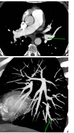
considered the gold standard for characterizing vessel morphology in CTEPH but is being challenged by advances in non-invasive modalities. CTPA (Figure 1) is currently widely used for assessment of operability and can also be valuable by evaluating bronchial artery collaterals, lung parenchyma (mosaic oligemia with minIP reconstruction, Figure 2) and mediastinum. Advanced CT technologies including dual-energy CT, ECG-gated area detector CT, cone-beam CT and contrast-enhanced Magnetic Resonance Pulmonary Angiography are emerging as valuable modalities for detailing the pulmonary vasculature.
Figure 2 ß Figure 1
Pag. 6 di 26 Therapy
Patients with untreated CTEPH are at high risk of progressive PH, right heart failure and death 22.
Early diagnosis and treatment by a multidisciplinary team are essential 23-24. Pulmonary
endarterectomy (PEA) is the gold-standard treatment, it is a potentially curative intervention, which in expert centres carries an in-hospital mortality rate <5% and leads hemodynamic and functional improvement with good long-term survival 25-26. However, less than 60% of patients with CTEPH
can undergo PEA, and PH persists or recurs after the procedure in 17–31% of patients 27-28.
For patients who are deemed inoperable or who have persistent or recurrent PH following PEA, targeted medical therapies developed for PAH can be given 29. The soluble guanylate cyclase
stimulator Riociguat and endothelin receptor antagonists (ERA) Macitentan are currently the only medical therapy for the treatment of CTEPH that has been shown to improve hemodynamics and exercise capacity 29-30. Phosphodiesterase 5 inhibitors (PDE5-I) and prostanoids are not approved for
this indication, because of a lack of positive randomised study evidence.
Percutaneous balloon pulmonary angioplasty (BPA) is an emerging option for patients with CTEPH who are not eligible for surgery or who have recurrent or persistent PH following PEA.
In this era of a comprehensive approach to CTEPH, an expert centre should be capable of evaluating and offering any/all established treatment modalities according to individual need
Treatment Algorithm67
#: multidisciplinary: PEA surgeon, PH expert, BPA interventionist and radiologist ¶: treatment assessment may differ depending on the level of expertise
Pag. 7 di 26
Balloon pulmonary angioplasty History, evolution and evidence
The first study of BPA was published in 2001 by Feinstein 31 in the USA, describing 18 patients with
inaccessible or “nonsurgical” CTEPH. Reperfusion pulmonary edema (RPE) occurred in 11 patients and 30-day mortality was 5.5%. Follow-up (mean 36 months) reported improvements in mean pulmonary arterial pressure (mPAP), New York Heart Association (NYHA) functional class and 6-min walking distance (6MWD), and all vessels previously dilated were patent at angiographic reassessment.
Following Feinstein’s report, BPA was abandoned for some years, mainly because of high complication rates. However, more recent reports, mainly from Japan, have revived interest. Sugimura 33 performed BPA in 12 patients with distal-type CTEPH stabilized for 1–3 months with
prostanoids, sildenafil and/or bosentan. Pulmonary hemodynamics were improved, and survival was better than for historical controls. Kataoka 32 performed BPA in 29 patients with inoperable CTEPH,
most of whom had previously been treated with an ERA, PDE-5 inhibitor or prostanoid. Functional class, brain natriuretic peptide levels and hemodynamics were improved at follow-up; medication was unchanged in most patients. Notably, hemodynamic effects took time to develop after BPA. Shortly after these studies were published, Andreassen 34 reported results with BPA for segmental
and subsegmental pulmonary arteries in 20 patients in Norway with inoperable disease or persistent PH after PEA. NYHA functional class and hemodynamic values were significantly improved, but periprocedural mortality was 10% and RPE occurred in seven patients. After 51 months’ follow-up, 85% of patients were still alive. Three further publications from European centres, one in 37 patients
24 and two others, each in nine patients 39-40, described improvements in mPAP, exercise capacity and
functional class after BPA, with low rates of complications.
Several Japanese centres have described improvements in hemodynamics, symptoms and exercise capacity after BPA, with generally low rates of major complications and post-procedural mortality
35- 37, 41-44, 46 with some follow-up over a year 36, 37, 44.
One report from Japan described outcomes of CTEPH managed using drug therapy (including pulmonary vasodilators), BPA or PEA 37. Despite significantly worse hemodynamic parameters at
baseline, patients managed with interventional therapies (PEA or BPA) had significantly superior survival at 5 years than those treated medically (98% versus 64%, respectively; p<0.0001). The efficacy and safety of BPA were similar in younger (<65 years) and older (⩾65 years) patients 38.
Data comparing outcomes of PEA in operable patients with those of BPA in inoperable patients suggest that the efficacy and safety of the two procedures are similar in their target cohorts 47.
In addition to the improvements in hemodynamics and functional capacity, there is accumulating evidence that BPA leads to improvements in right ventricular function, including volumes, ejection fraction, peak systolic strain, myocardial remodeling and alleviation of dyssynchrony 45, 46, 48-50.
It is difficult to compare results between countries and regions because of differences in patient selection and procedures, and possible differences in vascular structure and function related to ethnicity or diet.
Current practice
All patients with pulmonary hypertension, including CTEPH, should be evaluated and managed in a centre with specialist expertise and facilities 29, including a CTEPH team 5. A referral centre should
follow ⩾50 patients per month and receive at least 2 new referrals for PAH or CTEPH each month
29.
BPA should be reserved for expert centers (as for PEA) because it is a complex procedure that is not risk free and even with the technical refinements, there remains a steep learning curve in order to safely, effectively and consistently perform BPA 54. A successful BPA requires extensive training
Pag. 8 di 26
and case experience. BPA patient selection starts with a multidisciplinary review of all available and pertinent data and the first consideration in the evaluation of patients for BPA is the feasibility of PEA 29. The European Society of Cardiology/European Respiratory Society guidelines 29 state that
BPA may be considered for patients who are technically inoperable or who carry an unfavourable risk/benefit ratio for PEA (class IIb recommendation, level of evidence C).
This recommendation covers a very heterogeneous group and there are currently no data to identify those patients who might benefit most from BPA. Patient groups in whom BPA could potentially be used include those with inoperable CTEPH due to distal distribution of vascular obstructions, those with an unfavourable risk/benefit ratio for PEA (for example, few lesions but severe hemodynamics, or extreme surgical risk because of comorbidities, etc.), patients with persistent/recurrent PH after PEA, patients in need of rescue angioplasty after early failure of PEA 51, patients with residual lesions
after PEA, and patients with inoperable or persistent/recurrent CTEPH and an inadequate response to medical therapy. Some patients may have no distinct comorbidities that directly affect the surgical risk of PEA, but have limitations (e.g. general frailty, severe chronic obstructive pulmonary disease or inability to walk) such that the potential functional gain from surgery is very small (“low benefit” rather than “high risk”). BPA may be appropriate in these patients and is within the guideline recommendation. Conversely, BPA should not be offered to patients with large central clots or unilateral total occlusion 68 but can sometimes be justified as a rescue procedure for some
compromised patients that can be subjected to PEA but with severe comorbidities that contraindicate the surgery 62-63.
The rationale is to open obstructed vessels or widen stenotic lesions using a catheter-based intervention rather than the invasive surgery of PEA. The intention is to improve hemodynamics and pulmonary perfusion, ultimately reducing right ventricular afterload and preventing right ventricular failure.
Anatomical and functional assessment of pulmonary arteries and lung perfusion are critical to identify the target vessels 42. A selective pulmonary angiogram of the target vessels will show more details
and serves as confirmation prior to intervention during BPA but may not capture all the distal lesions potentially amenable to BPA, necessitating multiple complementary imaging modalities such as intravascular imaging and pressure gradient analysis to aid in lesion assessment and balloon sizing
40.
After identification of pulmonary artery lesions that can be treated a guide wire across the lesion or occlusion and balloon dilatation lead to restore pulmonary blood flow. Staged procedures with repeat catheterisations and dilatations are needed to achieve optimal hemodynamic results.
A comparison in terms of efficacy and complications between PEA and BPA is almost impossible: the two techniques are not mutually alternative 56. Instead the hybrid approach between the two
methods could be consider a treatment option for selected cases those who have a proximal localization of the thromboembolic material associated with a distal disease.
Technique
Percutaneous vascular access for BPA is femoral or jugular vein, using long sheaths through which pre-shaped guiding catheters (e.g. multipurpose shape, right or left coronary Judkins or left Amplatz shape) are inserted.
A long 6-, 7- or 8-French sheath containing a pre-shaped 6-French guiding catheter is placed in the pulmonary trunk and guided to the right or left main pulmonary artery. Usually the primary target for BPA (as for PEA) is the right lower lobe, because of its size and physiologically greater compartment of blood flow 52. In patients with low mPAP (i.e. <40 mmHg), more than one lobe may be treated in
a single session 32, 33, 53, 54.
It is important to have information about peripheral branches distal to the lesion in order to perform BPA safely 52. In severe cases, the primary goal is to reduce pressure by addressing simpler lesions
Pag. 9 di 26
first (i.e. web lesions or ring-like stenoses). The guide wire is passed carefully across the target lesion: proper guide wire placement must be proven before angioplasty can be performed (figure 3).
Figure 3
Recent practice in Japan has employed pressure wires to measure the ratio of distal to proximal pressures across the target lesion: this information can be used to reduce the risk of RPE 36, 53. In some
cases, soft hydrophilic guide wires are used, e.g. for the management of occlusive pulmonary artery lesions.
To begin the procedure, wires with tip loads <1 g should be used. Balloons should be undersized (i.e. a low balloon/artery ratio) to reduce the risk of vessel injury and reperfusion edema: a 2-mm balloon is typically used for initial dilatation, followed by larger balloons according to lesion size and residual pressure. The balloon is inflated by hand until the fluoroscopic waist disappears or until the balloon is expanded at 5–8 atm. For that reason, compliant balloons are preferred. Repeat dilatations with bigger balloons are performed if there is <50% increase in angiographic vessel size and an increase of pulmonary venous backflow is documented. In case of chronic total occlusions, microcatheters and wires with higher tip loads are used, similar to conventional chronic total occlusion techniques. Between three and 10 sessions of BPA are usually required for each patient to achieve the desired mPAP reduction and treat all amenable lesions. The number of sessions required depends on the extent of disease, the location and configuration of the lesions and the skills of the operator.
At each session, treatment is targeted to segmental and subsegmental pulmonary arteries of one pulmonary lobe. The need for repeated sessions is an important consideration when explaining the procedure, planning resources and evaluating potential hazards from repeated exposure to radiation and contrast media, including contrast-induced nephropathy.
After BPA, many patients still show elevated pressure and resistance values 24: hybrid therapy
(medical treatment combined with BPA) may be a promising option 24. This could include acute or
chronic “bridging” treatment before BPA, to improve hemodynamics and potentially reduce the risk of complications, or after BPA in patients whose hemodynamic response is unsatisfactory. Prospective studies need to be performed in this setting. In many BPA studies, some or all patients received additional targeted medical therapy to stabilize the patient or improve hemodynamics before the procedure, and this therapy was generally continued afterwards. However, medical therapy in conjunction with BPA has not been systematically evaluated and currently no robust data exist on the benefits of medical treatment before and after BPA.
Pag. 10 di 26 Lesions
In retrospective study it was found that different types of lesions correspond to different effects and a variable incidence of complications. For this reason it was necessary to create a new classification that took into account the angiographic aspect of pre-treatment lesions 42. Five categories have been
identified based on the lesion opacity and the blood flow distal to the lesion (figures below): A) ring stenosis
B) web
C) sub-occlusive lesions D) total vascular occlusions E) tortuous lesions
Ring stenoses and "webs" have the highest success rate and the least chance of causing complications
42. Despite the availability of different imaging techniques, the use of more advanced methods make
BPA a long, complex and expensive procedure, as well as impossible to implement in patients with a more compromised clinical status. Therefore, to standardize the procedure, it is necessary to identify the universal imaging method: selective pulmonary angiography is considered superior to any other modality 57.
Effects
Results demonstrating the impact of BPA on right ventricular function (analyzed both by echocardiogram and cardiac MRI) come from retrospective studies. There is a "reverse remodeling" of the right ventricular associated with an overall improvement of the hemodynamic parameters 48.
Cardiac MRI 3-6 months after the last session documents reduction of right ventricle dimension and thickening, increase of ejection fraction and an improvement in the straightening of the interventricular septum 35. Reduction in right ventricular dyssynchrony, recovery of left ventricular
function following decompression by the right ventricle and reduction of resting heart rate are also described as an effects of BPA 49-50.
BPA also has a beneficial effect in terms of ventilatory function: with the cardiopulmonary exercise test carried out immediately after the last procedure, the maximum oxygen consumption is significantly improved. It is interesting to note that it correlates more with the number of procedures performed than with the hemodynamic parameters: probably because, besides hemodynamics, BPA also produces a beneficial effect on the symptoms of heart failure and, consequently, induces a
Pag. 11 di 26
peripheral adaptation between one session and the other 58. Furthermore, the reduction of
intrapulmonary shunts and the parallel increase in venous saturation of oxygen contribute to improving hypoxemia.
At the systemic level, improvements in renal function, metabolism, nutritional status and vascular function have been described 59. In particular, despite the use of iodinated contrast agents during the
procedure, renal function is improved for two main reasons: first, BPA leads to a reduction in venous congestion, a major cause of worsening of renal function in patients with heart failure; second, the increase in cardiac output improves pre-renal component 64.
Although based on individual questionnaires, recent studies show that BPA also has a significant impact on the quality of life 65.
Complications
Different categories of complications were identified: During the procedure:
§ Vascular injury with/without haemoptysis § Wire perforation
§ Balloon overdilatation
§ High-pressure contrast injection § Vascular dissection
§ Allergic reaction to contrast
§ Adverse reaction to conscious sedation/local anaesthesia After the procedure:
§ Lung injury (radiographic opacity with/without haemoptysis, with/without hypoxaemia) § Renal dysfunction
§ Access site problems
Vascular injury and dissection can lead to signs and symptoms such as haemoptysis, coughing or hypoxia within 20 min, although they can be asymptomatic in some cases.
Wire perforation is the most common pulmonary artery injury and is usually caused by deep insertion of a guide wire into peripheral small branches, although if managed correctly. Wire perforation can be reduced by stabilizing the guiding catheter and avoiding deep insertion of the guide wire in lesions with unclear distality. The risk may also be reduced by avoiding BPA for complete obstructions, which require a heavy-weight-tip wire.
Balloon overdilatation and high-pressure contrast injection can lead to the rupture of a pulmonary arterial branch with a blood extravasation that is manifested by hemoptysis and worsening of oxygenation; over time, heart failure and shock may occur. The first method of intervention involves the use of a balloon at low pressure upstream of the breaking point for about 15 minutes. The procedure can be repeated several times and, if it does not lead to the resolution, an option is embolization with bioabsorbable gel or with metal spirals, which exclude the vessel irreversibly. Finally, although there are few data available on their long-term effectiveness 60, it may be necessary
to insert coated stents. Sometimes, to stop bleeding, it may be appropriate to evaluate the surgeon's intervention.
Damage caused by adhesion of the guides or catheter to the vascular wall may cause a pulmonary artery dissection. A close follow-up showed that, if left untreated, the dissection tends to disappear generally in one week 61.
The RPE represents the most frequent complication and was observed by Feinstein in 2/3 of the patients. Reperfusion edema / alveolar hemorrhage develops 24-72 hours (Figure 4) from the procedure for a number of factors: microtraumas caused by the guides, release of inflammatory mediators and due to the hemodynamic effect caused by high-pressure reperfusion of areas that for a
Pag. 12 di 26
long period received little amount or zero blood flow 52. Following the introduction of IVUS (led to
a more precise selection of the target segment and of the balloon size), the clinical-instrumental follow-up of patients undergoing BPA showed a clear reduction in severe lung injury, to the detriment of a relative increase in subclinical damage.
Figure 4
BPA is associated with a higher incidence of RPE compared to PEA. The reason is the fractionation of BPA treatment in several session: each distal pulmonary arterial branch mechanical opened receives a high pressure flow coming from the healthy district upstream; in PEA, most of the obstructions are removed in a single session, so the flow is redistribute in the pulmonary arterial tree downstream. Furthermore, the effect of pro-inflammatory cytokines seems to have an indirect role. Despite various efforts to reduce its incidence, the complication seems to be inevitable, especially following the first sessions. Therefore the focus has shifted to prevention: PEPSI (Pulmonary Edema Predictive Scoring Index) was created 52, obtained by multiplying the variation of the Pulmonary
Flow Grade score by the baseline value of pulmonary vascular resistance (PVR). This index has an high negative predictive value and correlates most with the risk of RPE. Pulmonary Flow Grade is a score based on the corresponding TIMI (Thrombolysis in Myocardial Infarction), used for the coronary angioplasty procedure. It divides the perfusion of the target vessels into four levels based on their angiographic appearance:
• Grade 0: absence of anterograde flow in the pulmonary arterial branches or minimal passage of contrast medium that remains trapped, with lack of opacification of the distal vascular bed.
• Grade 1: partial perfusion of the pulmonary arterial branches with passage of contrast medium in the circulation downstream of the lesion.
• Grade 2: complete perfusion of the distal pulmonary arterial branches with partial perfusion of the pulmonary venous branches, in which the flow appears slower compared to the areas not affected by obstruction.
• Grade 3: complete perfusion of arterial and pulmonary venous branches.
The treatment of a target lesion should lead to the achievement of grade 3: the grade improvement is significantly related to hemodynamic improvement. Furthermore, taking into account the basal PVR, PEPSI could play a role in defining the subgroup of patients candidates to benefit from a more aggressive approach, treating more vessels for each session: the therapeutic effect of BPA, in fact, is directly proportional to the number of treated vessels 53.
Since the development of RPE correlates with basal values of mean pulmonary arterial pressure, Inami et al. introduce pressure-wire guides capable of accurately detecting pressure values upstream and downstream of the target lesion. This reduces the reperfusion damage monitoring directly the blood pressure values and contributes to obtaining a significant hemodynamic improvement, minimizing the number of insufficiently treated vessels 36.
Pag. 13 di 26 PROJECT Background
In CTEPH, stenoses or obstructions of the pulmonary arteries due to unresolved organized thrombi cause an elevation of pulmonary pressure and PVR, which can result to progressive right heart failure
1-2. Traditionally, the treatment of choice for CTEPH is PEA 3-4; however, not all patients are eligible
for PEA and more than 30% of patients with CTEPH are deemed unsuitable due to distal lesion, advanced age, or comorbidities and have a poor prognosis and limited treatment options. Furthermore, of those who have undergone the surgery, 17%–31% experience recurrence or persistence of PH 5-6.
Recently, BPA has arisen as a promising alternative treatment strategy for CTEPH patients with inoperability or recurrence of PH after PEA: previous studies from Japan and Europe showed favorable results with regard to hemodynamics 7-8, right ventricular function and exercise capacity 9-11, quality of life 12 and respiratory function13 in CTEPH patients after BPA and risk/benefit imbalance
appear to be safe in experimented centres.
There have been no prior studies on the efficacy and safety of BPA in CTEPH patients from Italy. Therefore, we sought to compare clinical and hemodynamic parameters at the baseline and last BPA session in CTEPH patients who were inoperable or who experienced recurrence or persistent PH after PEA referred to Bologna Centre.
Purpose
The aim of this study was to compare clinical and hemodynamic parameters at the baseline and after last BPA session in inoperable CTEPH patients and in who experienced recurrence or persistent PH after PEA.
Methods Study population
The present study was single center, observational study of CTEPH patients who received BPA between June 2015 and September 2019 at Sant’Orsola Malpighi Hospital of Bologna.
All symptomatic patients (NYHA ≥ II), incident or prevalent, with inoperable CTEPH or with residual PH post PEA with a hemodynamic profile characterized by mPAP > 25 mmHg and PVR > 3.0 WU at the time of evaluation were considered eligible for BPA.
Baseline assessment included the NYHA functional class, 6-minute walk distance (6MWD), high resolution chest CT (HRCT), hemodynamic parameters by right heart catheterization (RHC) and non selective pulmonary angiography.
For this study we collected clinical and hemodynamic parameters in three different moments of the clinical history of our patients: at baseline (for inoperable patients we consider baseline RHC and for operable patients that underwent PEA we consider the first RHC post PEA that show residual PH), immediately before the first BPA session and after 3-6 months after the last BPA session; in patients who have not yet completed the treatment (target goals not yet been reached) and therefore have not performed a reassessment after the end of BPA program, we consider as post-BPA data the last RHC, 6MWD and functional evaluation performed immediately before the last session.
The day after every session HRCT is performed to identify "clinically silent" adverse events (radiographic opacity in the absence of hemoptysis with or without desaturation)
Pag. 14 di 26 Technique
All patients undergoing BPA receive written information regarding the procedure and provided informed consent. The BPA procedure is carried out by at least two interventional cardiologists. Oral anticoagulation (OAC) with vitamin K antagonist is withdrawn at least 3 days before the procedures and replaced with low weight molecular heparin, while patients treated with Non-vitamin K antagonist oral anticoagulants withdrawn OAC one or two days before the procedure (according to the number of administrations of the drug).
Firstly, we placed a 7F indwelling sheath into a mainly femoral vein and a 6F long sheath was brought into the main pulmonary artery. We performed RHC and then selected a branch of the pulmonary artery using an appropriate 6F guiding catheter and performing selective angiography to detect target lesions: the choice was optimized by the possibility of using bi-plane angiographic room in association with the 3D-CT reconstruction (Figure 5) of pulmonary circulation available in our centre.
Figure 5
Unfractionated heparin was additionally infused after introduction of the guiding catheter according to the activated clotting time (target during procedure: 200-300). A 0.014-inch wire crossed to the targeted lesion, and then the lesion was dilated using balloon catheters of the appropriate size. The balloon was inflated by hand until the indentation disappeared or until the balloon is fully expanded. If the vessel was dilated for the first time, a small-sized balloon was used regardless of reference diameter of the segmental artery to prevent procedure related complications and a slightly bigger balloon was used subsequently (or in the subsequent session). After inflation, angiography was performed to confirm sufficient dilation and the presence of ruptured vessels. Dilatation was repeated if deemed insufficient.
The day after BPA each patient underwent high resolution chest CT, irrespective of the clinical events that occurred during the session, to detects “silent” side effects and to manage the resumption of OAC.
A second session of BPA was done within 30-60 days of the first BPA session. We performed additional sessions of BPA if patients were still symptomatic (NYHA more than II) or had a mean pulmonary artery pressure greater than 30 mmHg and/or PVR more than 4 WU at follow-up.
Pag. 15 di 26 Statistical analysis
Data, including number of sessions and vessels treated, are expressed as median and interquartile range. Comparisons between baseline, pre-BPA and post-BPA data were conducted with Friedman test with two-by-two comparison and the pairwise post hoc analysis was performed with Bonferroni correction. Survival was calculated starting from the catheterization performed before the first BPA session and for the survival analysis we used the Kaplan-Meier method, considering mortality for all causes and comparing BPA group with other group according to different therapeutic strategies [PEA, medical therapy, no therapy (historical group)], taking advantage of the historical data of our register.
Results
47 patients with CTEPH [40 with inoperable disease (85%) and 7 with PH residual/recurrence after PEA (15%)] were considered eligible for BPA treatment; male 45%, median age 68 (51-74) years. The population had the following clinical, functional and hemodynamic characteristics at baseline:
NYHA III-IV (%) 89 6MWD (m)* 414 (348-523) RAP (mmHg) 6 (5-8) mPAP (mmHg) 45 (40-50) PAWP (mmHg) 8 (7-10) CI (L/min/m2) (2.2-3.0)2.6 PVR (WU) 7.5 (5.6-11.5) PA O2 Sat (%) 67 (62-71) PAC (ml/mmHg) 1.1 (0.8-1.5)
CI, Cardiac Index; mPAP, mean Pulmonary Arterial Pressure; PVR, Pulmonary Vascular Resistance; PA O2 Sat, Pulmonary Artery Oxygen Saturation; RAP, Right Atrial Pressure; 6MWD, 6 Minute Walking Distance; WHO-FC, World Health Organization Functional Class; PAC, pulmonary artery compliance. *40 patients
At baseline current PAH specific therapies and anticoagulation strategy were: PAH specific therapy N (%)
None 34 (72%)
MonoTx 8 (17%)
Combination Tx 5 (11%)*
*One in triple combination therapy with iv epoprostenol
Anticoagulation drug N (%)
Warfarin 28 (60%)
NOACs 17 (36%)
Pag. 16 di 26
Baseline assessments were conducted approximately 6 months (2-42) before the observation period; the reason for this extended range of time is that we included in BPA program both incident and prevalent patients (who were followed in Bologna Center and perform first RHC since before June 2015 and meet criteria for our study).
The medical therapy at the time of insertion in the study were: PAH specific therapy N (%)
None 1 (2%)
MonoTx 28 (60%)
Combination Tx 18 (38%)*
*One in triple combination therapy with iv epoprostenol
Our population at the time of inclusion in the study had the following characteristics: NYHA III-IV (%) 85 6MWD (m)* 425 (350-496) RAP (mmHg) 6 (5-8) mPAP (mmHg) 41 (34-48) PAWP (mmHg) 9 (7-11) CI (L/min/m2) (2.1-3.0)2.6 PVR (WU) 7.1 (4.7-10.3) PA O2 Sat (%) 68 (63-73) PAC (ml/mmHg) 1.2 (0.8-1.8) *38 patients
None
2%
ERA
7%
PDE5-I
53%
Combo
38%
Pag. 17 di 26 The following table shows changes from baseline to pre-BPA:
N° 47 patients Baseline Pre-BPA p
NYHA III-IV (%) 89 85 N.S. 6MWD (m)* (348-523)414 (350-496)425 N.S. RAP (mmHg) (5-8)6 (5-8)6 N.S. mPAP (mmHg) (40-50)45 (34-48)41 <0.05 PAWP (mmHg) (7-10)8 (7-11)9 N.S. CI (L/min/m2) 2.6 (2.2-3.0) 2.6 (2.1-3.0) N.S. PVR (WU) (5.6-11.5)7.5 (4.7-10.3)7.1 <0.05 PA O2 Sat (%) (62-71)67 (63-73)68 N.S. PAC (ml/mmHg) (0.8-1.5)1.1 1.2 (0.8-1.8) N.S. *38 patients
During the study period 136 sessions of BPA were performed and each patient went through an average of 2 (2-3.5) sessions of BPA, with 2 (2-3) vessels dilated for each session.
0 2 4 6 8 10 12 14 16 1 2 3 4 5 6 7 8 N °pat ie nt s N° sessions
Pag. 18 di 26
At the end of the observation period 42 patients (89.4%) performed at least a post-BPA clinical, functional and hemodynamic evaluation [time 11 (5-19) months after the pre-BPA assessment]. 5 patients did not perform a reassessment of the clinical and hemodynamic picture for the following reasons:
• A lung lesion compatible with heteroplasia was found in 1 patient after the first BPA session. For this reason, the BPA program has been suspended.
• 1 patient delayed the hemodynamic evaluation after first BPA session because, in the meantime, he underwent coronary angioplasty due to acute coronary syndrome and then he died for sudden death during follow up before the post-BPA revaluation.
• 1 patient died for sepsis before the post-BPA revaluation
• 1 patient performed the first BPA session near the end of the observation period. For this reason, it was not possible to have a revaluation.
• 1 patient refused to perform any other invasive test after the first session of BPA
There was no modification of PAH specific drugs between pre-BPA and the end of the observation period
Clinical, functional and hemodynamics effects of BPA are shown in the following table:
N° 42 patients Pre-BPA Post-BPA p
NYHA III-IV (%) 83 42 <0.05 6MWD (m)* (344-493)425 (370-543)446 <0.05 RAP (mmHg) (5-8)6 (4-7)6 N.S. mPAP (mmHg) (36-48)43 (29-40)35 <0.05 PAWP (mmHg) (7-11)9 (8-11)10 N.S. CI (L/min/m2) 2.7 (2.2-3.0) 2.9 (2.4-3.5) <0.05 PVR (WU) (4.8-9.9)7.2 4.7 (3.3-6.3) <0.05 PA O2 Sat (%) (63-73)68 69 (61-72) N.S. PAC (ml/mmHg) (0.8-1.7)1.2 1.7 (1.2-2.4) <0.05 *36 patients
Treatment goals (mPAP <30 mmHg and/or PVR <4 WU) was reached within the study period for 18 (38%) patients. The other patients have been scheduled for additional BPAs in the near future except for the patients who refused further invasive treatment.
Pag. 19 di 26 Results summary is shown in the following table
median (interquartile range) Baseline (n= 47) Baseline ÷ Pre-BPA 6 (2-42) months Pre-BPA (n= 47) Pre-BPA ÷ Post-BPA 11 (5-19) months Post-BPA (n= 42) p-value Global WHO-FC III-IV (%) 89 N.S. 85 <0.05 42 <0.001 6MWD (m) (348-523)414 N.S. 425 (350-496) <0.05 446 (370-543) <0.001 RAP (mmHg) (5-8)6 N.S. 6 (5-8) N.S. 6 (4-7) 0.006 mPAP (mmHg) (40-50)45 <0.05 41 (34-48) <0.05 35 (29-40) <0.001 CI (L/min/m2) (2.2-3.0)2.6 N.S. (2.1-3.0)2.6 <0.05 (2.4-3.5)2.9 <0.001 PVR (WU) (5.6-11.5)7.5 <0.05 7.1 (4.7-10.3) <0.05 4.7 (3.3-6.3) <0.001 PA O2 Sat (%) (62-71)67 N.S. 68 (63-73) N.S. 69 (61-72) <0.001 PAC (ml/mmHg) (0.8-1.5)1.1 N.S. (0.8-1.8)1.2 <0.05 (1.2-2.4)1.7 <0.001 CI, Cardiac Index; mPAP, mean Pulmonary Arterial Pressure; PVR, Pulmonary Vascular Resistance; PA O2
Sat, Pulmonary Artery Oxygen Saturation; RAP, Right Atrial Pressure; 6MWD, 6 Minute Walking Distance; WHO-FC, World Health Organization Functional Class; PAC, pulmonary artery compliance.
Safety
During the observation period 44% of the sessions took place in the absence of clinical and/or radiological complications. The remaining 56% of the sessions were characterized by events for the most of the times mild and with spontaneous remission, which rarely required an active role by the medical-nursing staff.
All the clinical-radiological events occurring in the peri-procedural period were grouped into four main categories: hemoptysis, reperfusion edema/alveolar hemorrhage, pulmonary vascular dissections and complications at the level of vascular accesses.
Hemoptysis was the most frequent clinical complication, affecting 14 patients (30%) for a total of 24 procedures (18%). In most cases (92%) it resolved spontaneously as it was a mild hemoptysis / haemoftoe in the absence of significant desaturation or hemodynamic compromise; a case of moderate hemoptysis required administration e.v. of protamine, while in only one case the severity of the complication was such as to require, in addition to the recombination by protamine infusion, endotracheal intubation and subsequent admission to intensive care unit. No patient required percutaneous cardiopulmonary support.
As already illustrated above, in order to highlight aspects of RPE or alveolar hemorrhage, all patients (even in the absence of clinical events) underwent HRCT scan 24 hours after each single session. Overall, the radiological evidence of RPE involved 33 patients (70%) for a total of 80 sessions (59%). Complications at the level of vascular accesses following the execution of right cardiac catheterization by right femoral venous were documented in 7 patients (15%) with a total of 7 procedures (5%): in particular, we recorded four hematoma (two of them supplied from the superficial femoral artery), two arteriovenous fistula and a pseudoaneurysm.
Pag. 20 di 26
Pulmonary artery dissection/perforation was documented in five patients (11%), only one episode led to hemoptysis, desaturation and required direct medical intervention (use of a balloon at low pressure upstream of the breaking point); the other four remained a “silent” side effect because were observed at fluoroscopy during selective angiography of treated vessel at proximal level and did not correlate to any symptom or clinical event.
None of the patients required an extracorporeal membrane oxygenation. The following table resume the complication rate:
N° patients (%) N° sessions (%)
Hemoptysis 14 (30) 24 (18)
RPE 33 (70) 80 (59)
Vascular access complication 7 (15) 7 (5)
Dissection/perforation 5 (11) 5 (4)
Survival
During the follow-up 5 deaths were recorded: one patient for sepsis, one for heart failure, one for arrhythmic storm, one had an advanced cancer problem and one patient with known severe multivessel coronary artery disease died of sudden cardiac death; during BPA sessions and in the following 30 days no deaths (0%) occurred, given that it goes to confirm the safety of this procedure.
Kaplan Meier Curves
PEA 171 147 140 131 120 114 Drug 202 155 141 118 102 81 None 32 19 16 15 14 9 BPA 47 35 23 12 2 0 0.00 0.25 0.50 0.75 1.00 Su rvi va l 47 35 23 12 2 0 gruppi = 6 32 19 16 15 14 9 gruppi = 4 202 155 141 118 102 81 gruppi = 3 171 147 140 131 120 114 gruppi = 2 Number at risk 0 12 24 36 48 60 Follow-up (months) gruppi = 2 gruppi = 3 gruppi = 4 gruppi = 6
Survival
Pag. 21 di 26 Discussion
For CTEPH there are several therapeutic strategies, each of which is dedicated to selected patient. Surgical treatment, when possible, must be considered the gold standard, as it is potentially curative, and the long-term survival of the operated subjects is significantly higher than that of the non-operated subjects 66. The hemodynamic effects are immediate with a sudden drop in pulmonary
pressures. Therefore, the careful selection of operable patients remains fundamental, which starts from the diagnosis of CTEPH.
Despite the many advances in recent years, the high percentage of inoperable patients or those with residual PH after surgery 27 requires effective and safe alternative treatments: precisely for these
patients over the last few years, BPA has been emerging, an interventional method aimed at the unblocking of pulmonary arterial vessels that are affected by distal location of thromboembolic material. This method is still limited to a few expert centres at international level and in the process of optimization, both for long-term effectiveness and safety.
The aim of our study was to demonstrate efficacy and safety of BPA procedure in patients with inoperable CTEPH or with residual PH post PEA referred to Sant’Orsola-Malpighi Hospital of Bologna.
Our centre started in June 2015 the selection of patients to be submitted to BPA with a multi-step program of dilation sessions with the final goal of reaching mPAP values <30 mmHg and RAP <4.0 WU.
47 patients eligible for percutaneous treatment were identified: at the time of enrollment in the study, a part of the patients had already been followed at the PH center of this Hospital and was being treated with PAH specific therapy. Each patient was reassessed from a clinical, functional and hemodynamic point of view before underwent a percutaneous treatment. Comparing the baseline data obtained at the first evaluation in Bologna centre and those obtained before the first BPA session [distance 6 months (2-42)] we observed that medical therapy ("reverse remodeling" of distal open pulmonary circulation not involved in thromboembolic processes) determined the stabilization of symptoms, exercise capacity and main hemodynamic parameters.
At the end of the observation period 42 patients (89.4%) performed at least a post-BPA clinical, functional and hemodynamic evaluation [time 11 (5-19) months after the pre-BPA assessment]; after a total of 136 sessions [2 sessions (2-3) per patient] we documented a decrease in mPAP from 43 (36-48) to 35 (29-40) mmHg equal to 19 % (p- value <0.05), a reduction in PVR from 7.2 (4.8-9.9) to 4.7 (3.3-6.3) WU equal to 35% (p value < 0.05) and an increase in IC from 2.7 (2.2-3.0) to 2.9 (2.4-3.5) l/min/m2 equal to 7% (p value <0.05). Along with hemodynamic changes also exercise capacity had
improved, with an increase in distance at 6MWD of 21 meters, from 425 (344-493) to 446 (373-539) meters (p value <0.05). We also observed a reduction in the number of patients in NYHA class III-IV from 83% to 42% (p value <0.05).
Regarding the safety profile, we considered both clinical and "silent" events by performing HRCT scan 24 hours after each BPA session. During the 136 BPA sessions there were 24 episodes of hemoptysis (involving 14 patients) which still represents the most frequent peri-operative complication that occurred in our centre, although in most cases it is mild (in one case, protamine infusion was required to stop the bleeding and in another case it was also necessary endotracheal intubation); the event may be an expression of direct vascular damage or due to reperfusion.
We have documented five episodes of iatrogenic dissection/perforation of a segmental pulmonary artery (4% of the total number of sessions): only one of that was related to clinical and hemodynamic repercussions and require direct treatment.
From the radiological point of view, however, we have highlighted the appearance of reperfusion edema/alveolar hemorrhage in 59% of the sessions; radiological change has also been interpreted as an indication of successful reperfusion and not only as a complication. Data provided by the HRCT scan were useful also for management of anticoagulant therapy immediately after the procedure.
Pag. 22 di 26
From our experience, pulmonary angioplasty has proved to be an effective and safe procedure in patients with CTEPH who were deemed inoperable due to distal localization of the thromboembolic material or with residual PH post PEA, constituting a therapeutic option preferable to pharmacological therapy alone.
Comparing the patients of the study with the historical data present in our register relating to CTEPH patients not subjected to this treatment, although observing a peri-procedural mortality rate of zero in the short term, it is not possible to define a clear advantage on survival in the absence of consistent long-term data.
Other relevant limitation of this study is that the results obtained are negatively affected by many patients included in the study who have not yet achieved the therapeutic goals (62%) and require further sessions.
Conclusions
The current knowledge of CTEPH leads us to consider this condition as a complex disease comprising proximal or distal (or both) chronic obstruction by organized fibrotic thrombus emboli, small vessel disease and remodeling of the whole pulmonary vascular bed. Even if the gold standard treatment for this disease remain PEA, BPA has emerged as a promising safe and effective procedure in experienced hands, able to improve symptoms, exercise tolerance and hemodynamic profile for inoperable patients and for patients with residual PH after surgery.
An expert centre must be able to offer a multimodal, individualized approach to treatment integrating surgical, interventional, imaging and medical PH expertise with the development of clear outcomes analyses. Until now Sant’Orsola-Malpighi Hospital of Bologna is the only Italian centre able to offer all the different therapeutic options for a patient with CTEPH.
Pag. 23 di 26 Bibliography
1. Simonneau G, Montani D, Celermajer DS, Denton CP, Gatzoulis MA, Krowka M, Williams PG, Souza R. Haemodynamic definitions and updated clinical classification of pulmonary hypertension. Eur Respir J. 2019 Jan 24;53(1).
2. Lang IM, Pesavento R, Bonderman D, Yuan JX. Risk factors and basic mechanisms of chronic thromboembolic pulmonary hypertension: a current understanding. Eur Respir J 2013; 41: 462–468.
3. Moser KM, Bloor CM. Pulmonary vascular lesions occurring in patients with chronic major vessel thromboembolic pulmonary hypertension. Chest 1993;103:685–692.
4. Dorfmüller P, Günther S, Ghigna M-R, et al. Microvascular disease in chronic thromboembolic pulmonary hypertension: a role for pulmonary veins and systemic vasculature. Eur Respir J 2014;44:1275-1288.
5. Kim NH, Delcroix M, Jenkins DP, et al. Chronic thromboembolic pulmonary hypertension. J Am Coll Cardiol 2013; 62: Suppl. 25, D92–D99.
6. Fedullo P, Kerr KM, Kim NH, Auger WR. Chronic thromboembolic pulmonary hypertension. Am J Respir Crit Care Med 2011; 183: 1605–1613.
7. Simonneau G, Torbicki A, Dorfmüller P, Kim NH. The pathophysiology of chronic thromboembolic pulmonary hypertension. Eur Respir Rev 2017; 26: 160112.
8. Pengo V, Lensing AW, Prins MH, et al. Incidence of chronic thromboembolic pulmonary hypertension after pulmonary embolism. N Engl J Med 2004; 350: 2257–2264.
9. Becattini C, Agnelli G, Pesavento R, et al. Incidence of chronic thromboembolic pulmonary hypertension after a first episode of pulmonary embolism. Chest 2006; 130: 172–175. 10. Klok FA, van Kralingen KW, van Dijk AP, Heyning FH, Vliegen HW, Huisman MV.
Prospective cardiopulmonary screening program to detect chronic thromboembolic pulmonary hypertension in patients after acute pulmonary embolism. Haematologica 2010; 95: 970–975.
11. Martí D, Gómez V, Escobar C, et al. Incidencia de hipertensión pulmonar tromboembólica crónica sintomática y asintomática. [Incidence of symptomatic and asymptomatic chronic thromboembolic pulmonary hypertension]. Arch Bronconeumol 2010; 46: 628–633.
12. Poli D, Grifoni E, Antonucci E, et al. Incidence of recurrent venous thromboembolism and of chronic thromboembolic pulmonary hypertension in patients after a first episode of pulmonary embolism. J Thromb Thrombolysis 2010; 30: 294–299.
13. Surie S, Gibson NS, Gerdes VE, et al. Active search for chronic thromboembolic pulmonary hypertension does not appear indicated after acute pulmonary embolism. Thromb Res 2010; 125: e202–e205.
14. Berghaus TM, Barac M, von Scheidt W, et al. Echocardiographic evaluation for pulmonary hypertension after recurrent pulmonary embolism. Thromb Res 2011; 128: e144–e147. 15. Held M, Hesse A, Gött F, et al. A symptom-related monitoring program following pulmonary
embolism for the early detection of CTEPH: a prospective observational registry study. BMC
Pulm Med 2014; 14: 141.
16. Giuliani L, Piccinino C, D'Armini MA, et al. Prevalence of undiagnosed chronic thromboembolic pulmonary hypertension after pulmonary embolism. Blood Coagul
Fibrinolysis 2014; 25: 649–653.
17. Guérin L, Couturaud F, Parent F, et al. Prevalence of chronic thromboembolic pulmonary hypertension after acute pulmonary embolism. Thromb Haemost 2014; 112: 598–605. 18. Kayaalp I, Varol Y, Çimen P, et al.The incidence of chronic thromboembolic pulmonary
hypertension secondary to acute pulmonary thromboembolism. Tuberk Toraks 2014; 62: 199–206.
Pag. 24 di 26
19. Vavera Z, Vojacek J, Pudil R, Maly J, Elias P. Chronic thromboembolic pulmonary hypertension after the first episode of pulmonary embolism? How often? Biomed Pap Med
Fac Univ Palacky Olomouc Czech Repub 2016; 160: 125–129.
20. Klok FA, Tesche C, Rappold L, et al. External validation of a simple non-invasive algorithm to rule out chronic thromboembolic pulmonary hypertension after acute pulmonary embolism.
Thromb Res 2015; 135: 796–801.
21. He J, Fang W, Lv B, et al. Diagnosis of chronic thromboembolic pulmonary hypertension: comparison of ventilation/perfusion scanning and multidetector computed tomography pulmonary angiography with pulmonary angiography. Nucl Med Commun 2012;33: 459– 463.
22. Delcroix M, Lang I, Pepke-Zaba J, et al. Long-term outcome of patients with chronic thromboembolic pulmonary hypertension: results from an international prospective registry. Circulation 2016; 133: 859–871.
23. Jenkins D, Mayer E, Screaton N, et al. State-of-the-art chronic thromboembolic pulmonary hypertension diagnosis and management. Eur Respir Rev 2012; 21: 32–39.
24. Kurzyna M, Darocha S, Koteja A, et al. Balloon pulmonary angioplasty for chronic thromboembolic pulmonary hypertension. Postepy Kardiol Interwencyjnej 2015; 11: 1–4. 25. Jenkins D. Pulmonary endarterectomy: the potentially curative treatment for patients with
chronic thromboembolic pulmonary hypertension. Eur Respir Rev 2015; 24: 263–271. 26. Jenkins D, Madani M, Fadel E, et al. Pulmonary endarterectomy in the management of chronic
thromboembolic pulmonary hypertension. Eur Respir Rev 2017; 26: 160111.
27. Freed DH, Thomson BM, Berman M, et al. Survival after pulmonary thromboendarterectomy: effect of residual pulmonary hypertension. J Thorac Cardiovasc Surg 2011; 141: 383–387.
28. Mayer E, Jenkins D, Lindner J, et al. Surgical management and outcome of patients with chronic thromboembolic pulmonary hypertension: results from an international prospective registry. J Thorac Cardiovasc Surg 2011; 141: 702–710.
29. Galiè N, Humbert M, Vachiery JL, Gibbs S, Lang I, Torbicki A, et al. 2015 ESC/ERS Guidelines for the diagnosis and treatment of pulmonary hypertension: the Joint Task Force for the Diagnosis and Treatment of Pulmonary Hypertension of the European Society of Cardiology (ESC) and the European Respiratory Society (ERS): endorsed by: Association for European Paediatric and Congenital Cardiology (AEPC), International Society for Heart and Lung Transplantation (ISHLT). Eur Respir J 2015;46(4):903-75.
30. Ghofrani HA, Simonneau G, D'Armini AM, Fedullo P, Howard LS, Jaïs X, Jenkins DP, Jing ZC, Madani MM, Martin N, Mayer E, Papadakis K, Richard D, Kim NH; MERIT study investigators. Macitentan for the treatment of inoperable chronic thromboembolic pulmonary hypertension (MERIT-1): results from the multicentre, phase 2, randomised, double-blind, placebo-controlled study. Lancet Respir Med. 2017 Oct;5(10):785-794
31. Feinstein JA, Goldhaber SZ, Lock JE, et al. Balloon pulmonary angioplasty for treatment of chronic thromboembolic pulmonary hypertension. Circulation 2001; 103: 10–13.
32. Kataoka M, Inami T, Hayashida K, et al. Percutaneous transluminal pulmonary angioplasty for the treatment of chronic thromboembolic pulmonary hypertension. Circ Cardiovasc Interv 2012; 5: 756–762.
33. Sugimura K, Fukumoto Y, Satoh K, et al. Percutaneous transluminal pulmonary angioplasty markedly improves pulmonary hemodynamics and long-term prognosis in patients with chronic thromboembolic pulmonary hypertension. Circ J 2012; 76: 485–488.
34. Andreassen AK, Ragnarsson A, Gude E, et al. Balloon pulmonary angioplasty in patients with inoperable chronic thromboembolic pulmonary hypertension. Heart 2013; 99: 1415–1420. 35. Fukui S, Ogo T, Morita Y, et al. Right ventricular reverse remodelling after balloon
Pag. 25 di 26
36. Inami T, Kataoka M, Shimura N, et al. Pressure-wire-guided percutaneous transluminal pulmonary angioplasty: a breakthrough in catheter-interventional therapy for chronic thromboembolic pulmonary hypertension. JACC Cardiovasc Interv 2014; 7: 1297–1306. 37. Inami T, Kataoka M, Ando M, et al. A new era of therapeutic strategies for chronic
thromboembolic pulmonary hypertension by two different interventional therapies; pulmonary endarterectomy and percutaneous transluminal pulmonary angioplasty. PLoS One 2014; 9: e94587.
38. Yanagisawa R, Kataoka M, Inami T, et al. Safety and efficacy of percutaneous transluminal pulmonary angioplasty in elderly patients. Int J Cardiol 2014; 175: 285–289.
39. Velázquez Martín M, Albarrán González-Trevilla A, Alonso Charterina S, et al. Balloon pulmonary angioplasty for inoperable patients with chronic thromboembolic pulmonary hypertension. Preliminary experience in Spain in a series of 7 patients. Rev Esp Cardiol 2015; 68: 535–537.
40. Roik M, Wretowski D, Łabyk A, et al. Refined balloon pulmonary angioplasty driven by combined assessment of intra-arterial anatomy and physiology – multimodal approach to treated lesions in patients with non-operable distal chronic thromboembolic pulmonary hypertension – technique, safety and efficacy of 50 consecutive angioplasties. Int J Cardiol 2016; 203: 228–235.
41. Aoki T, Sugimura K, Nochioka K, et al. Effects of balloon pulmonary angioplasty on oxygenation in patients with chronic thromboembolic pulmonary hypertension – importance of intrapulmonary shunt. Circ J 2016; 80: 2227–2234.
42. Kawakami T, Ogawa A, Miyaji K, et al. Novel angiographic classification of each vascular lesion in chronic thromboembolic pulmonary hypertension based on selective angiogram and results of balloon pulmonary angioplasty. Circ Cardiovasc Interv 2016; 9: e003318.
43. Koike H, Sueyoshi E, Sakamoto I, et al. Quantification of lung perfusion blood volume (lung PBV) by dual-energy CT in patients with chronic thromboembolic pulmonary hypertension (CTEPH) before and after balloon pulmonary angioplasty (BPA): preliminary results. Eur J Radiol 2016; 85: 1607–1612.
44. Ogo T, Fukuda T, Tsuji A, et al. Efficacy and safety of balloon pulmonary angioplasty for chronic thromboembolic pulmonary hypertension guided by cone-beam computed tomography and electrocardiogram-gated area detector computed tomography. Eur J Radiol 2017; 89: 270–276.
45. Tsugu T, Murata M, Kawakami T, et al. Changes in right ventricular dysfunction after balloon pulmonary angioplasty in patients with chronic thromboembolic pulmonary hypertension. Am J Cardiol 2016; 118: 1081–1087.
46. Yamasaki Y, Nagao M, Abe K, et al. Balloon pulmonary angioplasty improves interventricular dyssynchrony in patients with inoperable chronic thromboembolic pulmonary hypertension: a cardiac MR imaging study. Int J Cardiovasc Imaging 2017; 33: 229–239. 47. Taniguchi Y, Miyagawa K, Nakayama K, et al. Balloon pulmonary angioplasty: an additional
treatment option to improve the prognosis of patients with chronic thromboembolic pulmonary hypertension. EuroIntervention 2014; 10: 518–525.
48. Broch K, Murbraech K, Ragnarsson A, et al. Echocardiographic evidence of right ventricular functional improvement after balloon pulmonary angioplasty in chronic thromboembolic pulmonary hypertension. J Heart Lung Transplant 2016; 35: 80–86.
49. Sato H, Ota H, Sugimura K, et al. Balloon pulmonary angioplasty improves biventricular functions and pulmonary flow in chronic thromboembolic pulmonary hypertension. Circ J 2016; 80: 1470–1477.
50. Tsugu T, Murata M, Kawakami T, et al. Significance of echocardiographic assessment for right ventricular function after balloon pulmonary angioplasty in patients with chronic thromboembolic induced pulmonary hypertension. Am J Cardiol 2015; 115: 256–261.
Pag. 26 di 26
51. Collaud S, Brenot P, Mercier O, et al. Rescue balloon pulmonary angioplasty for early failure of pulmonary endarterectomy: the earlier the better? Int J Cardiol 2016; 222: 39–40.
52. Inami T, Kataoka M, Shimura N, et al. Pulmonary edema predictive scoring index (PEPSI), a new index to predict risk of reperfusion pulmonary edema and improvement of hemodynamics in percutaneous transluminal pulmonary angioplasty. JACC Cardiovasc Interv 2013; 6: 725–736.
53. Mizoguchi H, Ogawa A, Munemasa M, et al. Refined balloon pulmonary angioplasty for inoperable patients with chronic thromboembolic pulmonary hypertension. Circ Cardiovasc Interv 2012; 5: 748–755.
54. Ogawa A, Matsubara H. Balloon pulmonary angioplasty: a treatment option for inoperable patients with chronic thromboembolic pulmonary hypertension. Front Cardiovasc Med 2015; 2: 4.
55. Mayer E. Surgical and post-operative treatment of chronic thromboembolic pulmonary hypertension. Eur Respir Rev 2010;19(115):64-7.
56. Kataoka M, Inami T, Hayashida K, Shimura N, Ishiguro H, Abe T, et al. Percutaneous transluminal pulmonary angioplasty for the treatment of chronic thromboembolic pulmonary hypertension. Circ Cardiovasc Interv 2012;5(6):756-62.
57. Kawakami, T. et al. Response by Kawakami et al to Letter Regarding Article, «Novel Angiographic Classification of Each Vascular Lesion in Chronic Thromboembolic Pulmonary Hypertension Based on Selective Angiogram and Results of Balloon Pulmonary Angioplasty». Circ Cardiovasc Interv 10, e004962 (2017).
58. Fukui, S. et al. Exercise intolerance and ventilatory inefficiency improve early after balloon pulmonary angioplasty in patients with inoperable chronic thromboembolic pulmonary hypertension. Int J Cardiol 180, 66–68 (2015).
59. Tatebe, S. et al. Multiple Beneficial Effects of Balloon Pulmonary Angioplasty in Patients With Chronic Thromboembolic Pulmonary Hypertension. Circ J 80, 980–988 (2016). 60. Ejiri, K., Ogawa, A. & Matsubara, H. Bail-out technique for pulmonary artery rupture with a
covered stent in balloon pulmonary angioplasty for chronic thromboembolic pulmonary hypertension. JACC Cardiovasc Interv 8, 752–3 (2015).
61. Inami, T. et al. Incidence, avoidance, and management of pulmonary artery injuries in percutaneous transluminal pulmonary angioplasty. Int J Cardiol 201, 35–7 (2015).
62. Inami, T. et al. Percutaneous Transluminal Pulmonary Angioplasty for Chronic Thromboembolic Pulmonary Hypertension with Severe Right Heart Failure. Am J Respir Crit
Care Med 189, 1437–1439 (2014).
63. Tsuji, A. et al. Rescue balloon pulmonary angioplasty in a rapidly deteriorating chronic thromboembolic pulmonary hypertension patient with liver failure and refractory infection.
Pulm Circ 4, 142–7 (2014).
64. Kimura, M. et al. Balloon pulmonary angioplasty using contrast agents improves impaired renal function in patients with chronic thromboembolic pulmonary hypertension. Int J Cardiol 188, 41–42 (2015).
65. Darocha, S. et al. Improvement in Quality of Life and Hemodynamics in Chronic Thromboembolic Pulmonary Hypertension Treated With Balloon Pulmonary Angioplasty.
Circ J 81, 552–557 (2017).
66. Riedel, M., Stanek, V., Widimsky, J. & Prerovsky, I. Longterm follow-up of patients with pulmonary thromboembolism. Late prognosis and evolution of hemodynamic and respiratory data. Chest 81, 151–158 (1982).
67. Kim NH, Delcroix M, Jais X, et al. Chronic thromboembolic pulmonary hypertension. Eur Respir J 2019; 53: 1801915 [https://doi.org/10.1183/13993003.01915-2018].
68. Lang I, Meyer BC, Ogo T, et al. Balloon pulmonary angioplasty in chronic thromboembolic pulmonary hypertension. Eur Respir Rev 2017; 26: 160119 [https://doi.org/10.1183/ 16000617.0119-2016].