Genome-wide activity of unliganded estrogen
receptor-
α in breast cancer cells
Livia Caizzia,b,c, Giulio Ferreroa,d, Santina Cutrupia,d, Francesca Corderoa,e, Cecilia Ballaréc,f, Valentina Mianoa,d, Stefania Reinerib, Laura Riccib, Olivier Friarda, Alessandro Testoria, Davide Coràa,g, Michele Casellea,h,
Luciano Di Crocec,f,i, and Michele De Bortolia,d,1
aCenter for Molecular Systems Biology, University of Turin, 10043 Orbassano, Turin, Italy;bBioindustry Park Silvano Fumero, 10010 Colleretto Giacosa, Turin, Italy;cDepartment of Gene Regulation, Stem Cells and Cancer, CRG Center for Genomic Regulation, 08003 Barcelona, Spain;dDepartment of Clinical and Biological Sciences, University of Turin, 10043 Orbassano, Turin, Italy;eDepartment of Computer Science, University of Turin, 10149 Turin, Italy;fUniversitat Pompeu Fabra, 08018 Barcelona, Spain;gDepartment of Oncology, Institute for Cancer Research and Treatment (IRCC), University of Turin, 10060 Candiolo, Turin, Italy;hDepartment of Physics, University of Turin, 10125 Turin, Italy; andiInstitució Catalana de Recerca i Estudis Avançats (ICREA), 08010 Barcelona, Spain
Edited* by Michael G. Rosenfeld, University of California at San Diego, La Jolla, CA, and approved February 20, 2014 (received for review August 19, 2013)
Estrogen receptor-α (ERα) has central role in hormone-dependent breast cancer and its ligand-induced functions have been extensively characterized. However, evidence exists that ERα has functions that are independent of ligands. In the present work, we investigated the binding of ERα to chromatin in the absence of ligands and its func-tions on gene regulation. We demonstrated that in MCF7 breast can-cer cells unliganded ERα binds to more than 4,000 chromatin sites. Unexpectedly, although almost entirely comprised in the larger group of estrogen-induced binding sites, we found that unliganded-ERα binding is specifically linked to genes with developmental functions, compared with estrogen-induced binding. Moreover, we found that siRNA-mediated down-regulation of ERα in absence of estrogen is accompanied by changes in the expression levels of hundreds of coding and noncoding RNAs. Down-regulated mRNAs showed enrich-ment in genes related to epithelial cell growth and developenrich-ment. Stable ERα down-regulation using shRNA, which caused cell growth arrest, was accompanied by increased H3K27me3 at ERα binding sites. Finally, we found that FOXA1 and AP2γ binding to several sites is decreased upon ERα silencing, suggesting that unliganded ERα participates, together with other factors, in the maintenance of the luminal-specific cistrome in breast cancer cells.
chromatin binding
|
transcriptome|
enhancer|
pioneer factors|
epigeneticsE
strogen receptor-α (ERα) expression in breast cancer defines the Luminal A phenotype, which represents the subset of tumors that are responsive to endocrine treatments. Spontane-ous or experimentally induced (1) loss of ERα elicits growth arrest or epithelial to mesenchymal transition in vitro, whereas estrogen withdrawal from culture media, albeit reducing pro-liferation rate, has no such effect. These data suggest that loss of ERα does not equal depletion of estrogen. ERα is a DNA-binding, ligand-activated transcription factor, but it can be acti-vated in absence of ligands by diverse mechanisms, especially by phosphorylation through different pathways, including protein kinase A, mitogen-activated protein kinases, and others (ref. 2 and references therein). Ligand-independent activity of ERα was reported by several groups on individual genes (3–5). Genome-wide ERα binding in the absence of estrogen was also described in breast cancer cells acquainted with growing in hormone-depleted media (6–8) and in mouse uterus (9). These data suggest that ERα may have a wide genomic function in breast cancer cells independent of its ligands. Estrogen response in breast cancer cells was extensively characterized in terms of chromatin binding and gene-expression regulation using both cell lines and human tumor biopsies. In vitro models were especially useful because they allowed correlating ERα-binding events, which are rarely located at gene promoters, with gene-expression data (10– 13). In these studies, the experimental setting, together with the fact that breast cancer cell lines show a high grade of genomicrearrangements (14), made it difficult to evaluate ERα binding in cells treated with vehicle alone. As a consequence, most authors have dismissed the question of hormone-independent binding as compromised peak calling or as unspecific background (15, 16). However, especially for the clinical problem concerning the re-sponse to aromatase inhibitors, identifying possible ERα geno-mic actions in the absence of ligands would be very relevant. In this work, we first identified bona fide genomic ERα binding sites in the absence of estrogen in breast cancer cells. We then eval-uated the effect of ERα silencing on gene transcription and binding of pioneer factors, demonstrating that ERα controls ge-nomic activity by binding to several chromatin sites independently of estrogen exposure. Thus, unliganded ERα may participate, together with other factors, in the definition of the chromatin landscape of hormone-dependent breast cancer cells.
Results
Unliganded ERα Cistrome in MCF7 Cells.To identify ERα binding in the absence of estrogen, MCF7 cells were maintained in hor-mone-depleted (HD) medium, transfected with control (siCTR) or ERα-specific (siERα) siRNA and subjected to chromatin immunoprecipitation followed by high-throughput sequencing
Significance
Estrogen receptor-α (ERα) is a key protein in breast cancer and treatments targeting ERα are among the most widely used and effective in clinics. Although the role of estrogen-stimulated ERα in breast cancer has been exhaustively described, the functions of ERα in the absence of estrogen is hill-defined. In this work, we show that ERα binds extensively to the genome of breast cancer cells in the absence of estrogen, where it regulates the expression of hundreds of genes endowed with developmental functions. Our data suggest that ERα has a fundamental role in the homeostasis of luminal epi-thelial cells also when estrogen is ablated physiologically or pharmacologically.
Author contributions: L.D.C. and M.D.B. designed research; L.C., G.F., S.C., F.C., and V.M. performed research; C.B., S.R., L.R., and M.C. contributed new reagents/analytic tools; L.C., G.F., S.C., F.C., C.B., O.F., A.T., and D.C. analyzed data; and L.C., G.F., L.D.C., and M.D.B. wrote the paper.
The authors declare no conflict of interest.
*This Direct Submission article had a prearranged editor. Freely available online through the PNAS open access option.
Data deposition: The data reported in this paper have been deposited in the Gene Ex-pression Omnibus (GEO) database,www.ncbi.nlm.nih.gov/geo(accession no.GSE53533).
1To whom correspondence should be addressed. E-mail: [email protected].
Man-uscript number: 201315445.
This article contains supporting information online atwww.pnas.org/lookup/suppl/doi:10.
(ChIP-seq) using antibodies against ERα or IgG as control. Analysis of ERα enrichment over IgG in siCTR conditions evi-denced 4,232 unliganded ERα binding sites (apo-ERα binding sites, aERBS) (P < 1e-05). These sites were almost entirely contained in the ERα cistrome reported in MCF7 cells cultured in full medium (FM-ERBS) or after 17β-estradiol (E2) treatment (E2-ERBS) (Fig. 1A) (15, 17). Accordingly, aERBS showed ge-nomic distribution similar to estrogen-induced events, with in-creased prevalence of intergenic location (Fig. S1A).
To verify the specificity of the signal, we examined how siERα, which reduced ERα protein level by 80% (Fig. S1B), affected these binding events. ChIP signal was strongly reduced upon ERα knockdown (Fig. 1B and Fig. S1C), confirming that these are bona fide ERBS in the absence of hormone. Comparison of ERα binding enrichment in siCTR over siERα allowed ranking aERBS by significance (Fig. 1B) and this unraveled diversity among aERBS. Analysis of top 25% aERBS revealed a higher average number of reads and a full estrogen-response element (fERE) as the most represented motif at peak center (63% fERE-positive, P< 6.2e-58), compared with bottom 25% (27% fERE-positive, P< 3.9e-07). Bottom aERBS presented a half-ERE as the most represented motif (Fig. 1C). In addition to this finding, distribution around the peak center of the fERE probability was also different in top and bottom peaks (Fig. 1D). Moreover, the calculated theoretical fERE affinity was significantly higher in top aERBS (Fig. 1E andFig. S1D).
Given that aERBS overlap extensively with those observed in the presence of E2 (Fig. 1A), an important issue is whether aERBS
may represent“residual binding” after estrogen deprival. Using ChIP-quantitative PCR (qPCR), we verified that apo-ERα binding to several sites was stable up to 12 d in HD medium (Fig. S1E), thus excluding simple estrogen carryover when cells were switched to HD medium. Furthermore, using GREAT analysis (18), we found that aERBS lie close to genes associated with development, cell differentiation, and morphogenesis, whereas E2-ERBS and FM-ERBS, not in common with aFM-ERBS, showed enrichment in me-tabolism, lipid metabolism and biosynthesis terms (Datasets S1and
S2). This difference was clearly shown by semantic analysis of the associated Gene Ontology (GO) terms (19), as shown in Fig. 2A. Thus, this result suggests that ERα chromatin binding in absence of hormone has different functions than estrogen-induced binding.
Transcription factor binding sites (TFBS) analysis confirmed that apo-ERα binding is most likely facilitated by cooperating factors, as previously shown for liganded ERα (11, 12, 20). aERBS are frequently accompanied by forkhead box protein A1 (FOXA1/ HNF3A), activating enhancer binding protein 2 gamma (AP2γ/ TFAP2C), glucocorticoid receptor, and other motifs (Fig. 2B, Left). Interestingly, predicted TFBS were different in the top 25% vs. bottom 25% aERBS, showing fERE and FOXA1 as the most enriched motifs, respectively (Fig. S1F). We then compared TFBS predictions with available ChIP-seq datasets in MCF7 cells (Fig. 2B, Right). The highest overlap was observed in the case of FOXA1, GATA binding protein 3 (Gata3), nuclear receptor subfamily 2 group F member 2 (NR2F2), and AP2γ (connecting arcs in Fig. 2B). Noteworthy, FOXA1 and AP2γ binding in HD medium were among the most overlapped data. As we recently reported for E2-ERBS (21), aE2-ERBS overlap significantly with transposable elements of the mammalian interspersed repetitive (MIR) and endogenous retroviral sequence 1 (ERV1) superfamilies (Fig. S1G), which have been proposed as tools to coevolve TFBS modules.
To investigate the relevance of the aERBS identified in our study, we performed comparative analyses with ERBS reported in other available datasets (Fig. S1HandDataset S3A). First we verified significant overlap with datasets of MCF7 (15, 22), T47D (22), and H3396 cells (23) cultured in HD medium, which was particularly consistent for the top 25% aERBS. We then in-vestigated whether aERBS are conserved in cells adapted to long-term estrogen deprival (LTED cells). This analysis showed extremely variable overlap, from 84.9% in MCF7:2A (7), to al-most none (0.32%) in other MCF7-derived LTED cell line (6), suggesting that alternative pathways contribute to adaptation to hormone deprivation. We also observed significant overlap with ERBS reported in hydroxy-tamoxifen (OH-T)–treated MCF7 cells in two studies (11, 17), as well as in OH-T–resistant MCF7-derived clones (Fig. S1 H and I) (11, 24). Semantic analysis of GO terms comparing ERBS described in OH-T–treated MCF7 cells (15), MCF7:2A LTED cells (7), and aERBS, showed again a clear association of aERBS with developmental terms (Fig. S1J). Interestingly, we found that 420 of the aERBS described in our study were present in the set of 484 ERBS identified in human breast tumor samples (24), further emphasizing the role of aERBS in breast tumor cells (Fig. S1H). Noteworthy, among those sam-ples, 264 overlapping peaks were in top 25% aERBS, whereas only 27 were in the bottom 25%.
Taken together, these results demonstrate that ERα is bound to chromatin in absence of hormone to sites that represent a func-tionally significant subset of estrogen-induced binding sites. aERBS Are Functional Sites.Individual ChIP-qPCR analysis of se-lected aERBS not only confirmed ERα binding in the absence of estrogen but showed consistent decrease after siERα transfection (Fig. 3A, blue bars). To rule out residual estrogenic activity in HD medium, we repeated the experiments in serum-free (SF) medium, showing essentially similar results (Fig. 3A, orange bars).
The fact that most aERBS overlap estrogen-stimulated ERBS poses the question of whether these sites are fully occupied by Fig. 1. (A) Venn diagram of aERBS, FM-ERBS (17), and in E2-treated MCF7
cells (E2-ERBS) (15). (B) Peak intensity heat map of aERBS in a± 5-Kbp ge-nomic window. The 4,232 aERBS significantly enriched over IgG (siCTR) are ranked on P value versus ERα ChIP-seq in siERα-treated cells. (C) Average read counts for top and bottom quartiles in siCTR and siERα peaks. The S-logo indicates the most enriched motif in this quartile. (D) Localization probability of a fERE within 200 bp around the peak center of top 25% (orange) and bottom 25% (green). (E) Box plot depicting the predicted af-finity of fERE as in D of the top (orange) and bottom (green) 25% (***P< 0.001; two-tailed unpaired t-test). Black arrow indicates ERα peak center.
CELL
ERα in the absence of hormone. As shown in Fig. 3B, treatment of cells with E2 for 45 min induced a significant increase of ERα binding, confirming that these sites presented a low occupancy in absence of ligands, yet maintained E2-inducibility. We also noted that induction was less pronounced for peaks having a higher basal level (see for example FKBP4 and RARA), as previously reported for the intronic RARA binding site (25).
Next, we asked whether ERα down-regulation affects tran-scription even in the absence of estrogen. Using qRT-PCR, we verified that mRNA expression of five of seven genes containing aERBS was indeed significantly decreased 48 h after siERα transfection. This down-regulation was also reproduced in SF medium (Fig. 3C). As expected, E2 treatment caused an increase in mRNA levels up to ninefold (Fig. 3D andFig. S2A). However,
we observed no correlation between the level of repression after siERα and the induction by E2.
We also examined the effects of silencing ERα to a greater extent by transducing MCF7 cells with an shRNA-expressing vector in different growth conditions: in the absence of hormone (HD) versus serum-containing medium (FM) or versus E2 treat-ment (E2) (Fig. 3D andFig. S2A). The results obtained indicate that ERα shRNA significantly impaired the response to E2 treat-ment. Of note, the mRNA level obtained in ERα-knockdown cells upon E2 treatment is below the level observed in control cells in HD medium. Taken together, these data indicate that in the ab-sence of hormone ERα binds to regulatory sites, where it maintains basal transcription of its target genes, which can be either stimulated after ligand administration or repressed upon ERα depletion.
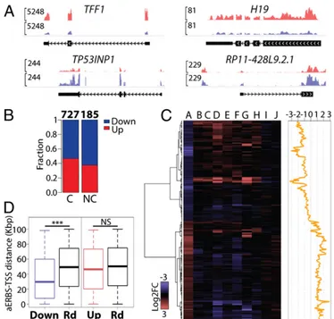
To evaluate the genome-wide effects of ERα depletion on the transcriptome of MCF7 cells, we performed polyA+ RNA-seq from cells cultured in absence of estrogen. RNAs were extracted 48 h after transfection of siCTR or siERα. To detect a broad range of variations in RNA levels we combined two comple-mentary strategies (SI Materials and Methods). This analysis led to the identification of 912 differentially expressed (DE) genes with at least 1.5-fold change (P< 0.05). ERα knockdown in the absence of hormone elicited both decrease (504 genes) and in-crease (408 genes) of coding and noncoding transcripts (Fig. 4 A and B). Even though most DE genes were protein coding (727), we found evidence of regulation of lncRNA expression, in par-ticular of lincRNAs (57 genes), antisense transcripts (48 genes), and pseudogenes (54 genes).
To understand whether transcriptional changes observed upon ERα knockdown overlap E2-stimulated genes, we compared our data to seven different public expression datasets from MCF7 cells treated with E2 for 4, 6, and 24 h (Dataset S3B). We found that 27.6% of the deregulated genes upon ERα knockdown were E2-regulated genes. Additional comparison with two time-course expression datasets (12, 26) did not increase this ratio (Fig. S2B). This analysis showed that ERα silencing causes, on average, a transcriptional effect with an opposite trend compared to E2-induction (Fig. 4C), confirming that genes controlled by unli-ganded ERα are a subset of estrogen-responsive genes. Finally, we examined two public MCF7 datasets measuring the effect of hormone withdrawal for 48–72 h (Dataset S3B) and we observed small overlap, though of coherent sign (Fig. 4C). In our experi-mental design, the time required for ERα down-regulation (48 h) accounts for the occurrence of indirect effects, in addition to primary ERα-mediated regulation. Nevertheless, we cannot ex-clude that some DE genes may represent, in part, an estrogen-independent gene expression response to ERα depletion.
Next, we sought to correlate aERBS with gene regulation. Nineteen percent of down-regulated and 5% of up-regulated genes had an aERBS within 20 kb from the transcription start site (TSS) and this ratio increased to 75% and 25%, respectively, extending the range up to 100 kb. In addition, we compared the distance from the TSS to the nearest aERBS in down-regulated vs. up-regulated genes. This analysis showed that aERBS accu-mulated significantly closer to down-regulated genes compared to random, whereas up-regulated genes did not (Fig. 4D andFig. S2C). Taken together, these observations suggest that down-regulated genes are directly down-regulated by apo-ERα binding, whereas the up-regulated set may contain secondary responders. Looking for functions of DE genes, pathway analysis (Ingen-uitySystems) showed“cell death and survival,” “cellular growth and proliferation,” and “cellular movement” as the most signifi-cant terms (Fig. S2D). However, unexpectedly, “interferon sig-naling” was indicated as the top canonical pathway and several interferon (IFN)-related molecules were predicted as activated upstream regulators (Dataset S4). This double-faced functional aspect became clear when we considered DE genes separately. In fact, all of the immune and IFN-related terms and upstream Fig. 2. (A) Semantic similarity of GO Biological Process terms enriched by
GREAT (18) in aERBS, FM-ERBS (17), or in E2-treated cells (E2-ERBS) (15). The heat map (Right) reports the semantic similarity computed between the following subsets: aERBS in common with FM or E2-treated conditions (aERBS/FM-ERBS shared; aERBS/E2-ERBS shared); ERBS detected in FM or E2 only (FM-ERBS only; E2-ERBS only); aERBS not present in FM or E2 (aERBS only/not E2-ERBS and aERBS only/not FM-ERBS). The three most represented GO categories for each cluster are indicated on the left. (B) Circos plot of TFBS predictions versus ChIP-seq datasets overlap relative to aERBS. Left heat map (red scale): predicted TFBS matrix frequency. Right heat map (blue scale): fraction of aERBS overlapped to TFBS reported by ChIP-seq. TFBS and ChIP-seq datasets characterized by the highest similarity are connected by lines of increasing color intensity, proportional to matrix similarity. Veh, untreated; E2, estrogen-treated; FM, full medium.
regulators were confined to up-regulated genes. Conversely, down-regulated genes showed cellular growth, survival, proliferation, development, and cell-cycle functions, together with the expected “tamoxifen,” “estradiol,” and “ESR1”, as most scored upstream regulators (Fig. S2 E–GandDataset S4). This function was clear-cut among down- and up-regulated genes and was confirmed by gene-set enrichment analysis (27) (Dataset S5A).
Taken together, these results suggest that unliganded ERα controls directly a set of genes related to cell growth and
survival and to the maintenance of the epithelial phenotype. Conversely, depletion of apo-ERα induces a stress-like response in the cell that is underpinned by the activation of immune and inflammatory-related genes. This idea was further confirmed by isolating DE genes possessing an aERBS within 100 kbp from the TSS. Noteworthy, functions associated with cell proliferation, death, migration, and invasion were segregated specifically to aERBS proximal genes (Fig. S2 H and IandDatasets S4
andS5A).
Unliganded ERα Binding Sites Function in Breast Cancer Cells. To appreciate phenotypic and epigenetic changes induced by ERα depletion, we used MCF7 cells cultured in HD medium and transduced with an shRNA-expressing vector, leading to a stable ERα down-regulation (Fig. S3A). Depletion of ERα completely
stopped cell growth in HD medium (Fig. 5A) and triggered a mesenchymal-like morphology (Fig. 5B), as previously reported (1, 5). Decreased apo-ERα binding and mRNA expression of target genes (Fig. 3D andFigs. S2AandS3B) was accompanied by increased level of the Polycomb-dependent histone modifi-cation H3K27me3 at ERα binding sites (Fig. 5C andFig. S3C), although we were not able to detect the occupancy of Polycomb components by ChIP at these sites. As described above, several TFBS accompany the ERE in aERBS, in particular AP2γ and FOXA1, which are considered pioneer factors and whose bind-ing is a necessary prerequisite for ERα function (11, 20). As expected, we observed that down-regulation of either AP2γ or FOXA1 reduced apo-ERα binding to aERBS, as reported for E2-induced binding (20) (Fig. S3D). In contrast to previous reports (11), FOXA1 siRNA, as well as AP2γ siRNA, reduced in part ERα protein level (Fig. S3 E and F). Contrary to expect-ations, though, we observed that ERα silencing resulted in marked decrease of FOXA1 and dramatic decrease of AP2γ occupancy in HD medium (Fig. 5 D and E). AP2γ is an ERα-dependent gene (28) and it is able to stabilize the binding of FOXA1 at colocalized ERBS (20). AP2γ expression decreases as a consequence of ERα silencing (Fig. 5F). To exclude the pos-sibility that the decrease of FOXA1 occupancy reflected AP2γ down-regulation, we investigated additional aERBS not pos-sessing AP2γ binding sites. We observed that in this case as well, markedly decreased apo-ERα binding (Fig. 5G) was followed by a decrease in FOXA1 occupancy (Fig. 5H), despite the absence of AP2γ binding at these sites (Fig. 5I).
Taken together, these data demonstrate that unliganded ERα is an essential factor for the maintenance of the luminal epithelial cistrome in unstimulated MCF7 breast cancer cells.
Fig. 3. ChIP-qPCR analysis of ERα target genes (A) following siCTR or siERα transfection, in HD and in SF medium; (B) after treatment with vehicle (NT) or 10 nM E2 for 45 min (E2). GAPDH promoter was used as a negative control region (Neg). (C) qRT-PCR mRNA analysis of target genes in HD and SF me-dium. Values are shown as ratios of relative mRNA level in siERα versus siCTR treated cells. (D) qRT-PCR mRNA analysis of TFF1 and FMN1 after siERα trans-fection or shERα transduction in HD, FM, and 10 nM E2 treatment in HD (E2). Error bars represent the SD of three independent biological replicates.
Fig. 4. (A) Genome-browser view of examples of DE protein coding (TFF1 and TF53INP1) and noncoding genes (H19 and RP11-428L9.2.1) after siCTR or siERα transfection. Only the main gene isoform is shown. (B) Fraction of up-regulated and down-up-regulated genes upon ERα silencing. C, coding genes; NC, noncoding genes. (C) Heat map representation of the overlap between DE genes (column A) and seven microarray gene expression datasets in E2-treated MCF7 cells (columns B–H) and two datasets in MCF7 switched to HD medium for 48 (column I) and 72 h (column J). Details of each dataset are reported inDataset S3B. (Right) Relative effect of siERα vs. E2-effect, calcu-lated as siERα Log2FC minus median Log2FC of E2-treated datasets. (D) Box plot distribution of the distance between the TSS of the DE genes and the closest aERBS center, compared with 1,000 random gene sets. (***P< 0.001; two-tailed unpaired t test). Down, down-regulated; Up, up-regulated; Rd, random genes.
CELL
Discussion
Previous work has exhaustively described chromatin binding of ERα in the presence of ligands and its effects on gene regulation (10–13, 15). Here, we report an unexpected role of ERα in breast cancer cells in the absence of estrogen. Unliganded ERα binding sites represent a nonrandom subset of the estrogen-induced cis-trome, which is connected with developmental functions. Moreover, unliganded ERα contributes to transcriptional activity, because its down-regulation causes changes in gene expression and chromatin modifications. Finally, we show that depletion of ERα in the ab-sence of estrogen leads to a reduced binding of pioneer factors, such as FOXA1 and AP2γ, to shared enhancers. This is an unexpected feature of ERα, whose binding to chromatin is thought to depend hierarchically from pioneer factors binding (29–31).
Genome-wide binding of ERα in the absence of hormones was observed previously in breast cancer (15, 22, 23) and recently in mouse uterus (9). Interestingly, we found that these sites, mostly located at enhancer regions, do not represent a random selection of high-affinity sites. In fact, the functions connected with aERBS neighboring genes are fundamentally related to cell development and differentiation that is strongly reminiscent of the primary action of ERα during development (32). In contrast, the analysis of ERBS specific for the estrogen treatment highlights the role of ERα in metabolic regulation, which is in agreement with the physiological action of estrogen on many tissues. To our knowledge, there was no previous support to differential functions of ERα in breast cancer cells, depending on the presence or absence of hormones.
The analysis of the transcriptome upon depletion of ERα allowed us to unveil the unexpected function of unliganded-ERα as modulator of specific loci. Indeed, we found that siERα down-regulated genes often present an aERBS, whereas those that are up-regulated do not, thus suggesting that the latter might be affected through an indirect mechanism. Our data suggest that unliganded-ERα contribute to enhancer activity, granting basal transcription of neighboring genes that are functionally related to cellular proliferation and development. In contrast, up-regu-lated genes are linked to different functions, in particular to the IFN/inflammatory response, most likely representing an indirect response to ERα ablation, in line with the protective role of ERα in several tissues (2). Interestingly, a similar signature can be observed also in data obtained during long-term hormone dep-rivation (8) (Dataset S5B). Almost 30% of the siERα-regulated
genes in HD medium was present among E2-responsive genes, further supporting the observation that aERBS represent a sub-set of the ERα-regulated cistrome.
We observed that stable ERα silencing by shRNA transduction induced complete growth arrest. This finding is consistent with the transcriptional program controlled by unliganded ERα. In similar conditions, establishment of cells with a complete epi-thelial to mesenchymal transition has been reported (1), mirror-ing the invasive phenotype of breast tumor cells escapmirror-ing ERα control in vivo. Genes controlled by unliganded ERα are not only related to cell growth, but several functional terms linked to the maintenance of the epithelial phenotype were also present. For example, the MIST1 transcription factor-encoding gene (BHLHA15), which is repressed 2.1-fold by siERα, is required to maintain mammary gland differentiation in transgenic mice (33). One important point in the context of antiestrogen treatments in breast cancer is whether aERBS are conserved in cells acquainted with estrogen deprivation (LTED). When we com-pared our aERBS dataset with those reported in two previous studies using LTED cell lines, we found that the overlap was almost complete in MCF7:2A (7) but null in the LTED cells reported by Miller et al. (6). Noteworthy, this latter case main-tains a proliferative response to estrogen, whereas the former does not. It is conceivable that in the latter case the unliganded ERα cistrome has been widely reprogrammed (6), but ERα activity is maintained on (some) genes linked to the pro-liferative response. In contrast, in the former case, unliganded ERα is possibly fully activated by other endogenous factors (e.g., coactivator amplification) as a main growth-sustaining axis. Noteworthy, GO terms analysis demonstrated that in LTED ERα is connected to metabolic functions, similarly to E2- or OH-T–induced sites but in contrast to aERBS.
Most aERBS colocalize with other TFBS. Extensive overlap with public ChIP-seq datasets confirmed that these sites are in-deed composite enhancers, possibly evolved from extensive spread of transposable elements in mammals (21). Significantly, top fac-tors comprise FOXA1 and AP2γ, known as “pioneer facfac-tors” for sex-steroid receptors (i.e., factors that open the chromatin allow-ing the landallow-ing of nuclear receptors on DNA) (29–31). We con-firmed that silencing of these factors also reduces unliganded ERα Fig. 5. (A) Growth curves of MCF7 cells transduced with control shRNA
(black line) or ERα shRNA (gray line) maintained in HD medium. (B) Mor-phological changes occurring in MCF7 cells after ERα silencing. (C) ChIP analysis of H3K27me3 at selected aERBS (*P< 0.05, **P < 0.01; one-tailed paired t test). (D and E) ChIP analysis of FOXA1 (D) and AP2γ (E) as above. (F) Western blot analysis of ERα, FOXA1 and AP2γ proteins. αtub, α-tubulin: loading control. (G–I) ChIP analysis of ERα (G), FOXA1 (H), and AP2γ (I) at selected aERBS predicted to contain FOXA1 binding but not AP2γ bind-ing sites. Intg1, -2, -3: anonymous intergenic aERBS. For ChIP experi-ments, GAPDH promoter was used as negative control (Neg). For all of the experiments shown (B–I), MCF7 cells transduced with shCTR or shERα were cultured for three days in HD medium before analysis. Error bars are SD of three independent biological replicates. (Magnification: B, 10×.)
occupancy. Unexpectedly, we observed that ERα silencing brought about a reduction of either AP2γ or FOXA1 binding at several loci, although not completely. The full interpretation of these results is complicated by the observation that AP2γ transcription is directly controlled by ERα (28) and its level is reduced in shRNA-transduced cells. However, we detected a reduction of FOXA1 binding also at enhancers that do not contain AP2γ, in contrast to previous data (34). A possible reason for this discrepancy is the use of stable, rather than transient, ERα silencing used in our study.
The data presented here may in part argue against the concept of pioneer factors as primary drivers of nucleosome remodeling, leading to ERα binding. Indeed, the elegant demonstration of progesterone receptor binding to nucleosome PRE, recently pub-lished (35), further challenges this view. Our results are consistent with a model where ERα, in the absence of estrogen stimulation, collaborates with other transcription factors to maintain the lu-minal epithelial enhancer landscape. When one of these factors is suppressed, enhancers progressively collapse, also because of the coordinated decrease of expression of other transcription factors. It is important to note, in this scenario, that a feed-forward loop exists involving AP2γ and ERα, which sustain each other’s ex-pression in breast cancer cells (28, 36).
Ligand-independent functions of ERα have been described in several tissues in addition to breast cancer cells (2–5). Results reported here provide a frame to understand why ERα is required to respond to aromatase inhibitors in breast cancer. Luminal epithelial cancer cells are stable and survive until ERα is present. The absence of estrogen keeps these cells growing at a very low rate that is presumably controlled by the host. Loss of ERα exposes the cells to immune control, removes a brake on reprogramming, which results, in vivo, in the emergence of other growth-sustaining pathways.
Materials and Methods
Detailed protocols are provided inSI Materials and Methods.
SiRNA and shRNA Interference. Cells were transfected with siERα (Stealth RNAi Invitrogen), siAP2γ (Qiagen), siFOXA1 (Santa Cruz), and siCTR (Invitrogen). Transfection was performed using Lipofectamine 2000 (Invitrogen). MCF7 cells were infected with control shRNA (MISSION shRNA; Sigma-Aldrich) or shRNA against ERα (MISSION shRNA; Sigma-Aldrich) and selected with 2 μg/mL puromycin for 3 d.
ChIP and ChIP-Seq. ChIP was performed as previously described (37, 38). Antibodies and PCR primers used in this assay are reported inSI Materials and Methods. For ChIP-seq, library preparation for sequencing was per-formed starting with 10 ng of immunoprecipitated DNA (GAIIX, Illumina).
RNA-Seq. RNA-seq libraries were prepared from poly(A)+ selected, gel-purified> 200 bp RNA. Sequencing was performed on Illumina HiSeq2000. Bioinformatic Analysis. Published algorithms were used to analyze ChIP-seq and gene expression datasets. Ingenuity pathway analysis (IngenuitySystems) was used for gene ontological analysis. ChIP-seq and RNA-seq data are ac-cessible in the Gene Expression Omnibus database (accession no. GSE53533).
ACKNOWLEDGMENTS. We thank all the members of the L.D.C. laboratory for discussions, the Center for Genomic Regulation Genomic and Bioinfor-matic Units, and Dr. He and Dr. Liu for providing data. This work was supported by Associazione Italiana per la Ricerca sul Cancro Grant IG9397 (to M.D.B.); Italian Ministry of Education, University and Research PRIN Grant 2008PC9CFW_002 (to M.D.B.); Fondazione SanPaolo grant GeneRNet (to M.C.); the Spanish“Ministerio de Educación y Ciencia” (BFU2010-18692) and Agència de Gestió d’Ajuts Universitaris i de Recerca (L.D.C.); and in part by an International Union against Cancer short-term International Cancer Technol-ogy Transfer fellowship (to L.C.).
1. Al Saleh S, Al Mulla F, Luqmani YA (2011) Estrogen receptor silencing induces epi-thelial to mesenchymal transition in human breast cancer cells. PLoS ONE 6(6):e20610. 2. Maggi A (2011) Liganded and unliganded activation of estrogen receptor and
hor-mone replacement therapies. Biochim Biophys Acta 1812(8):1054–1060.
3. Cvoro A, et al. (2006) Distinct roles of unliganded and liganded estrogen receptors in transcriptional repression. Mol Cell 21(4):555–564.
4. Dhasarathy A, Kajita M, Wade PA (2007) The transcription factor snail mediates ep-ithelial to mesenchymal transitions by repression of estrogen receptor-alpha. Mol Endocrinol 21(12):2907–2918.
5. Cardamone MD, et al. (2009) ERalpha as ligand-independent activator of CDH-1 regulates determination and maintenance of epithelial morphology in breast cancer cells. Proc Natl Acad Sci USA 106(18):7420–7425.
6. Miller TW, et al. (2011) ERα-dependent E2F transcription can mediate resistance to estrogen deprivation in human breast cancer. Cancer Discov 1(4):338–351. 7. He HH, et al. (2012) Differential DNase I hypersensitivity reveals factor-dependent
chromatin dynamics. Genome Res 22(6):1015–1025.
8. Aguilar H, et al. (2010) Biological reprogramming in acquired resistance to endocrine therapy of breast cancer. Oncogene 29(45):6071–6083.
9. Hewitt SC, et al. (2012) Research resource: Whole-genome estrogen receptorα binding in mouse uterine tissue revealed by ChIP-seq. Mol Endocrinol 26(5):887–898. 10. Carroll JS, et al. (2006) Genome-wide analysis of estrogen receptor binding sites. Nat
Genet 38(11):1289–1297.
11. Hurtado A, Holmes KA, Ross-Innes CS, Schmidt D, Carroll JS (2011) FOXA1 is a key determinant of estrogen receptor function and endocrine response. Nat Genet 43(1): 27–33.
12. Cicatiello L, et al. (2010) Estrogen receptor alpha controls a gene network in luminal-like breast cancer cells comprising multiple transcription factors and microRNAs. Am J Pathol 176(5):2113–2130.
13. Kwon Y-S, et al. (2007) Sensitive ChIP-DSL technology reveals an extensive estrogen receptor alpha-binding program on human gene promoters. Proc Natl Acad Sci USA 104(12):4852–4857.
14. Neve RM, et al. (2006) A collection of breast cancer cell lines for the study of func-tionally distinct cancer subtypes. Cancer Cell 10(6):515–527.
15. Welboren W-J, et al. (2009) ChIP-Seq of ERalpha and RNA polymerase II defines genes differentially responding to ligands. EMBO J 28(10):1418–1428.
16. Madak-Erdogan Z, Lupien M, Stossi F, Brown M, Katzenellenbogen BS (2011) Genomic collaboration of estrogen receptor alpha and extracellular signal-regulated kinase 2 in regulating gene and proliferation programs. Mol Cell Biol 31(1):226–236. 17. Ross-Innes CS, et al. (2010) Cooperative interaction between retinoic acid
receptor-alpha and estrogen receptor in breast cancer. Genes Dev 24(2):171–182.
18. McLean CY, et al. (2010) GREAT improves functional interpretation of cis-regulatory regions. Nat Biotechnol 28(5):495–501.
19. Yu G, et al. (2010) GOSemSim: An R package for measuring semantic similarity among GO terms and gene products. Bioinformatics 26(7):976–978.
20. Tan SK, et al. (2011) AP-2γ regulates oestrogen receptor-mediated long-range chro-matin interaction and gene transcription. EMBO J 30(13):2569–2581, corrected in EMBO J (2011), 30(13):2750.
21. Testori A, et al. (2012) The role of transposable elements in shaping the combinatorial interaction of transcription factors. BMC Genomics 13:400.
22. Joseph R, et al. (2010) Integrative model of genomic factors for determining binding site selection by estrogen receptor-α. Mol Syst Biol 6:456.
23. Ceschin DG, et al. (2011) Methylation specifies distinct estrogen-induced binding site repertoires of CBP to chromatin. Genes Dev 25(11):1132–1146.
24. Ross-Innes CS, et al. (2012) Differential oestrogen receptor binding is associated with clinical outcome in breast cancer. Nature 481(7381):389–393.
25. Laganière J, Deblois G, Giguère V (2005) Functional genomics identifies a mechanism for estrogen activation of the retinoic acid receptor alpha1 gene in breast cancer cells. Mol Endocrinol 19(6):1584–1592.
26. Hah N, et al. (2011) A rapid, extensive, and transient transcriptional response to es-trogen signaling in breast cancer cells. Cell 145(4):622–634.
27. Subramanian A, et al. (2005) Gene set enrichment analysis: a knowledge-based ap-proach for interpreting genome-wide expression profiles. Proc Natl Acad Sci USA 102(43):15545–15550.
28. Orso F, et al. (2004) Activator protein-2gamma (AP-2gamma) expression is specifically induced by oestrogens through binding of the oestrogen receptor to a canonical element within the 5′-untranslated region. Biochem J 377(Pt 2):429–438. 29. Magnani L, Lupien M (2014) Chromatin and epigenetic determinants of estrogen
receptor alpha (ESR1) signaling. Mol Cell Endocrinol 382(1):633–641.
30. Garcia-Bassets I, Wang D (2012) Cistrome plasticity and mechanisms of cistrome re-programming. Cell Cycle 11(17):3199–3210.
31. Green KA, Carroll JS (2007) Oestrogen-receptor-mediated transcription and the in-fluence of co-factors and chromatin state. Nat Rev Cancer 7(9):713–722.
32. Hewitt SC, Harrell JC, Korach KS (2005) Lessons in estrogen biology from knockout and transgenic animals. Annu Rev Physiol 67:285–308.
33. Zhao Y, et al. (2006) Identification of a basic helix-loop-helix transcription factor expressed in mammary gland alveolar cells and required for maintenance of the differentiated state. Mol Endocrinol 20(9):2187–2198.
34. Lupien M, et al. (2008) FoxA1 translates epigenetic signatures into enhancer-driven lineage-specific transcription. Cell 132(6):958–970.
35. Ballaré C, et al. (2013) Nucleosome-driven transcription factor binding and gene regulation. Mol Cell 49(1):67–79.
36. McPherson LA, Baichwal VR, Weigel RJ (1997) Identification of ERF-1 as a member of the AP2 transcription factor family. Proc Natl Acad Sci USA 94(9):4342–4347. 37. Strutt H, Paro R (1999) Mapping DNA target sites of chromatin proteins in vivo by
formaldehyde crosslinking. Methods Mol Biol 119:455–467.
38. Vicent GP, et al. (2011) Four enzymes cooperate to displace histone H1 during the first minute of hormonal gene activation. Genes Dev 25(8):845–862.
CELL