PhD in Clinical & Experimental Medicine
UNIVERSITÀ DEGLI STUDI DEL PIEMONTE ORIENTALE
“AMEDEO AVOGADRO”
Department of Clinical and Experimental Medicine
Corso di Dottorato di Ricerca in Medicina Clinica e Sperimentale
Ciclo XXVIII
Exploration of new uracil-based compounds as novel inhibitors of Hepatitis C Virus replication
SSD (Settore Scientifico Disciplinare) MED/09
Coordinatore Tutor Prof. Marisa GARIGLIO Prof. Mario PIRISI
Dottorando Andrea MAGRI
Contents
INTRODUCTION ... 6
Hepatitis C virus (HCV): ... 7
Physical and chemical properties of HCV particles ... 7
Molecular virology of HCV ... 8
5’ NTR ... 10
3’ NTR ... 10
Core protein ... 10
E1E2 envelope glycoproteins ... 13
p7 Protein ... 15 NS2 protein ... 16 NS3-NS4A complex ... 16 NS4B protein ... 18 NS5A protein ... 18 NS5B protein ... 19 Lifecycle ... 20
Attachment and entry ... 21
HCV entry: a model ... 30
Fusion mechanism ... 32
Translation ... 33
Replication ... 35
Viral assembly, maturation and release ... 38
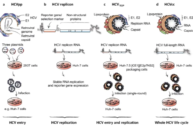
In vitro models to study HCV cell cycle ... 42
HCV pseudo-typed particles (HCVpp). ... 43
HCV replicons ... 44
HCV trans-complemented particles (HCVTCP) ... 45
The disease ... 47
HCV variability and worldwide distribution. ... 47
Modes of transmission ... 48
Natural History ... 49
The Acute Phase of HCV infection ... 50
The chronic phase of HCV infection ... 51
Current and novel HCV therapies ... 53
Preventive and therapeutic Vaccination ... 53
HCV therapy with Interferon ... 55
HCV therapy and DAAs ... 55
Aim of the study ... 57
MATERIAL AND METHODS... 60
Materials ... 61
Chemicals ... 61
Kits ... 61
Cells ... 62
Cell Culture Growth Medium ... 63
Drugs ... 63 Clones ... 65 Antibodies ... 66 Bacterial Strains... 66 Solutions ... 66 Oligonucleotides ... 67 Methods ... 68 DNA Manipulation ... 68 RNA Manipulation ... 70 Reverse Transcription ... 71 Real-Time PCR... 73
Tissue Culture Maintenance ... 75
Cell culture of infectious HCV (HCVcc) ... 75
HCV pseudo-typed particles (HCVpp) ... 79
HCV replicons ... 80
HCV trans-complemented particles (HCVTCP) ... 82
Evaluation on Influenza Virus ... 83
Statistical Analysis ... 83
RESULTS ... 84
Antiviral effect on HCVcc ... 85
Inhibition of HCV entry with HCVpp ... 87
Entry inhibition of different virus using pseudo-particles ... 87
Inhibitory effect on HCV replication ... 88
Transient Replicon ... 88
Generation of a replicon cell line ... 89
Replicon cell line inhibition ... 90
Determining IC50 and CC50 values ... 92
Evaluation of drug resistance mutations ... 95
RNA inhibition on HCVcc ... 96
Analysis of single-cycle infection on HCVcc ... 97
Single-cycle infection inhibition on HCVTCP ... 99
Effect on an early step of viral replication ... 100
Effect on HCV translation ... 100
Antiviral effect on Influenza Virus ... 103
Development of an PCR-based assay for negative strand quantification ... 104
Primer Evaluation ... 104
Real Time Testing ... 105
Detection of positive RNA in viral particles ... 106
Optimising RT Primer Concentration ... 109
Viral kinetic based on negative strand synthesis ... 110
Positive and negative strand RNA inhibition ... 111
Evaluation on single-cycle infection ... 111
Determining positive and negative strand RNA on infection ... 112
DISCUSSION ... 113
Hepatitis C virus (HCV):
HCV is a member of the Hepacivirus genus, along with the pestiviruses (i.e. bovine viral diarrhea virus or BVDV) and flavivirus (including yellow fever virus, dengue and Zika); they constitute the Flaviviridae family1.
Physical and chemical properties of HCV particles
HCV particles from serum of infected patients are characterised by a diameter ranging between 30 and 80 nm2-4; surprisingly, in vitro produced HCVcc particles showed a diameter
between 60 and 75 nm5,6. Analysing by density gradients the physical properties of HCV
particles, emerged that in patient serum-derived particles the HCV RNA is distributed on a rage of densities from 1,03 g/ml to 1,20 g/ml and the peak of RNA is between 1,04 g/ml and 1,12 g/ml7,8. This heterogeneity is dependent on the association of viral particles with
different classes of lipoproteins (VLDL, LDL and HDL) and/or immunoglobulins7,9-12.
HCV particles with higher density are less infectious, confirming how the association of HCV with lipoproteins is crucial for infectivity. Moreover, virus particles isolated from serum of infected chimpanzee have been found to have densities between 1,03 and 1,10 g/ml 9,13,14.
Noteworthy, the peak of infectivity has been described at low density < 1.10 g/ml15-17,
whereas HCV particles, produced in in vitro model, showed a higher density, of approximately 1,15 g/ml. This difference has also been shown in-patient sera, exhibiting lower values compared to in vitro densities, probably because the hepatoma cell line currently used to produce HCV has deficient lipoprotein metabolism18. To support the
hypothesis that this low buoyant density of infectious HCV particles is the result of the association of the virions with serum lipoproteins7,10,12,19, ApoAI, ApoB, ApoC1, and ApoE
have been demonstrated to be associated with serum-derived HCV particles6,20-23. Based
serum as a hybrid lipoviral particles (LVPs) following VLDL (very low density lipoprotein) production and secretion pathway24. Although the exact structure of the infectious lipo-viral
particles and their interactions with HCV has been debated for many years, initially, two models were proposed (Fig. 1): in the first one viral particles interact transiently with lipoproteins, while in the second one they contribute into forming an integrated particle. However, recent studies, using electron microscopy, demonstrated that HCV is part of a bigger structure containing lipoprotein (figure 1B) 25. This configuration of the LVPs may
provide a shield, protecting virus from neutralizing antibodies19,26,27.
Figure 1. Two models for lipo-viral particle (LVP). In the first the two-particle model (A), HCV particles and serum lipoproteins transiently interact. In the second one (B), the single-particle model for LVP structure, a low-density lipoprotein (LDL) single-particle is integrated in an HCV particle24.
Molecular virology of HCV
HCV particles contains one molecule of 9.6 kb single stranded positive RNA genome. It is composed of the 5’ non-translated region (NTR), characterized by the presence of an IRES that drives the translation of a single open reading frame (ORF), coding for structural and non-structural proteins, and the 3’ NTR25. Structural proteins have been shown to form the
capsid of the viral particles (core protein) and to be expressed on the envelope as glycoproteins, specifically E1 and E2. The non-structural proteins (NS), which are necessary for viral replication and assembly of infectious virus particles, comprised the ion channel p7,
the NS2 protease, the helicase/protease NS3-4A complex, the cofactor NS4B, the phosphoprotein NS5A and the NS5B RNA-dependent RNA polymerase (RdRp)28.
IRES-mediated translation leads to the synthesis of a polyprotein of approximately 3000 aminoacids (aa) residing on endoplasm reticulum (ER) that undergoes a processing by cellular and viral proteases. Specifically, core protein is cleaved sequentially by the cellular signal peptidase (SP) and the cellular signal peptide peptidase (SPP), while glycoproteins E1, E2 and p7 are cleaved by SP; non-structural proteins are processed by NS2 and NS3/NS4 proteases29,30 (Fig. 2). Interestingly, the HCV proteins showed multifunctional
roles on different viral stages; in particular the NS3-4A complex, that has been shown to be involved in the HCV replication and pathogenesis31-36. In addition, the function of HCV
proteins may be influenced by interaction with other viral or cellular molecules, which may induce different structural conformations37.
Figure 2. HCV genome organization and polyprotein processing38. HCV RNA is translated
5’ NTR
The 5′ NTR extends between nucleotides 1 to 342, is highly conserved through HCV genotypes and it is characterised by the presence of 4 major stem loops; furthermore, it has been shown playing two key functions in the HCV replication cycle. First, in the positive strand it contains a specific internal ribosome entry site (IRES), necessary to drive RNA translation, and thus polyprotein synthesis. Second, it has been proposed that the 5’NTR is essential to direct the synthesis of progeny positive-strand RNA using as template the negative strand RNA. The hypothesis has been supported by the finding that the 5’NTR adopts very different secondary structures between positive and negative strands39,40.
3’ NTR
The 3′NTR is mainly involved in RNA replication41, presumably playing a key role in the
initiation and regulation of negative-strand synthesis42. It is constituted by three main
regions: a variable region, a polyU/UC tract of variable length (ranging between 30 and 90 base pair depending on HCV isolates), whom function still needs to be clarified, and a highly conserved element known as X-tail or 3′X (98 nts) 43,44. Deletions in the variable region
impair viral replication but they do not determine its abrogation, suggesting that this sequence is not essential41,45. The X-tail has been found to be relatively conserved among
HCV isolates and has been propose to contain the main regulatory elements required for the negative-strand synthesis43,44. Since mutations in this region are affect viral replication,
both its conserved sequence and structure are critical45,46.
Core protein
The core protein is the first structural protein encoded by HCV genome and it is responsible for the nucleocapsid formation. A short region comprising the C-terminus of the core protein acts as a signal sequence that targets the nascent polyprotein to translocate into the ER membrane. In addition, the core signal sequence has been reported to be recognised and
cleaved by the host signal peptidase (SP) with production of an immature, 191 aa core. An additional cleavage is required at the C terminus of immature core, by the signal peptide peptidase (SPP), to obtain the mature core protein (21 kDa) 47,48. Although the C-terminal
(aa177-191) domain D3 is immediately cleaved after translation, and thus is absent from the mature form of HCV core, it has been shown to be important for core stability and correct function. The mature core is a dimeric membrane protein and may be stabilised by an intramolecular disulphide bonds49,50.
The mature core protein is composed of two domains: the N-terminal hydrophilic D1 domain and the C-terminal hydrophobic domain D2. The D1 has been shown to be involved in RNA binding and homo-oligomerization. Indeed, core D1 possess RNA chaperone activity, which has been reported to be required for the structural remodelling and packaging of the viral RNA51. Moreover, it has also been described that D1 can interact with several host factors
and thus altering cellular functions upon HCV infection37,52. D2 is more hydrophobic and it
is thought to interact with phospholipids on lipid droplets (LDs) through amphipathic regions53.
The HCV core protein, besides its role in formation of the virus nucleocapsid, has been reported to be involved in the modulation of different host pathways and to interact with a variety of cellular factors. Those include numerous transcription factors, such as heterogeneous nuclear ribonucleoprotein K54, leucine zipper transcription factor (LZIP)55,
14-3-3 protein56, NF-kB57 and RNA helicases, such as DDX3 protein58, involved in HCV RNA
replication59.
It has also been suggested that core protein might be involved in apoptosis and cell cycle regulation, contributing to the pathogenesis of HCC37. For this purpose, HCV core protein is
considered as a major viral factor inducing development of hepatocellular carcinoma during HCV infection. To support this hypothesis, it has been demonstrated that HCV core can directly modulate the expression tumor-associated genes, such as the cyclin-dependent
inhibitor p21 60, involved in cell-cycle control and tumor formation, and it can also interfere
with Wnt/β-catenin pathway, which play a major role in the initiation of carcinogenesis61.
In addition, it has been shown that HCV core protein expression might induce development of hepatic steatosis, particularly in genotype 3 infected patients62. Indeed, it has been proved
that HCV core enhances the transcriptional activity of sterol regulatory element binding protein 1 (SREBP1) and peroxisome proliferators-activated receptor gamma (PPARγ) 63.
Although it has been largely reported that the core protein predominantly resides in the cell cytoplasm, associated with lipid droplets, which represent the site of virus morphogenesis, it has also been detected in the nucleus64,65 and mitochondria66,67. However, whereas the
presence of core on the LDs is mandatory for the virus assembly, the significance of its presence in the nucleus and mitochondria for the virus life cycle or pathogenesis is still not clear.
Alternative open reading frame protein (Core+1; ARFP)
Normally, the correct HCV translation leads to the synthesis of a polyprotein of 3000 aa; however, an overlapping ORF has been reported, which may generate an alternate reading frame product68. Indeed, a novel HCV protein (named protein F, core +1 or alternative
reading frame protein) has been shown in in vitro models, encoded by an alternative open reading frame in the core region69. The existence of T-cell response70 or antibody
production71 against core+1 in HCV infected individuals suggests that this protein is also
produced in natural HCV infection. Nevertheless, ARFP has not yet been detected either in patients’ sera or in infected tissues and its biological role remains unknown72.
Moreover, it has been suggested that HCV core+1 synthesis might negatively regulate production of the canonical HCV core protein; furthermore, Core+1 protein seems to not be necessary for HCV life cycle72, but the presence of specific antibodies in HCV patients
E1E2 envelope glycoproteins
HCV encodes for two glycoproteins: E1 and E2. They are classified as type I membrane proteins, with a molecular weight of approximately 35 kDa and 70 kDa, respectively. Both contain a large N-terminal ectodomains (of 160 and 334 aa in E1 and E2, respectively) and a short C-terminal transmembrane domain. The transmembrane domain (TMD) is responsible for anchoring E1 and E2 in the membrane and for their localization into the endoplasmic reticulum (ER). The TMD is characterised by two stretches of hydrophobic residues separated by a short segment containing at least one positively charged amino acid. These positively charged residues have an important role alongside with the N-terminal part of E1 TMD in E1-E2 heterodimerization and retention in the ER, whereas the entire TMD sequence seems to be necessary for anchoring to the membrane73-75. Six and seven
ectodomains have been described in E1 and E2, respectively, which undergo an extensive N-glycosylation during the translocation into the endoplasmic reticulum lumen. These sites, highly conserved among HCV genotypes, are involved in glycoprotein folding and have been reported to play a specific role in HCV entry76. Moreover, a segment containing the
N-terminal 27 aa residues of E2, defined as hyper variable region 1 (HVR1), is characterized by a high content of basic aminoacids known to influence HCV pseudo particles (HCVpp see below) infectivity77,78. HVR1 has been shown to be partially responsible for HCV escape
from the immune response79; furthermore, viruses lacking HVR1 have been described as
less infectious and exhibited impaired fusion, showing an enhanced neutralization and precipitation by E2 specific antibodies and soluble CD8180, one of the main factors involved
in HCV entry, as described below. Initially, newly-synthesized E1 and E2 are assembled as a non-covalent heterodimer on the ER membrane, then the HCV glycoproteins are subjected
to post-translational changes. Interestingly, on the viral particles they exist as large covalent complexes stabilised by disulfide bridges81.
HCV glycoproteins E1 and E2 are present in tandem and this organization has also been found in alphaviruses and flaviviruses that encode class II fusion proteins, although for HCV the prediction of which glycoprotein acts as main fusion protein, remains unclear. In fact, over the last 10 years many studies, supported by contrasting evidences, led to the generation of many hypothesis, trying to explain fusogenic activity. Interestingly, to support the idea that E1 is the main protein involved in fusion, sequence analysis revealed that the E1 ectodomain contains a fusion peptide-like motif similar to the homologous peptide of paramyxoviruses and flaviviruses82. In addition, has been demonstrated that the structure
of this motif is not required to mediate cell fusion, while the presence of specific residues is essential for viral entry83. However, comparing the E2 structure with other members of the
Flaviviridae family, led the authors hypothesized that fusion is mediated by E2, suggesting
that E2 belongs to the class II fusion protein84,85. To support this idea, Lavillette et al
described at least three putative regions involved in virus-cell fusion by a mutagenesis approach, suggesting that E2 may contribute to the fusion step, either by direct interaction with the lipid membrane or by induction of conformational change of E1E2 complex86.
Moreover, several residues responsible for CD81-E2 interaction have been described, supporting the idea that E2 plays a major role in the fusion step87,88. However, very recently,
Kong et al succeeded in obtaining a crystal structure of E2 89. Surprisingly, the authors
described E2 as a globular structure that contains many regions with no regular secondary structure despite the presence of eight disulfide bonds. Comparing E2 structure with other class II fusion proteins of different Flaviviruses, it emerged that the only shared element is the Ig-fold beta sandwich and that E2 does not adopt the putative three-domain fold described for other class II fusion proteins88,89. Furthermore, data obtained allowed to
Despite these controversial data concerning fusion properties, observing the nature of E1 and E2 glycoproteins and their capacity to form E1-E2 heterodimer seems to suggest that they may have a synergic role in regulating HCV entry and fusion. Indeed, it has been shown that the E1E2 heterodimer can bind to CD81 stronger than E2 alone indicating that E1 may modulate E2 activity91,92.
p7 Protein
p7 is a 63 aa, integral trans-membrane protein that is usually listed as NS proteins even though there is not a concrete evidence whether it can be directly associated to viral particle. Its structure is characterised by two transmembrane α-helices (named M1 and M2) linked by a positively charged cytosolic loop with the N and C termini both oriented towards the ER lumen93. The first experiments, conducted with electron microscopy, showed that p7
monomers assemble into hexamer or heptamer complexes in artificial membranes94.
Furthermore, another study, based on electron microscopy, revealed the presence of a hexameric complex characterized by a flower-shaped architecture and six protruding petals oriented toward the ER-lumen95. Subsequently, a third α-helix, located upstream of M1, has
been identified96. A more recent model, developed using similar experimental strategies,
suggested that hexameric and heptameric complexes might coexist; in this situation they may form a functional ion channels97. Interestingly, analysing p7 structure, some analogies
have been found with viral proteins of other viruses such as 6k of alphaviruses, M2 of influenza A virus, vpu of HIV-1 which all belong to the viroporin family97. Typically, viroporins
are not essential for viral RNA replication but they are involved in assembly and release of virus particles. Surprisingly, some recent data seem to suggest that p7 might also have a role on HCV replication98, even though this eventuality has still to be discussed.
Other studies, conducted in vitro and in vivo, confirmed that p7 is essential for assembly and release of HCV particles99-101, moreover, p7 acts in the assembly stage through the
interaction with NS2. During viral release, p7 acts mainly to form ion channels that are essential to equilibrate pH gradients within the endo-lysosomal compartments, in order to protect HCV particles from pH-dependent uncoating during egress95,96,101-104.
NS2 protein
NS2 is a transmembrane protein, containing three putative transmembrane domains at its N-terminal, and one cysteine protease domain at C-terminal. Interestingly, It has been shown that homo-dimerisation of NS2 induces folding of the catalytic domain and generation of two active sites at dimer interface105. To date, only one substrate, the junction NS2/NS3,
has been identifies for the NS2 protease activity. NS2 is necessary for polyprotein processing and full-length HCV genome replication but it is not required for RNA replication of the subgenomic replicon106,107.
Interestingly, NS2 has been recently identified also as a key factor for the assembly of new infectious HCV particles and this function has been demonstrated that does not depend on its protease activity108,109. Specifically, NS2 has been shown to act in combination with
E1-E2 glycoproteins, p7, and the NS3–4A enzyme complex at an early stage during virus assembly109-114. Furthermore, Counihan et al demonstrated that the recruitment of the core
protein, from lipid droplets (LDs) to the putative sites of virus assembly, is strictly dependent on the NS2 and NS3–4A interaction115. In particular, it has been shown that this interaction
is required to localize NS2 and core-associated LDs to putative sites of the virus assembly109,111,113.
NS3-NS4A complex
NS3-4A is a non-covalent protein complex composed of NS3 and its cofactor NS4A. NS3 is a 70 kDa protein characterised by two well defined enzymatic activities. NS3/4A structure
has been solved by Yao116 et al. in 1999, showing that the N-terminal contains the serine
protease activity responsible for the processing of the HCV polyprotein to generate the NS proteins, whereas the C-terminus has been reported as the NTPase/RNA helicase catalytic site117-119. The N-terminal domain encodes for the viral protease, which belongs to the
trypsin/chymotrypsin protease superfamily120,121. The protease activity is exerted by the NS3
catalytic triad, and the NS4A cofactor has been shown to stabilise the interaction between the catalytic site inside the NS3 and its substrates, enhancing the efficacy and specificity122.
The process of NS3-substrate recognition is dependent on the presence of specific multiple aminoacid residues; however, recent experiments in order to investigate potential NS3-4A substrates, has not been able to solve this enigma. In fact, only few of a large number of cellular proteins, that contain the specific consensus sequence, are effectively cleaved by the NS3-4A. On the other hand, several cellular substrates have been identified as effective substrates, although they present some differences in the recognition sequence compared to the canonical one119.
The NS3 helicase belongs to the RNA helicase super family 2, and shares six helicase motifs with proteins belonging to the same family123. Furthermore, the NS3 exerts its
helicase activity, nucleic acid unwinding, through ATP consumption; indeed, an APT-binding domain has been well described124. However, in experiments were NS3 protein was
expressed alone, it showed an impaired activity compared to the entire NS3-NS4A complex. Hence, although the NS3 helicase is essential for HCV RNA replication, more recently it has also been proposed that NS3-4A can play a role in the virus assembly and immune response125-130 .
Interestingly, an “in-plane amphipathic α-helix” at the N terminus of NS3, and the transmembrane α-helix harboured in the NS4A N-terminal have been shown to be the key factors in the association of NS3-4A with membranes and the structural organization131.
Surprisingly, the authors described that the NS3-NS4A complex can be found on mitochondrial or mitochondria-associated membranes. It has been hypothesized that this atypical localisation is responsible for interfering with immune response, inactivating MAVS132,133.
NS4B protein
NS4B is a hydrophobic integral membrane protein of approximately 27 kDa. NS4B has been characterised by its effect to induce the formation of specific membrane alterations, forming a characteristic formation called membranous web; it consists of locally confined membranes in which HCV NS proteins accumulate, interact and drive viral replication134,135.
NS4B has been demonstrated to be involved in several distinct functions: NTPase activity, interactions with other HCV NS proteins, direct binding to RNA and finally playing a role in the assembly of viral particles34,136-138. As already shown for other HCV NS proteins, NS4B
can associate to form oligomers139,140. Furthermore, NS4B is essential to stabilise the
functional replication complex: in fact, mutations affecting its oligomerization impair the membranous web formation and, as consequence, they affect HCV replication. It has also been suggested that the NS4B expression induces membrane curvature and vesicle formation141.
NS5A protein
NS5A is a membrane-associated phosphoprotein of 447 aa that is involved in modulation of HCV RNA replication and particle formation during assembly. It is characterised by a membrane anchor in the N-terminal and three main domains, which are separated by two low complexity sequence (LCS) 142. The first two domains, named D1 and D2 have been
shown to be involved in RNA replication whereas D3 is required for virus assembly31,33.
Interestingly, HCV NS5A has been described in two different states: basally phosphorylated (56 kDa) and hyper-phosphorylated (58 kDa). While basal phosphorylation is mainly focused
on NS5A C-terminal and central residues, the hyper-phosphorylation has been described in a specific residue in the LCS. Based on the evidence showing that cell-culture adaptive mutations mostly affect these residues and data obtained with kinase inhibitors, it has been hypothesized that NS5A regulates HCV RNA replication, probably interacting with specific host factors143-145. NS5A is anchored to the ER membrane through an N-terminal
amphipathic alpha helix that is located into the cytosolic face of the membrane. NS5A has been shown to associate with phospholipid monolayer allowing its interaction with the core protein located on LDs or on the LDs-ER interface. Analysing the crystal structure of D1, it has been demonstrated that the dimerization, characterised by an extended shape, is able to form a channel capable to host both ssRNA and dsRNA142. Moreover, the NS5A D1
domain has been found capable to bind RNA as a dimer146. Hence, it has been proposed
that NS5A dimers might bind RNA and drive it through the HCV replication147. Interestingly,
D2 and D3 domains have been found natively unfolded suggesting that to reach their stable conformation (mainly alpha helical), several interactions with specific cellular or viral proteins are required. Furthermore, D2 and D3 have been reported as substrate of Cyclophilin A (CypA)148,149; CypA, an isomerase enzyme, which have been shown essential for HCV
replication; moreover, CypA can be inhibited by cyclosporine A (CsA)150. In particular, it has
been reported that D2 and D3 can directly bind to the active site of CypA and specifically to proline residues, which have been shown to be substrates for the isomerase activity 148,149.
Finally, it is important to highlight that most of the known mutations conferring CsA resistance, have been found in the NS5A D2 domain151.
NS5B protein
NS5B is the RNA-dependent RNA polymerase (RdRp) that has been isolated more than 20 years ago; for this reason, it has been largely studied and crystalised152-155. The NS5B, a
530, a linker domain of 40 aa and a short C-terminal membrane anchor (CMA) of 21 aa. This anchor has been reported to be essential for viral replication in vivo but, in in vitro experiments, it is dispensable for RNA synthesis156. In vivo the CMA has been reported to
be responsible for cytosolic orientation of the RdRp catalytic domain157. The N-terminal
catalytic domain, is characterised by a well-defined right hand shape, which can be found in many other RNA-dependent RNA polymerases. In this hand-like folding, different structural regions were identified, referred as fingers, thumb and palm154,158,159. In addition, authors
have identified a beta-flap on the thumb region that has been shown also in other Flaviviridae RdRp160. Hence, two different conformations have been reported: a closed conformation
has been described during the de novo initiation step, while the open folding has been reported during the elongation step. Interestingly, the closed conformation is able to use ssRNA as template recognising the two initiating nucleotides155. Interestingly, on de novo
replication, the RNA template and nucleotides are surrounded by an encircled active site, which is closed from one side by fingers and on the other by interaction between linker and beta-flap. Once the RNA synthesis is started, the NS5B undergoes a significant change during which the linker and the beta-flap are removed and the RdRp might begin the elongation. Interestingly, from the recent structural models, it has not been completely clarified whether the newly synthesized RNA can emerge as double stranded, paired to the template, or if it is forced to unwind in order to leave the active site161.
Lifecycle
The HCV life cycle is a complicated process based on different well-distinguished steps: entry, translation, replication, assembly and release. Below, all of these steps have been described individually.
Attachment and entry
The HCV entry is a multistep process that involves several factors and co-factors sequentially interacting with the virus that promote initiation of productive infection. Below are described all known factors involved in viral attachment.
Glycosaminoglycans (GAGs)
GAGs are long unbranched polysaccharides, characterised by repeated disaccharide units that show several degrees of heterogeneity including molecular weight, disaccharide construction, and sulfation. Based on core disaccharide features, GAGs can be classified into four groups: heparin/heparan sulfate, chondroitin/dermatan sulfate, keratan sulfate and hyaluronic acid GAGs. They are commonly associated with cellular proteins to form a panel of molecules exposed on cell surfaces, known as proteoglycans (PG). Interestingly, it has been reported that many viruses use PG for their attachment to host cells. Specifically, a heparan sulfate proteoglycan (HSPG) has been shown to be crucial for the early steps of HCV infection. The direct interaction between the HCV E2 glycoprotein and cell surface GAGs, via the positively charged residues at the N terminus, has been demonstrated and extensively studied using HCV-like particles produced in insect cell lines and recombinant E2 glycoprotein162. Nevertheless, it is generally accepted that the primary site of interaction,
between HCV and the liver, is the heparan sulphate, especially Syndecan 1 163. This
interaction is mediated by lipoproteins associated to LVPs and especially Apolipoprotein E (ApoE). For this reason, the role of Apo E exposed on the HCV surface in virus attachment to heparin/heparin sulfate PG has been demonstrated by showing that antibodies targeting ApoE and synthetic peptides derived from ApoE were able to inhibit HCV cell binding164.
Moreover, Shi et al demonstrated the role of different heparin/heparin sulfate proteoglycan (HSPG) core proteins in the HCV binding process, showing that Syndecan 1 plays a major
role in virus attachment compared to other member of the Syndecan family and others HSPG163.
DC-SIGN and L-SIGN
Dendritic cell-specific intercellular adhesion molecule 3-grabbing non-integrin (DC-SIGN) and the related protein liver/lymph node-specific (L-SIGN or DC-SIGNR) are calcium-dependent lectins expressed on dendritic and endothelial cells in the liver and lymph nodes, respectively. DC-SIGN and L-SIGN have been initially investigated due to their role in mediating human immunodeficiency virus (HIV) binding and internalisation. It has also been shown that the interaction between these molecules and HIV is dependent on the presence of mannose N-linked chains in the HIV envelope protein (Env). Based on these observations, it has been reported that glycosylation of HCV E1-E2 glycoproteins is comparable to that of HIV Env, and, moreover, L-SIGN is expressed in the liver. Interestingly, results obtained from early studies, conducted with soluble E2 (sE2) and HCVpp, confirmed that virus-associated E1 and E2 were able to bind DC-SIGN and L-SIGN165. These data have been subsequently confirmed with the HCV cell-cultured (HCVcc,
see below) system166. Based on these data, it has been hypothesized that L-SIGN,
expressed on sinusoidal endothelium cells, might be used as a docking site for circulating HCV within sinusoids, facilitating the virus transfer to hepatocytes. Furthermore, the absence of DC-SIGN and L-SIGN on hepatocyte membranes suggests that they do not play a role as direct virus entry factors; rather, these molecules could enhance infection by promoting virus attachment. Finally, Pöhlmann et al suggested that sinusoidal endothelial cells might be involved in capturing and concentrating circulating virions in the liver, allowing their presentation to the hepatocyte167.
Several human proteins have been identified as factors or co-factors involved in HCV entry; they are briefly described below.
Low-density lipoprotein receptor (LDLR).
LDLR, a member of the low-density lipoprotein family168, is a cell-surface receptor involved
in cholesterol homeostasis. It has been shown to recognise ApoB100 embedded in LDL and ApoE in intermediate lipoprotein (IDL), and thus, mediate endocytosis of these lipoproteins169. The presence of lipoprotein components on the virus surface and the role of
LDLR in HCV entry were first suggested by V. Agnello170. Several studies confirmed that
indeed ApoB100 and ApoE are present on the HCV surface23,26,171. This hypothesis was
also strengthened by the finding that cell entry of patient-derived HCV strains required LDLR172. Although several studies clearly demonstrated the role of LDLR in HCV cell
entry170,172-174, in one particular study the role of LDL-R in HCV RNA replication has been
suggested in addition to virus entry process175. Cluster of Differentiation 81 (CD81).
CD81 is a 26 KDa ubiquitously expressed transmembrane protein belonging to the
tetraspanin family. It is involved in regulation of cell morphology, motility and signalling176.
CD81 is a type III membrane glycoprotein characterised by four transmembrane domains producing two extracellular loops and one short intracellular domain. CD-81 was initially identified as an HCV entry factor by Pileri et al, based on its interaction with E2 protein177
and subsequently confirmed using HCVpp model178. Several experiments confirmed that
disulphide bonds between cysteine residues in the large extracellular loop (LEL) are responsible for the stabilization and integrity of CD81 enabling its interaction with E290,179.
Specifically, the LEL has been demonstrated to be crucial for E2 binding180-182; moreover,
the residues on CD81, involved in the interaction with E2, have been identified87,90,177. HCV
E2 alone91 and thus suggesting that CD81 might induce a conformational change in E1E2
heterodimers to promote low pH-dependent fusion and endocytosis183.
It has also been shown, thanks to new high-resolution fluorescence microscopy techniques, that CD81 is present on the cell surface in particular features, known as dot-like tetraspanin-enriched microdomains (TEMs), in which tetraspanins are present at higher concentrations184,185. These areas, which have been equally reported in experiments with
other viruses186, suggested that CD81 clustering on the cell membrane might be linked to
susceptibility to HCV infection187.
Moreover, a new regulatory ligand of CD81 has been described, EWI-2wint (EWI-2 without its N-terminus), whose expression could inhibit HCV entry in cells non-susceptible to infection188. Furthermore, silencing CD81 expression by small interfering RNA can efficiently
inhibit HCVpp and HCVcc entry into hepatoma cell lines189, while, on the other hand, CD81
expression in non-hepatic cells conferred susceptibility to HCV infection178,190. Specific
anti-CD81 antibodies effectively prevent HCV entry but not its binding, confirming the role of CD81 as co-receptor required for the virus cell entry after attachment step 190-192.
Importantly, CD81 has been found to be present in cholesterol rich-microdomains (lipid rafts) and in cell junctions, where it co-localises with SR-BI and CLDN1, respectively193.
Scavenger Receptor Class B type I (SR-BI).
SR-BI is a 509 amino acids glycoprotein of 82 kDa, which is expressed on the membrane of several cell types including hepatocytes. It is involved in the bidirectional transport of cholesterol and it has been characterised as a receptor for various classes of lipoproteins194.
It has a large extracellular loop anchored at the membrane at both N- and C- termini195.
SR-BI has been identified as a potential receptor for viral entry based on its interaction with soluble E2 protein196. Furthermore, it has been proposed that it could interact with the
binding has been largely investigated, identifying a region on SR-BI between amino acids 70-87 necessary for E2 recognition198. However, the experiments conducted with
serum-derived (thus lipoprotein-associated) HCV have demonstrated that the effective interaction between SR-BI and associated ApoB-containing lipoproteins is mediated by virus-associated VLDL26.
Interestingly, the endogenous function of SR-BI as the lipid transporter has been investigated to determine whether it could confer susceptibility to HCV infection: in fact, using HCVpp and HCVcc, it has been shown that high-density lipoproteins (HDL) enhanced HCV infection197,199, while oxidized LDLs reduced HCV entry200,201. Moreover, Ploss et al
have demonstrated that human CD81 and Occludin (OCLN) expression is essential to render mouse cells susceptible to infection, whereas the expression of murine SR-BI and CLDN1 homologues may function similarly to the human proteins in promoting HCV entry202.
Finally, inhibition experiments have shown that anti-SR-BI antibodies block HCV infection of hepatic cell lines203 and of chimeric mice with transplanted human hepatocytes204,
confirming that SR-BI is an essential factor for HCV infection.
Recent studies have validated the hypothesis that SR-BI is involved in HCV cell entry at binding and post-binding steps205 as well as in the cell-to-cell virus transmission198.
Claudin 1 (CLDN1).
CLDN1 is a 211 amino acid, transmembrane protein of 23 KDa. It is an important component of tight junctions and is involved in cellular permeability and polarity206. It is expressed in all
epithelial tissues and especially in the liver but it can also be found at the basolateral membrane of hepatocytes, as non-junctional CLDN1207. This molecule has been identified
as a novel entry factor for HCV infection by screening a library of cellular proteins with HCVpp208. Although there is no homology between CLDN1 and tetraspanins209, CLDN1 has
shown to complex with CD81, forming a well characterised complex that play a role in the post-binding step211,212. The critical region of CLDN1 for HCV entry is the extracellular loop
1208 and especially the domain containing the highly conserved motif W30-GLW51-C54-C64 213. Moreover, some studies have demonstrated that lateral diffusion of CD81-CLDN1
complexes is crucial for HCV entry in vitro211,214,215; however, it has not been completely
clarified if these complexes are pre-existing in the cells or are induced by HCV infection216.
It has also been hypothesized that CLDN1 expression in the tight junctions might be related with HCV permissiveness217, thus suggesting that tight junctions play a critical role for HCV
entry. In fact, in infected cells CLDN1 is generally down regulated to avoid superinfection218.
Moreover, the role of the cell polarisation in the HCV cell entry has been largely investigated: the polarisation affected tight junction formation interfering with the correct localisation of CLDN1 and other proteins219. As consequence, a non-junctional CLDN1 pool is produced,
with impaired capacity of binding CD81 and thus, reduced HCV entry219. Equally, it has been
proposed that HCV entry might be mediated by other proteins belonging to the Claudin family, such as CLDN6 and CLDN9, although with different efficiency220,221.
Occludin (OCLN)
OCLN is a 65 KDa transmembrane protein highly expressed in the tight junctions of polarised cells222. This protein has been shown to have an important function in cell-cell
adhesion and in anchoring the junctional complex to the cytoskeleton223,224. After that this
protein has been identified as a new cellular receptor for HCV entry202, it has been
demonstrated that OCLN is required for late, post-binding entry events218,225. According to
the most recent findings, OCLN and CD81 are considered the two critical cellular factors responsible for human HCV tropism. In fact, the expression of these human proteins confers susceptibility to HCV infection of mouse cells202. It has also been demonstrated that
entry, as already shown for SR-BI226. However, it has not been clarified whether OCLN has
a direct interaction with viral proteins and further studies are necessary to fully understand the role of OCLN in this process209.
EGFR and EphA2.
The epidermal growth factor receptor (EGFR) is a 170 kDa transmembrane glycoprotein characterised by an intracellular domain with tyrosine kinase activity. EGFR overexpression has been detected in a large proportion of hepatocellular carcinoma cases (40-70%) 227.
Recently, EGFR has been identified as a co-factor for HCV entry by RNA interference kinase screening228. Moreover, a second entry co-factor, the ephrin type A receptor 2 (EphA2), has
been reported; EphA2 is a transmembrane tyrosine kinase protein involved in cell positioning, cell morphology, polarity and motility229.
From several experiments, performed with HCVpp, HCVcc and replicon systems on several lines of hepatic origin and primary hepatocytes, it emerged that EGFR and EphA2 have no a direct interaction with HCV particles, but that they modulate CD81-claudin-1 association, affecting viral glycoprotein–dependent membrane fusion and facilitating virus entry228. This
mechanism seems to be dependent on the synergistic expression and the activity of both EGFR and EphA2. Binding of HCV with CD81 but not with CLDN1 activates EGFR, triggering the internalisation of the HCV-CLDN1-CD81 complex.
HCV entry is apparently mediated by EGFR activation but seems to not be related to its kinase activity230. On contrary, the complex HCV-CD81-CLDN1 has been shown to
associate with two other proteins: CD81-associated protein ITGB1 and Rap2B, which have been reported as putative cofactors for HCV entry. This complex activates a GTPase protein, HRas that represents the link between the HCV entry complex and the signalling pathway of EGFR. EGFR, trough HRas activation, is thought to promote HCV entry via the MAPK pathway, regulating CD81-CLDN1 complex assembly231. Furthermore, it has been
suggested that Rap2B, another GTPase protein, acts by regulating tetraspanin-enriched microdomains formation promoting CD81 and ITGB1 clustering231.
Niemann Pick C1-like 1 receptor (NPC1L1).
NPC1L1 is a glycoprotein with a molecular mass of 170 to 200 kDa, which is supposed containing 13 transmembrane domains with three large extracellular loops (LEL); between them, LEL1 has been shown to bind cholesterol. NPC1L1 is a protein naturally involved in cellular cholesterol absorption and, in humans, it is highly expressed in the liver and in the gastrointestinal tract. In hepatocytes, during steady state NPC1L1 has been found mainly in the endocytic recycling compartment, whereas, in the case of cholesterol depletion, it is translocated to the canalicular membrane. Once exposed on the plasma membrane, NPC1L1 activity is controlling the uptake of biliary cholesterol into the cells. Recently, a model on NPC1L1 function has been proposed232; the authors suggested that NPC1L1 can
bind to cholesterol present on bile micelles and transfer it to form a NPC1L1-flotillin-cholesterol microdomain that subsequently undergoes endocytosed via clathtrin coated vesicles. This mechanism and its role in cholesterol homeostasis suggests that this cell surface cholesterol-sensing receptor might be involved in HCV entry, based on the evidence that cholesterol is present on virus particles233. It has also been shown that knock down of
NPC1L1, through the inhibition of pharmacological endocytosis or blockage of its LEL1 by antibodies, dramatically reduced HCV entry234. Furthermore, it has been reported that
NPC1L1 is likely to be a HCV-specific entry cofactor, since no effects on vesicular stomatitis virus G protein pseudotyped particles were detected. Finally, it has been suggested that cholesterol content in HCV particles might highly influence viral entry via NPC1L1. It has not been completely clarified whether NPC1L1 may interact directly with HCV by removing cholesterol associated with virions, thus revealing a binding site on E1-E2, or conferring required conformational changes for optimal fusion. It has been proposed that NCPC1L1
might play a role similar to that suggested for EGFR, i.e. the cholesterol triggered endocytosis of NPC1L1 and consequently entry of HCV particles.
Transferrin receptor 1 (TfR1).
TfR1 is involved is iron homeostasis, mediating the iron-transferrin complex uptake; it is widely expressed in most of the human tissues. TfR1 is a 760 amino acid single pass type II membrane protein that undergoes endocytosis in a clathrin-dependent way. Once iron is delivered inside the cells, TfR1 is recycled and return to the cell membrane to collect more iron235. TfR1 has been proposed as entry factor for several arenaviruses including Machupo
virus, Guanarito virus and Sabiá virus236,237. A possible correlation between iron metabolism
and HCV infection has been proposed recently, supported by several evidences: first, a significant proportion of HCV patients have altered iron levels, suggesting iron overload238;
in addition, microarray analysis revealed that changes in genes involved in iron metabolism may occur during HCV infection. Based on these observations, TfR1 has been suggested as putative factor in HCV entry. Interestingly, preliminary studies showed that TfR1 might interact with viral envelope glycoproteins; the authors also proposed that such interaction may take place after virus binding to CD81239.
Cluster of differentiation 63 (CD63).
CD63 is a member of the tetraspanin superfamily, but it does not belong to the CD subfamily. In fact, CD63 constitutes its own subfamily because it has been shown that it originated before other CD molecules240. CD63 is ubiquitously expressed and it resides either on the
cell surface or in the endosomal system. This protein is characterised by a lysosome-targeting motif that is recognised by AP-2 and AP-3, which are adaptor proteins involved in processing of clathrin-coated vesicles, and respectively mediate endocytosis from the plasma membrane and redistribution from endosomes to lysosomes241.
Recently CD63 has been identified as a new entry factor by a novel approach based on computational prediction and data integration242. The authors further analysed CD63 and
discovered that it is able to bind directly to HCV E2. In addition, HCV infection can be efficiently inhibited by anti-CD63 antibody and, in particular, by a polypeptide corresponding to the extracellular domain 2 of CD63243.
HCV entry: a model
As represented in figure 3, HCV present in the blood stream might interact with endothelial cells of the liver sinusoids, where molecules of the lectin family, in particular L-SIGN, might act as capture receptors for the transmission of the viral particles to the hepatocytes244.
Subsequently, virions are supposed to be trafficked to the basolateral membrane of the hepatocytes162,245. The first site of attachment is believed to be the HSPG192, especially
Syndecan-1 163. This might be due to the capacity of the LVPs to interact with both GAGs
and LDLR through the virus associated lipoproteins allowing the attachment of HCV virions to the hepatocytes170. Post-binding events have not been completely determined, but it is
supposed that, after the primary attachment, the viral particles interact with SR-BI that is able to bind HCV indirectly via VLDL associated to the virus26 and then facilitate their uptake
due to its cholesterol transfer function246. In sequence, the entry process probably involves
the virus interaction with CD81 through E1E2 heterodimers91. Then, CD81 forms a complex
with proteins of the CLDN family211, though it has not been completely understood if these
interactions are pre-existing or induced by HCV binding. After these early steps, the HCV entry may be altered by the presence of molecules circulating in the blood such as HDL and LDL 247, VLDL 26, or Lipoprotein Lipase (LPL) 248, facilitating (HDL) or inhibiting (VLDL, LDL,
LPL) HCV infectivity. The role of other factors is still unclear, although, according to the most recent data, HCV entry co-factors such EGFR and EphA2, once activated by their ligands, are responsible for HCV-CD81-CLDN1 complex modulation and transport into the tight
junctions220. This step is supposed to trigger viral glycoprotein–dependent membrane fusion
and endocytosis by an actin-dependent mechanism249,250. The role of TfR1 is not completely
clear; however, it could play a role during endocytosis239. The tight junctions have been
identified as necessary for protein localisation and virion internalization202,219,251; at this step
of the entry process the complexes constituted by virion-CD81-CLDN most probably interact with other co-factors such as OCLN and NPC1L1234 leading to a clathrin-mediated
endocytosis208,220, a common internalization mechanism for different viruses252-255.
Figure 3. Putative mechanism of HCV entry showing the interactions between the virus and entry factors256. Viral particles interact with L-SIGN on sinusoids to be translocated in the
Space of Disse where they directly face hepatocytes. Entry process involves several host receptors, such as LDLR, Gags, SR-BI, CD81, CLND1, EGFR. These interactions allow the transport of viral particles to tight junctions, where other proteins like OCLN and NPC1L1 are responsible to mediate clathrin-mediated endocytosis. After the entry, the fusion step that leads to HCV uncoating (not shown).
Fusion mechanism
The HCV fusion is a process that has not been completely described; for many years it has been suggested that it was mediated by the presence of a class II fusion protein, similar to those of other viruses possessing this class of protein257. Class II fusion proteins have been
shown to induce membrane fusion through a clathrin-mediated endocytosis in a process that is highly dependent on the acidic environment208,220,252. For viruses belonging to
Flaviviridae family, it has been demonstrated that low pH induces conformational changes
in the glycoproteins and in the heterodimer dissolution, resulting in the formation of a fusion-competent homo-trimer258,259.
Moreover, cholesterol has been shown to facilitates HCV-mediated fusion, depending on the presence of functional E1 and E2 proteins260. The fusion protein has been reported to
act synergistically with lipid and cholesterol during the virus-cell fusion step. Noteworthy, the level of virion-associated cholesterol is significantly important; indeed, the most fusion-competent HCV particles showed the same density as the predominant cholesterol rich lipoprotein LDL 261,262. Depletion of cholesterol from the virus almost completely abolished
HCV infectivity, affecting internalization but not attachment263.
Interestingly, it has been reported that other cellular lipids, in particular glycerophospholipids, sphingolipids and sterols, might be involved in HCV fusion thank to their physical, mechanical and/or chemical properties, whereas it has been demonstrated that cholesterol might bind to certain viral envelope protein after its organisation in cholesterol-rich microdomains, which have been shown to be implicated in the entry of many virus species such as Ebola and Marburg viruses, Vaccinia virus, murine Hepatitis virus, lymphocytic choriomeningitis virus and Herpes Simplex virus264,265.
Translation
The HCV positive-strand RNA genome is directly used as a template for translation in the cytosol, immediately after infection and uncoating. This process is driven by an IRES-mediated translation, whom presence has been largely documented in the 5'-NTR of the viral RNA; its presence has been shown to be crucial to start the translation process, bypassing the cellular mRNA processing events and recruiting all the translation factors to the viral RNA.
The HCV IRES is characterized by the presence of several stem-loops (I, IIa, IIb, IIIa, IIIb,, IIIc, IIId, IIIe, IIIf, IV, V, VI). Interestingly, stem-loops IIId, IIIe,IIIf and IV have been shown to constitute the IRES core, forming a double pseudoknot structure, with a transfer RNA (tRNA) like structure266,267. This structure has been reported to bind strongly to the small
ribosomal 40S subunit thank to multiple contacts. These contacts are responsible for the IRES binding to the 40S without any contribution from other initiation factors268-271. It has
been suggested that the interaction between the pseudoknot and the 40S ribosomal subunit contribute to the positioning of the AUG codon in the mRNA binding cleft of the 40S ribosome272. Interestingly, the stem-loop II has been shown to be required for translation,
while the stem-loop I presence is not necessary273,274; moreover, the stem-loop II, through
the interaction with stem-loop IV, is thought to be responsible for placing the region with AUG into the 40S channel275. Moreover, it has been demonstrated that only three eukaryotic
initiation factors (eIF) are required to form the 48S ribosomal complex and subsequently the 80S, triggering the IRES-mediated translation. The eIF3 interacts with 40S by binding to the apical part of stem-loop III 276-278; furthermore, eIF2, in combination with the initiator tRNA
(tRNAi) and the guanosine-5’-triphosphate (GTP), forms the eIF2-GTP-Methionine (Met)-tRNAiMet, that is responsible for transferring the Met-tRNAi to 40S in a GTP dependent way. Noteworthy, it has been shown that the recognition of the ORF start codon, driven by the eIF5, is supported by the aforementioned complex and is able to induce GTP,
eIF2-mediated, hydrolysis. Finally, the addition of the 60S to the already established large complex leads to the formation of the translation-competent 80S 279-281. Probably, the core
sequence and the secondary structures, present in the protein, might positively affect translation282. Moreover, the 3’ NTR has been reported to enhance the translation, especially
through elements mapped in its variable region, including poly U/C tract and the 3’-X region283. Usually, eIF2 activity is suppressed in case of a host cell-response to virus
infections and, in this condition, an alternative eIF has been found, able to sustain the IRES-mediated translation, In particular, eIF2, eIF5 or the eIF2a can be replaced by eIF5b that is able to promote delivery of tRNA in a GTP-independent way281. Several other factors,
including the La protein have been suggested to contribute to the efficient translation but their mechanisms of action need to be clarified284,285. Recent studies have identified the
expression of miR-122 as a novel key factor in hepatitis C virus translation286. miR-122 has
been shown to regulate HCV by binding directly to two adjacent sites close to the 5' end of HCV RNA287. Although these experiments were performed using genotype 1a and 1b HCV
RNA, the miR-122 binding sites are located in a highly conserved region; moreover, it has been reported that miR-122 is required for replication of infectious type 2a HCV288. As
miRNAs generally function to repress gene expression by binding to 3'UTR sites, this positive regulation of viral replication via a 5'UTR represents a novel function for miR-122. The mechanism of regulation is not yet known. miR-122 stimulates translation of HCV RNA, but not to a sufficient extent to explain its effects on viral replication, indicating that a second stage of the viral replication cycle must also be regulated289. HCV RNA synthesis is not
affected by miR-122, suggesting that regulation of other processes such as RNA stability may occur285.
Replication
Immediately after translation and the subsequent processing of the polyprotein, the NS proteins, comprised between NS3 and NS5B, rapidly constitute the replication complex on the ER membrane. Interestingly, the establishment of the replication complex leads to membrane alterations135,290; altered membranes have been reported since the first studies
on the human and chimpanzee liver tissues291-293. Expressing the NSs comprised in the
replication complex, in particular NS4B, leads to a massive vesicles modifications and induces their accumulations, forming a characteristic feature, reported as membranous web134. According to several studies, it has been reported that the HCV RNA replication
sites are protected by membranes294-296. Based on other findings, it has been hypothesized
that these vesicles are membrane invagination with a pore that allows the exchange of hydrophilic molecules like nucleotides297. Aside single vesicles, more complex structures
have been described in HCV infected cells. As shown in figure 4, these structures are defined as double-membrane vesicles (DMVs), massively predominant, together with multiple-membrane vesicles (MMVs) 290,298; however, their functions have not been
completely clarified38,299. The morphology of membranous web is not affected by RNA
synthesis, although it depends on the expression of NS3-N5B module that is likely to interact with host factors298-300. NS4B plays also an important role in the formation of replication
complexes; indeed, its expression alone is sufficient to generate a membranous web resembling to the one generated by NS3 to NS5 protein expression134. Interestingly, the
expression of NS3/4A or NS5B alone has been reported to induce membranous web morphogenesis; moreover, the NS5A expression occasionally produces vesicles, which present the same morphology of DMVs 300. In addition, HCV is known to alter the expression
of genes involved in lipid metabolism; as consequence, an intracellular lipid accumulation has been observed, that is crucial for optimal replication301-303. Specifically, it has been
replication site through membrane proliferation and inducing protein modifications like geranyl-geranylation and palmitoylation304; however, very recently has been shown that in
replicating cells the majority of NS4B is not palmitoylated305.
Figure 4. (A) RNA translation and formation of the membranous web24. Viral RNA is
immediately translated to generate a polyprotein, which is processed to obtain viral proteins. HCV proteins are expressed on the ER membrane, where they form double-membrane vesicles, sites in which RNA replication takes place.
Once membranous web is completely established, RNA replication takes place. This step is a complicated process that has not been yet completely elucidated. Almost all data have been collected from in vitro experiments, thus the mechanism observed in vivo remains unclear, mainly due to lack of appropriate model systems. Initially, from studies conducted with purified NS5B, it has been proposed that RNA synthesis may initiate in two ways: by a primer-dependent mechanism or by the de novo synthesis152,153,306-308; however, recent
studies seemed to confirm that de novo synthesis is the physiological mode of RNA synthesis in HCV infected cells309.
The first step of HCV replication is the synthesis of the intermediate negative-strand RNA, in a ratio to positive RNA comprised between 10-100 310,311, which is then used as template
for the synthesis of the positive-strand RNA; the newly synthesized RNA is either packaged into virions or re-used for negative strand synthesis312. Interestingly, negative-strand
synthesis appears to be rate limiting, suggesting it may represent a mechanism to control replication efficiency38.
The 3’NTR on the positive strand has been shown to be crucial for viral RNA replication41,
probably for its role in the initiation and regulation of negative-strand synthesis313. Moreover,
the 3’ of the HCV negative strand has been shown to represent the template for RNA synthesis initiation, whereas the 3’ end of the positive strand is part of a stable structure that cannot be accessed by the NS5B in the closed conformation42,314. These observations
suggest that the synthesis of the intermediate negative strand, starting from the positive strand, requires other factors, such as the NS3 helicase.
RNA synthesis in vitro can be divided in four different steps: RNA binding, initiation, elongation and termination. NS5B polymerase has been shown to bind to a various number of RNA template, in a slow and inefficient process315. Interestingly, the NS5B enzymatic core
is able to bind with high affinity to single strand RNAs characterised by more than seven nucleotides316. Following the binding of a single-stranded template, a dinucleotide primer is
synthesized; for this process, a high concentration of GTP nucleotide is required317.
Noteworthy, the synthesis of the dinucleotide can generate an accumulation of it in vitro, probably due to the closed conformation of NS5B, suggesting that it dissociate rapidly from the NS5B-template complex155. Furthermore, it has been hypothesized that the dinucleotide
primer is subsequently addressed in a “platform” where the addition of the third base takes place. It has been proposed that the C-terminal linker or the beta flap might represent the site for this “platform” as already shown for other pestiviruses160,318. Switching to the
elongation requires a high concentration of the third base incorporated and it is facilitated by high GTP concentration319,320. Moreover, the switch to elongation requires a
dsRNA and “fingers” shift adapting contacts with the “thumb” 321,322. Interestingly, it has been
demonstrated that the amino acid in position 405 in the thumb is crucial to switch from initiation to elongation, stabilising the close conformation first, and then facilitating the transition to the open conformation323. During elongation, it has been estimated that NS5B
can incorporate between 100-400 nucleotides per minute324,325; interestingly it has been
reported that NS5B is able to replicate the full HCV genome in vitro, suggesting that the NS3 helicase is not required at this stage155,324,325. In this process, the NS5B polymerase is
strictly associated to its template and surprisingly, low nucleotide concentrations are required compared to initiation stage326. Termination step of RNA synthesis is almost
unknown, although it has been suggested that the polymerase might dissociated when reach the end of the template. NS5B-mediated synthesis has been largely documented as error prone, providing evidences for the high variability of HCV isolates. Powdrill et al showed that the NS5B error rate is approximately of 10-3 per site, with a strong bias toward G:U/U:G
mismatches327.
Viral assembly, maturation and release
The exact mechanisms underneath the assembly of infectious virus particles remain poorly understood but it has been demonstrated that these events are driven by a complex interaction between viral and cellular factors. HCV core protein, thanks to the presence of amphipathic regions, acts like a membrane protein and this feature allows its association to the surface of cytosolic LDs (cLD), which derive from the outer leaflet of the ER53.
Interestingly, specific mutations in Domain 2 of core protein seemed to impair interaction with cLDs, reducing virus production, presumably affecting the assembly stage15,49,328. In
particular, a specific mutation in D2, phenylalanine 130, heavily compromises the protein stability and blocks the interaction between core and LDs 49. The trafficking of core to cLD