Giuseppe Resta, Gabriele Anania, Federico Messina, Damiano de Tullio, Gloria Ferrocci, Federico Zanzi, Davide Pellegrini, Rocco Stano, Giorgio Cavallesco, Gianfranco Azzena, Savino Occhionorelli
are linked to a baleful prognosis, with a survival average of 6-10 mo after surgery[3,4].
Many studies have demonstrated that only surgery can lead to a control of chronic anemia related to intestinal melanoma bleeding and resolution of the episodes of intestinal sub-occlusion. Surgery on melanoma metastases moreover, can guarantee an increase of survival, in addition to an excellent improvement in quality of life[5-7].
Intestinal metastases represent the occurrence of an occult skin melanoma in only 3%-5% of cases, in which a spontaneous regression of the cutaneous lesion happens[8].
Intestinal metastasis bleeding is extremely rare[9,10].
In order to add more information about surgical presentation of intestinal occult melanoma herein we describe a case of a young woman affected by bloody jejunal metastasis of occult cutaneous melanoma, complicated by intestinal invagination-an extremely rare case in the adult population.
CASE REPORT
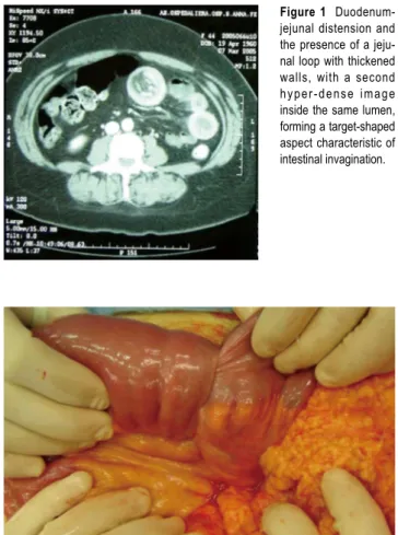
A 45-year old woman complained of continual nausea and biliary vomiting, associated with a weight loss of 5 kg. Due to localized abdominal pain, mainly in the right hypo-chondrium and episodes of hematemesis, the patient was admitted to our hospital. Blood tests revealed sideropenic anemia with 81 g/L haemoglobin, serum iron 100 pg/L, ferritin 23 µg/L, and fecal occult blood test (FOBT) posi-tive in three fecal samples. A gastroscopy was performed, which showed the presence of gradeⅠesophagitis, moder-ate hiatal hernia and chronic erosive gastritis. The colonos-copy was incomplete due to the presence of colic stools. Abdominal ultrasonography highlighted a distension of the intestinal loops without signs of parenchymatous or-gan pathology. The patient therefore received an abdomi-nal CT, which suggested the presence of a gastric disten-sion with duodenum-jejunal distendisten-sion and the presence of a jejunal loop with thickened walls. A second hyper-dense image inside the intestinal lumen, forming a target-shaped image was also present: typical feature of intestinal invagination (Figure 1). We therefore decided to proceed to urgent surgical operation after blood transfusion. Dur-ing surgery, intra-peritoneal fluid was found and samples were removed for cytological testing. Invagination at the third jejunal loop (Figure 2) was evidenced. The presence of hypertrophic lymphatic tissue with intestinal mesenteric lymphoadenomegalia was also present. Manual resolu-tion of the invaginaresolu-tion was carried out. This procedure highlighted the presence of a hyperchromic ulcerated
CASE REPORT
Jejuno-jejunal invagination due to intestinal melanoma
www.wjgnet.com
Giuseppe Resta, Gabriele Anania, Federico Messina, Damiano de Tullio, Gloria Ferrocci, Federico Zanzi, Davide Pellegrini, Rocco Stano, Giorgio Cavallesco, Gianfranco Azzena, Savino Occhionorelli, Università degli Studi di Ferrara, Dipartimento di Scienze Chirurgiche Anestesiologiche e Radiologiche, Istituto di Clinica Chirurgica, Arcispedale “S. Anna”, Ferrara, Italy
Correspondence to: Dr. Federico Messina, Università degli Studi di Ferrara, Dipartimento di Scienze Chirurgiche Anestesiologiche e Radiologiche, Istituto di Clinica Chirurgica. Arcispedale “S. Anna”, Corso Giovecca, Ferrara 203-44100,
Italy. [email protected]
Telephone: +39-532-236316 Fax: +39-532-209819 Received: 2006-10-22 Accepted: 2006-12-07
Abstract
Cutaneous melanoma is one of the most studied neoplastic lesions in biology and clinical oncology. It has been well documented that this type of neoplasm presents a high metastatic rate, and is able to involve nearly every tissue. Non-cutaneous melanoma represents an unusual pattern of melanoma, and the small intestine is an uncommon anatomic localization. Herein we report an extremely rare clinical case of a young woman affected by a bleeding jejunal melanoma, whose early clinical presentation was an intestinal invagination. © 2007 The WJG Press. All rights reserved.
Key words: Cutaneous melanoma; Intestinal obstruction;
Intestinal melanoma; Invagination
Resta G, Anania G, Messina F, de Tullio D, Ferrocci G, Zanzi F, Pellegrini D, Stano R, Cavallesco G, Azzena G, Occhionorelli S. Jejuno-jejunal invagination due to intestinal melanoma. World J Gastroenterol 2007; 13(2): 310-312
http://www.wjgnet.com/1007-9327/13/310.asp
INTRODUCTION
Cutaneous melanoma is a malignancy characterized by a high mortality rate. It can metastasize to all organs, although the gastrointestinal tract is an unusual metastatic localization. In 50% of cutaneous melanomas, in fact, metastases in the gastroenteric tract are diagnosed upon autopsy, and only in 5% of cases, they are diagnosed clinically[1,2]. Intestinal metastases of cutaneous melanomas
PO Box 2345, Beijing 100023, China World J Gastroenterol 2007 January 14; 13(2): 310-312 www.wjgnet.com World Journal of Gastroenterology ISSN 1007-9327 [email protected] © 2007 The WJG Press. All rights reserved.
Resta G et al. Intestinal obstruction due to intestinal melanoma 311
www.wjgnet.com
formation, with obvious signs of recent bleeding coming from the serosa (Figure 3).
Thorough exploration of the abdominal cavity did not detect further replicative lesions. Resection of the third jejunal loop containing the neoformations and the whole underlying mesentery with its lymph nodes, was per-formed. The intestinal continuity was restored through a latero-lateral jejuno-jejunal anastomosis.
The post-operative course was uneventful and the patient was discharged after 10 postoperative days. The definitive histological examination showed the presence of an intestinal metastasis of cutaneous melanoma of unknown origin (Figures 4 and 5). The patient was then
re-ferred to an oncologic centre for the search of the primary melanoma localization. At three, six months and a year fol-low-up, the patient is alive and no signs of skin melanoma have been detected.
DISCUSSION
The peculiar rarity of this clinical case represents the principal reason for our interest. In spite of the clinical manifestation and the diagnostic-therapeutic approach adopted, this case presents many conditions that have been previously poorly documented in international literature.
Our patient presented a chronic anemia and repeated biliary vomiting that were related to the erosive gastritis and esophagitis identified by the gastroscopy, even if this did not necessarily exclude chronic bleeding from neo-plastic lesions. The pre-operative CT scan of parajejunal lymphoadenomegalia prompted us to suspect the presence of a neoplastic lesion causing the jejunal invagination. Je-junal invagination in an adult, in fact, can cause 1%-3% of surgically treated intestinal occlusion but is mainly due to peritoneal adhesion or to the presence of intestinal anas-tomosis[11] and less frequently to a neoplasm. Melanoma
intestinal metastasis is clinically diagnosed only in 3%-5% of cases, and is usually found to affect the stomach or the colon. Jejunal location is much less frequent[12] and,
if present, does not show clinical signs till diagnosed at autopsy. A diagnosis based on the finding of metastatic Figure 2 Invagination at the third jejunal loop.
Figure 5 Strong positivity at immunohistochemical assay for HMB45.
Figure 4 Ileal wall infiltrated with metastatic melanomatous cells with nuclear
pseudoinclusions and nucleoli, in nest and trabecular arrangement (HE x 10).
Figure 3 Presence of a hyperchromic ulcerated neoformation, with signs of recent
bleeding of the serosa.
Figure 1
Duodenum-jejunal distension and the presence of a jeju-nal loop with thickened walls, with a second hyper-dense image inside the same lumen, forming a target-shaped aspect characteristic of intestinal invagination.
lesions, without identification of the primary cutaneous source, as described in our report, is carried out in only 3% of melanomas. Even if the etiology of primary gas-trointestinal melanomas remain undefined, some authors suggest that primary gastrointestinal melanomas are de-rived from melanoblastic cells of the neural crest which, migrating through the omphalomesenteric canal or APUD cells, reach the intestinal tract, undergoing neoplastic transformation[13]. Non-cutaneous melanomas represent
a rare form of melanoma. In a review of 84 836 cases of melanoma, 91.2% were cutaneous, 5.2% ocular, 2.2% of unknown primary site and only 1.3% of gastrointestinal mucosa[14]. Melanomas that arise on mucosal surfaces
ap-pear to be more aggressive and are associated with worse prognosis than cutaneous melanomas. The poorer progno-sis may be related to the delay in diagnoprogno-sis, to their more aggressive behaviour, or to earlier dissemination because of the rich lymphatic and vascular supply of the gastroin-testinal mucosa[15,16].
Our case shows that the presence of an early complica-tion due to intestinal occlusion, through a sequential and appropriate instrumental diagnostic evaluation, gave us the opportunity to identify a rare melanotic lesion. Moreover, the right surgical approach has given us the opportunity to improve the length and quality of the life of this unlucky patient.
REFERENCES
1 Reintgen DS, Thompson W, Garbutt J, Seigler HF. Radiologic,
endoscopic, and surgical considerations of melanoma metastatic to the gastrointestinal tract. Surgery 1984; 95: 635-639
2 de la Monte SM, Moore GW, Hutchins GM. Patterned
distribution of metastases from malignant melanoma in humans. Cancer Res 1983; 43: 3427-3433
3 Ihde JK, Coit DG. Melanoma metastatic to stomach, small
bowel, or colon. Am J Surg 1991; 162: 208-211
4 Caputy GG, Donohue JH, Goellner JR, Weaver AL. Metastatic
melanoma of the gastrointestinal tract. Results of surgical management. Arch Surg 1991; 126: 1353-1358
5 Khadra MH, Thompson JF, Milton GW, McCarthy WH. The
justification for surgical treatment of metastatic melanoma of the gastrointestinal tract. Surg Gynecol Obstet 1990; 171: 413-416 6 Agrawal S, Yao TJ, Coit DG. Surgery for melanoma metastatic
to the gastrointestinal tract. Ann Surg Oncol 1999; 6: 336-344 7 Ollila DW, Essner R, Wanek LA, Morton DL. Surgical
resection for melanoma metastatic to the gastrointestinal tract. Arch Surg 1996; 131: 975-979; 979-980
8 Reintgen DS, McCarty KS, Woodard B, Cox E, Seigler HF.
Metastatic malignant melanoma with an unknown primary. Surg Gynecol Obstet 1983; 156: 335-340
9 Loualidi A, Spooren PF, Grubben MJ, Blomjous CE, Goey
SH. Duodenal metastasis: an uncommon cause of occult small intestinal bleeding. Neth J Med 2004; 62: 201-205
10 Wulf V, Schröder HJ. Metastasis to the small intestine of malignant melanoma as a rare cause of intestinal hemorrhage. Zentralbl Chir 1994; 119: 515-516
11 Begos DG, Sandor A, Modlin IM. The diagnosis and management of adult intussusception. Am J Surg 1997; 173: 88-94
12 Pacovský Z, Fait V. Distant metastasis of malignant melanoma in the small intestine. Rozhl Chir 1992; 71: 424-428
13 Elsayed AM, Albahra M, Nzeako UC, Sobin LH. Malignant melanomas in the small intestine: a study of 103 patients. Am J Gastroenterol 1996; 91: 1001-1006
14 Chang AE, Karnell LH, Menck HR. The National Cancer Data Base report on cutaneous and noncutaneous melanoma: a summary of 84,836 cases from the past decade. The American College of Surgeons Commission on Cancer and the American Cancer Society. Cancer 1998; 83: 1664-1678
15 Sachs DL, Lowe L, Chang AE, Carson E, Johnson TM. Do primary small intestinal melanomas exist? Report of a case. J Am Acad Dermatol 1999; 41: 1042-1044
16 Lagoudianakis EE, Genetzakis M, Tsekouras DK, Papadima A, Kafiri G, Toutouzas K, Katergiannakis V, Manouras A. Primary gastric melanoma: a case report. World J Gastroenterol 2006; 12: 4425-4427
S- Editor Wang GP L- Editor Zhu LH E- Editor Liu WF
312 ISSN 1007-9327 CN 14-1219/R World J Gastroenterol January 14, 2007 Volume 13 Number 2