REVIEW
Autologous haematopoietic stem cell mobilisation in multiple
myeloma and lymphoma patients: a position statement from
the European Group for Blood and Marrow Transplantation
M Mohty
1,27, K Hübel
2,27, N Kröger
3,27, M Aljurf
4,27, J Apperley
5,27, GW Basak
6,27, A Bazarbachi
7,27, K Douglas
8,27, I Gabriel
5,27,
L Garderet
1,27, C Geraldes
9,27, O Jaksic
10,27, MW Kattan
11,27, Z Koristek
12,27, F Lanza
13,27, RM Lemoli
14,27, L Mendeleeva
15,27, G Mikala
16,27,
N Mikhailova
17,27, A Nagler
18,27, HC Schouten
19,27, D Selleslag
20,27, S Suciu
21,27, A Sureda
22,27, N Worel
23,27, P Wuchter
24,27,
C Chabannon
25,27and RF Duarte
26,27Autologous haematopoietic SCT with PBSCs is regularly used to restore BM function in patients with multiple myeloma or
lymphoma after myeloablative chemotherapy. Twenty-eight experts from the European Group for Blood and Marrow
Transplantation developed a position statement on the best approaches to mobilising PBSCs and on possibilities of optimising graft
yields in patients who mobilise poorly. Choosing the appropriate mobilisation regimen, based on patients
’ disease stage and
condition, and optimising the apheresis protocol can improve mobilisation outcomes. Several factors may in
fluence mobilisation
outcomes, including older age, a more advanced disease stage, the type of prior chemotherapy (e.g.,
fludarabine or melphalan),
prior irradiation or a higher number of prior treatment lines. The most robust predictive factor for poor PBSC collection is the CD34
+cell count in PB before apheresis. Determination of the CD34
+cell count in PB before apheresis helps to identify patients at risk of
poor PBSC collection and allows pre-emptive intervention to rescue mobilisation in these patients. Such a proactive approach
might help to overcome de
ficiencies in stem cell mobilisation and offers a rationale for the use of novel mobilisation agents.
Bone Marrow Transplantation (2014)
49, 865–872; doi:10.1038/bmt.2014.39; published online 31 March 2014
INTRODUCTION
Autologous haematopoietic SCT (auto-HSCT) aims to restore BM
function after high-dose chemotherapy. In the context of
auto-HSCT, mobilised PBSCs are currently the preferred source
of HSCs worldwide for adult patients with multiple myeloma (MM)
and lymphoma (non-Hodgkin
’s and Hodgkin’s lymphoma).
1–3Auto-HSCT with PBSCs is favoured because it leads to faster
engraftment
and
haematologic
reconstitution
versus
BM
infusion, resulting in potentially improved patient outcomes.
1Moreover, some studies demonstrated that the use of PBSCs
was associated with better quality of life and reduced total
costs.
1,2,4,5HSCs usually circulate in a very small number in PB;
6therefore,
their mobilisation from BM to PB is an essential part of auto-HSCT
programs. Cytokines such as G-CSF, alone or in combination with
chemotherapy, are typically used for PBSC mobilisation.
1,2,7Compared with G-CSF-based mobilisation, chemo-mobilisation
(i.e., chemotherapy+G-CSF) has advantages in terms of putative
antitumour effect and higher probability of obtaining grafts with a
suf
ficient CD34
+cell count with lower numbers of aphereses.
1,7However, disadvantages of chemo-mobilisation include increased
toxicity and morbidity, and the need for hospitalisation
(depend-ing on the chemotherapy schedule; e.g., cyclophosphamide-based
mobilisation is possible in an outpatient setting).
1,7Despite widespread and established practice, current
mobilisa-tion strategies vary between centres and differ in terms of
feasibility and outcome.
2,7Although the majority of patients are
able to mobilise suf
ficient CD34
+cells for at least a single
autograft, approximately 15% fail to do so.
8If two autografts are
needed for a specific treatment strategy, even more patients fail to
reach their individual collection goal. Newer approaches aiming to
optimise mobilisation procedures include off-label use of
pegy-lated G-CSF,
9–12erythropoietin,
13SCF
14,15and plerixafor.
16–18Nevertheless, it is necessary to optimise the current mobilisation
approaches and to identify upfront the patients at risk of
mobilisation failure.
1
Department of Haematology, Saint Antoine Hospital, Paris, France;2
University Hospital Cologne, Cologne, Germany;3
University Hospital Hamburg-Eppendorf, Hamburg, Germany;4
King Faisal Specialist Hospital and Research Centre, Riyadh, Saudi Arabi;5
Imperial College London, London, UK;6
The Medical University of Warsaw, Warsaw, Poland; 7
American University of Beirut, Beirut, Lebanon;8
Beatson West of Scotland Cancer Centre, Glasgow, UK;9
University Hospital Coimbra, Coimbra, Portugal;10
University Hospital Dubrava, Zagreb, Croatia;11Quantitative Health Sciences Cleveland Clinic, Cleveland, OH, USA;12Department of Haematooncology, University Hospital Ostrava, Ostrava, Czech Republic;13
Cremona Hospital, Cremona, Italy;14
University of Genoa, Genoa, Italy;15
National Research Centre for Haematology, Moscow, Russia;16
St Istvan and St Laszlo Hospital, Budapest, Hungary;17Institute of Children Haematology and Transplantation n.a. R Gorbacheva, St Petersburg State Pavlov Medical University, St Petersburg, Russia;18Chaim Sheba Medical Center, Tel Hashomer, Israel;19
Maastricht University Medical Centre, Maastricht, The Netherlands;20
Department of Haematology, AZ Sint-Jan, Brugge-Oostende, Belgium;21
EORTC Headquarters, Brussels, Belgium;22
Addenbrooke's Hospital, Cambridge, UK;23
Medical University Vienna, Vienna, Austria;24
Department of Medicine V, Heidelberg University, Heidelberg, Germany;25
Institut Paoli-Calmettes and Inserm CBT-510, Marseille, France and26
Catalan Institute of Oncology, L'Hospitalet de Llobregat, Barcelona, Spain. Correspondence: Professor M Mohty, Department of Haematology, Saint Antoine Hospital, 184, rue du Faubourg Saint Antoine, Paris 75571, France. E-mail: [email protected]
27
All these authors contributed equally to this work.
Received 20 October 2013; revised 19 January 2014; accepted 28 January 2014; published online 31 March 2014
PURPOSE AND METHOD
This review generated by 28 experts from the European Group for
Blood and Marrow Transplantation (EBMT) attempted to develop a
position statement on best approaches for auto-HSC mobilisation
in patients with MM and lymphoma, on factors predictive of poor
mobilisation or mobilisation failure and on potential optimisation
options for poor mobilisers. The position statement is based on
currently
available
literature
and
clinical
practice
of
the
expert group.
OVERVIEW OF CURRENT AUTO-HSC MOBILISATION
APPROACHES
Steady-state (cytokines only)
Monotherapy with G-CSFs remains the only available option for
steady-state mobilisation, as GM-CSF is no longer available in
many countries after commercial failure and withdrawal. G-CSF
treatment leads to granulocyte activation/expansion and release
of various proteases,
eventually resulting in cleavage of
cell
–extracellular adhesion molecules that retain HSCs in BM.
6Currently, the G-CSF cytokines
filgrastim and lenograstim have
marketing
authorisation
for
mobilisation
of
auto-HSCs
in
Europe.
19,20The
approved
doses/schedules
are
filgrastim
10
μg/kg per day subcutaneously for 5–7 consecutive days and
lenograstim 10
μg/kg per day subcutaneously for 4–6 days;
leukapheresis should be performed on days 5 or 6 (
filgrastim)
and between days 5 and 7 (lenograstim). Mobilisation with
cytokines alone (Figure 1a) is well tolerated, but their use can be
limited by the feasibility of embedding them into individual,
study-based treatment plans or by suboptimal PBSC yields.
Chemo-mobilisation
Adding chemotherapeutic agents to cytokines may increase PBSC
yields and can further decrease tumour burden.
1,7However, the
PBSC mobilisation window is less predictable compared with
steady-state approaches,
1,7causing potential problems with
apheresis scheduling. In addition, the incidence and severity of
side effects with chemotherapy+G-CSF is increased compared
with G-CSF alone.
21–23The approved doses for PBSC mobilisation
after myelosuppressive chemotherapy are
filgrastim 5 μg/kg per
day subcutaneously and lenograstim 150
μg/m
2per day (i.e.,
therapeutically equivalent of 5
μg/kg per day subcutaneously),
starting within 1
–5 days after completion of chemotherapy until
last leukapheresis.
19,20Chemotherapy-based mobilisation may be part of the
disease-specific chemotherapy (Figure 1b) or of separate
chemo-therapeutic course(s) in addition to disease-specific treatment
(Figure 1c). The choice of a speci
fic chemo-mobilisation approach
is based on patient disease characteristics and on local clinical
practice guidelines (Table 1). In MM patients, high-dose
cyclopho-sphamide+G-CSF is probably the most commonly used
chemo-mobilisation strategy,
21,23whereas some studies also suggest
etoposide-based mobilisation approaches.
24With the advent of
new therapeutic agents, such as proteasome inhibitors and
immunomodulatory drugs,
25the role of high-dose
cyclopho-sphamide as therapeutic agent in MM now becomes more
questionable given its relatively small antitumour effect.
26In
lymphoma patients, chemotherapy+G-CSF as part of the
disease-speci
fic induction and salvage regimens has always been the
preferred method. Such approach can eliminate the need for
additional chemo-mobilisation or steady-state mobilisation before
auto-HSCT in these heavily treated patients.
27–30Furthermore,
these disease-speci
fic chemotherapy combinations have been
shown to be more effective than cyclophosphamide-based
chemo-mobilisation.
29,30Plerixafor
Plerixafor, a novel chemokine-receptor (CXCR4) antagonist,
disrupts the interaction between stromal-deriving factor 1 and
CXCR4, thereby enhancing the HSC mobilisation effect of G-CSF.
31In Europe, plerixafor is approved for use in combination with
G-CSF for auto-HSC mobilisation in patients with lymphoma or
MM whose cells mobilise poorly.
32The approved dose is 240
μg/
kg per day subcutaneously 6
–11 h before initiation of apheresis
following 4-day G-CSF pretreatment.
Plerixafor plus G-CSF alone or G-CSF+chemotherapy has been
shown to be effective and well tolerated in patients with MM or
lymphoma, including poor mobilisers, with superior ef
ficacy to
G-CSF alone or G-CSF+chemotherapy.
13,16,17,33–37Numerous
studies reported that plerixafor plus G-CSF is effective as rescue
therapy during HSC mobilisation.
36,38–41Whether the addition of
plerixafor results in a benefit for patients in terms of clinical
outcomes is still under investigation.
Disease-specific (chemo)therapy
a
b
c
Apheresis G-CSF Apheresis Apheresis High-dose chemotherapy G-CSF High-dose chemotherapy G-CSF Disease-specific chemotherapy Mobilisation chemotherapy Disease-specific (chemo)therapy High-dose chemotherapy Auto-HSCT Auto-HSCT Auto-HSCTFigure 1. Current auto-HSCT mobilisation strategies: steady-state
mobilisation (
a; cytokines alone), chemotherapy-based mobilisation
using disease-speci
fic chemotherapy (b) or separate mobilisation
chemotherapy (
c).
Table 1.
Current chemo-mobilisation approaches (selection based on clinical practice of the expert group)Disease-specific chemo-mobilisation Separate mobilisation chemotherapy MM
Cyclophosphamide-based CAD, DPACE, VDT-PACE
(Relapsed) lymphoma Etoposide-based ABVD, BEACOPP, (R)-CHOP, (R)-DA-EPOCH,
(R)-DHAP, carbo-DHAP, dexa-BEAM, (R)-ESHAP, (R)-mini-BEAM, (R)-ICE, IVE, R-ACVBP, R-bendamustine, VIM
Abbreviations: ABVD= doxorubicin, bleomycin, vinblastine, dacarbazine; ACVBP= doxorubicin, cyclophosphamide, vindesine, bleomycin, prednisone; BEACOPP= bleomycin, etoposide, doxorubicin, cyclophosphamide, vincristine, procarbazine, prednisone; BEAM= BCNU, etoposide, ara-C (cytarabine), melphalan; CAD= cyclophosphamide, doxorubicin, dexa-methasone; carbo= carboplatin; CHOP = cyclophosphamide, doxorubicin, vincristine, prednisone; DA-EPOCH= dose-adjusted etoposide, prednisone, vincristine, cyclophosphamide, doxorubicin; Dexa= dexamethasone; DHAP= dexamethasone, ara-C, cisplatin; DPACE= dexamethasone, platinum, doxorubicin, cyclophosphamide, etoposide; ESHAP= etoposide, methylprednisolone, ara-C, cisplatin; ICE= ifosfamide, carboplatin, etoposide; IVE= ifosfamide, etoposide, epirubicin; MM = multiple myeloma; R= rituximab; VDT-PACE = bortezomib, dexamethasone, thalidomide, cisplatin, doxorubicin, cyclophosphamide, etoposide; VIM= etoposide, ifosfamide, mitoxantrone.
OPTIMISING GRAFT ‘QUANTITY’ (CD34
+CELL YIELD)
Mobilisation regimen
In clinical practice,
filgrastim and lenograstim may be used
interchangeably and modi
fications of the approved label may be
applied depending on local guidelines and treatment plans.
Higher G-CSF doses (⩾10 μg/kg per day) or alternate application
schedules (two times 5
μg/kg per day instead of one time
10
μg/kg per day) have been investigated to further enhance the
number of harvested auto-HSCs.
42,43However, further research is
required. In addition, high G-CSF doses may increase the risk
of splenic rupture.
44As mentioned above, CD34
+cell yields can be
further increased by mobilisation with chemotherapy+G-CSF
compared with G-CSF monotherapy.
21–23The addition of
plerixafor to standard mobilisation strategies has been shown
to increase CD34
+cell yields in known or predicted poor
mobilisers.
13,16,17,33–37,39,40Timing
The timing of mobilisation regimen administration and apheresis
may also in
fluence CD34
+cell yields. Recent data suggest that
auto-HSC collection efficacy is higher and the proportion of
patients with optimal harvest is larger when G-CSF is given
3 h before apheresis versus administration on the evening before
apheresis.
45Nevertheless, further research is needed to confirm
these data. Plerixafor was shown to mobilise adequate auto-HSC
yields when administered as rescue treatment before or during
apheresis in patients with insuf
ficient auto-HSC mobilisation after
G-CSF monotherapy or chemo-mobilisation.
38–41Earlier detection
of PBSC mobilisation (based on CD34
+cell-count assessments on
day 4 versus 5) can help determine whether plerixafor should be
administered, and has been suggested to further decrease the risk
of mobilisation failure.
46Technical aspects
The
flow cytometry protocol for CD34
+cell measurement is
a critical step in monitoring the HSC mobilisation process.
47A
validated
protocol
and
external
quality
control
are
recommended.
48CD34
+cell collection has been shown to be
more ef
ficient with larger apheresis volumes (4.0–5.3 times the
patient
’s total blood volume)
49,50and no difference in CD34
+cell
viability was observed compared with normal-volume apheresis
(2.7
–3.5 times the patient’s total blood volume).
49Therefore,
enhanced volumes are recommended for apheresis in relatively
poor mobilisers or patients with high individual CD34
+cell
collection goal (⩾3 transplants). For patients who still mobilise
poorly with larger-volume approaches, plerixafor addition to
standard mobilisation strategies may sufficiently enhance
mobi-lisation efficacy.
51Nevertheless, not all patients are eligible for
enhanced volume strategies. Larger transfusion volumes and
related higher DMSO contents have been associated with
increased risk of cardiac side effects.
52Position statement on current auto-HSC mobilisation approaches
and their optimisation
Auto-HSC mobilisation approaches for MM and lymphoma
patients suggested by the expert group are shown in Figure 2. If
required, strategies are recommended to be optimised by
remobilising with cytokines alone or by changing the previously
chosen chemo-mobilisation approach (e.g., switch from
steady-state to mobilisation, or choose an alternative
chemo-mobilisation strategy if patients failed to mobilise after an initial
chemotherapy-based approach). In addition, new agents such as
plerixafor or the use of large-volume apheresis can further
improve mobilisation outcomes. For the latter, processing of up
to three times the total blood volume is suggested as feasible
without impairing the patient
’s tolerance.
The optimal time to start apheresis is more predictable for
patients mobilised with G-CSF alone than for those mobilised with
chemotherapy+G-CSF. For patients who were mobilised with
chemotherapy+G-CSF and showed too low CD34
+counts on the
estimated day of apheresis, the expert group suggests reassessing
CD34
+counts after 1 day. This ensures that leukapheresis is
started on the optimal day. Up to four leukapheresis sessions can
be recommended as feasible. However, the group raised the issue
of cost-effectiveness of such practice.
MM
Steady-state mobilisation*
Chemotherapy-based mobilisation†
(cyclophosphamide or etoposide)
Decision to use steady-state or chemo-mobilisation should be based on local guidelines However, sufficient CD34+ cell yields are less likely with steady-state mobilisation
• •
• Cyclophosphamide monotherapy: range of 1.5–4.0 g/m2 is feasible
Lymphoma
Disease-specific chemotherapy approaches†
Steady-state mobilisation*
Disease-specific chemotherapy approaches are suggested to avoid the burden of additional chemotheraphy cycles
Steady-state mobilisation may be an option for selected patients: - Patients in complete remission
- Patients ineligible for chemo-mobilisation •
•
a
b
Figure 2. Position statement: PBSC mobilisation strategies for MM (a) and lymphoma patients (b). *G-CSF only;
†G-CSF+chemotherapy;
§higher
doses of cyclophosphamide may be used based on available data; however, the aim should be to keep the duration of neutropenia as short as
possible.
AUTOGRAFT ‘QUALITY’
Cell subsets other than CD34
+cells
Recent data suggest that the quality of CD34
+cells from poor
mobilisers is comparable to those from adequate mobilisers in
patients treated with
filgrastim;
53the dose of mobilised CD34
+cells per kg mainly determines neutrophil and platelet
engraft-ment after auto-HSCT.
54The addition of plerixafor to G-CSF alone
or G-CSF+chemotherapy not only mobilises more CD34
+cells but
also seems to increase the proportion of more-primitive HSC
subsets, the absolute lymphocyte count and the numbers of
various lymphocyte subsets (CD19
+B lymphocytes, CD3
+T cells and natural killer (NK) cells) in the autograft.
55–59Preliminary data suggest a positive correlation between the
number of reinfused NK cells and early absolute lymphocyte
recovery after auto-HSCT.
60However, further investigation is
needed to evaluate potential effects of autograft cell subsets on
the patients
’ clinical outcomes.
Tumour cell contamination
Auto-HSCT is associated with the risk of tumour cell contamination
of the graft. Current mobilisation strategies with G-CSF or G-CSF
+chemotherapy vary not only in auto-HSC yields and safety, but
also in levels of autograft contamination with tumour cells.
61–66Whether the antitumour effect of chemo-mobilisation also
translates into a lower risk of tumour cell contamination,
compared to steady-state mobilisation, remains controversial.
62The integration of novel agents into mobilisation regimens so far
does not appear to increase tumour cell contamination of the
graft.
67,68A large currently ongoing trial (Collaboration between
EBMT and Genzyme to collect Autologous transplant outcomes in
Lymphoma and Myeloma patients (CALM)) will compare the
outcomes of patients transplanted with plerixafor-mobilised cells
between 2008 and 2011 to those of equivalent patients
transplanted without plerixafor to determine whether plerixafor
mobilises increased rates of malignant cells (data to be published;
patients will be followed until 2014). The impact of autograft
tumour cell contamination on long-term safety and clinical
outcome is still controversial.
62Randomised phase III studies
suggest that tumour cell contamination of the graft does not
signi
ficantly affect PFS or OS.
5,62–64,69–71On the other hand, results
from a report of the Centre for International Blood and Marrow
Transplant Research indicate that syngeneic transplants lead to
better outcome than autologous transplants, suggesting that
contamination is a problem in myeloma and probably also in
lymphoma.
72,73It should also be noted that most clinical trials in
MM patients were performed before the recent implementation of
novel treatments. Therefore, in vivo tumour debulking may be
much higher today, resulting in a higher potential of
contami-nated autografts and reinfused tumour cells inducing relapse. This
makes it dif
ficult to draw definitive conclusions on the role of
residual plasma cells and ex vivo purging.
Position statement on autograft
‘quality’
Determination of cell subsets other than CD34
+cells is not
routinely performed in clinical practice, but only in clinical trials.
Accordingly, assessment of tumour cell contamination in routine
clinical practice may not be valuable but can be of interest in
clinical trials.
FACTORS PREDICTIVE OF POOR MOBILISATION OR
MOBILISATION FAILURE
Factors described as predictive of impaired HSC collection or
mobilisation failure include: older age; female sex; diagnosis
(lymphoma worse than MM); longer time since diagnosis; more
advanced disease; previous radiotherapy and/or chemotherapy
(especially
fludarabine and other purine analogues, and
melpha-lan); higher number of previous therapy lines; longer time from
last chemotherapy to mobilisation initiation; previous auto-HSCT;
low haemoglobin, WBC, or platelet levels before mobilisation; and
low CD34
+cell counts in BM before mobilisation and in PB before
apheresis.
74–88Whether prior treatment with new therapies such
as lenalidomide and rituximab negatively affects the mobilisation
outcomes in MM and lymphoma patients, respectively, is
controversial.
77,78,82,84,89–91Adaptation of the mobilisation
strat-egy and/or the addition of novel agents (e.g., plerixafor) to
conventional regimens may overcome the negative effect
of
prognostic
factors
for
poor
mobilisation.
24,27,34–36,92–94Nevertheless, there is an urgent need to define which patient
population might benefit from optimised mobilisation approaches
to help clinical decision-making.
Algorithms to de
fine poor mobilisers
In a retrospective analysis of 840 patients with MM or
non-Hodgkin
’s lymphoma, 129 patients (15%) were identified as poor
mobilisers and divided into three categories based on CD34
+levels in PB before leukapheresis:
‘borderline’ poor mobilisers
(11
–19/μL at maximum stimulation), ‘relative’ poor mobilisers
(6
–10/μL) and ‘absolute’ poor mobilisers (o5/μL).
8Diagnosis, sex,
age, body weight and previous irradiation made no signi
ficant
difference in HSC mobilisation capacity. Only the number of
previous chemotherapy cycles and prior melphalan treatment had
a signi
ficant impact on the ability to mobilise HSCs. In another
retrospective analysis of 1556 patients with lymphoproliferative
disorders initially mobilised with G-CSF alone, sensitivity–
specificity analysis was used to identify ideal PB CD34
+count
cut-points that would allow early intervention and prevent
collection failure.
86In patients with plasma cell disorders, PB
CD34
+counts of 11, 17, 21 and 28/
μL by day 4 or 5 were required
to collect a minimum of 2, 4, 8 or 12 × 10
6CD34
+cells per kg,
respectively. A CD34
+yield
o0.8 × 10
6cells per kg on day 1 of
apheresis was predictive of
o2 × 10
6CD34
+cells per kg. For
patients with non-Hodgkin
’s or Hodgkin’s lymphoma, PB CD34
+counts of
o6 and o15/μL on day 4 or 5 predicted failure
to achieve a target collection of 2 and 4 × 10
6cells per kg,
respectively.
The Gruppo Italiano Trapianto di Midollo Osseo proposed a
de
finition of poor mobilisers in lymphoma and MM patients using
an analytic hierarchical process.
94Patients are de
fined as ‘proven’
poor mobilisers when (1) after adequate mobilisation (G-CSF
10
μg/kg if used alone or ⩾ 5 μg/kg after chemotherapy),
the circulating CD34
+cell peak is
o20/μL for up to 6 days
after mobilisation with G-CSF or up to 20 days after G-CSF
+chemotherapy, or (2) they yield
o2.0 × 10
6CD34
+cells per kg in
⩽ 3 aphereses. Patients are ‘predicted’ poor mobilisers if they
(1) failed a previous collection attempt, (2) previously received
extensive radiotherapy or full courses of therapy affecting HSC
mobilisation and (3) meet two of the following criteria: advanced
disease (
⩾2 lines of chemotherapy), refractory disease, extensive
BM involvement or cellularity
o30% at the time of mobilisation,
and age
⩾ 65 years.
Several groups have developed algorithms to guide the use of
plerixafor.
85,95–97Costa et al.
97developed and validated a
decision-making algorithm based on the PB CD34
+cell count on
day 4 of G-CSF administration and the collection target of CD34
+cells to guide cost-effective use of plerixafor for auto-HSC
mobilisation (continuing G-CSF only or adding plerixafor).
Subse-quently, they showed that patient-adapted plerixafor use based
on this algorithm was superior to cyclophosphamide plus growth
factor
98and successfully mobilised HSCs in MM patients
previously treated with lenalidomide.
99Abhyankar et al.
95devel-oped a risk-based approach to optimise HSC collection with
plerixafor by identifying potential poor mobilisers upfront. The
868
plerixafor algorithm takes into account the number of circulating
CD34
+cells per
μL on day 5 of G-CSF mobilisation, the desired
number of CD34
+cells per kg needed per transplant (
⩾2.5 × 10
6/
kg for 1 transplant and
⩾ 5 × 10
6/kg for >1 transplants), and the
day-1 CD34
+collection yield. A day-5 CD34
+circulating level of
o10 cells per μL (for 1 transplant), or o20 cells per μL (for >1
transplant), or a day-1 CD34
+collection of less than one-half of the
total CD34
+dose needed, prompted the use of plerixafor. Data
demonstrated that this approach helped decrease the need for
remobilisation and reduce the number of collection days needed.
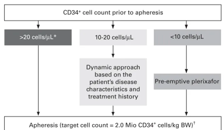
Position statement on predictive factors for poor mobilisation,
CD34
+cell-count determination and proactive intervention to
rescue PBSC collection
Factors considered by the expert group to be predictive of poor
mobilisation in MM and lymphoma patients in daily clinical
practice are listed in Table 2. Of these, the CD34
+cell count in PB
before apheresis is the most robust predictor for poor PBSC
collection.
8,85,86,100Thus, determination of CD34
+cell counts in PB
before apheresis is suggested by the expert group to estimate the
patient
’s risk of poor PBSC collection and to consider additional
intervention for patients at risk (Figure 3). For patients with
>20 CD34
+cells per
μL in PB before apheresis, no proactive
intervention is needed, whereas the group suggests pre-emptive
use of plerixafor to reach a minimum cell target of 2 × 10
6CD34
+cells per kg body weight for patients with
o10 CD34
+cells per
μL
in PB before apheresis. For patients with 10–20 CD34
+cells per
μL
at the mobilisation peak before apheresis (i.e., grey zone), a
dynamic approach is suggested, also taking into account other
previously published predictive factors (Table 2) and the target
number of aphereses before applying plerixafor (e.g., when
collecting PBSCs for
⩾ 2 transplants in MM patients, pre-emptive
intervention with plerixafor may become mandatory for patients
with 10
–20 CD34
+cells per
μL before apheresis). These
sugges-tions are to be considered the minimum number of CD34
+cells
for auto-HSCT. Higher CD34
+cell counts before auto-HSCT may
reduce the need of post-HSCT support. The EBMT
‘Haematopoietic
Stem Cell Mobilisation and Apheresis
’ handbook recommends
optimal levels being
⩾ 5 × 10
6CD34
+cells per kg for a
single
transplant
(http://www.ebmt.org/Contents/Resources/
Library/Resourcesfornurses/Documents/Haematopoietic%20Stem
%20Cell%20Mobilisation%20and%20Apheresis%20Handbook.pdf;
accessed on 19 November 2013).
In the opinion of the expert group robust validation of these
factors in a prospective registration trial is desirable. Although
several algorithms were recently proposed to predict PBSC
collection failure using PB CD34
+counts before leukapheresis as
threshold to stratify patients at risk and to trigger proactive
intervention with plerixafor,
8,85,86,95–97,100there is a need for
optimised algorithms to predict apheresis yields based on
circulating CD34
+cell numbers. Also, readily available, robust,
and harmonised techniques for such CD34
+cell number
determination are required.
CONCLUSIONS
PBSC mobilisation can be optimised with an appropriate strategy
adapted to each patient, based on the patient
’s disease and
treatment features and the individual collection goal. A low CD34
+cell count in PB before apheresis is a candidate predictor for poor
PBSC collection. The expert group suggested that determination
of CD34
+cell counts before apheresis may estimate the patient
’s
risk for poor PBSC collection and may allow proactive intervention
to rescue mobilisation failure.
CONFLICT OF INTEREST
MM: Research support and honoraria from Sanofi, Amgen and Chugai, whose products are discussed here. KH: Consulting fee or honorarium and support for travel to meetings from Genzyme/Sanofi; board membership for Pfizer and Noxxon; payment for lectures including service on speakers’ bureaus from Janssen; and travel/ accommodations/meeting expenses unrelated to activities listed from Roche. NK: Honorarium for lectures from Sanofi; and consultant fee from Noxxon. MA: Consulting fee or honorarium from ETICHO. GWB: Sponsorship for participation in scientific conferences from Sanofi; membership of Mozobil advisory boards for Sanofi; and reimbursements for trainings from Sanofi. AB: Honoraria from Sanofi whose products are discussed here. KD: Speaker’s fees and honoraria for medical advisory board work from Genzyme and Sanofi Europe between 2009 and 2012 inclusive. GL: Consulting fee or honorarium from ETICHO. CG: None disclosed. OJ: Consulting fee or honorarium from ETICHO; and consultancy for Sanofi. MWK: consultancy for GlaxoSmithKline, Bayer and Merck. ZK: Consultancy for CEEOR; payment for lectures including service on speakers’ bureaus from Sanofi-Aventis; and travel/accommoda-tions/meeting expenses unrelated to activities listed from Sanofi-Aventis. RML: Payment for lectures including service on speakers’ bureaus from Sanofi; and consultancy and board membership for Sanofi. GM: Grant and fees for participation in review activities such as data monitoring boards, statistical analysis, end point committees and the like from ETICHO; support for travel to meetings or other purposes from Genzyme; expert testimony for Janssen and ETICHO; and payment for lectures including service on speakers’ bureaus from Janssen. AN: Research grant from Genzyme/Sanofi; and honorarium for participating in a scientific advisory board. HCS: Membership of an advisory board for Sanofi. DS: Honoraria and research grants from Genzyme and Amgen; and membership of advisory boards for Genzyme and Amgen. AS: Participation in Sanofi-organised advisory boards. NW: Speaker’s fee from Sanofi and membership of the advisory board. PW: Honorarium for lectures from Sanofi; and consulting fee or honorarium from ETICHO. CC: Consultancy for Terumo BCT, Novartis and Sanofi-Genzyme; and membership of an advisory board for
Table 2.
Factors described as predictive of poor mobilisation ormobilisation failure
Predictive factors References Age
Older patients 81,83,100 Disease
More advanced stage 83,85,100 Prior chemotherapy
Higher no. of prior treatment lines 8,75,83,85,88,100
Type of chemotherapy (fludarabine, lenalidomide (controversial) or melphalan)
8,75–78,82–85,87,88,91,100
Prior irradiation 75,83,100 Low CD34+cell count in PB before apheresis 8,75,85,86 Low platelet count before mobilisation
(controversial)
75,79–81,88
CD34+ cell count prior to apheresis
>20 cells/µL* 10-20 cells/µL Dynamic approach based on the patient’s disease characteristics and treatment history <10 cells/µL Pre-emptive plerixafor
Apheresis (target cell count = 2.0 Mio CD34+ cells/kg BW)†
Figure 3. Position statement: proactive intervention to rescue
mobilisation failure. *No proactive intervention required;
†a target
cell count of
>2.0 Mio CD34
+cells per kg body weight (BW) may be
needed depending on the patient
’s disease and treatment features,
and the individual collection goal.
Sanofi-Genzyme. RFD: Consultancy for Sanofi-Genzyme, Amgen and Italfarmaco; and payment for lectures including service on speakers’ bureaus from Sanofi-Genzyme, Amgen and Italfarmaco. JA, IG, FL, NM, LM and SS: no relevant conflicts of interest.
ACKNOWLEDGEMENTS
Medical writing and editorial assistance was provided by Marianne Jenal-Eyholzer, PhD, CMPP, from ETICHO (European Training in Clinical Hematology and Oncology), The Hague, The Netherlands, funded by an unrestricted grant from Sanofi. The authors are fully responsible for the content of this manuscript.
REFERENCES
1 Gertz MA. Current status of stem cell mobilization. Br J Haematol 2010;150: 647–662.
2 Mohty M, Ho AD. In and out of the niche: perspectives in mobilization of hematopoietic stem cells. Exp Hematol 2011;39: 723–729.
3 Passweg JR, Baldomero H, Gratwohl A, Bregni M, Cesaro S, Dreger P et al. The EBMT activity survey: 1990-2010. Bone Marrow Transplant 2012;47: 906–923. 4 Vellenga E, van Agthoven M, Croockewit AJ, Verdonck LF, Wijermans PJ, van
Oers MH et al. Autologous peripheral blood stem cell transplantation in patients with relapsed lymphoma results in accelerated haematopoietic reconstitution, improved quality of life and cost reduction compared with bone marrow transplantation: the Hovon 22 study. Br J Haematol 2001;114: 319–326. 5 Vose JM, Sharp G, Chan WC, Nichols C, Loh K, Inwards D et al. Autologous
transplantation for aggressive non-Hodgkin's lymphoma: results of a rando-mized trial evaluating graft source and minimal residual disease. J Clin Oncol 2002;20: 2344–2352.
6 Pusic I, DiPersio JF. The use of growth factors in hematopoietic stem cell transplantation. Curr Pharm Des 2008;14: 1950–1961.
7 Bensinger W, DiPersio JF, McCarty JM. Improving stem cell mobilization strategies: future directions. Bone Marrow Transplant 2009;43: 181–195. 8 Wuchter P, Ran D, Bruckner T, Schmitt T, Witzens-Harig M, Neben K et al. Poor
mobilization of hematopoietic stem cells-definitions, incidence, risk factors, and impact on outcome of autologous transplantation. Biol Blood Marrow Transplant 2010;16: 490–499.
9 Isidori A, Tani M, Bonifazi F, Zinzani P, Curti A, Motta MR et al. Phase II study of a single pegfilgrastim injection as an adjunct to chemotherapy to mobilize stem cells into the peripheral blood of pretreated lymphoma patients. Haematologica 2005;90: 225–231.
10 Putkonen M, Rauhala A, Pelliniemi TT, Remes K. Single-dose pegfilgrastim is comparable to dailyfilgrastim in mobilizing peripheral blood stem cells: a case-matched study in patients with lymphoproliferative malignancies. Ann Hematol 2009;88: 673–680.
11 Russell N, Mesters R, Schubert J, Boogaerts M, Johnsen HE, Canizo CD et al. A phase 2 pilot study of pegfilgrastim and filgrastim for mobilizing peripheral blood progenitor cells in patients with non-Hodgkin's lymphoma receiving chemotherapy. Haematologica 2008;93: 405–412.
12 Tricot G, Barlogie B, Zangari M, van Rhee F, Hoering A, Szymonifka J et al. Mobilization of peripheral blood stem cells in myeloma with either pegfilgrastim orfilgrastim following chemotherapy. Haematologica 2008; 93: 1739–1742. 13 Hart C, Grassinger J, Andreesen R, Hennemann B. EPO in combination with
G-CSF improves mobilization effectiveness after chemotherapy with ifosfamide, epirubicin and etoposide and reduces costs during mobilization and trans-plantation of autologous hematopoietic progenitor cells. Bone Marrow Trans-plant 2009;43: 197–206.
14 Johnsen HE, Geisler C, Juvonen E, Remes K, Juliusson G, Hörnsten P et al. Priming with r-metHuSCF andfilgrastim or chemotherapy and filgrastim in patients with malignant lymphomas: a randomized phase II pilot study of mobilization and engraftment. Bone Marrow Transplant 2011;46: 44–51.
15 Lapierre V, Rossi JF, Heshmati F, Azar N, Vekhof A, Makowski C et al. Ancestim (r-metHuSCF) plus filgrastim and/or chemotherapy for mobilization of blood progenitors in 513 poorly mobilizing cancer patients: the French compassionate experience. Bone Marrow Transplant 2011;46: 936–942.
16 DiPersio JF, Stadtmauer EA, Nademanee A, Micallef IN, Stiff PJ, Kaufman JL et al. Plerixafor and G-CSF versus placebo and G-CSF to mobilize hematopoietic stem cells for autologous stem cell transplantation in patients with multiple myeloma. Blood 2009;113: 5720–5726.
17 DiPersio JF, Micallef IN, Stiff PJ, Bolwell BJ, Maziarz RT, Jacobsen E et al. Phase III prospective randomized double-blind placebo-controlled trial of plerixafor plus granulocyte colony-stimulating factor compared with placebo plus granulocyte colony-stimulating factor for autologous stem-cell mobilization and transplan-tation for patients with non-Hodgkin's lymphoma. J Clin Oncol 2009; 27: 4767–4773.
18 Jantunen E, Lemoli RM. Preemptive use of plerixafor in difficult-to-mobilize patients: an emerging concept. Transfusion 2012;52: 906–914.
19 Neupogen [Summary of Product Characteristics]. Amgen Europe BV: Breda, The Netherlands, 2013. Available at: www.medicines.org.uk/emc/medicine/27485/ SPC/Neupogen+30+MU+%280.3+mg+ml%29+solution+for+injection/ (accessed on 28 August 2013).
20 Granocyte [Summary of Product Characteristics]. Chugai Pharma UK Ltd: London, UK, 1993. Available at: www.medicines.org.uk/emc/medicine/8347/SPC/ Granocyte+13+million+IU%2c+and+34+million+IU/ (accessed on 28 August 2013).
21 Gertz MA, Kumar SK, Lacy MQ, Dispenzieri A, Hayman SR, Buadi FK et al. Comparison of high-dose CY and growth factor with growth factor alone for mobilization of stem cells for transplantation in patients with multiple myeloma. Bone Marrow Transplant 2009;43: 619–625.
22 Pusic I, Jiang SY, Landua S, Uy GL, Rettig MP, Cashen AF et al. Impact of mobilization and remobilization strategies on achieving sufficient stem cell yields for autologous transplantation. Biol Blood Marrow Transplant 2008;14: 1045–1056.
23 Alegre A, Tomás JF, Martínez-Chamorro C, Gil-Fernández JJ, Fernández-Villalta MJ, Arranz R et al. Comparison of peripheral blood progenitor cell mobilization in patients with multiple myeloma: high-dose cyclophosphamide plus GM-CSF vs G-CSF alone. Bone Marrow Transplant 1997;20: 211–217.
24 Wood WA, Whitley J, Moore D, Sharf A, Irons R, Rao K et al. Chemomobilization with etoposide is highly effective in patients with multiple myeloma and over-comes the effects of age and prior therapy. Biol Blood Marrow Transplant 2011; 17: 141–146.
25 NCCN Clinical Practice Guidelines in Oncology (NCCN Guidelines): Multiple Myeloma; version 2.2013. Available at: http://www.nccn.org (accessed on 19 August 2013).
26 Desikan KR, Barlogie B, Jagannath S, Vesole DH, Siegel D, Fassas A et al. Comparable engraftment kinetics following peripheral-blood stem-cell infusion mobilized with granulocyte colony-stimulating factor with or without cyclo-phosphamide in multiple myeloma. J Clin Oncol 1998;16: 1547–1553. 27 Fox CP, McMillan AK, Bishton MJ, Haynes AP, Russell NH. IVE (ifosfamide,
epirubicin and etoposide) is a more effective stem cell mobilisation regimen than ICE (ifosphamide, carboplatin and etoposide) in the context of salvage therapy for lymphoma. Br J Haematol 2008;141: 244–248.
28 Moskowitz CH, Bertino JR, Glassman JR, Hedrick EE, Hunte S, Coady-Lyons N et al. Ifosfamide, carboplatin, and etoposide: a highly effective cytoreduction and peripheral-blood progenitor-cell mobilization regimen for transplant-eligible patients with non-Hodgkin's lymphoma. J Clin Oncol 1999; 17: 3776–3785.
29 Pavone V, Gaudio F, Guarini A, Perrone T, Zonno A, Curci P et al. Mobilization of peripheral blood stem cells with high-dose cyclophosphamide or the DHAP regimen plus G-CSF in non-Hodgkin's lymphoma. Bone Marrow Transplant 2002; 29: 285–290.
30 Watts MJ, Ings SJ, Leverett D, MacMillan A, Devereux S, Goldstone AH et al. ESHAP and G-CSF is a superior blood stem cell mobilizing regimen compared to cyclophosphamide 1.5 g m(-2) and G-CSF for pre-treated lymphoma patients: a matched pairs analysis of 78 patients. Br J Cancer 2000;82: 278–282. 31 Jantunen E. Novel strategies for blood stem cell mobilization: special focus on
plerixafor. Expert Opin Biol Ther 2011;11: 1241–1248.
32 Mozobil [Product Information]. Genzyme Ltd: Suffolk, UK, 2009. Available at: www.ema.europa.eu/docs/en_GB/document_library/EPAR_-_Product_Information/ human/001030/WC500030686.pdf (accessed on 28 August 2013).
33 Attolico I, Pavone V, Ostuni A, Rossini B, Musso M, Crescimanno A et al. Plerixafor added to chemotherapy plus G-CSF is safe and allows adequate PBSC collection in predicted poor mobilizer patients with multiple myeloma or lymphoma. Biol Blood Marrow Transplant 2012;18: 241–249.
34 Basak GW, Jaksic O, Koristek Z, Mikala G, Basic-Kinda S, Mayer J et al. Haematopoietic stem cell mobilization with plerixafor and G-CSF in patients with multiple myeloma transplanted with autologous stem cells. Eur J Haematol 2011; 86: 488–495.
35 Cashen A, Lopez S, Gao F, Calandra G, MacFarland R, Badel K et al. A phase II study of plerixafor (AMD3100) plus G-CSF for autologous hematopoietic pro-genitor cell mobilization in patients with Hodgkin lymphoma. Biol Blood Marrow Transplant 2008;14: 1253–1261.
36 D’Addio A, Curti A, Worel N, Douglas K, Motta MR, Rizzi S et al. The addition of plerixafor is safe and allows adequate PBSC collection in multiple myeloma and lymphoma patients poor mobilizers after chemotherapy and G-CSF. Bone Marrow Transplant 2011;46: 356–363.
37 Dugan MJ, Maziarz RT, Bensinger WI, Nademanee A, Liesveld J, Badel K et al. Safety and preliminary efficacy of plerixafor (Mozobil) in combination with chemotherapy and G-CSF: an open-label, multicenter, exploratory trial in
870
patients with multiple myeloma and non-Hodgkin's lymphoma undergoing stem cell mobilization. Bone Marrow Transplant 2010;45: 39–47.
38 Basak GW, Mikala G, Koristek Z, Jaksic O, Basic-Kinda S, Cegledi A et al. Plerixafor to rescue failing chemotherapy-based stem cell mobilization: it’s not too late. Leuk Lymphoma 2011;52: 1711–1719.
39 Calandra G, McCarty J, McGuirk J, Tricot G, Crocker SA, Badel K et al. AMD3100 plus G-CSF can successfully mobilize CD34+ cells from non-Hodgkin’s lym-phoma, Hodgkin’s disease and multiple myeloma patients previously failing mobilization with chemotherapy and/or cytokine treatment: compassionate use data. Bone Marrow Transplant 2008;41: 331–338.
40 Duarte RF, Shaw BE, Marín P, Kottaridis P, Ortiz M, Morante C et al. Plerixafor plus granulocyte CSF can mobilize hematopoietic stem cells from multiple myeloma and lymphoma patients failing previous mobilization attempts: EU compassio-nate use data. Bone Marrow Transplant 2011;46: 52–58.
41 Micallef IN, Stiff PJ, DiPersio JF, Maziarz RT, McCarty JM, Bridger G et al. Successful stem cell remobilization using plerixafor (mozobil) plus granulocyte colony-stimulating factor in patients with non-hodgkin lymphoma: results from the plerixafor NHL phase 3 study rescue protocol. Biol Blood Marrow Transplant 2009;15: 1578–1586.
42 Carrión R, Serrano D, Gómez-Pineda A, Díez-Martín JL. A randomised study of 10 microg/kg/day (single dose) vs 2 × 5 microg/kg/day (split dose) G-CSF as stem cell mobilisation regimen in high-risk breast cancer patients. Bone Marrow Transplant 2003;32: 563–567.
43 Romeo A, Chierichini A, Spagnoli A, Vittori M, Vacca M, Gozzer M et al. Standard-versus high-dose lenograstim in adults with hematologic malignancies for peripheral blood progenitor cell mobilization. Transfusion 2010;50: 2432–2446. 44 Nuamah NM, Goker H, Kilic YA, Dagmoura H, Cakmak A. Spontaneous splenic rupture in a healthy allogeneic donor of peripheral-blood stem cell following the administration of granulocyte colony-stimulating factor (g-csf). A case report and review of the literature. Haematologica 2006;91: ECR08.
45 Kim JE, Yoo C, Kim S, Lee DH, Kim SW, Lee JS et al. Optimal timing of G-CSF administration for effective autologous stem cell collection. Bone Marrow Transplant 2011;46: 806–812.
46 Micallef IN, Sinha S, Gastineau DA, Wolf R, Inwards DJ, Gertz MA et al. Cost-effectiveness analysis of a risk-adapted algorithm of plerixafor use for auto-logous peripheral blood stem cell mobilization. Biol Blood Marrow Transplant 2013;19: 87–93.
47 Keeney M, Brown W, Gratama J, Papa S, Lanza F, Sutherland DR et al. Single platform enumeration of viable CD34(pos) cells. J Biol Regul Homeost Agents 2003;17: 247–253.
48 Whitby A, Whitby L, Fletcher M, Reilly JT, Sutherland DR, Keeney M et al. ISHAGE protocol: are we doing it correctly? Cytometry B 2012;82: 9–17.
49 Abrahamsen JF, Stamnesfet S, Liseth K, Hervig T, Bruserud O. Large-volume leukapheresis yields more viable CD34+ cells and colony-forming units than normal-volume leukapheresis, especially in patients who mobilize low numbers of CD34+ cells. Transfusion 2005;45: 248–253.
50 Coluccia P, Montefusco V, Tunesi S, Avella M, Bompadre A, Longoni P et al. Peripheral blood stem cell collection in multiple myeloma: a retrospective analysis of 6 years leukapheresis activity in 109 patients treated at the Istituto Nazionale dei Tumori of Milan. J Clin Apher 2009;24: 134–140.
51 Jaksic O, Basic-Kinda S, Maricic I, Bojanic I, Nemet D, Pejsa V et al. Effective stem cell mobilization with plerixafor+G-CSF followed by large volume leukapheresis in poor mobilizers: the experience of the Croatian cooperative group for haematologic diseases (KROHEM). Bone Marrow Transplant 2010; 45: S321 (abstract P1018).
52 Donmez A, Tombuloglu M, Gungor A, Soyer N, Saydam G, Cagirgan S. Clinical side effects during peripheral blood progenitor cell infusion. Transfus Apher Sci 2007;36: 95–101.
53 Jiang L, Malik S, Litzow M, Gastineau D, Micallef I, Roy V et al. Hematopoietic stem cells from poor and good mobilizers are qualitatively equivalent. Transfu-sion 2012;52: 542–548.
54 Weaver CH, Hazelton B, Birch R, Palmer P, Allen C, Schwartzberg L et al. An analysis of engraftment kinetics as a function of the CD34 content of peripheral blood progenitor cell collections in 692 patients after the administration of myeloablative chemotherapy. Blood 1995;86: 3961–3969.
55 Holtan SG, Porrata LF, Micallef IN, Padley DJ, Inwards DJ, Ansell SA et al. AMD3100 affects autograft lymphocyte collection and progression-free survival after autologous stem cell transplantation in non-Hodgkin lymphoma. Clin Lymphoma Myeloma 2007;7: 315–318.
56 Fruehauf S, Veldwijk MR, Seeger T, Schubert M, Laufs S, Topaly J et al. A combination of granulocyte-colony-stimulating factor (G-CSF) and plerixafor mobilizes more primitive peripheral blood progenitor cells than G-CSF alone: results of a European phase II study. Cytotherapy 2009;11: 992–1001. 57 Taubert I, Saffrich R, Zepeda-Moreno A, Hellwig I, Eckstein V, Bruckner T et al.
Characterization of hematopoietic stem cell subsets from patients with
multiple myeloma after mobilization with plerixafor. Cytotherapy 2011; 13: 459–466.
58 Varmavuo V, Mäntymaa P, Silvennoinen R, Nousiainen T, Kuittinen T, Jantunen E. CD34+ cell subclasses and lymphocyte subsets in blood grafts collected after various mobilization methods in myeloma patients. Transfusion 2013; 53: 1024–1032.
59 Varmavuo V, Mäntymaa P, Kuittinen T, Nousiainen T, Jantunen E. Blood graft lymphocyte subsets after plerixafor injection in non-Hodgkin's lymphoma patients mobilizing poorly with chemotherapy plus granulocyte-colony-stimulating factor. Transfusion 2012;52: 1785–1791.
60 Porrata LF, Gastineau DA, Padley D, Bundy K, Markovic SN. Re-infused auto-logous graft natural killer cells correlates with absolute lymphocyte count recovery after autologous stem cell transplantation. Leuk Lymphoma 2003;44: 997–1000.
61 Demirkazik A, Kessinger A, Armitage JO, Bierman PJ, Lynch J, Vose J et al. Progenitor and lymphoma cells in blood stem cell harvests: impact on survival following transplantation. Bone Marrow Transplant 2001;28: 207–212. 62 DiPersio JF, Ho AD, Hanrahan J, Hsu FJ, Fruehauf S. Relevance and clinical
implications of tumor cell mobilization in the autologous transplant setting. Biol Blood Marrow Transplant 2011;17: 943–955.
63 Ho J, Yang L, Banihashemi B, Martin L, Halpenny MAtkins H et al. Contaminating tumour cells in autologous PBSC grafts do not influence survival or relapse following transplant for multiple myeloma or B-cell non-Hodgkin's lymphoma. Bone Marrow Transplant 2009;43: 223–228.
64 Kopp HG, Yildirim S, Weisel KC, Kanz L, Vogel W. Contamination of autologous peripheral blood progenitor cell grafts predicts overall survival after high-dose chemotherapy in multiple myeloma. J Cancer Res Clin Oncol 2009;135: 637–642. 65 Lemoli RM, Fortuna A, Motta MR, Rizzi S, Giudice V, Nannetti A et al. Concomitant mobilization of plasma cells and hematopoietic progenitors into peripheral blood of multiple myeloma patients: positive selection and transplantation of enriched CD34+ cells to remove circulating myeloma cells. Blood 1996;87: 1625–1634.
66 Vogel W, Kopp HG, Kanz L, Einsele H. Myeloma cell contamination of peripheral blood stem-cell grafts can predict the outcome in multiple myeloma patients after high-dose chemotherapy and autologous stem-cell transplantation. J Cancer Res Clin Oncol 2005;131: 214–218.
67 Fruehauf S, Ehninger G, Hübel K, Topaly J, Goldschmidt H, Ho AD et al. Mobilization of peripheral blood stem cells for autologous transplant in non-Hodgkin's lymphoma and multiple myeloma patients by plerixafor and G-CSF and detection of tumor cell mobilization by PCR in multiple myeloma patients. Bone Marrow Transplant 2010;45: 269–275.
68 Tricot G, Cottler-Fox MH, Calandra G. Safety and efficacy assessment of plerixafor in patients with multiple myeloma proven or predicted to be poor mobilizers, including assessment of tumor cell mobilization. Bone Marrow Transplant 2010; 45: 63–68.
69 Bourhis JH, Bouko Y, Koscielny S, Bakkus M, Greinix H, Derigs G et al. Relapse risk after autologous transplantation in patients with newly diagnosed myeloma is not related with infused tumor cell load and the outcome is not improved by CD34+ cell selection: long term follow-up of an EBMT phase III randomized study. Haematologica 2007;92: 1083–1090.
70 Lemoli RM, Martinelli G, Zamagni E, Motta MR, Rizzi S, Terragna C et al. Engraftment, clinical, and molecular follow-up of patients with multiple myeloma who were reinfused with highly purified CD34+ cells to support single or tandem high-dose chemotherapy. Blood 2000;95: 2234–2239.
71 Stewart AK, Vescio R, Schiller G, Ballester O, Noga S, Rugo H et al. Purging of autologous peripheral-blood stem cells using CD34 selection does not improve overall or progression-free survival after high-dose chemotherapy for multiple myeloma: results of a multicenter randomized controlled trial. J Clin Oncol 2001; 19: 3771–3779.
72 Bashey A, Pérez WS, Zhang MJ, Anderson KC, Ballen K, Berenson JR et al. Comparison of twin and autologous transplants for multiple myeloma. Biol Blood Marrow Transplant 2008;14: 1118–1124.
73 Bierman PJ, Sweetenham JW, Loberiza FR Jr, Taghipour G, Lazarus HMRizzo JD et al. Syngeneic hematopoietic stem-cell transplantation for non-Hodgkin's lymphoma: a comparison with allogeneic and autologous transplantation—The Lymphoma Working Committee of the International Bone Marrow Transplant Registry and the European Group for Blood and Marrow Transplantation. J Clin Oncol 2003;21: 3744–3753.
74 Eve HE, Seymour JF, Rule SA. Impairment of peripheral blood stem-cell mobilisation in patients with mantle-cell lymphoma following primary treat-ment withfludarabine and cyclophosphamide +/ − rituximab. Leuk Lymphoma 2009;50: 463–465.
75 Han X, Ma L, Zhao L, He X, Liu P, Zhou S et al. Predictive factors for inadequate stem cell mobilization in Chinese patients with NHL and HL: 14-year experience of a single-center study. J Clin Apher 2012;27: 64–74.
76 Janikova A, Koristek Z, Vinklarkova J, Pavlik T, Sticha M, Navratil M et al. Efficacious but insidious: a retrospective analysis of fludarabine-induced myelotoxicity using long-term culture-initiating cells in 100 follicular lymphoma patients. Exp Hematol 2009;37: 1266–1273.
77 Kumar S, Dispenzieri A, Lacy MQ, Hayman SR, Buadi FK, Gastineau DA et al. Impact of lenalidomide therapy on stem cell mobilization and engraftment post-peripheral blood stem cell transplantation in patients with newly diagnosed myeloma. Leukemia 2007;21: 2035–2042.
78 Mazumder A, Kaufman J, Niesvizky R, Lonial S, Vesole D, Jagannath S. Effect of lenalidomide therapy on mobilization of peripheral blood stem cells in pre-viously untreated multiple myeloma patients. Leukemia 2008;22: 1280–1281. 79 Mendrone AJr, Arrais CA, Saboya R, Chamone Dde A, Dulley FL. Factors affecting
hematopoietic progenitor cell mobilization: an analysis of 307 patients. Transfus Apher Sci 2008;39: 187–192.
80 Nakasone H, Kanda Y, Ueda T, Matsumoto K, Shimizu N, Minami J et al. Retro-spective comparison of mobilization methods for autologous stem cell trans-plantation in multiple myeloma. Am J Hematol 2009;84: 809–814.
81 Ozsan GH, Micallef IN, Dispenzieri A, Kumar S, Lacy MQ, Dingli D et al. Hematopoietic recovery kinetics predicts for poor CD34+ cell mobilization after cyclophosphamide chemotherapy in multiple myeloma. Am J Hematol 2012;87: 1–4.
82 Paripati H, Stewart AK, Cabou S, Dueck A, Zepeda VJ, Pirooz N et al. Compro-mised stem cell mobilization following induction therapy with lenalidomide in myeloma. Leukemia 2008;22: 1282–1284.
83 Perseghin P, Terruzzi E, Dassi M, Baldini V, Parma M, Coluccia P et al. Management of poor peripheral blood stem cell mobilization: incidence, predictive factors, alternative strategies and outcome. A retrospective analysis on 2177 patients from three major Italian institutions. Transfus Apher Sci 2009; 41: 33–37.
84 Popat U, Saliba R, Thandi R, Hosing C, Qazilbash M, Anderlini P et al. Impairment offilgrastim-induced stem cell mobilization after prior lenalidomide in patients with multiple myeloma. Biol Blood Marrow Transplant 2009;15: 718–723. 85 Sancho JM, Morgades M, Grifols JR, Juncà J, Guardia R, Vives S et al. Predictive
factors for poor peripheral blood stem cell mobilization and peak CD34(+) cell count to guide pre-emptive or immediate rescue mobilization. Cytotherapy 2012; 14: 823–829.
86 Sinha S, Gastineau D, Micallef I, Hogan W, Ansell S, Buadi F et al. Predicting PBSC harvest failure using circulating CD34 levels: developing target-based cutoff points for early intervention. Bone Marrow Transplant 2011;46: 943–949. 87 Waterman J, Rybicki L, Bolwell B, Copelan E, Pohlman B, Sweetenham J et al.
Fludarabine as a risk factor for poor stem cell harvest, treatment-related MDS and AML in follicular lymphoma patients after autologous hematopoietic cell transplantation. Bone Marrow Transplant 2012;47: 488–493.
88 Duarte R, Apperley JF, Basak G, Douglas K, Gabriel IH, Geraldes C et al. Patient and disease characteristics influencing the outcome of mobilization with pler-ixafor: large compassionate-use analysis by ECOSM (European Consortium for Stem Cell Mobilization). Bone Marrow Transplant 2011;46: S60.
89 Copelan E, Pohlman B, Rybicki L, Kalaycio M, Sobecks R, Andresen S et al. A randomized trial of etoposide and G-CSF with or without rituximab for PBSC mobilization in B-cell non-Hodgkin's lymphoma. Bone Marrow Transplant 2009; 43: 101–105.
90 Cavallo F, Bringhen S, Milone G, Ben-Yehuda D, Nagler A, Calabrese E et al. Stem cell mobilization in patients with newly diagnosed multiple myeloma after lenalidomide induction therapy. Leukemia 2011;25: 1627–1631.
91 Sinha S, Gertz MA, Lacy MQ, Dispenzieri A, Hayman SR, Buadi FK et al. Majority of patients receiving initial therapy with lenalidomide-based regimens can be successfully mobilized with appropriate mobilization strategies. Leukemia 2012; 26: 1119–1122.
92 Mark T, Stern J, Furst JR, Jayabalan D, Zafar F, LaRow A et al. Stem cell mobili-zation with cyclophosphamide overcomes the suppressive effect of lenalido-mide therapy on stem cell collection in multiple myeloma. Biol Blood Marrow Transplant 2008;14: 795–798.
93 Nazha A, Cook R, Vogl DT, Mangan PA, Gardler M, Hummel K et al. Stem cell collection in patients with multiple myeloma: impact of induction therapy and mobilization regimen. Bone Marrow Transplant 2011;46: 59–63.
94 Lanza F, Lemoli RM, Olivieri A, Laszlo D, Martino M, Specchia G et al. Factors affecting successful mobilization with plerixafor: an Italian prospective survey in 215 patients with multiple myeloma and lymphoma. Transfusion 2013; 54: 331–339.
95 Abhyankar S, DeJarnette S, Aljitawi O, Ganguly S, Merkel D, McGuirk J. A risk-based approach to optimize autologous hematopoietic stem cell (HSC) collection with the use of plerixafor. Bone Marrow Transplant 2012;47: 483–487.
96 Chen AI, Bains T, Murray S, Knight R, Shoop K, Bubalo J et al. Clinical experience with a simple algorithm for plerixafor utilization in autologous stem cell mobi-lization. Bone Marrow Transplant 2012;47: 1526–1529.
97 Costa LJ, Alexander ET, Hogan KR, Schaub C, Fouts TV, Stuart RK. Development and validation of a decision-making algorithm to guide the use of plerixafor for autologous hematopoietic stem cell mobilization. Bone Marrow Transplant 2011; 46: 64–69.
98 Costa LJ, Miller AN, Alexander ET, Hogan KR, Shabbir M, Schaub C et al. Growth factor and patient-adapted use of plerixafor is superior to CY and growth factor for autologous hematopoietic stem cells mobilization. Bone Marrow Transplant 2011;46: 523–528.
99 Costa LJ, Abbas J, Hogan KR, Kramer C, McDonald K, Butcher CD et al. Growth factor plus preemptive ('just-in-time') plerixafor successfully mobilizes hemato-poietic stem cells in multiple myeloma patients despite prior lenalidomide exposure. Bone Marrow Transplant 2012;47: 1403–1408.
100 Olivieri A, Marchetti M, Lemoli R, Tarella C, Iacone A, Lanza F et al. Proposed definition of 'poor mobilizer' in lymphoma and multiple myeloma: an analytic hierarchy process by ad hoc working group Gruppo Italiano Trapianto di Midollo Osseo. Bone Marrow Transplant 2012;47: 342–351.