Pro-resolving lipid mediators (SPMs) and their actions in
regulating miRNA in novel resolution circuits in
inflammation
Antonio Recchiuti†and Charles N. Serhan*
Center for Experimental Therapeutics and Reperfusion Injury, Department of Anesthesiology, Perioperative and Pain Medicine, Brigham and Women’s Hospital, Harvard Medical School, Harvard Institutes of Medicine, Boston, MA, USA
Edited by:
Janos G. Filep, University of Montreal, Canada Reviewed by:
Egle Solito, Queen Mary University London, UK
Paola Allavena, Clinical Institute Humanitas, Italy
*Correspondence: Charles N. Serhan, Center for Experimental Therapeutics and Reperfusion Injury, Brigham and Women’s Hospital, 77 Avenue Louis Pasteur, Harvard Institutes of Medicine 829, Boston, MA 02115, USA.
e-mail: [email protected]. harvard.edu
†Present address:
Antonio Recchiuti , Department of Biomedical Sciences “G. d’Annunzio” University of Chieti and Center of Excellence on Aging (CeSI) “G. d’Annunzio” University Foundation, Chieti, Italy.
Unresolved inflammation is associated with several widely occurring diseases such as arthritis, periodontal diseases, cancer, and atherosclerosis. Endogenous mechanisms that curtail excessive inflammation and prompt its timely resolution are of considerable interest. In recent years, previously unrecognized chemical mediators derived from polyunsaturated fatty acids were identified that control the acute inflammatory response by activating local resolution programs. Among these are the so-called specialized pro-resolving lipid media-tors (SPMs) that include lipoxins (LX), resolvins (Rv), protectins (PD), and maresins (MaR), because they are enzymatically biosynthesized during resolution of self-limited inflamma-tion. They each possess distinct chemical structures and regulate cellular pathways by their ability to activate pro-resolving G-protein coupled receptors (GPCRs) in a stereospecific manner. For instance, RvD1 controls several miRNAs of interest in self-limited acute inflam-mation that counter-regulate the mediators and proteins that are involved in inflaminflam-mation. Here, we overview some of the biosynthesis and mechanisms of SPM actions with focus on the recently reported miR involved in their pro-resolving responses that underscore their beneficial actions in the regulation of acute inflammation and its timely resolution. The elucidation of these mechanisms operating in vivo to keep acute inflammation within physiologic boundaries as well as stimulate resolution have opened resolution pharma-cology and many new opportunities to target inflammation-related human pathologies via activating resolution mechanisms.
Keywords: resolution, resolvin, protectin, n-3 PUFA, lipoxin
ACUTE INFLAMMATION: A PROTECTIVE HOST RESPONSE THAT CAN TURN HARMFUL
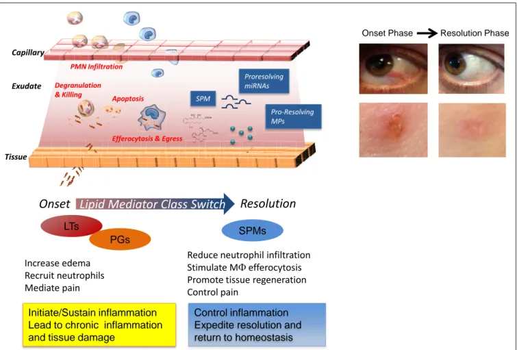
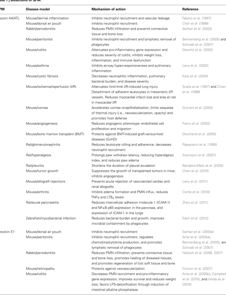
Acute inflammation is a defensive physiological response occur-ring in vascularized tissues to protect the host against injuries (Majno and Joris, 1996). This formidable ally manifests its impor-tant role, for instance, in the early phase after a microbial infection, when it fights against invading pathogens before the adaptive immune system is engaged (Abbas et al., 2011). The characteristic “cardinal signs” of inflammation, described by the Roman physi-cian Celsus in the first century, rubor (redness), tumor (swelling), calor (heat), and dolor (pain), are the macroscopic manifesta-tion of changes that occur at molecular and cellular levels in inflamed tissues. Tissue edema is one of the earliest events in the acute inflammatory response that arises from increased vascular permeability of the microvasculature (Figure 1). Leukocytes are then recruited at sites of inflammation and traverse postcapillary venules. Polymorphonuclear neutrophils (PMN) are among the first leukocyte responders that accumulate in the inflamed site. As they are the first line of defense of the innate immune system, these cells kill pathogens by engulfing them via phagocytosis and release of microbicidal proteins stored in their intracellular gran-ules and reactive oxygen species into phagolysosomal vacuoles
to kill invaders (Majno and Joris, 1996). Next, in experimental acute inflammation, mononuclear cells enter the inflammatory site. They can differentiate into macrophages (MΦs) and clear microbes, cellular debris, and apoptotic PMN by phagocytosis in a non-phlogistic process termed efferocytosis (Honn et al., 1989; Gordon, 2007;Serhan et al., 2007).
Ultimately, the clearance and efflux of phagocytes allow for resolution of the tissue and the return to homeostasis, namely catabasis (Figure 1). In order to maintain a healthy status, both the initiation of acute inflammation and its resolution must be efficient. Notably, it is not how often or how extensive an acute inflammatory reaction starts, but how effectively and quickly it resolves that determines whether the battle of inflammation is detrimental or the ideal favorable outcome for the host. Indeed, uncontrolled or unresolved inflammation is now recognized as a major driver of human pathologies, including arthritis, asthma, cancers, and cardiovascular diseases (Serhan, 2004;Serhan and Savill, 2005; Nathan and Ding, 2010). Given the high occur-rence of these and many other diseases, understanding how acute inflammation resolves is of wide interest.
This review focuses on the specialized pro-resolving media-tors (SPM) that are biosynthesized from essential polyunsaturated
Reduce neutrophil infiltration Stimulate MF efferocytosis Promote tissue regeneration Control pain
Lipid Mediator Class Switch
Increase edema Recruit neutrophils Mediate pain
Initiate/Sustain inflammation Lead to chronic inflammation and tissue damage
SPMs LTs
PGs
Control inflammation Expedite resolution and return to homeostasis
Onset
Resolution
Capillary Exudate Tissue Degranulation & KillingEfferocytosis & Egress PMN Infiltration Apoptosis tio tio tio tio tio tio tio tio tio tio tio tio aryyyyyyyyyyyyyyyyyyyyyyyyyyyyyyyyyyyyy Proresolving miRNAs Pro-Resolving MPs SPM ue
Onset Phase Resolution Phase
FIGURE 1 | Cellular and molecular mediators in acute inflammation and resolution. Surgical intervention, tissue injuries, or microbial infections in
vascularized tissues evoke a rapid acute inflammatory response
characterized by a rapid exudate formation with edema, leukocyte infiltration, and serum proteins. Polymorphonuclear leukocytes (PMN) are among the first responders that fight microbes followed by monocytes that differentiate locally into pro-resolving macrophages (MΦs). Efferocytosis of apoptotic PMN and microbes by pro-resolving MΦs and subsequent egress via lymphatics are hallmarks of tissue resolution. Redness and swelling, two of the cardinal signs of inflammation, can be easily appreciated in the example of eye and skin inflammation shown in the right panel. A few days later,
inflammation is almost completely resolved and homeostasis re-established. (Pictures were taken using a Lumix SZ7 digital camera). Lipid autacoids prostaglandins (PGs) and leukotrienes (LTs) are classical mediators of the onset phase of inflammation, promoting edema, PMN recruitment, and pain. By sustaining inflammation, PGs and LTs can lead to chronic inflammation and tissue damage. Specialized pro-resolving lipid mediators (SPMs) are biosynthesized within resolving exudates and proved to be very potent in reducing further PMN infiltration, stimulating non-phlogistic MΦ efferocytosis, promoting tissue regeneration, and controlling pain. Recent advances have demonstrated that specific miRNAs and microparticles can possess pro-resolving properties.
fatty acids (PUFAs), arachidonic acid (AA), eicosapentaenoic acid (EPA), and docosahexaenoic acid (DHA), namely lipoxins (LX), resolvins (Rv), protectins (PD), and maresins (MaR) and on their biosynthetic pathways, receptors, and miRNAs that act to control self-limited inflammation and promote its timely resolution. For readers interested in the biosynthesis of Rv and PD, this subject was recently reviewed in detail inBannenberg and Serhan (2010), and the confirmation and total organic synthesis inSerhan and Petasis (2011).
RESOLUTION IS AN ACTIVE PROCESS CONTROLLED BY SPM: SELF-LIMITED EXPERIMENTAL SYSTEM
At the histological level, resolution was well described by pathol-ogists for more than 100 years as the time when the neutrophils that infiltrated the inflamed tissue sites leave or are lost from the
site (Majno and Joris, 1996). Traditionally, resolution was thought to be a passive process, simply due to the attenuation/dissipation of chemotactic and pro-inflammatory signals. Our results ( Ser-han et al., 2000a; Levy et al., 2001), followed by those from many others worldwide (reviewed in a consensus report in Ser-han et al., 2007) demonstrated that resolution is instead an active process orchestrated by special novel chemical mediators that turn on biochemical and cellular pathways to enable the return to homeostasis.
Lipid mediators (LM) from PUFA play essential roles in distinct phases of acute inflammation, with prostaglandins (PGs;Flower, 2006;Samuelsson, 2012) and cysteinyl leukotrienes (cysLTs) pro-moting early increase in vascular permeability and leukotriene (LT) B4 acting as a potent leukocyte chemoattractant (
(von Euler, 1973). Chronic inflammation is widely viewed as an excess of pro-inflammatory mediators (Figure 1;Nathan and Ding, 2010). Results from our laboratory first demonstrated that the resolution phase is characterized by the active biosynthesis of specific LM that operate as “resolution agonists” to a) keep inflammation within physiological boundaries and b) expedite the complete return to homeostasis (Figure 1). The identifica-tion of this new array of LM was achieved using self-limited or naturally resolving acute inflammation models in vivo and a systems approach (Serhan et al., 2000a, 2002). The pharma-cologic impact of the Rv and PD was reviewed in (Serhan and Chiang, 2008). This new array of LM is now recognized as a genus of SPM (Serhan and Chiang, 2008) that have two broad functions and are anti-inflammatory and pro-resolving via stimulating multi-level actions. Accruing evidence indicates that failure or disruption of the endogenous pro-resolution path-ways governed by SPM can be detrimental and underlie some of the mechanisms of chronic inflammatory diseases (Gilroy et al., 1999; Karp et al., 2004; Schwab et al., 2007; Chan and Moore, 2010). SPM exert their potent dual anti-inflammatory and pro-resolving activities in the low nano- to microgram dose range when added back into experimental inflammatory disease models (Serhan and Chiang, 2008) and provide biotemplates for the design of novel therapeutics currently in clinical trials (see http://Clinicaltrials.Gov Identifier: NCT00799552). There-fore, harnessing these SPM may provide fascinating opportunities in the new and uncharted terrain of resolution pharmacology, with a substantial shift from a depletion pharmacology (i.e., via inhibitors, blockers, antagonists) toward a new approach based on resolution agonists that activate endogenous protective and clearance mechanisms.
Additional chemical mediators are operative in inflamed tissues to switch off leukocyte infiltration and restore their physiolog-ical functions. Among these are several cytokines (e.g., TGFβ, IL-10) that accumulate in resolving exudates (Bannenberg et al., 2005); glucocorticoids and the glucocorticoid-induced annexin-1 protein, which tune the inflammatory response and bring about homeostasis (for a recent review see (Perretti and Dalli, 2009)); and the transcription factor NF-κB, which also carries some anti-inflammatory properties (Lawrence et al., 2001). Moreover, inducing PMN apoptosis as well as lymphoid cells while stimulat-ing their prompt removal by MΦs also can promote resolution (Honn et al., 1989; Ariel et al., 2006). Recent results indicate that small inhibitors of cyclin-dependent kinases fulfill this goal (Leitch et al., 2012) as do annexin-1 peptides (Perretti and Dalli, 2009). Therefore, the resolution process can be pharmacologically targeted.
Importantly, resolution is not synonymous with endogenous anti-inflammation. This is because, in order to be considered a “pro-resolver,” a chemical and/or molecular entity, in addition to serving as a “stop signal” for neutrophil trafficking and other cardi-nal signs of inflammation (e.g., swelling, pain), must also stimulate efferocytosis by MΦ,favor the antibacterial activities,and promote tissue repair and regeneration to achieve homeostasis. Along these lines, PGE2can have anti-inflammatory properties in certain set-tings via stimulation of cAMP, but is not acting as pro-resolver since it does not enhance the uptake and clearance of apoptotic
cells by MΦs (Kunkel et al., 1981). Also, although cyclooxygenase (COX) inhibitors as well as certain lipoxygenase (LO) inhibitors reduce some of the cellular events of the inflammatory reaction (e.g., edema formation, PMN recruitment, and pain), they dra-matically impact the endogenous pro-resolution circuits and may delay or even derange this ideal outcome of acute inflammation and thus are “resolution toxic” (Gilroy et al., 1999;Schwab et al., 2007). In contrast, aspirin and glucocorticoids work synergisti-cally with endogenous pro-resolution pathways (Perretti et al., 2002).
Complete resolution also requires the clearance of the rem-nants of damaged tissues and activated or apopotic cells, so-called microparticles (MPs). Originally viewed merely as empty vesi-cles, MPs are now recognized as “specialized shuttles” used by the organism to transfer bioactive molecules from cell to cell. Their role in inflammation and resolution is now being appreci-ated. Recently, the anti-inflammatory properties of a PMN-derived sub-population of MPs were uncovered, where they appear to signal to activate resolution mechanisms (Gasser and Schifferli, 2004; Dalli et al., 2008; Norling et al., 2011). Mimicking this new endogenous mechanism in resolution, novel human PMN-derived nanoparticles containing AT-RvD1 or a LXA4stable ana-log, termed humanized pro-resolving nanomedicines, were pre-pared. These SPM-enriched nanohumanized particles possessed beneficial bioactivities in reducing acute inflammation in vivo, expediting resolution, and promoting wound healing (Norling et al., 2011). Hence, NPs can serve as mimetics of endogenous pro-resolution pathways (Figure 1).
Active resolution includes gene expression regulation of several soluble chemical mediators (e.g., cytokines, chemokines), recep-tors (e.g., Toll-like receprecep-tors), as well as transcription facrecep-tors. An emerging line of investigation indicates that many genes are under tight control of miRNAs, short non-coding RNA molecules that act as translational repressors of mRNA transcripts (Bartel, 2009). They are involved in many physiological and pathological processes, including cell development, cancer (Iorio and Croce, 2009), and inflammation (Sheedy and O’Neill, 2008; Alam and O’Neill, 2011;O’Neill et al., 2011). Our recent results uncovered roles of miRNAs in self-limited inflammation and in the resolution phase, specifically, RvD1-G-protein coupled receptor-dependent gene networks in resolution of acute inflammation as part of the endogenous circuitry that controls this active process (Recchiuti et al., 2011;Krishnamoorthy et al., 2012).
IDENTIFYING SPM IN RESOLUTION
An unbiased systems approach was taken to identify the endoge-nous SPM and decode their mechanisms of action in resolution. For this, the murine dorsal air pouch and self-limited acute inflammation was ideal because it permitted isolation of con-tained inflammatory exudates (Serhan et al., 2000a, 2002) and also enabled direct LM lipidomics of bioactive products, as well as their inactive precursors and further metabolites, proteomics, and analyses of cellular composition of the resolving exudate; namely the natural means by which inflammation returns to resolution and homeostasis. With this systems approach it was also possible to establish the local and temporal dissociation of LM biosyn-thesis (Bannenberg et al., 2005). For example, upon initiation
of inflammation with TNF-α, there was a typical acute-phase response characterized by rapid PMN infiltration preceded by local generation of both PGs and LTs. Unexpectedly, the eicosanoids undergo what was termed earlier a “class switch” and the profiles of LM made within this milieu switched with time (Levy et al., 2001). Indeed, the potent chemoattractants LT were deactivated and 15-LO required for LX and Rv production was transcription-ally activated (Levy et al., 2001). Notably, the omega-3 essential fatty acids DHA and EPA are precursors of Rv, PD, and MaR, are rapidly carried into the exudates via plasma edema and are then made available for conversion for the congregated exudate cells (Kasuga et al., 2008). Of interest, this LM class switching is driven in part by COX-derived PGE2and D2, via transcriptional regu-lation of enzymes involved in LX biosynthesis (Levy et al., 2001). Hence, the concept that “alpha signals omega,” namely the begin-ning signals the end in inflammation, was introduced by Sir John Savill and one of us to emphasize this finding (Serhan and Sav-ill, 2005) to note that at time zero mediators are biosynthesized that signal to limit PMN influx and terminate the contained acute inflammatory response.
WHAT IS A LIPID MEDIATOR?
To qualify as a LM, a product must be stereoselective in its actions and be produced in amounts that are commensurate with its potency and range of action (Serhan et al., 1996). Along this line, LX, Rv, and PD are present in human serum in pM to nM amount (e.g., LXA4, ∼1.4 nM; RvD1, ∼50 pM; RvE1, ∼0.5 nM; values from the Serum Metabolome Project;Psychogios et al., 2011; see also Oh et al., 2011 for RvE1 andOh et al., 2012 for RvE2 values) in human peripheral blood samples from healthy donors. LM lipidomics using liquid chromatography-tandem mass spectrom-etry (LC-MS/MS) coupled with informatics permit profiling of closely related compounds and identification of new molecules. Retrograde synthesis, both biogenic and total organic, allows the complete elucidation of chemical structure, stereochemistry, and physical properties, along with the recapitulation of the in vivo biosynthetic pathway (for examples seeSun et al., 2007;Serhan et al., 2009). The matching/identification of LM is usually carried out with at least two different instruments and/or mobile phase solvent systems and the criteria to identify a known LM are the following: (a) LC retention time should match by coelution with the LM authentic standard; (b) UV chromophore should match the synthetic and authentic LM (i.e.,λmaxand band shape); as well as (c) ≥6 diagnostic ions of tandem MS/MS spectrum (Figure 2). Also, the LC-MS/MS fragmentation mechanisms for the Rv and PD D1 and related DHA-derived products have been studied using deuterium-labeled compounds that facilitated their identification in vivo (Hong et al., 2007).
LIPOXINS
Lipoxin A4 and B4 were the first anti-inflammatory LM recog-nized to possess pro-resolving actions (Serhan et al., 1984a,b; Maddox et al., 1997; Takano et al., 1998;Godson et al., 2000). Although LXs were first isolated and identified in the 1980s in the Samuelsson laboratory (Serhan et al., 1984a), their potent bioac-tions were uncovered some years later with the identification of the aspirin-triggered LX (ATL;Claria and Serhan, 1995) and the
design of ATL stable analogs (Serhan et al., 1995; vide infra), when it became clear that they act as “braking signals” of further PMN infiltration (Takano et al., 1998) and as potent stimuli for the non-phlogistic recruitment of monocytes (Maddox et al., 1997) and MΦ efferocytosis (Godson et al., 2000; recently reviewed in Serhan, 2005;Spite and Serhan, 2011). LXs are lipoxygenase inter-action products derived from the enzymatic conversion of AA via transcellular biosynthesis during cell–cell interactions occur-ring duoccur-ring inflammation (Samuelsson et al., 1987). In humans, sequential oxygenation of AA by 15-LO and 5-LO, followed by enzymatic hydrolysis, leads to the biosynthesis of LXA4and B4in mucosal tissues, such as airways, gastrointestinal tract, and oral cavity (Edenius et al., 1990;Levy et al., 1993;Gronert et al., 1998;
Figure 3 and reviewed inRomano (2010). Blood vessels represent a second site for LX biosynthesis, with the conversion of 5-LO-derived LTA4into LXA4and B4by 12-LO in platelets (Serhan and
Sheppard, 1990;Romano and Serhan, 1992;Romano, 2010).
ATL: THE FIRST ASPIRIN-TRIGGERED MEDIATORS
A third LX synthetic pathway is initiated by aspirin, the well-known derivative of salicylates, by acetylation of COX-2. This covalent modification shifts the enzyme activity from endoperoxi-dase to lipoxygenase-like, and COX-2 converts AA into 15R-HETE, which is a substrate of leukocyte 5-LO for the biosynthesis of 15R-epi-LXA4 and B4 (Claria and Serhan, 1995). Hence, among the non-steroidal anti-inflammatory drugs (NSAID), aspirin has the unique capability to “jump start” resolution by its ability to trig-ger endogenous biosynthesis of so-called “aspirin-trigtrig-gered” LX (Figure 3) Notably, ATL produced in vivo in human subjects taking aspirin (Chiang et al., 2004) proved to mediate the local anti-inflammatory actions of low-dose aspirin in healthy individuals (Morris et al., 2009).
SPM BIOSYNTHESIZED FROM OMEGA-3 POLYUNSATURATED FATTY ACIDS: LOCAL MEDIATORS
The essential roles of omega-3 PUFA in health were evident in 1929 (Burr and Burr, 1929), andω-3, also known as n-3 PUFA EPA and DHA, have beneficial effects in human diseases including poten-tial antithrombotic, immunoregulatory, and anti-inflammatory properties (Iigo et al., 1997;De Caterina, 2011). Also, the Gruppo Italiano per lo Studio della Sopravvivenza nell’Infarto Miocardico-Prevenzione trial evaluated the effects ofω-3 PUFA supplementa-tion with>11,000 patients surviving myocardial infarction taking >1 g of ω-3 PUFA daily along with recommended preventive treat-ments including aspirin, and reported a significant benefit with a decrease in cardiovascular death (GISSI-Prevenzione Investiga-tors, 1999). It is believed that the actions of the major lipid of fish oil EPA (C20:5) are based upon (a) preventing conversion of AA to proinflammatory and prothrombotic eicosanoids; (b) serv-ing as an alternate substrate for the five-series LTs that are less potent than four-series LTs; and (c) conversion by COX to three-series prostanoids (i.e., PGI3) that also maintain antithrombotic actions. These and other explanations offered (Iigo et al., 1997; Calder, 2009;De Caterina, 2011) have not been generally accepted because of the lack of molecular evidence in vivo and the high con-centrations ofω-3 PUFA required to achieve putative “beneficial actions” in in vitro cell culture experiments.
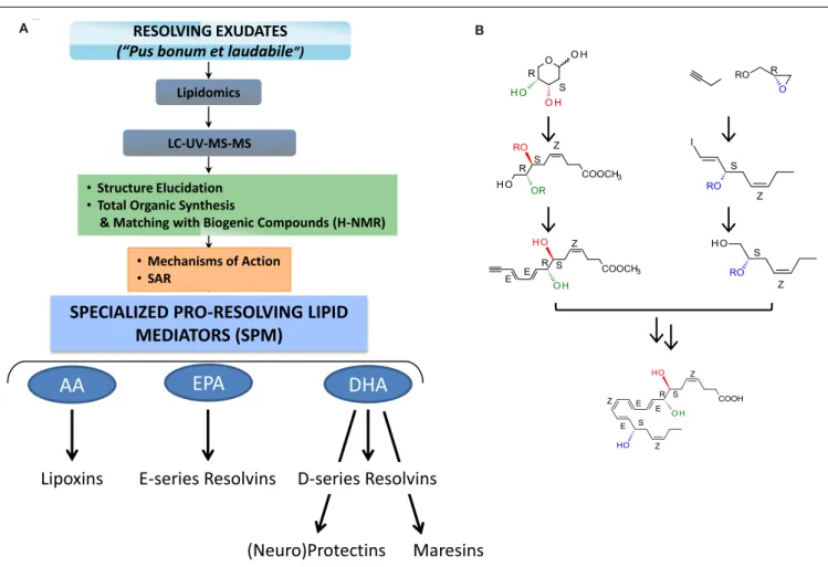
RESOLVING EXUDATES (“Pus bonum et laudabile”)
LC-UV-MS-MS Lipidomics
• Structure Elucidation • Total Organic Synthesis
& Matching with Biogenic Compounds (H-NMR)
AA
EPA
Lipoxins
E-series Resolvins
DHA
Maresins
(Neuro)Protectins
SPECIALIZED PRO-RESOLVING LIPID
MEDIATORS (SPM)
Fig. 2
B
• Mechanisms of Action • SARA
D-series Resolvins
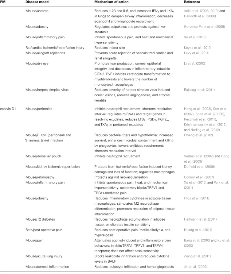
A BFIGURE 2 | Strategy for functional profiling of SPM in resolving exudates. (A) During self-limited inflammation, murine exudates (a “good
and laudable” pus according to ancient physicians;Majno, 1991) as well as human leukocytes biosynthesize SPM, which include the lipoxins, E-series resolvins, D-series resolvins, protectins (neuroprotectin D1), and maresins, which work to keep the inflammatory response within physiological boundaries and help to expedite the return to homeostasis. Functional profiling takes advantage of liquid chromatography-ultraviolet
spectrometry-tandem mass spectrometry (LC-UV-MS/MS) for identifying and quantifying SPM. Gas chromatography-mass spectrometry (GC-MS) is also useful to provide additional information together with LC-UV-MS/MS to
support structural identification and proposed structures. Retrograde analysis with biogenic synthesis using isolated human cells and total organic synthesis allows the assignment of chirality and double bond geometries using H-NMR with synthetic materials and matching studies (see text;Fiore et al., 1991; Serhan et al., 2000a, 2002, 2006, 2012;Sun et al., 2007;Spite et al., 2009a; Krishnamoorthy et al., 2010for details). Bioactions of SPM are assessed in both animal models and human cell systems. They must be stereoselective and evident at concentrations/doses that are commensurate with the amount of SPM produced.(B) Example of RvD1 stereoselective total organic
synthesis (reported inSun et al., 2007); for further details (seeSerhan and Petasis, 2011) for a recent review of organic synthesis.
To address the molecular basis for anti-inflammatory proper-ties ofω-3 fatty acids, an unbiased LC-MS/MS-based informatics approach was developed to identify novel mediators generated from ω-3 precursors during acute inflammation in vivo. Using this approach, EPA and DHA were found to be enzymatically converted into novel potent LMs coined Rv, an acronym of resolution phase interaction products, because they: (a) are pro-duced during cell–cell interactions occurring in the resolution phase of acute inflammatory response; (b) “stop” further neu-trophil entry to sites of inflammation, and (c) reduce exudates (Serhan et al., 2000a, 2002, 2006; Hong et al., 2003; Bannen-berg et al., 2005). Rv represented an entirely new family of mediators produced from the ω-3 fatty acids and importantly they appeared during the resolution phase via active biosyn-thetic processes. The biosynthesis of Rv gives rise to stereospecific
local mediators that have potent actions and activate specific receptors.
E-SERIES RESOLVINS
EPA-derived E-series Rv are endogenously biosynthesized in vivo in resolving murine exudates and in isolated human cells systems by isolated cells (e.g., endothelial cell-leukocyte interaction) and in whole blood (vide infra). The complete stereochemistry of the first member of this family, RvE1, has been established as 5S,12R,18R-trihydoxy-6Z,8E,10E,14Z,16E-EPA (Arita et al., 2005a). For fur-ther details on the total organic synthesis (seeSerhan and Petasis, 2011). Within vascular endothelial cells, aspirin-acetylated COX-2 converts EPA into 18R-hydro(peroxy)-eicosapentaenoic acid (HEPE), which is rapidly taken up by activated leukocytes (e.g., PMN) and further metabolized into RvE1. Notably, quantitative
AA
15-LO LXA4 15-epi-LXA4 S R S R 15S-H(p)-ETE 15R-H(p)-ETE COX-2/ASA 5-LO 17S-H(p)-DHADHA
RvD1 17R-H(p)-DHA AT-RvD1 S R S R COX-2/ASA 15-LO 5-LO 5-LOEPA
18S-RvE1 18S-H(p)-EPE S S RvE1 18R-H(p)-EPE R R COX-2/ASA 5-LO 5-LO 16R,17R-epoxide intermediate AT-(NPD1)/PD1 16S,17S-epoxide intermediate (NPD1)/PD1 Enzymatic Hydrolysis Enzymatic HydrolysisB
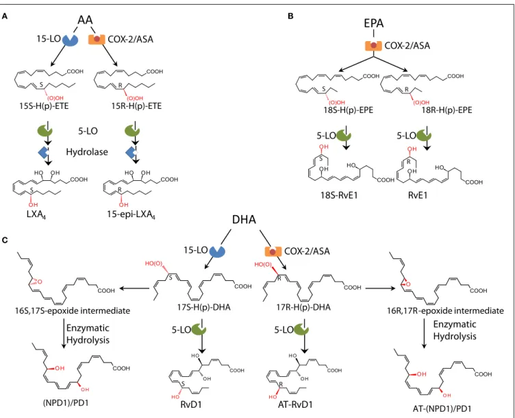
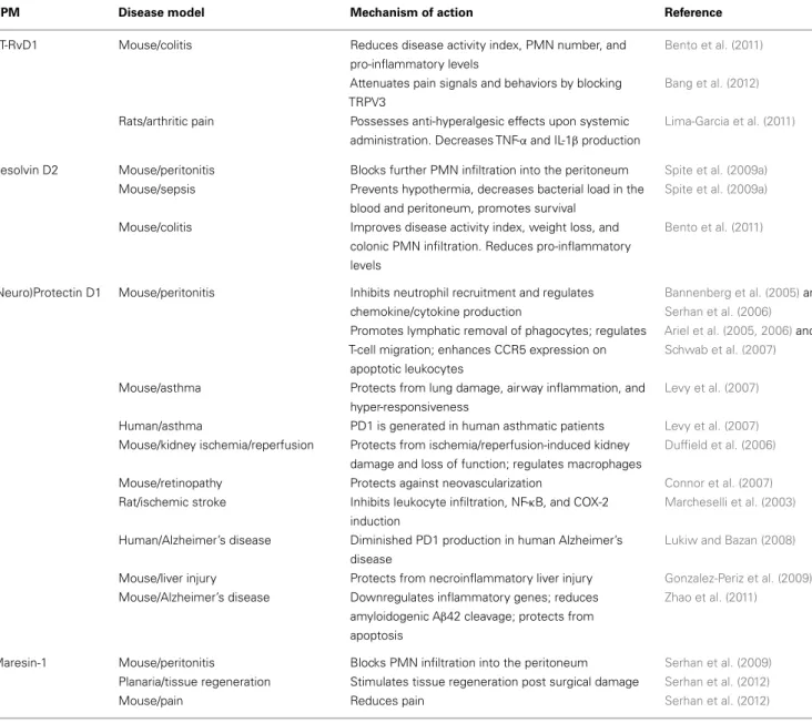
Hydrolase A C BFIGURE 3 | Biosynthetic schemes of SPM. (A) In humans, AA can be
converted into 15S-H(p)-ETE through 15-LO and into 15R-H(p)-ETE by aspirin (ASA)-acetylated COX-2. Both intermediates can be further metabolized through 5-LO and enzymatic hydrolysis yielding LXA4 or 15-epi-LXA4.(B) E-series
resolvins are biosynthesized via conversion of EPA by ASA-acetylated COX-2. Products of these reactions, 18S-H(p)-EPE and 18R-H(p)-EPE, are rapidly taken up by 5-LO and converted to 18S-RvE1 and RvE1.(C) The DHA metabolome
includes several SPM biosynthesized via 15/5-LO and ASA-acetylated COX-2. Each SPM is biosynthesized via distinct biochemical routes involving
stereocontrolled oxygenation, epoxide formation, and enzymatic hydrolysis. The main structures of key SPM and their biosynthetic routes (with precursors and main enzymes involved) are depicted (see text andSerhan and Petasis, 2011for further details). The complete stereochemistry of each of these SPM is established, total organic synthesis achieved, and bioactions confirmed.
chiral HPLC analysis indicated that the 18R-HEPE isomer was dominant to its epimer 18S-HEPE in human plasma from healthy subjects taking EPA (Oh et al., 2011). In contrast, human subjects who were administered aspirin before EPA had more 18S- than 18R-HEPE, indicating that aspirin might promote 18S-HEPE pro-duction as well as 18R-HEPE from ingested EPA (Oh et al., 2011). This 18S-HEPE can also be converted to epimeric RvE1 and RvE2 by human recombinant 5-LO and LTA4hydrolase (LTA4H), known as pro-inflammatory LTB4-synthesizing enzymes (Oh et al., 2011). RvE1 is also produced in vivo through an aspirin-independent pathway via cytochrome P450-driven oxygenation of EPA (Serhan et al., 2000b). Of interest, RvE1 was also found to be produced by Candida albicans and appears to be involved in clearance of
this organism (Haas-Stapleton et al., 2007). Thus RvE1 has multi-ple biosynthetic routes. RvE2 (5S,18-dihydroxy-EPE) is biosyn-thesized in resolving exudates and in human whole blood via reduction of 5S-hydroperoxy,18-hydroxy-EPE, an intermediate in the biosynthetic pathway of RvE1 (Tjonahen et al., 2006;Ogawa et al., 2009;Oh et al., 2012; Figure 3).
D-SERIES RESOLVINS
Earlier investigations using LC-MS/MS lipidomics of resolving exudates from mice given DHA and aspirin provided the first evidence of novel endogenous routes that lead to the forma-tion of 17-hydroxy-containing mediators. Gaining informaforma-tion on how human tissue and cells may produce D-series Rv involved
the in vitro recapitulation of biosynthetic pathways using isolated human cells and recombinant enzymes establishing potential ori-gins of novel compounds isolated from resolving exudates in vivo. Along these lines, hypoxic human endothelial cell COX-2 con-verted DHA to 13-hydroxy-DHA, which switched with ASA to 17R-HDHA. Human neutrophils transformed 17R-hydroxy-DHA into two series of di- and trihydroxy products; one initiated via oxygenation at carbon 7 and the other at carbon 4 (Serhan et al., 2002). The conversion of 17R-HDHA by human PMNs displayed similar features as those established for the conversion of AA to LTB4or LXs as well as the 18R series of EPA products. These were termed the “aspirin-triggered” D-series Rv (Serhan et al., 2002). Remarkably, in the absence of aspirin, D-series Rv carrying the 17S-hydroxy group were identified in murine exudates and iso-lated human cells (Serhan et al., 2002;Hong et al., 2003). The enzy-matic pathway leading to the formation of 17S- and 17R-RvD1 is shown in Figure 3. Following the complete organic synthesis, the stereochemistry of 17S-, 17R-RvD1, and RvD2 were established as 7S,8R,17S-trihydroxy-4Z,9E,11E,13Z,15E,19Z-DHA (17S-RvD1), 7S,8R,17R-trihydroxy-4Z,9E,11E,13Z,15E,19Z-DHA (17R-RvD1; Sun et al., 2007), and 7S, 16R, 17S-trihydroxy-4Z, 8E, 10Z, 12E, 14E, 19Z -DHA (RvD2;Spite et al., 2009a). Additional members of this family were identified (RvD3–RvD6). Each of these arises by similar biosynthetic routes, but has distinct chemical structures and potentially additional bioactions that are now being unveiled (Chiang et al., 2012). Importantly, both RvE1 and RvD1 were iden-tified in circulating blood of healthy donors by (Psychogios et al., 2011) as part of the Serum Metabolome Project.
THE (NEURO)PROTECTINS
In addition to D-series Rvs, DHA also serves as precursor of a new family of LM characterized by a conjugated triene system and two alcohol groups called PD. The name PD accounts for their pro-tective actions observed in neural tissues and within the immune system, while the prefix neuro PD gives the tissue localization and site of action. The structure of the founding member of this family, PD1, was first disclosed in a report on the isolation and elucida-tion of Rv (Serhan et al., 2002;Hong et al., 2003), and its complete stereochemistry later established as 10R,17S-dihydroxy-docosa-4Z,7Z,11E,13E,15Z,19Z -hexaenoic acid (Serhan et al., 2006). In addition to PD1, several stereo- and positional isomers that also possess lower bioactivity than PD1 were identified in human and mouse tissues. These include 10S,17S-diHDHA, 4S,17S-diHDHA, 7S,17S-diHDHA, and 22-hydrox-10,17S-docosatriene (a putative inactivation product of PD1; Serhan et al., 2002; Hong et al., 2003). The geometry of the double bonds in PD1, their posi-tions during biosynthesis, and chirality of C10 indicate that PD1 biosynthesis proceeds through a C16(17)-epoxide intermediate and requires specific enzymatic steps to generate the potent bioac-tive molecule from DHA, as confirmed by the isolation of alcohol trapping products as well as two vicinal diol 16,17-docosatrienes as minor products of PD1 biosynthesis (Hong et al., 2003; Ser-han et al., 2006). Recently, a novel aspirin-triggered COX-2 dri-ven pathway was reported that biosynthesizes the 17R-epimeric form of PD1 from DHA (Marcheselli et al., 2003; Figure 3). The total organic synthesis and complete stereochemical assignment of ATPD1 (10R,17R dihydroxydocosa4Z,7Z,11E,13E,15Z,19Z -hexaenoic acid) were recently achieved. Both PD1 and AT-PD1
reduced leukocyte infiltration in murine peritonitis, reduced PMN transmigration with endothelial cells, and enhanced efferocytosis of apoptotic PMN by human MΦ (Serhan et al., 2006). These are the hallmark actions of a SPM carrying both anti-inflammatory and pro-resolving actions demonstrable both in vitro and in vivo.
MARESINS
MΦs have pivotal roles in orchestrating the return to home-ostasis (Gordon, 2007) and biosynthesize SPM that enhance their pro-resolving and homeostatic functions. For example, MΦ ingesting apoptotic cells initiate the biosynthesis of LXA4, RvE1, and PD1 but not LTB4(Freire-de-Lima et al., 2006;Schwab et al.,
2007). In addition, a new family of SPM biosynthesized by MΦs was identified (Serhan et al., 2009). Unbiased LM LC-MS/MS-based metabololipidomics during self-limited peritonitis led to the identification of a novel pathway that converted DHA into 14-hydroxy DHA (HDHA). Experiments with 12/15-LO−/−mice or with LO inhibitor confirmed that 14-HDHA production was via a DHA carbon 14 lipoxygenation pathway. Freshly prepared 14-H(p)DHA is rapidly converted by isolated human and mouse MΦ into a new set of bioactive products, whose molecular struc-ture was established (Serhan et al., 2009) and recently confirmed by matching of biogenic material with those prepared by total organic synthesis (Serhan et al., 2012). The major product of this new pathway proved to be 7,14dihydroxydocosa4Z,8E,10E,12Z,16Z,19Z -hexaenoic acid, denoted MaR (macrophage mediator in resolving inflammation) 1 (first in the family; MaR1;Serhan et al., 2009). Similar to that of other potent SPM, MaR1 biosynthesis proceeds via an epoxide intermediate, specifically the MaR1 13,14-epoxide intermediate, that was demonstrated by trapping experiment and with18O-containing molecular oxygen O2that opens during enzy-matic conversion to MaR1 keeping the double bond geometry and chirality of carbon 7 and 14.
In addition to MaR1 in resolving murine exudates, a novel double dioxygenation product was isolated and identified, 7S,14S-dihydroxydocosa-4Z,8E,10Z,12E,16Z,19Z-hexaenoic acid (denoted 7S,14S-diHDHA), formed by consecutive lipoxygena-tion of 14-HDHA, was also identified using molecular oxygen incorporation, and proved bioactive but less potent in activity than MaR1 in stimulating efferocytosis with human cells (Serhan et al., 2009, 2012). MaR1 and RvE1 are also potent stimulators of organ regeneration using a planaria regeneration system (Serhan et al., 2012). Hence, SPM are primordial molecules that signal from the inflammatory site to generate the aftermath of inflammation and tissue injury.
GPCRs FOR SPM IN ANTI-INFLAMMATION AND RESOLUTION
GPCRs FOR LXs
The first evidence for receptor-mediated actions of LXA4arises from studies with Santosh Nigam when he was on sabbatical in the Serhan Lab at BWH in the late 1980s, which demonstrated stimula-tion of rapid lipid remodeling and pertussis toxin (PTX)-sensitive release of arachidonate in PMN treated with LXA4(Nigam et al.,
1990). To examine the molecular basis of these actions, synthetic [11,12-3H]-LXA4was prepared and used to demonstrate specific and reversible binding to intact human PMN with a Kd∼ 0.5 nM. [3H]-LXA
4binding was stereoselective as LXB4, LTB4, 6S-LXA4, or 11-trans-LXA4did not compete for LXA4binding, while cysteinyl
LT C4and D4partially displaced bound labeled LXA4(Fiore et al.,
1992). Screening of cDNA clones from differentiated HL60 human cells lines led to the identification of formyl peptide receptor like-1, a homolog of formyl receptor, as putative LXA4GPCR (Fiore
et al., 1994). This receptor has recently been coined ALX/FPR2 by the international nomenclature committee in light of its high affinity for LXA4(Ye et al., 2009).
Human FPR2/ALX is highly expressed in myeloid cells and at a lower extent in lymphocytes, dendritic cells, and resident cells (Chiang et al., 2006). Orthologs of the human FPR2/ALX have been identified in mice (Takano et al., 1997) and rats (Chiang et al., 2003). In addition to LXA4, FPR2/ALX is activated by the glucocorticoid-induced protein annexin-1 and its N-terminal pep-tides (Perretti et al., 2002), representing the prototype of GPCR able to coordinate anti-inflammatory and pro-resolving activi-ties of both lipid and peptide ligands. Genetic manipulation of ALX/FPR2 and its ortholog in mice has provided evidence for the essential role of this GPCR in controlling immune responses. Indeed, myeloid-driven overexpression of human FPR2/ALX in transgenic mice resulted in a reduced neutrophil infiltration dur-ing zymosan-induced peritonitis (Devchand et al., 2003), whereas ALX/FPR2−/−mice have an exacerbated inflammatory phenotype and delayed resolution (Dufton et al., 2010).
More strikingly, ATL and FPR2/ALX expression levels dic-tate both the magnitude and duration of acute inflammation in humans (Morris et al., 2010). Hence, mechanisms that regulate this expression are of wide interest. Recent results fromSimiele et al. (2012)uncovered the molecular basis of ALX/FPR2 transcription machinery, with the identification of the core promoter sequence, the elucidation of transcription factors and epigenetic mecha-nisms that regulate promoter activity, and the identification of the first inheritable SNP that impairs promoter activity in individuals at high cardiovascular risk. Notably, LXA4upregulates ALX/FPR2 levels by activating its promoter, suggesting an additional mecha-nism by which LXA4exerts its bioactivities (Simiele et al., 2012). This is particularly relevant in considering LX roles in stimulating resolution. In addition, earlier studies demonstrated that radiola-beled 15-epi-LXA4binds at cysteinyl LT receptor 1 (CysLT1) with equal affinity to LTD4, providing additional molecular mecha-nisms for ATL dampening CysLT signals in the vasculature as well as regulating leukocyte trafficking via ALX/FPR2 (Gronert et al., 2001).
GPCRs FOR E-SERIES RESOLVINS
At least two GPCRs are involved in mediating RvE1 actions, namely ChemR23 and BLT1 (Arita et al., 2005a, 2007). RvE1 binding to ChemR23 was assessed with [3H]-labeled RvE1, which was pre-pared by catalytic hydrogenation from synthetic diacetylenic RvE1. [3H]-RvE1 bound to ChemR23 transfectants with high affinity (Kd=11.3 ± 5.4 nM) and stereoselectivity, since RvE1 biogenic precursors EPA and 18R-HEPE did not compete with [3H]-RvE1. Also, the synthetic peptide fragment (YHSFFFPGQFAFS) derived from human chemerin that was earlier reported to be a ligand for this same receptor (Wittamer et al., 2003) displaced [3H]-RvE1 binding by ∼70% when tested at 10 µM concentration, suggesting that RvE1 and chemerin share recognition sites on ChemR23 (Arita et al., 2005a;Ohira et al., 2009). [3H]-RvE1 specific binding was also demonstrated with membrane fractions isolated from
human PMN. Radiolabeled RvE1 bound human PMN with Kd of ∼50 nM and was displaced by homoligand RvE1 (Ki∼ 34 nM), LTB4(Ki=0.08 nM), and LTB4receptor 1 (BLT1) selective antag-onist U-75302 (Ki=1.5 nM), but not by the chemerin peptide (Arita et al., 2007). These results strikingly demonstrated that RvE1 binding sites are pharmacologically distinct from ChemR23 on human PMN and prompted us to investigate whether RvE1 binds to LTB4receptors.
In these studies, Arita et al. found that [3H]-RvE1 also gave high affinity binding to recombinant BLT1 (Kd∼ 45 nM) that was competed by unlabeled LTB4(Ki=3 nM). In contrast, BLT2-overexpressing cells did not show [3H]-RvE1 binding at concen-trations up to 10 nM. These results clearly demonstrated that RvE1 binds to BLT1 on human PMN and acts as a partial agonist to atten-uate LTB4incoming signals in both mouse and human leukocytes (Arita et al., 2007).
Profiling for tissue distribution of human ChemR23 showed expression of this GPCR in brain, kidney, cardiovascular, gastroin-testinal, and myeloid tissues (Arita et al., 2005a). More recently, direct evidence for ligand-receptor interactions of RvE1 and its epimer 18S-RvE1 was provided using ChemR23 and BLT1 β-arrestin cells. In this system, cells were engineered to co-express a β-arrestin protein tagged with an inactive moiety ofβ-galactosidase enzyme together with a candidate GPCR fused to the short Pro-Link peptide derived fromβ-galactosidase. In the presence of lig-and, in this context RvE1 activates GPCR interacts withβ-arrestin, bringing to proximity two inactive portions of β-galactosidase and reconstituting the fully active enzyme. The activity of this enzyme, which is stoichiometrically dependent on GPCR-ligand interaction, is monitored with a chemiluminescence detection sys-tem. With ChemR23β-arrestin cells, 18S-RvE1 dose-dependently activated ChemR23 receptor, with EC50 (∼6.3 pM) lower than that obtained with RvE1 (∼0.14 nM). 18S-RvE1 also antago-nized LTB4-mediated BLT1 activation with higher potency and efficacy than RvE1 in BLT1 β-arrestin cells (Oh et al., 2011). Hence, RvE1 and 18S-RvE1 can share the same site(s) of spe-cific binding to human ChemR23 as well as BLT1 and thus suggest location-dependent mechanisms in their signaling capabilities.
RvE2 exerts potent and cell-specific bioactions on human leukocytes (Tjonahen et al., 2006; Oh et al., 2012). Recently, tritium-labeled [3H]-RvE2 was synthesized and gave comparable Kd(∼25 nM) with other SPM in isolated human PMN. In addi-tion, using ChemR23 and BLT1β-arrestin cells, RvE2 was found to share, at least in part, receptors with RvE1 (Oh et al., 2012).
GPCRs FOR D-SERIES RESOLVINS
RvD1 activates its own GPCR and does not activate ChemR23. RvD1 exerts specific bioactivities on human PMN, namely reduc-tion of F-actin polymerizareduc-tion, that are inhibited by PTX but not cholera toxin, whereas it did not stimulate Ca2+release nor activate cAMP in human PMN (Krishnamoorthy et al., 2010). For the purpose of investigating direct binding of RvD1 to human PMN, [3H]-RvD1 was prepared by catalytic hydrogena-tion of synthetic [13, 14]-acetylenic RvD1 methyl ester by custom tritiation (American Radiolabel; Krishnamoorthy et al., 2010). This procedure was followed by de-esterification and isolation of the RvD1 label using RP-HPLC. [3H]-RvD1 specifically bound to human PMN with high affinity (Kd∼ 0.17 nM) and was competed
by homoligand cold RvD1 (100%) and LXA4 (∼60%) but not the ALX-ligand annexin-1-derived Ac2-12 peptide. In parallel, [3H]-RvD1 also showed specific binding with human monocytes (Krishnamoorthy et al., 2010). Since RvD1 counteracts TNF-α
in vivo, luciferase-based reporter system (Arita et al., 2005a) was employed in functional screenings to assess the ability of selected GPCRs (Figure 4) to transduce RvD1 signals that block NF-κB activity in response to TNF-α.
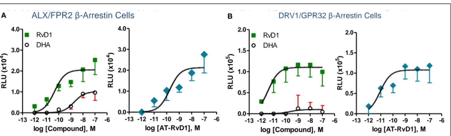
Phylogenetically related GPCR linked to inflammation and chemoattraction were overexpressed into HeLa cells together with a reporter vector consisting of NF-κB promoter sequence linked to the luciferase gene. RvD1 significantly reduced TNF-α-stimulated NF-κB response in cells overexpressing either the LX receptor ALX/FPR2 or the orphan, GPR32, but not other GPCRs (e.g., BLT1, BLT2, CB1, GPR-1, FPR, and ChemR23; Krish-namoorthy et al., 2010). Moreover, RvD1 dose-dependently acti-vated ALX/FPR2 and GPR32 in recombinantβ-arrestin cells with EC50in the low picomolar range (EC50∼ 1.2 pM for ALX/FPR2; 8.8 pM for GPR32; Figure 5). In contrast, RvD1 did not acti-vate RvE1 receptor ChemR23, demonstrating the high selectivity of these ligands for their specific GPCR (Krishnamoorthy et al., 2010). In comparison, at equimolar concentrations, RvD1, its epimer AT-RvD1, RvD1-carboxy-methyl ester, and a metaboli-cally more stable analog 17 (R/S)-methyl RvD1-ME activated both ALX/FPR2 and GPR32 with similar potencies and EC50, whereas the biosynthetic precursor native DHA was not active with GPR32 and ALX/FPR2 in this concentration (Krishnamoorthy et al., 2012). Of interest, the known anti-inflammatory ALX/FPR2 ago-nist compound 43 identified by traditional medicinal chemistry screening also activated GPR32 (EC50∼ 2.2 pM) and ALX/FPR2 (EC50∼ 2.0 pM) in β-arrestin cells but not the ADP receptor P2Y12 (Krishnamoorthy et al., 2010). Hence, RvD1, AT-RvD1, and the derivatives carboxy methyl ester and 17(R/S)-RvD1 directly acti-vate ALX/FPR2 and GPR32, hereafter referred to as RvD1 tor (DRV1) following the IUPAC recommendations for recep-tor nomenclature (Brink et al., 2003; Figure 5). Overexpression of either ALX/FPR2 or GPR32 in human MΦs gave further enhancement of efferocytosis in response to RvD1, while knock-down of ALX/FPR2 or DRV1/GPR32 determined a decrease in RvD1-stimulated phagocytosis response (Krishnamoorthy et al., 2010).
In keeping with this,Norling et al. (2012)demonstrated that the ability of RvD1 to reduce human PMN-endothelial cell inter-actions is absolutely dependent on ALX/FPR2 and DRV1/GPR32. Interestingly, actions of low concentration (1 nM) RvD1 were dampened by DRV1/GPR32 blocking antibody, whereas at high concentrations (10 nM) they appeared ALX/FPR2-specific. These two receptors also have distinct responses in an activated cell envi-ronment in that upon activation human PMN rapidly mobilized ALX/FPR2 stored in secretory granules, but not DRV1/GPR32, to the cell membrane. In addition, in ALX/FPR2 knockout mice RvD1 did not exert anti-inflammatory (e.g., stop PMN infiltra-tion) nor pro-resolving (e.g., enhancing MΦ efferocytosis) actions (Norling et al., 2012). Hence, specific GPCRs selectively mediate RvD1 actions with ALX/FPR2 being rapidly upregulated in PMN that are exposed to pro-inflammatory stimuli and DRV1/GPR32 possibly conveying more homeostatic functions. With respect to cell and tissue distribution, ALX/FPR2 is present on leukocytes
and resident cells (including MΦ, synovial fibroblasts, mesangial cells, endothelial, and epithelial cells;Krishnamoorthy et al., 2010). Human DRV1/GPR32 was identified in peripheral blood leuko-cytes and arterial and venous tissues using a cDNA array. It is mostly abundant on PMN, monocytes, and macrophages and is also present on vascular endothelial cells (Krishnamoorthy et al., 2010). The murine ortholog of DRV1/GPR32 is currently not known but is present in chimpanzees. Regulatory mechanisms of DRV1/GPR32 are of interest while those of ALX/FPR2 have recently been uncovered (Simiele et al., 2012). Although specific receptors for RvD2, RvD3, and RvD4 have not yet been identi-fied, the stereoselective actions of RvD2 were inhibited by PTX (Spite et al., 2009a), implicating the involvement of GPCRs. More recently,Chiang et al. (2012)reported activation of RvD1-receptor DRV1/GPR32 by RvD5, which is related biosynthetically to RvD1, with the recombinant human DRV1.
GPCRs FOR NEURO (N)PD1/PD1
Biosynthesis of (N)PD1 occurs in neural tissues in response to injury, ischemia-reperfusion, and exposure to β-amyloid pep-tides (Marcheselli et al., 2003; Mukherjee et al., 2004; Bazan, 2007). In addition (N)PD1 shows protective anti-inflammatory and pro-resolving actions within the immune system (Serhan et al., 2002;Hong et al., 2003). Hence, it was of interest to determine the molecular basis of (N)PD1/PD1 actions. Specific binding of tritium-labeled (N)PD1 obtained by catalytic tritiation of syn-thetic 15-acetylenic NPD1 methyl ester was demonstrated with both retinal pigment cells (RPE) and human PMN. [3H]-(N)PD1 bound RPE with Kd∼ 30 pmol/mg of cell protein. However, at high concentration of radio-ligand (>10 nM), non-specific bind-ing was evident, in line with the highly hydrophobic nature of this compound. In these experiments, competitive binding studies with unlabeled ligand demonstrated 90–100% displacement for the free acid form of cold (N)PD1. In these studies (N)PD1-ME showed a lower affinity for binding sites and ∼74% displace-ment, while other structurally related omega-3 fatty acid-derived compounds (17S-hydroxy-DHA, RvE1, ∆15-trans-NPD1, and ∆15-trans-NPD1-carboxy methyl ester) gave only minimal or no displacement. Specific binding experiments with [3H]-NPD1 and isolated human PMN proved high affinity specific binding and showed a two-site fit for sites of high and low affinity binding (Kd≈25 and 200 nM). Two other SPM that bind specific receptors on human PMN, namely LX A4(LXA4) and RvE1, did not compete with [3H]-NPD1 binding on these cells (Marcheselli et al., 2010). HOW DO THE PRO-RESOLVING MEDIATORS WORK?
By definition, SPM: (a) are generated within the resolution phase; (b) limit leukocyte infiltration; (c) enhance phago-cytic activity of pro-resolving MΦ to remove apoptotic cells and/or microbes; (d) stimulate the clearance of PMN from mucosal surfaces and their anti-microbial actions. If a LM fulfills each of these bioactivities, then it belongs to the genus of SPM. At the cellular and molecular levels, SPM dis-play distinct modes of action on PMN and monocyte/MΦs, which can be demonstrated with isolated cells. Each SPM (RvE1, RvE2, and RvD1) stimulates a rapid shape change of human leukocytes that reflects ligand-receptor responses and cytoskeletal events that ultimately limit the PMN to diapedesis
LXA4, ATL RvD1, AT-RvD1 DP2(GPR44) BLT1 BLT2 ChemR23 DRV1/GPR32 ALX/FPR2 FPR FFAR1 FFAR2 FFAR3 EP4 EP2 DP1 IP EP3 FP FP EP1 TP FP S1PR3 S1PR1 S1PR2 ADORA3 ADORA1 ADORA2B ADORA2A PAFR P2Y13 P2Y14 P2Y12 P2Y11 P2Y1 P2Y4 P2Y2 P2Y6 GPR17 CysLT1 CysLT2 CXCR4 CXCR3 CXCR5 CXCR2 CXCR1 CCR6 CXCR6 CCR7 CCR9 CCR10 CCR8 CCR3 CCR1 CCR5 CCR2 CCR4 CX3CR1 XCR1 CXCR7 CXCR Chemokine Receptors Purinergic Receptors Adenosine Receptors Lysophospholipid Receptors Prostanoid Receptors BLT1 LTB4 Receptos RvE1, 18(S)-RvE1 SPM RECEPTORS
Free Fatty Acid Receptors CysLTC CysLT Receptors * * 0 5 10 15 20 25 % In h ib iti o n T NF -α s ti m u la te d N F -κ B A ct ivi ty NF-kB Response Element Luciferase TNF- TNF-R ALX GPR32 RvD1
A
AB
B αFIGURE 4 | Identification of RvD1 GPCR. (A) Phylogenetic tree shows
similarities in the amino acid sequences of human GPCRs closely related to LXA4, RvE1, and LTB4receptors (left panel). Cluster was generated with the ClustalW2 software (www.ebi.ac.uk/Tools/clustalw2). Protein sequences were deduced from the NCBI database and receptor nomenclature followed the IUPAR classification for GPCR.(B) For functional screening and
identification of RvD1 receptor, GPCRs were cloned in pcDNA3 vector and overexpressed in human HeLa cells cotransfected with pNF-κB luciferase
plasmid. Cells were treated with RvD1 and TNF-α (see inset in the right panel). RvD1 reduces TNF-α-stimulated NF-κB activation in DRV1/GPR32 and ALX/FPR2 receptor-overexpressing cells, while cells transfected with other related GPCRs (e.g., BLT1, BLT2, CB1, GPR-1, FPR, and ChemR23) did not significantly inhibit TNF-α-induced NF-κB luciferase activity on addition of the ligand RvD1. The results illustrated are expressed in luminescence units subtracted from pNF-κB and pcDNA3 empty vector (*P < 0.05 vs. BLT2-transfected cells).
ALX/FPR2 β-Arrestin Cells
-13 -12 -11 -10 -9 -8 -7 -6 0.0 1.0 2.0 3.0 4.0 RvD1 DHA log [Compound], M R L U ( x 1 0 4) -13 -12 -11 -10 -9 -8 -7 -6 0.0 1.0 2.0 3.0 4.0 log [AT-RvD1], M R L U ( x 1 0 4) -13 -12 -11 -10 -9 -8 -7 -6 0.0 0.5 1.0 1.5 2.0 RvD1 DHA log [Compound], M R L U ( x 1 0 5) -13 -12 -11 -10 -9 -8 -7 -6 0.0 0.5 1.0 1.5 2.0 log [AT-RvD1], M RL U (x 1 0 5)
DRV1/GPR32 β-Arrestin Cells
ALX/FPR2 β-Arrestin CellsA B DRV1/GPR32 β-Arrestin Cells
FIGURE 5 | Structure-activity relationship of RvD1, AT-RvD1, and DHA on ALX/FPR2 and DRV1/GPR32 receptors. Activation of
ALX/FPR2 and DRV1/GPR32 was determined using theβ-arrestin cell system. This system is engineered by stably expressing the target ALX/FPR2 or DRV1/GPR32 tagged with theβ-galactosidase Pro-Link peptide. Cells also co-expressed theβ-arrestin protein linked to the β-galactosidase EA fragment. In the presence of ligand, activated GPCR interacts withβ-arrestin, bringing to proximity the EA and Pro-Link
fragments, forming a functional enzyme.β-galactosidase activity is measured by adding the substrate and generating a chemiluminescent signal that is stoichiometrically associated to ligand dependent GPCR activation (see text andKrishnamoorthy et al., 2010for further details). Dose-response curves show activation of ALX/FPR2(A) and
DRV1/GPR32(B) receptors by RvD1, AT-RvD1, but not DHA determined
using theβ-arrestin cell system. Results are mean (±SEM) from n = 4 to 6. RLU, relative luminescence unit.
in vivo, hence reducing inflammation and tissue damage (Dona et al., 2008; Kasuga et al., 2008; Oh et al., 2012) and pre-pare the macrophages to enhance phagocytosis of both apop-totic cells and microbes (Schwab et al., 2007; Ohira et al., 2009).
The best examples of these pro-resolving cellular mechanisms studied to date are for the Rv ligands RvE1 and RvD1. In the case of RvE1, its direct binding to the human recombinant GPCR denoted ChemR23, an RvE1 receptor, activates the receptor with the sub-sequent regulation of Akt intracellular signaling pathway and
phosphorylation signaling of proteins that increase phagocytosis (Ohira et al., 2009). In the case of RvD1, the ligand reduces actin polymerization in isolated human PMN in a PTX-sensitive man-ner (Krishnamoorthy et al., 2010), diminishes surface expression of CD11b involved in leukocyte adhesion to endothelial cells (Krishnamoorthy et al., 2010) as does RvE1 (Dona et al., 2008), evokes a shape change and stops chemoattractant-initiated PMN migration observable at a single cell level with human cells iso-lated within microfluidic chamber systems (Kasuga et al., 2008). Of note, LXA4also stops PMN chemotaxis in vitro (reviewed in
Serhan, 2005), while it induces rapid activation of small GTPases and redistribution of cytoskeletal proteins (e.g., MYH9, F-actin) in human monocyte-derived MΦs during phagocytosis (Maderna et al., 2002;Reville et al., 2006). Hence, there appears to be a general mechanism on how pro-resolving molecules work to achieve their potent anti-inflammatory and pro-resolution actions, namely via SPM coupling to distinct high affinity receptors that evoke cell-type and specific intracellular pathways.
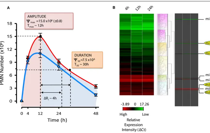
Besides these general actions, each SPM possesses additional specific activities (see Table 1;Fredman and Serhan, 2011). Given the important protective function of acute inflammation to fight infections or dangers from within and the need to safeguard the host from an uncontrolled reaction, it is not surprising that SPM have some bioactions overlapping in target tissues and spe-cific cell types. In addition, the sites of biosynthesis for each SPM and the degree of cell distribution of their GPCRs may underlie selectivity and specificity of the pro-resolving system. In experimental models of inflammation and resolution, SPM proved to be stereoselectively active in the nano- to low micro-gram dose range to control inflammation, limit tissue damage, shorten resolution intervals, promote healing, and alleviate pain (Table 1). In order to define resolution in unbiased, quantitative terms, mathematical resolution indices were introduced for deter-mining the cellular changes in exudates, i.e., Tmax, time point of maximum PMN infiltration (Ψmax); T50, time necessary to achieve 50% reduction in PMN number (Ψ50) fromΨmax; res-olution interval (Ri=T50−Tmax), time interval between Tmax and T50 (Bannenberg et al., 2005). The introduction of reso-lution indices permits the evaluation of pro-resoreso-lution bioac-tions of endogenous chemical mediators or pharmacological agents (Schwab et al., 2007;Haworth et al., 2008;Navarro-Xavier et al., 2010). These results first demonstrate the possibility to pharmacologically manipulate resolution and stimulate resolu-tion. Along these lines, results from the first human Phase I– II clinical trials demonstrated the safety and efficacy of a Rv analog that reduces both signs and symptoms of dry eye syn-drome (http://Clinicaltrials.Gov Identifier: NCT00799552) and have moved forward to Phase III clinical trial with Celtic Ther-apeutics. Dye eye syndrome is a chronic illness commonly treated with the immune-suppressant cyclosporine, providing evidence that SPM have the potential to treat a broad array of human diseases.
Acute inflammation following tissue injury, surgery, or infec-tions causes pain (Majno and Joris, 1996). Peripheral sensitization of primary sensory neurons is induced by inflammatory mediators released after tissue insults, such as bradykinin, prostaglandins, nerve growth factors (NGF), pro-inflammatory cytokines such as TNF-α, interleukin (IL)-1β and IL-6, and pro-inflammatory
chemokines (Stein et al., 2009). The contribution of PGE2and I2 led to the use of NSAID (e.g., naproxen, ibuprofen) and selective COX-2 inhibitors as analgesic. Since SPM are potent regulators of acute inflammation and pro-inflammatory mediators (including PGs, TNF-α,and IL-1β),and since COX-2 inhibitors are resolution toxic (Schwab et al., 2007), it was of interest to investigate whether SPM could control inflammation associated and chronic pain. The initial report fromSvensson et al. (2007)on the antinociceptive actions of LXA4 was followed by further studies demonstrat-ing that Rvs, PD, and also MaR1 have potent analgesic activities when administered both locally and systematically (Xu et al., 2010; Huang et al., 2011;Lima-Garcia et al., 2011;Park et al., 2011; Ser-han et al., 2012). Notably, the exquisite potent actions of SPM, which proved as effective as morphine and COX-2 inhibitor NS-398 at much lower doses (Xu et al., 2010), occur without altering basal sensitive perception, unlike other anesthetics used to con-trol pain during surgery. Hence it appears possible to resolve pain signaling as well as inflammation.
Complete resolution requires regeneration of destroyed tissues without affecting their functionality as in the case of fibrosis or scarring. Pro-resolving MΦ play key functions in tissue remodel-ing under both homeostatic (e.g., post parturition) and patholog-ical (e.g., removal of microbes from infected tissues) conditions (Honn et al., 1989;Majno and Joris, 1996;Gordon, 2007). In this regard, SPM are of considerable interest in view of their roles in regulating MΦ activities. For instance, LX, Rv, and PD stimu-late the non-phlogistic efferocytosis by MΦ (Godson et al., 2000; Schwab et al., 2007; Hong et al., 2008; Krishnamoorthy et al., 2010;Oh et al., 2011). In addition, RvD1 regulates MΦ accumu-lation in diabetic obese mice (Hellmann et al., 2011) and reduces arthritic pain (Xu et al., 2010;Lima-Garcia et al., 2011). Failure in the MΦ-driven pro-resolution program can support persis-tent inflammation associated with many human diseases, such as periodontitis. In keeping with this, recent reports indicate that MΦ from localized aggressive periodontitis have impaired phago-cytosis and persistent inflammation that is rescued with RvE1 (Fredman et al., 2011). In addition to enhancing MΦ phagocy-tosis, MaR1 biosynthesized in vivo during tissue injury repair also accelerated tissue regeneration in planaria (D. tigrina) after sur-gical head removal (Serhan et al., 2012). Of note, these actions of MaR1 were inhibited by PTX, indicating the involvement of GPCR and related signaling in this process (Serhan et al., 2012). miRNAs IN RESOLUTION CIRCUITS
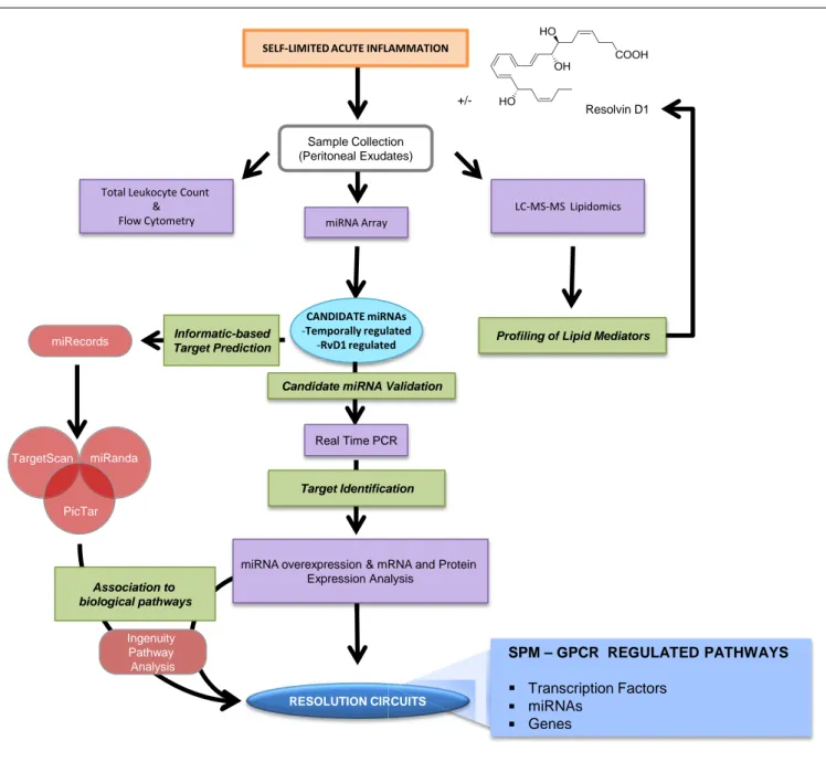
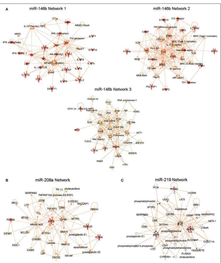
Results from the Serhan laboratory at Brigham and Women’s Hospital-Harvard Medical School demonstrated for the first time that SPM are operative in resolution and act locally to control leukocyte trafficking, regulate chemical mediators (e.g., cytokines, chemokines, and lipid autacoids), and expedite the return to homeostasis. Since microRNA (miRNAs) have emerged as the fine tuners of many cellular processes, including immune responses and cancer, it was of interest to investigate whether they also played roles in resolution and SPM-regulated specific miRNA as part of their mechanisms of action. To identify a miRNA signature of resolution in self-resolving exudates, a strategy with resolving exudates from murine peritonitis was used (Figure 6). Zymosan A particles from S. cerevisiae, a Toll-like receptor 2 and 4 ligand, were injected i.p. and peritoneal exudates collected to
Table 1 | Bioactions of SPM.
SPM Disease model Mechanism of action Reference
Lipoxin A4/ATL Mouse/dermal inflammation Inhibits neutrophil recruitment and vascular leakage Takano et al. (1997) Mouse/dorsal air pouch Inhibits neutrophil recruitment Clish et al. (1999) Rabbit/periodontitis Reduces PMN infiltration and prevents connective
tissue and bone loss
Serhan et al. (2003)
Mouse/peritonitis Inhibits neutrophil recruitment and lymphatic removal of phagocytes
Bannenberg et al. (2005)and Schwab et al. (2007) Mouse/colitis Attenuates pro-inflammatory gene expression and
reduces severity of colitis, inhibits weight loss, inflammation, and immune dysfunction
Gewirtz et al. (2002)
Mouse/asthma Inhibits airway hyper-responsiveness and pulmonary inflammation
Levy et al. (2002)
Mouse/cystic fibrosis Decreases neutrophilic inflammation, pulmonary bacterial burden, and disease severity
Karp et al. (2004)
Mouse/ischemia/reperfusion (I/R) Attenuates hind limb I/R-induced lung injury. Detachment of adherent leukocytes in mesenteric I/R vessels. Reduces myocardial infarct size and area at risk in myocardial I/R
Scalia et al. (1997)andChiang et al. (1999)
Mouse/cornea Accelerates cornea re-epithelialization, limits sequelae of thermal injury (i.e., neovascularization, opacity) and promotes host defense
Gronert et al. (2005)
Mouse/angiogenesis Reduces angiogenic phenotype: endothelial cell proliferation and migration
Fierro et al. (2002)
Mouse/bone marrow transplant (BMT) Protects against BMT-induced graft-versus-host diseases (GvHD)
Devchand et al. (2005)
Rat/glomerulonephritis Reduces leukocyte rolling and adherence; decreases neutrophil recruitment
Papayianni et al. (1995)
Rat/hyperalgesia Prolongs paw withdraw latency, reducing hyperalgesic index, and reduces paw edema
Svensson et al. (2007)
Rat/pleuritis Shortens the duration of pleural exudation Bandeira-Melo et al. (2000) Mouse/tumor growth Suppresses the growth of transplanted tumors in mice;
inhibits angiogenesis
Chen et al. (2010)
Mouse/allograft rejections Prevents acute rejection of vascularized cardiac and renal allografts
Levy et al. (2011)
Mouse/arthritis Inhibits edema formation and PMN influx, reduces TNFα and LTB4levels
Conte et al. (2010)
Rat/acute pancreatitis Reduces intercellular adhesion molecule 1 (ICAM-1) and NF-κB p65 expression in the pancreas, and expression of ICAM-1 in the lungs
Zhou et al. (2011)
Zebrafish/mycobacterial infection Reduces bacterial burden and growth; improves microbial containment by phagocytes
Tobin et al. (2012)
Resolvin E1 Mouse/dorsal air pouch Inhibits neutrophil recruitment Serhan et al. (2000a) Mouse/peritonitis Inhibits neutrophil recruitment, regulates
chemokine/cytokine production, and promotes lymphatic removal of phagocytes
Arita et al. (2005a),
Bannenberg et al. (2005), and Schwab et al. (2007) Rabbit/periodontitis Reduces PMN infiltration, prevents connective tissue
and bone loss, promotes healing of diseased tissues, and promotes regeneration of lost soft tissue and bone
Hasturk et al. (2006, 2007)
Mouse/retinopathy Protects against neovascularization Connor et al. (2007) Mouse/colitis Decreases PMN recruitment and pro-inflammatory
gene expression; improves survival and reduces weight loss; favors LPS-detoxification through induction of intestinal alkaline phosphatase
Arita et al. (2005b),Campbell et al. (2010), andIshida et al. (2010)
Table 1 | Continued
SPM Disease model Mechanism of action Reference
Mouse/asthma Reduces IL-23 and IL-6, and increases IFNγ and LXA4 in lungs to dampen airway inflammation; decreases eosinophil and lymphocyte recruitment
Aoki et al. (2008, 2010)and Haworth et al. (2008)
Mouse/obesity Regulates adipokines and protects against liver steatosis
Gonzalez-Periz et al. (2009)
Mouse/inflammatory pain Inhibits spontaneous pain, and heat and mechanical hypersensitivity
Xu et al. (2010)
Rat/cardiac ischemia/reperfusion injury Reduces infarct size Keyes et al. (2010) Mouse/allograft rejections Prevents acute rejection of vascularized cardiac and
renal allografts
Levy et al. (2011)
Mouse/dry eye Promotes tear production, corneal epithelial integrity, and decreases in inflammatory inducible COX-2. RvE1 inhibits keratocyte transformation to myofibroblasts and lowers the number of monocytes/macrophages
Li et al. (2010)
Mouse/herpes simplex virus Reduces severity of herpes simplex virus-induced ocular lesions, reduces angiogenesis, and stromal keratitis
Rajasagi et al. (2011)
Resolvin D1 Mouse/peritonitis Inhibits neutrophil recruitment; shortens resolution interval; regulates miRNAs and target genes in resolving exudates; reduces LTB4, PGD2, PGF2α, and TXA2in peritoneal exudates
Hong et al. (2003),Sun et al. (2007),Spite et al. (2009b), Recchiuti et al. (2011), Krishnamoorthy et al. (2012), andNorling et al. (2012) Mouse/E. coli (peritoneal) and
S. aureus (skin) infection
Reduces bacterial titers and hypothermia; increased survival; enhances microbial containment and killing by phagocytes; lowers antibiotic requirement; shortens resolution interval
Chiang et al. (2012)
Mouse/dorsal air pouch Inhibits neutrophil recruitment Serhan et al. (2002)andHong et al. (2003)
Mouse/kidney ischemia-reperfusion Protects from ischemia/reperfusion-induced kidney damage and loss of function; regulates macrophages
Duffield et al. (2006)
Mouse/retinopathy Protects against neovascularization Connor et al. (2007) Mouse/inflammatory pain Inhibits spontaneous pain, heat, and mechanical
hypersensitivity; selectively blocks TRPV1 and TRPA1-mediated pain
Xu et al. (2010)andPark et al. (2011)
Mouse/obesity Reduces inflammatory cytokines in adipose tissue macrophages; stimulates M2 macrophage
differentiation; promotes resolution of adipose tissue inflammation
Titos et al. (2011)
Mouse/T2 diabetes Reduces macrophage accumulation in adipose tissue; ameliorates insulin sensitivity
Hellmann et al. (2011)
Rats/post-operative pain Reduces post-operative pain, tactile allodynia, and hyperalgesia
Huang et al. (2011)
Mouse/pain Attenuates agonist-induced and inflammatory pain behaviors; inhibits TRPA1, TRPV3, and TRPV4 receptors; does not affect basal sensitivity
Bang et al. (2010)andXu et al. (2010)
Mouse/acute lung injury Blocks leukocyte infiltration and reduces cytokine levels in BALF
Wang et al. (2011)
Mouse/corneal inflammation Reduces leukocyte infiltration and hemangiogenesis Jin et al. (2009)