*Corresponding author: Victor NGU NGWA, School of Veterinary Medicine and Sciences, University of Ngaoundéré, P.O. Box 454, Ngaoundéré, Cameroon, Tel: +237 673198092; E-mail: [email protected]
Citation: Ngu Ngwa V, Cuteri V, Awah-Ndukum J, Tangwa BV, Manchang KT, et al. (2020) Bacterial Pathogens Involved in Bovine Mastitis and Their Antibiotic Resistance Patterns in the Adamawa Region of Cameroon. J Dairy Res Tech 3: 012.
Received: January 25, 2020; Accepted: February 25, 2020; Published: March 03, 2020
Copyright: © 2020 Ngu Ngwa V, et al. This is an open-access article distributed under the terms of the Creative Commons Attribution License, which permits un-restricted use, distribution, and reproduction in any medium, provided the original author and source are credited.
Introduction
Mastitis, an inflammatory mammary gland condition, is the most common, troublesome and the most expensive disease of dairy ru-minants worldwide as it is responsible for heavy economic losses in terms of reduction in milk yield, profit margins, and quality of milk and milk products [1-4]. Although physical and chemical injuries may cause inflammation of the mammary gland, infections most of-ten caused by bacteria or other microorganisms (fungi, viruses, algae) are the primary cause of mastitis [5]. Thus, based on etiopathological investigations, it is usually classified as subclinical, acute, subacute, chronic or gangrenous [6,7].
The causative organisms are well adapted to survive in the mam-mary glands and in most cases, establish mild subclinical infection of long duration during which pathogens of public health significance might be shed into milk from the infected quarters [8]. Furthermore, mastitis is associated with a number of zoonotic diseases including Tuberculosis, Brucellosis, Campylobacteriosis and streptococcal sore throat in which milk acts as a vehicle of infection [7,9]. Public haz-ards associated with the consumption of antibiotic contaminated milk and products cause allergic responses, changes in intestinal flora and development of antibiotic resistant pathogenic bacteria [10,11]. The dairy industry in Cameroon is rudimentary [12] and masti-tis is becoming a significant constrained in its development. Gram positive and Gram negative bacteria are involved as major pathogens causing mastitis worldwide, such as Staphylococcus aureus, Esche-richia coli, Streptococcus spp., Klebsiella spp. [13]. S. aureus and E. coli are the most commonly isolated pathogen from clinical mastitis [14]. Staphylococcus spp. is a major pathogen causing various forms of subclinical and clinical mastitis in cattle [15]. Coagulase negative staphylococci remain the most frequently isolated pathogens from the subclinical mastitis in dairy cows [14].
An important aspect in the appropriate control of infectious dis-eases is identification of the causative agents. Antimicrobial therapy aiming against infectious agents causing mastitis is usually recom-mendable [16]. The indiscriminate use of antimicrobial drugs with-out testing in vitro sensitivity, as commonly practice in the country, may be considered the primary cause of lack of success in treatment. Transmission of resistant pathogens to humans via bulk milk with subclinical mastitis is of major public health interest [17]. In addition,
Dairy Research & Technology
Research Article
Victor Ngu Ngwa1*, Vincenzo Cuteri2, Julius Awah-Ndukum1,
Bernard Viban Tangwa3, Kingsley Tanyi Manchang3 and
Anna-Rita Attili2
1School of Veterinary Medicine and Sciences, University of Ngaoundéré,
Cameroon
2School of Biosciences and Veterinary Medicine, University of Camerino,
Italy
3Veterinary Research Laboratory, IRAD Wakwa, Ngaoundéré, Cameroon
Bacterial Pathogens Involved in
Bovine Mastitis and Their
Anti-biotic Resistance Patterns in the
Adamawa Region of Cameroon
Abstract
Data on the sensitivity pattern of bacteria are scarce in sub-Saha-ran Africa, especially in Cameroon. This paper reports the prevalence of bovine mastitis and major bacterial pathogens associated with the disease and their antimicrobial profiles in the Adamawa Region of Cameroon. It was conducted to investigate the sensitivity pattern of bacteria isolated from mastitis cases that could be helpful in the ap-plication of appropriate therapeutic measures. For this study, 224 lactating cows were examined. A high average prevalence (59.8%) in subclinical mastitis was recorded as compared to clinical mastitis (3.6%; χ2=163.7, P=10-4). Out of the 135 clinical and subclinical mas-titis cases recorded, bacteria were cultured from 115 milk samples (85.2%, n=135). In all, 14 different bacterial pathogens were isolated including: coagulase negative Staphylococci (27.5%),
Staphylococ-cus aureus (23.3%), Escherichia coli (11.3%), StreptococStaphylococ-cus aga-lactiae (7.1%), Streptococcus dysagaaga-lactiae (4.2%), Enterococcus faecalis (2.8%), Klebsiella pneumoniae (2.8%), Enterobacter aero-genes (2.1%), Pseudomonas aeruginosa (2.1%), Corynebacterium
spp. (1.4%), Proteus spp. (1.4%), Brucella spp. (1.4%),
Mycoplas-ma spp. (0.7%), and Mycobacterium spp. (0.7%). A Mycoplas-major variation
in the sensitivity of isolated bacteria against 14 different antibiotics was noticed. Overall the sensitivity test revealed that Enrofloxacin, Gentamicin, and to a lesser extent Oxacillin and Amoxicillin/Clavu-lanic acid, were most efficacious. The study gives a significant con-tribution to the epidemiology and contributes to reducing the lack of knowledge about the antibiotic resistance patterns of major bacterial mastitis in Cameroon. The application of these antibiotics could be beneficial in resolving the cases of bovine mastitis in dairy herds. Keywords: Antibiotic resistance; Cameroon: Cattle; Mastitis; Patho-genic bacteria
the risk to human health for Mycobacterium avium subsp. paratuber-culosis [18], Mycobacterium bovis, the causal agent of Tuberculous, mastitis, and other milk zoonoses is of great concern particularly in developing countries where there is an increase in the consumption of untreated milk [19]. Therefore, it is important to investigate the sensi-tivity pattern of the different bacteria isolated from mastitis as well as apply the appropriate therapeutic measures. Such data are very scarce in sub-Saharan Africa, especially in Cameroon.
In this context this study was carried out to identify the causative bacterial agents of bovine mastitis in Adamawa region of Cameroon as well as evaluate their antibiotic susceptibility profiles. The investi-gation also attempt to provide epidemiological data which are key to the formulation of antimicrobials therapeutic measures against bovine mastitis in the country.
Materials and methods
Study design and sampling population
In this study, 224 lactating cows from 16 different smallholder
dairy farms located in the Adamawa Region of Cameroon were exam-ined to determine the prevalence of mastitis, and to identify the major bacterial pathogens associated with the disease and their antimicro-bial patterns. The cows enrolled were randomly chosen from farms practicing the semi intensive husbandry system and included 64 Hol-stein-Friesian breed, 50 Adamawa Gudali hybrid breed, 32 Adamawa Gudali breed, 34 White Fulani breed, 24 Red Fulani breed, and 20 Banyo Gudali breed. Of the total number of cows sampled, 103 cows were less than or equal to 5 years of age and 124 were more than 5 years of age.
Detection of mastitis
To determine clinical and subclinical mastitis in the lactating cows,
clinical examination of the udder was performed [7,20]. Screening was done using the California mastitis test (CMT) (ImmuCell® CMT, Portland, USA) as previously described [12,20].
Microbiological analysis
Collection of milk samples
Before milk collection from the CMT positive animals, the teats
of the udders were wiped thoroughly with 70% ethyl alcohol, with particular attention to the teat orifice. The first streams of milk were discarded and sterile test tubes were used in collecting the milk in a strictly aseptic manner. Approximately 10 ml of milk were collected per cow. The samples were delivered to the microbiology laboratory in an ice-cooled box within 4 hours and processed immediately for the isolation, characterization and identification of bacteria.
Direct microscopy
The milk samples were centrifuged and the obtained pellet was
swiped on a slide and then stained. A Gram- and Ziehl Neelsen stains were used routinely [20].
Bacteriological culture
The bacteriological culture was carried out following standard
microbiological technique and microbiological procedures for the di-agnosis of bovine mastitis infection [20]. Briefly, a loop full of milk streaked on 7% sheep blood agar plates are checked for growth after
24, 48 and up to 72 hours to rule out slow growing microorganisms. A sample was considered negative if there is no growth after 72 hours. Suspected bacteria were sub-cultured onto different selective/differ-ential bacteriological media and incubated at 37°C for 24 hours. Pure cultures were achieved as per procedures described by [21,22]. Colony morphology, hemolytic characteristics, Gram staining, catalase test, motility test, triple sugar iron reaction, CAMP test, IM-ViC (Indole, Methyl red, Voges-Proskauer, Citrate), coagulase and cytochrome oxidase tests were conducted to identify the isolates ac-cording to the procedures adopted by Quinn et al. [20]. Furthermore, biochemical identifications by commercial kits were carried out (Inte-gral System Enterobacteria, Inte(Inte-gral System Staphylococci, Inte(Inte-gral System Streptococci, Liofilchem®, Abruzzo, Italy).
Standard specific culturing techniques were applied in the sus-pected cases of Paratuberculosis, Tuberculosis, Brucellosis and CBPP (Contagious Bovine Pleuropneumonia) for the isolation of Mycobac-terium spp., Brucella spp., and Mycoplasma spp., respectively.
Antimicrobial susceptibility testing
Selected bacterial isolates were tested for susceptibility to
differ-ent antimicrobials using in vitro disk diffusion (Kirby-Bauer) method as described by Quinn et al. [20]. Cultured broth was cross-checked with McFarland standard before applying on Mueller Hinton agar and disk application. Fourteen different antimicrobial disks obtained from commercial sources (Oxoid Ltd, Baring-stoke, Hampshire, England, and Liofilchem®, Abruzzo, Italy) were selected for the testing and they included: Enrofloxacin (5μg), Amoxicillin (10μg), Streptomycin (10μg), Erythromycin (15μg), Ampicillin (10μg), Gentamicin (30μg), Doxycycline (30µg), Oxytetracycline (30µg), Penicillin G (10 IU), Trimethoprim/sulphamethoxazole (1.25/23.75µg), Neomycin (30µg), Amoxicillin/clavulanic acid (20/10µg), Ceftiofur (15µg), and Oxacil-lin (1µg).
In all, 12 isolated bacteria were subjected to antimicrobial suscep-tibility testing with the exception of Mycoplasma and Mycobacterium species. Brucella species were tested for antimicrobials susceptibility using five antimicrobial agents [Enrofloxacin (5µg), Streptomycin (10µg), Gentamicin (30µg), Doxycycline (30µg), Oxytetracycline (30µg)]. Based on the susceptibility to antimicrobials, the bacteria were categorized into three groups: sensitive, intermediate and resis-tant. For statistical analysis, the intermediate group was considered as resistant.
The interpretation on susceptibility was done according to the guidelines of Clinical and Laboratory Standard Institute [23].
Statistical analysis
The qualitative data were analyzed using Statistical software
STA-TA version 13 (SSTA-TASTA-TA Corporation, College Station, Texas, USA). Univariate analyses on prevalence percentages were performed. Sta-tistical differences were calculated by Chi Square test and P-values less than 0.05 were considered statistically significant.
Results
Clinical and subclinical mastitis prevalence
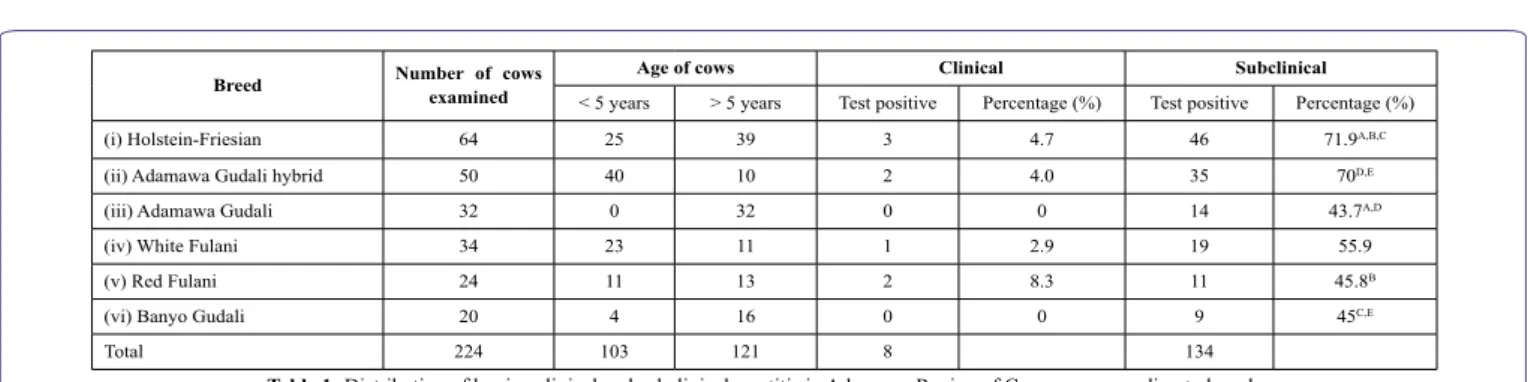
Out of the 224 lactating cows examined, a high average
preva-lence in subclinical mastitis (59.8%) was recorded as compared to clinical mastitis (3.6%; χ2=163.7, P=10-4) (Table 1).
The subclinical and clinical mastitis were most represented in the bovine population aged less than or equal to 5 years, 69.9% and 5.8% (n=103), respectively. In relation to age, a significant difference was observed only for subclinical mastitis (69.9% vs 51.2%, n=121; χ2=8.06, P=0.0045). In relation to the farm, prevalence rate ranged from 25.0% to 81.8%, for subclinical mastitis and from 0% to 9.1% for clinical mastitis. The farms with the highest prevalence rates for subclinical mastitis also showed the highest prevalence rates for clin-ical mastitis.
Bacteria isolates
From the 135 clinical and subclinical mastitis cases recorded, bacteria were successfully cultured from 115 milk samples (85.2%, n=135). In one hundred and four samples (77.0%, n=135) grew pure cultures. Eleven samples (8.1%, n=135) had mixed growth, of which one isolate per sample was considered for further analyses based on medical/veterinary importance judgment taking into consideration the morphology of the colonies. Twelve samples presented no growth (8.9%, n=135), four samples (3.0%, n=135) were contaminated with manure at the site of collection hence were discarded, and fungi grew in four other samples (3.0%, n=135), so they were not included in the analyses. Mastitis of viral origin or uncultivable bacterial species may be responsible for the negative cultures.
In all, 14 different bacterial pathogens were isolated (Table 2).
CoNS (Coagulase negative Staphylococci) had the highest prev-alence (39 cases) overall, followed by Staphylococcus aureus (33 cases), Escherichia coli (16 cases), Streptococcus agalactiae (10 cas-es), Streptococcus dysagalactiae (6 cascas-es), Enterococcus faecalis (4 cases), Klebsiella pneumoniae (4 cases), Enterobacter aerogenes (3 cases), Pseudomonas aeruginosa (3 cases), Corynebacterium spp. (2 cases), Proteus spp. (2 cases), Brucella spp. (2 cases), Mycoplasma spp (1 case), and Mycobacterium spp. (1 case). Brucella and Myco-plasma species were cultured from clinical mastitis cases while My-cobacterium spp. was cultured from a case of subclinical mastitis, and were all isolated from milk samples gotten from the local indigenous cattle.
Therefore, the predominant bacteria involved in clinical and sub-clinical mastitis in the Adamawa Region of Cameroon, were iden-tified as Coagulase Negative Staphylococci, Staphylococcus aureus, Escherichia coli, and Streptococci.
Antimicrobial susceptibility testing
The in-vitro antimicrobial susceptibility assays showed high
resis-tance patterns (Figure 1).
The resistance percentages ranged from 18.5% (n=124) for Enro-floxacin (5µg) to 99.0% (n=122) for Erythromycin (15µg).
Breed Number of cows examined Age of cows Clinical Subclinical
< 5 years > 5 years Test positive Percentage (%) Test positive Percentage (%) (i) Holstein-Friesian 64 25 39 3 4.7 46 71.9A,B,C
(ii) Adamawa Gudali hybrid 50 40 10 2 4.0 35 70D,E
(iii) Adamawa Gudali 32 0 32 0 0 14 43.7A,D
(iv) White Fulani 34 23 11 1 2.9 19 55.9 (v) Red Fulani 24 11 13 2 8.3 11 45.8B
(vi) Banyo Gudali 20 4 16 0 0 9 45C,E
Total 224 103 121 8 134
Table 1: Distribution of bovine clinical and subclinical mastitis in Adamawa Region of Cameroon according to breed.
A: χ2 = 7.20, P = 0.007; B: χ2 = 5.19, P = 0.0227; C: χ2 = 4.87, P = 0.0273; D: χ2 = 5.59, P = 0.018; E: χ2 = 3.82, P = 0.0505
Table 2: Frequency of occurrence of the isolated bacteria.
Figure 1: In-vitro resistance patterns (%) observed for each antibiotic tested.
AMX = Amoxicillin (10 µg), Co-AMX = Amoxicillin/Clavulanic acid (20 / 10 µg), P = Penicillin G (10 IU), AMP = Ampicillin (10 µg), OXA = Oxacillin (1 µg), ENR = Enrofloxacin (5 µg), CEF = Ceftiofur (15 µg), E = Erythromycin (15 µg), S = Streptomycin (10 µg), G = Gentamicin (30 µg), N = Neomycin (30 µg), DOX = Doxycyclin (30 µg), OXY = Oxytetracycline (30 µg), SUL= Trimethoprim/Sulpha-methoxazole (1.25 / 23.75 µg).
Bacteria isolated Frequency Prevalence rate (%)
Coagulase Negative Staphylococci (CoNS) 39 27.5
Staphylococcus aureus 33 23.3 Escherichia coli 16 11.3 Streptococcus agalactiae 10 7.1 Streptococcus dysagalactiae 6 4.2 Enterococcus faecalis 4 2.8 Klebsiella pneumoniae 4 2.8 Enterobacter aerogenes 3 2.1 Pseudomonas aeruginosa 3 2.1 Corynebacterium spp. 2 1.4 Proteus vulgaris 2 1.4 Brucella spp. 2 1.4 Mycoplasma spp. 1 0.7 Mycobacterium spp. 1 0.7 Fungi 4 2.8 Negative samples 12 8.4 Total 142 100
Fluoroquinolones resistance rate was the lowest recorded, and sig-nificant differences were observed between Enrofloxacin vs Strepto-mycin (98.4%; χ2=162.7, P=10-4), Enrofloxacin vs Sulfamethoxazole
plus Trimethoprim (95.1%; χ2=146.6, P= 10-4), Enrofloxacin vs
Dox-ycycline (94.3%; χ2=144.9, P= 10-4).
Significant differences were observed between classes of anti-biotics, in particular between Aminoglycosides (99.2%, n=122) vs Fluoroquinolones (19.3%, n=124; χ2=161.9, P=10-4), and within the
same class of antibiotics: Amoxicillin (83.6%) vs Amoxicillin-Clavu-lanic acid (52.5%; χ2=27.2, P=10-4), Amoxicillin vs Oxacillin (47.5%;
χ2=38.1, P=10-4), Penicillin (88.3%) vs Ampicillin (91.0%; χ2=5.6,
P=0.0176), Penicillin vs Amoxicillin-Clavulanic acid (χ2=21.2, P=10 -4), Penicillin vs Oxacillin (χ2=28.4, P=10-4), Ampicillin vs
Amox-icillin-Clavulanic acid (χ2=44.6, P=10-4), Ampicillin vs Oxacillin
(χ2=54.1, P=10-4), Streptomycin (98.4%) vs Gentamicin (37.1%;
χ2=106.6, P=10-4), Streptomycin vs Neomycin (87.7%; χ2=10.9,
P=0.001), Gentamicin vs Neomycin (χ2=67.0, P=10-4), Doxycycline
(94.3%) vs Oxytetracycline (79.8%; χ2=11.6, P=0.0007).
In relation to the Gram affinity, Gram positive bacteria showed a significant higher resistance rate (86,2%) only for Cephalosporins (χ2=4.9, P=0.0289). Gram negative bacteria revealed a high
resis-tance rates for Beta-Lactam antibiotics (100%), Macrolides (92.9%), and Tetracyclines (100%), but the differences were not significant
(P>0.05) (Figure 2). Table 3 shows the antibiotic resistance (%) profiles recorded for each isolated bacterium.
Figure 2: Antibiotic resistance patterns (%) observed for each antibiotic in relation
to the Gram affinity.
AMX =Amoxicillin (10 µg), Co-AMX = Amoxicillin/Clavulanic acid (20 / 10 µg), P = Penicillin G (10 IU), AMP = Ampicillin (10 µg), OXA = Oxacillin (1 µg), ENR = Enrofloxacin (5 µg), CEF = Ceftiofur (15 µg), E = Erythromycin (15 µg), S = Streptomycin (10 µg), G = Gentamicin (30 µg), N = Neomycin (30 µg), DOX = Doxycyclin (30 µg), OXY = Oxytetracycline (30 µg), SUL= Trimethoprim/Sulpha-methoxazole (1.25 / 23.75 µg).
Table 3: In-vitro antibiotic resistance patterns (%) observed for each bacterial isolates.
SA = Staphylococcus aureus, CoNS = Staphylococci coagulase negative, EC = Escherichia coli, STREP. A. = Streptococcus agalactiae, STREP. D. = Streptococcus dysgalactiae, EF = Enterococcus faecalis, KP = Klebsiellapneumoniae, EA = Enterobacter aerogenes, PA = Pseudomonas aeruginosa, C = Corynebacterium spp., PV = Proteus vulgaris, B = Brucella spp., ENR = Enrofloxacin (5 µg), AMX =Amoxicillin (10 µg), S = Streptomycin (10 µg), E =
Erythromycin (15 µg), AMP = Ampicillin (10 µg), G = Gentamicin (30 µg), DOX = Doxycyclin (30 µg), OXY = Oxytetracycline (30 µg), P = Penicillin G (10 IU), SUL= Trimethoprim/Sulphamethoxazole (1.25 / 23.75 µg), N = Neomycin (30 µg), Co-AMX = Amoxicillin/Clavulanic acid (20 / 10 µg), CEF = Ceftiofur (15 µg), OXA = Oxacillin (1 µg).
1(χ2 = 5.11, P = 0.024); 2(χ2 =4.0, P = 0.046); 3(χ2 = 20.0, P = 10-4); 4(χ2 = 17.0, P = 10-4); 5(χ2 = 5.1, P = 0.0238); 6(χ2 = 4.2, P = 0.0402); 7(χ2 = 5.2, P = 0.0231); 8(χ2 = 7.1, P = 0.0078); 9(χ2 = 4.3, P = 0.0383); 10(χ2 = 12.2, P = 0.0005); 11(χ2 = 14.5, P = 0.0001); 12(χ2 = 7.9, P = 0.0049); 13(χ2 = 11.6, P = 0.0007); 14(χ2 = 7.6, P = 0.0060); 15(χ2 = 5.9, P = 0.0154); 16(χ2 = 4.2, P = 0.0398); 17(χ2 = 9.0, P = 0.0027); 18(χ2 = 17.0, P = 10-4); 19(χ2 = 5.2, P = 0.0228); 20(χ2 = 6.4, P= 0.0111); 21(χ2 = 12.2, P = 0.0005); 22(χ2 = 4.5, P = 0.0347); 23(χ2 = 8.7, P = 0.0033); 24,25(χ2 = 5.6, P = 0.0175); 26(χ2 = 5.5, P = 0.0195); 27(χ2 = 13.1, P = 0.0003); 28(χ2 = 17.0, P = 10-4); 29(χ2 = 4.3, P = 0.0383); 30(χ2 = 4.6, P= 0.0313); 31(χ2 = 9.9, P = 0.0016); 32(χ2 = 13.0, P = 0.0003); 33(χ2 = 8.7, P = 0.0033); 34(χ2 = 8.9, P = 0.0029); 35(χ2 = 4.2, P = 0.0402); 36(χ2 = 4.0, P = 0.0460); 37,38(χ2 = 13.3, P = 0.0003); 39(χ2 = 5.7, P = 0.0174); 40(χ2 = 5.8, P = 0.0160); 41(χ2 = 8.0, P = 0.0047); 42(χ2 = 4.3, P = 0.0373); 43(χ2 = 6.1 P = 0.0134); 44(χ2 = 12.8 P = 0.0003); 45(χ2 = 17.6 P = 10-4); 46(χ2 = 5.7 P = 0.0173); 47(χ2 = 9.3 P = 0.0023); 48(χ2 = 11.4 P = 0.0007); 49(χ2 = 16.3 P = 0.0001); 50(χ2 = 6.5 P = 0.0107); 51(χ2 = 10.2 P = 0.0014). The superscripts highlighted italic numbers (1- 51) represent significant values.
Isolates No ENR AMX S E AMP G DOX OXY PEN SUL N Co-AMX CEF OXA
SA 33 9.11 87.9 1004 90.9 97.0 24.28,9 100 11,13 78.8 16,19 75.826,29,30 93.9 90.940 57.641 93.7 12.145,47,49,51 CoNS 39 17.9 84.6 100 2,3 94.9 5 97.46 30.87 97.4 10,12 94.9 16,17,18,21,23,24,25 94.9 26,27,28,33 10036,37,38 89.739 69.2 41,43 84.2 20.544,46,48,50 EC 16 12.5 93.7 100 87.5 87.5 31.2 100 14,15 87.5 20,22 10030,31,32,34,35 93.7 62.539,40 50 56.2 -STREP. A. 10 20 70 902 80 806 70 7,8 6010, 11,14 6017 50 27,31 9036 90 2041,42 80 8044,45 STREP. D. 6 16.7 66.7 100 66.75 83.3 66.79 66.7 12,13, 15 33.3 18,19, 20 33.328,29,32 100 83.3 16.743 83.3 66.7 46,47 EF 4 501 75 100 100 0 50 100 5023 50 33,34 100 100 25 50 100 48,49 KP 4 501 75 100 100 100 25 100 100 7535 100 100 50 75 -EA 3 0 66.7 100 100 100 33.3 100 33.3 21,22 100 66.737 100 33.3 66.7 -PA 3 33.3 66.7 100 100 100 33.3 100 100 100 66.738 100 33.3 100 -C 2 0 100 100 100 100 100 100 100 0 100 100 50 100 100 50,51 PV 2 50 100 100 100 100 50 100 5024 100 100 100 50 100 -B 2 100 - 503,4 - - 100 100 5025 - - - - -
High resistance to Beta-Lactam antibiotics was recorded from Gram negative (100%, n=28) nevertheless no significant difference resulted towards Gram positive (96.8%, n=94; χ2=0.9 P=0.3385). In
particular, a significant difference was demonstrated for Penicillin (96.4%, n=28, vs 75.5%, n=94; χ2=6.0 P=0.0146).
The 16.7% of Staphylococcus species (n=72) showed Methicillin resistance phenotypically, and no significant differences were record-ed between Staphylococcus spp. coagulase negative (20.5%) and S. aureus (12.1%).
Oxacillin resistant Streptococci isolates were 75% (n=16), while 81.2% were Ampicillin resistant (vs penicillin: χ2=4.8, P=0.0285; vs
Amoxicillin and Clavulanic acid: χ2=12.5, P=0.0004), 68.7% were
Amoxicillin resistant, 43.7% were Penicillin-resistant, 18.7% were Amoxicillin and Clavulanic-acid resistant (vs Oxacillin: χ2=10.2,
P=0.0014; vs Amoxicillin: χ2=8.1, P=0.0044
Discussion
In most sub-Saharan countries including Cameroon,
sub-clini-cal mastitis received little or no attention and efforts are focused on the treatment of clinical cases while high productive and economic losses could come from sub-clinical mastitis. In the present study, there were overwhelming cases of sub-clinical mastitis (59.8%) com-pared to clinical mastitis (3.6%). Our findings are similar to those of many studies [12,24]. In the current study, fourteen different bacte-rial pathogens were isolated from milk samples collected from 135 mastitis cows. The isolated bacteria were Coagulase negative Staph-ylococci (27.5%), Staphylococcus aureus (23.3%), Escherichia coli (11.3%), Streptococcus agalactiae (7.1%), Streptococcus dysagalac-tiae (4.2%), Enterococcus faecalis (2.8%), Klebsiella pneumoniae (2.8%), Enterobacter aerogenes (2.1%), Pseudomonas aeruginosa (2.1%), Corynebacterium spp. (1.4%), Proteus spp. (1.4%), Brucel-la spp. (1.4%), MycopBrucel-lasma spp (0.7%), and Mycobacterium spp. (0.7%). The study showed that Staphylococcus spp, Escherichia coli, and Streptococcus spp, are the major cause of mastitis in Adamawa Region Cameroon. This finding is in agreement with those of many studies carried out in many parts of the world [7,25-28].
The in vitro antibiotic susceptibility testing of twelve different types of bacterial isolates to 14 different antibiotics such as Enro-floxacin, Amoxicillin, Streptomycin, Erythromycin, Ampicillin, Gen-tamicin, Doxycycline, Oxytetracycline, Penicillin G, Trimethoprim/ sulphamethoxazole, Neomycin, Amoxicillin/Clavulanic acid, Cef-tiofur, and Oxacillin showed overall effective drug therapy against isolated pathogens, in the following order: Enrofloxacin, Gentami-cin, and to a lesser extent by Oxacillin and Amoxicillin/Clavulanic acid was observed but resistance of most of the isolates to the other antibiotics were noticed. The variation in the sensitivity of common antibiotics could be the result of extensive and indiscriminate use of these in the treatment of udder infection.
In the past two decades, a significant increased of antimicrobial resistance among Gram-positive bacteria has been observed, includ-ing multidrug-resistant staphylococci, penicillin-resistant streptococ-ci, and among Gram-negative bacteria, including the emergence and spread of resistance in Enterobacteriaceae. Klebsiella pneumoniae and Enterobacter spp. infections now involve strains not suscepti-ble to third-generation cephalosporins. Such resistance in K. pneu-moniae to third-generation cephalosporins is typically caused by the
acquisition of plasmids containing genes that encode for extend-ed-spectrum β-lactamases (ESBLs), and these plasmids often carry other resistance genes as well. ESBL-producing K. pneumoniae and Escherichia coli are now relatively common in healthcare settings and often exhibit multidrug resistance. ESBL-producing Enterobac-teriaceae have now emerged in the community as well [29].
In the currently study, bacteria of the family Enterobacteriaceae recorded 100% resistance to Beta-Lactams. Moreover, Enterobacter aerogenes showed over 66.7% to the third generation rins. Resistance of Enterobacter spp. to third-generation Cephalospo-rins was the most typically caused by overproduction of AmpC β-lac-tamases, and treatment with third-generation cephalosporins may select for AmpC-overproducing mutants. Some Enterobacter cloacae strains are now ESBL and AmpC producers, conferring resistance to both third- and fourth-generation cephalosporins [30].
Fluoroquinolones resistance Enterobacteriaceae was 17.4% (n=23). Quinolone resistance in Enterobacteriaceae is usually the result of chromosomal mutations leading to alterations in target en-zymes or drug accumulation. More recently, however, plasmid-medi-ated quinolone resistance has been reported in K. pneumoniae and E. coli, associated with acquisition of the qnr gene [30].
Oxacillin-resistant Staphylococcus aureus (MRSA) represents an important problem worldwide, and its prevalence may vary signifi-cantly in human and veterinary medicine. Most MRSA isolates show resistance to virtually all Beta-lactams by production of penicillinase and a low-affinity penicillin-binding protein (PBP) called PBP 2a [31].
Since its detection in Papua New Guinea and Australia, Penicillin resistance in Streptococcus spp. has now been reported worldwide [32]. In the present study the Penicillin resistance rate observed for Streptococci isolates was 43.7% (n=16), lower when compared to other beta-lactams, in particular to Ampicillin (81.2%, n=16; χ2=4.8
P=0.0285).
Further investigations will be needed to study the beta lactamase production by Gram negative isolates, and Oxacillin/Methicillin re-sistance from Staphylococcus genus.
In a summary, the different bacteria isolated from sub-clinical and clinical mastitis cases in this study showed that Staphylococci were the most common, followed by Streptococcus species and Escherich-ia coli. Thus, for effective treatment of bovine mastitis, medicinal for-mulations should contain antibiotics with good inhibition spectrum of against most species of bacteria. In this context, it is interesting to note that Enrofloxacin especially, and to a lesser extent, Gentamicin, Oxacillin and Amoxicillin/Clavulanic acid showed the highest sensi-tivity among almost all of the bacteria isolates in this study and should considered among the choice antibiotics for effective treatment of bo-vine mastitis in the study area to yield the best possible result. Other studies [33,34] have shown similar susceptibility pattern regarding the use of Fluoroquinolones against bovine mastitis pathogens. Finally, due to logistical reasons we were unable to perform the antibiotic susceptibility test for the isolated Mycobacterium and My-coplasma species. Nevertheless, this will be carryout in subsequent studies when the situation will have been resolved.
In conclusion, potential drug resistant pathogens in otherwise nor-mal dairy herd may be a serious concern for public health. Current findings suggest further studies with the isolated strains of bacteria. This study revealed the existence of alarming levels of resistance of Staphylococcus spp., Gram negative bacteria and to a lesser extent, Streptococcus spp. to commonly used antimicrobial agents. The re-sults suggest a possible development of resistance from prolonged and indiscriminate usage of some antimicrobials. Thus, it is very im-portant to implement a systemic application of an in vitro antibiotic susceptibility test prior to the use of antibiotics in both treatment and prevention of intra-mammary infections.
Acknowledgements
The authors are grateful to the management of the Microbiology
and Infectious Diseases Laboratory of the School of Biosciences and Veterinary Medicine, University of Camerino, Italy for donating the bacteriological culture media, identification kits, antibiotics disks, and biochemical reagents used in the study. The authors wish to thank dairy farm owners in the Adamawa Region for their unfailing cooper-ation.
Conflict of Interest
The author declares that there is no conflict of interests regarding
the publication of this paper.
References
1. Seegers H, Fourichon C, Beaudeau F (2003) Production effects related to mastitis and mastitis economics in dairy cattle herds. Vet Res 34: 475-491. 2. Halasa TK, Huijps O, Osteras H, Hogeveen H (2007) Economic effects
of bovine mastitis and mastitis management: a review. Vet Q 29: 18-31. 3. FAO (2014) Impact of mastitis in small scale dairy production systems.
Animal production and health working paper. 13.
4. Elva C, Rebecca LS, Anders RK, Julia AH, Ynte HS, et al. (2016) The value of pathogen information in treating clinical mastitis. J Dairy Res 83: 456-463.
5. Reugg LP, Radostits OM (2001) Health and production management in dairy herds. Herd health, food animal production. B Saunders Company (3rd Edn), Philadelphia, Pennsylvania, Pg no’s: 211- 244.
6. Erskine RJ1, Bartlett PC, Johnson GL 2nd, Halbert LW (1996) Intramus-cular administration of ceftiofur sodium versus intrammamary infusion penicillin/novobiocin for treatment of Streptococcus agalactiae mastitis in dairy cows. J Am Vet Med Assoc 208: 258-260.
7. Radostitis OM, Gay CC, Blood DC, Hinchllif KW (2007) Mastitis. In: Vet-erinary Medicine: A textbook of the diseases of cattle, horses, sheep, pigs and goats. Haracourt Ltd (9th edn.), London, Pg no’s. 603-700.
8. Bradley A (2002) Bovine mastitis an evolving disease. Vet J 164: 116-128. 9. Morgan G, Chadwick P, Lander KP, Gill KP (1985) Campylobacter jejuni
mastitis in a cow: a zoonosis-related incident. Vet Rec 122: 262-268. 10. Kassa T, Wirtu G, Tegegne A (1999) Survey of mastitis in dairy herds in
the Ethiopian central highlands. SINET: Ethiopian J Sci 22: 291-301. 11. Moon JS, Lee AR, Kang HM, Lee ES, Kim MN, et al. (2007)
Phenotyp-ic and genetPhenotyp-ic antibiogram of methPhenotyp-icillin-resistant staphylococci isolated from Bovine mastitis in Korea. J Dairy Sci 90: 1176-1185.
12. Ngu Ngwa V, Awah-Ndukum J, Cuteri V, Tanyi KM, Souaibou A, et al. (2018) Prevalence study on bovine mastitis in the Adamawa Region of Cameroon. Large Ani Rev 24: 21-29.
13. Olde Riekerink RG, Barkema HW, Kelton DF, Scholl DT (2008) Incidence rate of clinical mastitis on Canadian dairy farms. J Dairy Sci 91: 1366-1377.
14. Contreras A, Luengo C, Sanchez A, Corrales JC (2003) The role of intra-mammary pathogens in dairy goats. Livestock Prod Sci 79: 273-283. 15. Ibrahim A, Kursat K, Haci AC (2009) Identification and antimicrobial
sus-ceptibility of subclinical mastitis pathogens isolated from Hair goat’s milk. J Ani Vet Adv 8: 1086-1090.
16. Constable P, Pyorala S, Smith G (2008) Guidelines for antimicrobial use in cattle. In: Guide to Antimicrobial Use in Animals. Guardabassi L, Jensen LB & Kruse H ed. Blackwell Publishing Ltd., Oxford, UK 12: 143–160. 17. Sol J, Sampimon OC, Barkema HW, Schukken YH (2000) Factors
associ-ated with cure after therapy of clinical mastitis caused by Staphylococcus aureus. J Dairy Sci 83: 278-284.
18. Attili AR, Ngu Ngwa V, Preziuso S, Pacifici L, Domesi A, et al. (2011) Ovine paratuberculosis: seroprevalence study in dairy flocks reared in the Marche region of Italy. Vet Med Int: 782875.
19. Grange JM (2001) Mycobacterium bovis infection in human beings. Tu-berculosis TuTu-berculosis (Edinb) 81: 71-77.
20. Quinn P, Carter ME, Markey BK, Carter GR (2002) Clinical Veterinary Microbiology. Harcourt Publishers, Virginia, USA, Page no’s. 331-344. 21. OIE (2000) Manual of Standards for Diagnostic Tests and Vaccines. OIE
(4th Edn.), Paris, France.
22. Cowan ST (1985) Cowan and steel’s manual for identification of medical bacteria. Cambridge University Press (2nd Edn.), Cambridge, London 2: 138-139.
23. CLSI VET 08 (2018) Performance Standard for Antimicrobial Disk and Dilution Susceptibility Tests for Bacteria Isolated from Animals. Clinical and Laboratory Standards Institute (4th Edn.), USA.
24. Dego OK, Tareke F (2003) Bovine mastitis in selected areas of southern Ethiopia. Trop Ani Health Prod 35: 197-205.
25. Shitandi A, Anakalo G, Galgalo T, Mwangi M (2004) Prevalence of bovine mastitis amongst small holder dairy herds in Kenya, Israel. J Vet Med 59: 1-2.
26. Lakew M, Tolosa T, Tigre W (2009) Prevalence and major bacterial causes of bovine mastitis in Asella, South Eastern Ethiopia. Trop Ani Health Prod 41: 1525-1530.
27. Zadoks N, Fitzpatrick L (2009) Changing trends in mastitis. Irish Vet J 62: 59-70.
28. Kayano M, Itoh M, Kusaba N, Hayashiguchi O, Kida K, et al. (2018) As-sociations of the first occurrence of pathogen-specific clinical mastitis with milk yield and milk composition in dairy cows. J Dairy Res 85: 309-316. 29. Jayarao B, Almeida R, Oliver SP (2019) Antimicrobial Resistance on
Dairy Farms. Foodborne Pathog Dis 16: 1-3.
30. Paterson DL (2006) Resistance in gram-negative bacteria: Enterobacteria-ceae. Am J Med 34: 20-28.
31. Deshpande LM, Jones RN (2003) Bactericidal activity and synergy stud-ies of BAL9141, a novel pyrrolidinone-3-ylidenemethyl cephem, tested against streptococci, enterococci and methicillin-resistant staphylococci. Clin Microbiol Infect 9: 1120-1124.
32. Dowson CG, Johnson AP, Cercenado E, George RC (1994) Genetics of Oxacillin Resistance in Clinical Isolates of Streptococcus pneumoni-ae That Are Oxacillin Resistant and Penicillin Susceptible. Antimicrob Agents Chemother 38: 49-53.
33. Lehtolainen T, Shwimmer A, Shpigel NY, Honkanen-Buzalski T, Pyörälä S (2003) In vitro antimicrobial susceptibility of Escherichia coli isolates from clinical bovine mastitis in Finland and Israel. J Dairy Sci 86: 3927-3932.
34. Grobbel A, Lubke-Beckera WL, Froymanb R, Frienderichsb S, Filiosb S (2007) Comparative quantification of the in vitro activity of veterinary flu-oroquinolones. Vet Microbiol 124: 73-81.
Advances In Industrial Biotechnology | ISSN: 2639-5665 Advances In Microbiology Research | ISSN: 2689-694X Archives Of Surgery And Surgical Education | ISSN: 2689-3126 Archives Of Urology
Archives Of Zoological Studies | ISSN: 2640-7779 Current Trends Medical And Biological Engineering
International Journal Of Case Reports And Therapeutic Studies | ISSN: 2689-310X Journal Of Addiction & Addictive Disorders | ISSN: 2578-7276
Journal Of Agronomy & Agricultural Science | ISSN: 2689-8292 Journal Of AIDS Clinical Research & STDs | ISSN: 2572-7370
Journal Of Alcoholism Drug Abuse & Substance Dependence | ISSN: 2572-9594 Journal Of Allergy Disorders & Therapy | ISSN: 2470-749X
Journal Of Alternative Complementary & Integrative Medicine | ISSN: 2470-7562 Journal Of Alzheimers & Neurodegenerative Diseases | ISSN: 2572-9608 Journal Of Anesthesia & Clinical Care | ISSN: 2378-8879
Journal Of Angiology & Vascular Surgery | ISSN: 2572-7397 Journal Of Animal Research & Veterinary Science | ISSN: 2639-3751 Journal Of Aquaculture & Fisheries | ISSN: 2576-5523
Journal Of Atmospheric & Earth Sciences | ISSN: 2689-8780 Journal Of Biotech Research & Biochemistry
Journal Of Brain & Neuroscience Research
Journal Of Cancer Biology & Treatment | ISSN: 2470-7546 Journal Of Cardiology Study & Research | ISSN: 2640-768X Journal Of Cell Biology & Cell Metabolism | ISSN: 2381-1943 Journal Of Clinical Dermatology & Therapy | ISSN: 2378-8771 Journal Of Clinical Immunology & Immunotherapy | ISSN: 2378-8844 Journal Of Clinical Studies & Medical Case Reports | ISSN: 2378-8801 Journal Of Community Medicine & Public Health Care | ISSN: 2381-1978 Journal Of Cytology & Tissue Biology | ISSN: 2378-9107
Journal Of Dairy Research & Technology | ISSN: 2688-9315 Journal Of Dentistry Oral Health & Cosmesis | ISSN: 2473-6783 Journal Of Diabetes & Metabolic Disorders | ISSN: 2381-201X
Journal Of Emergency Medicine Trauma & Surgical Care | ISSN: 2378-8798 Journal Of Environmental Science Current Research | ISSN: 2643-5020 Journal Of Food Science & Nutrition | ISSN: 2470-1076
Journal Of Forensic Legal & Investigative Sciences | ISSN: 2473-733X Journal Of Gastroenterology & Hepatology Research | ISSN: 2574-2566
Journal Of Genetics & Genomic Sciences | ISSN: 2574-2485 Journal Of Gerontology & Geriatric Medicine | ISSN: 2381-8662 Journal Of Hematology Blood Transfusion & Disorders | ISSN: 2572-2999 Journal Of Hospice & Palliative Medical Care
Journal Of Human Endocrinology | ISSN: 2572-9640
Journal Of Infectious & Non Infectious Diseases | ISSN: 2381-8654 Journal Of Internal Medicine & Primary Healthcare | ISSN: 2574-2493 Journal Of Light & Laser Current Trends
Journal Of Medicine Study & Research | ISSN: 2639-5657 Journal Of Modern Chemical Sciences
Journal Of Nanotechnology Nanomedicine & Nanobiotechnology | ISSN: 2381-2044 Journal Of Neonatology & Clinical Pediatrics | ISSN: 2378-878X
Journal Of Nephrology & Renal Therapy | ISSN: 2473-7313 Journal Of Non Invasive Vascular Investigation | ISSN: 2572-7400
Journal Of Nuclear Medicine Radiology & Radiation Therapy | ISSN: 2572-7419 Journal Of Obesity & Weight Loss | ISSN: 2473-7372
Journal Of Ophthalmology & Clinical Research | ISSN: 2378-8887 Journal Of Orthopedic Research & Physiotherapy | ISSN: 2381-2052 Journal Of Otolaryngology Head & Neck Surgery | ISSN: 2573-010X Journal Of Pathology Clinical & Medical Research
Journal Of Pharmacology Pharmaceutics & Pharmacovigilance | ISSN: 2639-5649 Journal Of Physical Medicine Rehabilitation & Disabilities | ISSN: 2381-8670 Journal Of Plant Science Current Research | ISSN: 2639-3743
Journal Of Practical & Professional Nursing | ISSN: 2639-5681 Journal Of Protein Research & Bioinformatics
Journal Of Psychiatry Depression & Anxiety | ISSN: 2573-0150
Journal Of Pulmonary Medicine & Respiratory Research | ISSN: 2573-0177 Journal Of Reproductive Medicine Gynaecology & Obstetrics | ISSN: 2574-2574 Journal Of Stem Cells Research Development & Therapy | ISSN: 2381-2060 Journal Of Surgery Current Trends & Innovations | ISSN: 2578-7284 Journal Of Toxicology Current Research | ISSN: 2639-3735 Journal Of Translational Science And Research
Journal Of Vaccines Research & Vaccination | ISSN: 2573-0193 Journal Of Virology & Antivirals
Sports Medicine And Injury Care Journal | ISSN: 2689-8829 Trends In Anatomy & Physiology | ISSN: 2640-7752