TUSCIA UNIVERSITY - VITERBO
DEPARTMENT OF SCIENCES AND TECHNOLOGIES FOR AGRICULTURE, FORESTRY, NATURE AND ENERGY
-DAFNE-
PhD IN PLANT PROTECTION XXVII CYCLE
S.S.D. AGR/12
LIFE OF PSEUDOMONAS SYRINGAE PV. ACTINIDIAE IN ACTINIDIA SAP
AND ITS INTERACTION WITH RESIDENT BACTERIAL COMMUNITY
PhD dissertation of Dr. Vincenzo Tagliavento
Tutor
Dr. Giorgio Mariano Balestra
Coordinator Prof. Leonardo Varvaro
2
Summary
1 - Introduction... 4
1.1 - Botanical and agronomic characteristics of Actinidia spp. ... 4
1.2 - Cultivars of Actinidia spp. ... 8
1.3 - Commercial aspects of Actinidia spp. ... 9
2. - Diseases ... 10
2.1 - Main diseases of Actinidia spp. ... 10
2.2 - Bacterial Canker of Actinidia ... 11
2.3 - Control of bacterial diseases ... 16
3 – Aim of work ... 19
4. - Bacterial populations in the sap of Actinidia spp. ... 20
4.1 - Materials and Methods ... 20
4.1.1 - Sap collection... 20
4.1.2 - Bacterial populations into the sap ... 21
4.1.3 - Molecular identification ... 22
4.2 - Results... 24
4.2.1 - Bacterial populations into the sap ... 24
4.2.2 - Molecular identification ... 24
5. - Characterization of bacterial populations ... 29
5.1 - Materials and methods. ... 29
5.1.1 - Production of fluorescent pigments on King’s B ... 29
5.1.2 - Levan production ... 29
5.1.3 - Solubility assay in KOH ... 30
5.1.4 - Tobacco hypersensitivity ... 30
5.2 - Results... 31
6. - Antagonism: spot tests ... 32
6.1 - Materials and methods ... 32
3
6.2 - Results... 32
7. Antagonism: broth Minimal Medium test ... 35
7.1 - Materials and methods ... 35
7.1.1 - Minimal medium ... 35
7.1.2 - Inoculum preparation ... 35
7.2 - Results... 36
8. - Antagonism: filtration spot ... 39
8.1 – Materials and methods ... 39
8.1.1 - Inoculum preparation ... 39
8.1.2 – Biological activity of Pseudomonas fluorescens II and Pseudomonas sp. P25 culture filtrate ... 39
8.1.3 - Ethanol extraction of cultur filtrates ... 39
8.2 - Results... 40
8.2.1 – Biological activity of Pseudomonas fluorescens II and Pseudomonas sp. P25 culture filtrate ... 40
8.2.2 - Ethanol extraction of culture filtrates ... 41
9. – Spread on Actinidia spp. leaves of Antagonists ... 43
9.1 - Materials and methods ... 43
9.2 - Results... 44
10. Discussion ... 45
4
1 - Introduction
Actinidia is a very old genus that has its geographic origin centre in the mountains and hills of
South China between Chang Jiang Basin (Yangtze River) and Xi Jiang Basin, a narrow strip located between latitudes 25° and 30° N. The species of Actinidia can be found in Siberia, Korea and Japan, passing through China, to the Indus-China, Thailand, India and Indonesia. The genus is spread from latitude 50° N to the equator, i.e., from cold temperatures belts (arctic forests) to those of the Tropics (Ferguson, 1990).
Despite having been introduced in Europe about a century ago, fossil imprints of leaves and seeds of the Actinidia genus, along with plane trees and magnolias were found both in England and in the West Siberian Lowland (Zuccherelli, 1994). These paleobotanical evidences suggest that the genus could be very widespread, extending to the whole of Europe and even to Great Britain (Pearson, 1964).
Its current distribution area is bounded to the north by the 51st parallel, to the south by Borneo (18th parallel), to the west by Tibet (90th meridian) and to the East by Japan (Zuccherelli, 1994). This genus, although widely adaptable to different habitats, seem to prefer hot and humid environments (Ferguson, 1990).
While China has now become the largest producer of Actinidia, especially of the Actinidia deliciosa and Actinidia chinensis species, New Zealand has, instead, the merit of having made known the kiwi in the whole world and in all countries with fruit-growing tradition.
In fact, the word “kiwi”, of Anglo-Saxon origin, derives from the name of the typical New Zealand bird that feeds on the fruits of this plant. Its Chinese name is “Yang Tao” (from the Yangtze River), which means “fruit of health” (Valli, 2001).
Even if it was started more recently than in other countries, the cultivation of this plant in Italy has achieved great relevance in the international markets, to the point that, excluding China, Italy has become the world leader of the market (Zuccherelli, 1994; Macchi, 2012).
1.1 - Botanical and agronomic characteristics of Actinidia spp.
The constitution of the Actinidia genus dates back to 1821 made by the Danish Nathaniel Wallich, who carried out is first studies in Nepal on Actinidia callosa (Zuccherelli, 1994). Lindley in 1836 revised the cataloguing work of Wallich, reclassifying the specimens he studied, as belonging to a new family of Dilleniaceae and giving it the name of Actinidia (from the Greek aktis, “a ray”) (Ferguson, 1990).
5
In 1899, Van Tieghem proposed the creation of the Actinidiaceae family, which currently includes genres Actinidia, Clematiclethra, Sanzania and Sladenia.
The first real systematic organization of the genus was carried out in 1911 by Dunn who described 24 species, the only ones known at that time. This classification was continuously updated and today it lists about 60 species and 100 taxa belonging to the Actinidiaceae family (Ferguson, 1990). Among the species hitherto classified, those cultivated mostly belong to Actinidia chinensis,
Actinidia deliciosa and Actinidia arguta.
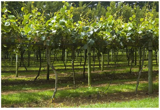
The plants of Actinidia are small tree climbing creepers (Fig. 1) that can reach a height of 8-10 m and require, for their bearing, supports for both trunk and branches.
Fig. 1 – Actinidia plants.
The root system has fleshy roots, branched and is rather superficial. The leaves are alternate, simple and petiolate, usually toothed, but sometimes with entire margins; shape and size are variable, but most commonly they are heart-shaped, leathery and hairy on the underside.
The fruiting takes place on a year old branches (shoots), coming from the previous year ones. The shoots growing in one season, come from axillary buds grown in the previous season and are protected, during the winter, by the cortical and cicatricial tissue of the previous year leaf.
The young shoots can be glabrous or tomentose, while the older branches are covered with a gray or brown bark that often has linear lenticels.
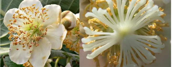
All plants belonging to this genus are dioecious with staminate and pistillate flowers brought by different individuals (Fig. 2). The plants with female flowers produce nonviable pollen, while the
6
male ones have a greatly reduced and underdeveloped ovary. The flowers are sometimes single, sometimes gathered in inflorescence (corymb) with thin, usually white, petals.
Fig. 2 – Male flower (left) and female flower (right) of kiwifruit plants.
The flower induction occurs during the spring, while the blossoming between May and July, depending on the variety and the place the orchard is located. Pollination, anemophilous or entomophilous, it is often difficult and must be helped with the shedding of pollen by artificial ventilation; flowers are in fact poorly attractive because they have no scent and, in some cases, of nectar.
To ensure adequate pollination normally a staminiferous individual is planted every 7-8 female individuals and it is important the presence of at least 8-10 bee hives per hectare.
Because there is a positive correlation between the number of seeds and the fruit size, on the flower stigmas are required at least 2,000 pollen grains (Pietropoli, 2004).
The fruit is an oval berry with a weight ranging from 40 to 150 g that has tomentose epicarp with soft or bristly green-brown trichomes. The mesocarp is fleshy and has many (more than 1,000 per fruit) and very small dark seeds, arranged around a white and edible heart, called “columella”. The flesh of the fruit is light green in A. deliciosa, while in A. chinensis there is a range of yellow, yellow-green or dark green colour (Ferguson, 1990; Testolin & Ferguson, 2009).
Being a sub-tropical plant, Actinidia requires appropriate temperatures, wide availability of water, high humidity of air and low summer temperatures. From the thermal point of view, the length of the growing season, which extends over 8-9 months, entails precise territorial limits, determined by anthesis-harvest period of about 160-180 days. This leads to exclude all areas where there is a high probability of autumn seasons with premature cold.
7
Planting density usually depends on the structures and the farming system adopted. The prevailing types of farming are pergola system (“tendone” in Italian), with 4x4 or 5x5 m planting layout, in southern areas, and T-bar system (“pergoletta” in Italian) in northern environments (Costa & Testolin, 1990; Dal Pane, 2002). The distance between rows should always be 4-5 m in order to ensure the passage of tractors, the one in the row varies in relation to soil fertility and vegetative vigor of the selected cultivar (Testolin et al., 1993).
This crop requires a medium exposure to lower temperatures (600-1200 h below +7°C) preferring a mild climate and to avoid excessive cold and heat. In fact, it appears to be very sensitive to cold in late winter and in early spring, albeit for short periods of time, below + 5-10°C. Adult plants with very woody branches and in complete dormancy better tolerate lower temperatures. Plants that have already satisfied the cold requirements are therefore more susceptible to outbreaks of winter frost. The high summer temperatures are usually not harmful, but are often associated with low humidity air, which must be corrected with irrigation (climatising) (Zuccherelli, 1994).
In fact, in this period, the leaves are particularly susceptible to burning and drying when the level of transpiration is very high (Xiloyannis et al., 1986).
The clear demonstration of the importance of irrigation practices is the development of specific irrigation techniques for this crop (No. drippers per plant, under foliage nebulisation, etc.) compared to other fruit tree crops in temperate climate (Xiloyannis et al., 1996).
The temperature stress can be aggravated by the wind, which causes a further reduction of air humidity leading to water deficit. The wind can also cause the breaking of branches, leaves and fruits rubbing them against pieces of wood or the support structure. It also disturbs the activity of the pollinating insects with negative effects on the fruit set (Zuccherelli, 1994).
To avoid problems of asphyxia and root rots, the soil should be dissolved and permeable, but also deep and rich in organic matter in order to ensure a sufficient supply of nutrients through the activity of the microbial flora (Marangoni et al., 2003).
Given the predisposition of Actinidia toward chlorosis phenomena related to iron deficiency, a sub-acidic reaction of soil (pH below 7.5) associated with a low content of active lime (CaCO3 < 5%) is to be preferred (Rombolà et al., 2000; Rombolà et al., 2003). Although the abundant vegetation of a kiwifruit orchard requires a high nutritional support, excessive nitrogen availability could be negative because it creates a micro-environment conducive to cryptogamic attacks and risks of poor lignification that could predispose to cold damage (Marangoni et al., 2003; Zuccherelli, 1994). The best soils for the planting of Actinidia is silty-sandy with permeable alluvial, deep, fertile, fresh subsoil and with a pH between 5.5 and 7.4 (Zuccherelli, 1994).
8 1.2 - Cultivars of Actinidia spp.
Italy began to cultivate Actinidia plants in 1971 and since then it has considerably increased the harvested area and, consequently, its production has became one of the most important in the world (Cacioppo, 2009).
The initial development of the Italian kiwifruit industry was based mainly on the cultivars of A.
deliciosa constituted in New Zealand, but currently also the cultivars belonging to A. chinensis play
an important commercial role.
The cultivars mostly cultivated that belong to A. deliciosa
Hayward
Moderately vigorous plant, later flowering than other selections, big size fruits with weight 90-150 g, green-brown tomentose epicarp, mesocarp light green in colour, good taste and exceptional preservation of fruits (Testolin & Ferguson, 2009);
Earligreen ®
Natural mutation of Hayward, anticipated harvest of 40-50 days compared to Hayward, all the fruits of a branch do not fill or ripen at the same time, fruits with reduced shelf life (Bucci & Costa, 2006);
Green Light ® Natural mutation of Hayward, fruits with similar characteristics to those of Hayward and that fill up in advance of 30-35 days (Spada & Spada, 2005);
Top Star® Less vigorous Hayward mutation, which therefore requires minimum summer pruning; Summer Kiwi® Precocious selection, fruits similar to those of Hayward but having a weight of 80-90 g,
short period of fruits storage;
Abbott Vigorous plant, precocious and productive, fruits of medium size;
Allison Vigorous and productive plant with later flowering compared to Abbott, fruits slightly larger than Abbott;
Bruno Productive and vigorous plant with flowering slightly delayed compared to Allison, large fruits of elongated-cylindrical shape;
Monty Very vigorous plant, very productive, sometimes excessively, oblong medium size fruits;
The four cultivars of A. chinensis
Hort16A
Vigorous plant that blooms a month before Hayward, the fruits are harvested about in the same period of Hayward, sweeter and aromatic flavour with low acidity, colour of flesh yellow-gold;
Gold3 (G3) New cultivar used to replace Hort 16A that showed great susceptibility to Kiwifruit Bacterial Canker, similar to Hort 16A;
Jintao
Less vigorous but more productive than Hayward, blooms a week before Hayward, fruits are harvested three weeks before, the flesh has a bright golden colour and a sweet taste, good preservation period but small fruits up to 90 g (Cipriani & Testolin, 2007); Soreli Fruits that ripen 30 days before Hayward, of large size, more than 100 g, flesh bright
yellow in colour, good flavour but short shelf-life (Fideghelli, 2012);
Another cultivar belonging to A. chinensis is the Chinese “Hong Yang” also known as red kiwifruit. More precisely, the flesh is two-tone green-yellow; the red colour is arranged radially around columella (Fideghelli, 2012; Testolin, 2014).
9 1.3 - Commercial aspects of Actinidia spp.
Due to the success that this species has obtained with consumers, in recent years the cultivation of kiwifruit has seen a sharp increase in many producing countries.
Data relating to 2010 showed that the harvested area of kiwifruit in the world was close to 160,000 ha with more than 70,000 ha concentrated in China, a country that in the last 20 years has given greater impulse to the increase of cultivation.
Excluding China, the areas planted with Kiwifruit in the 90s had decreased significantly, reaching around 54,000 ha; then the interest towards this crop is gradually increased to cover 98,656 ha and reaching 1,412,455 tonnes of product in 2012 (Faostat, 2015).
The kiwifruit production is highly concentrated and the top five producing countries are China with over 490,000 t (27% of the world total), Italy with approximately 384,844 t (24% of the total and 24,327 ha harvested), New Zealand with 376,400 t (20% of the total and 12,757 ha harvested), Chile with 240,000 t (10% of the total and 10,950 ha harvested) and Greece with 161,400 t (6% of the total and 7,300 ha harvested); followed at a distance by France with 65,253 t (3,952 ha harvested), Turkey with 36,781 t (28,500 ha harvested), Iran with 32,000 t (2,900 ha harvested), Japan with 28,000 t (2,300 ha harvested), the United States with 26,853 t (1,700 ha harvested), Portugal with 25,000 t (1,600 ha harvested), Spain 16,200 t (800 ha harvested) and South Korea with 10,600 t (820 ha harvested) (Faostat, 2015).
The Italian production is concentrated in few regions: Lazio, the Italian leader for the cultivation of Kiwifruit, with over 30% of the investments, Piedmont with about 20%, Emilia-Romagna with 16%, Veneto with 15% followed by Calabria, Campania and Friuli (Macchi, 2012; Faostat, 2015). Clearly, the attitude of a producer country is to satisfy consumer demand, increasing production and thus the supply; therefore it is necessary an extension of the sales calendar in respect of the consumers requirements.
In the EU Kiwifruit market Italy’s principal competitor is Greece, which has the same seasonality of production. The southern hemisphere countries, which have a complementary seasonality of production to Italy, do not represent particular problems for Italy’s export.
Chile’s export calendar of kiwifruits overlaps with the tail end of the Italian one and does not affect the Italian market. However, New Zealand’s export calendar clashes with the beginning of the Italian one reducing the marketing period of the Italian product.
At present, the main problem among Kiwifruit producing countries is the outbreak and spread of Bacterial canker of Actinidia, that since 2008 has heavily affected thousands of Kiwifruit hectares worldwide, determining numerous removals of harvested areas (Macchi, 2012).
10
2. - Diseases
Among the main reasons that have contributed to the success and, consequently to rapid expansion, of Actinidia orchards, not only in Italy but also in rest of the world, there is certainly the high economic convenience.
Its profitability is attributable not only to the productivity of the crop and to the price that the market confers to the fruits, but also to the sporadic phytosanitary management needed for its cultivation.
In general, fruit crops require several efforts with pesticides, in order to limit losses caused by pests. Until a few years ago the kiwifruit cultivation required rare phytosanitary treatments. However, today the “Bacterial Canker of Actinidia”, due to large economic loss caused, represents a tremendous parasitic problem that is still to be solved.
2.1 - Main diseases of Actinidia spp.
Between the most well-known Actinidia diseases there is the “wood decay”, a fungal disease with complex etiology that usually manifests in adult plants (8-10 years). It presents itself with visible symptoms on the leaf, more or less extensive necrotic areoles in the leaf surface. At the same time the wood attacked by different pathogens, is friable or necrotic mainly starting from pruning wounds distally located (Tosi et al., 2008).
Despite the infection progresses constantly in woody tissue, its manifestation is not progressive and constant impact over the years. Its control is related to the removal of diseased branches (Osti & Di Marco, 2011).
Other diseases of Actinidia spp. are caused by plant pathogenic bacteria such as Pseudomonas
syringae pv. syringae, agent of floral buds necrosis, and Pseudomonas viridiflava, agent of bacterial
blight. These were observed in Italy and recorded worldwide in the late 80s early 90s (Balestra & Varvaro, 1997; Varvaro et al., 1990).
P. s. pv. syringae is able to cause not only sporadic cankers, but also dark yellow-brown browning
of flowers and buds, followed by necrosis and subsequent drop of these organs. On leaves it causes necrosis of 1-2 mm in diameter, often surrounded by chlorotic halo. Furthermore, it is able to determine frost damages, especially during the period of vegetative resumption (Rossetti & Balestra, 2007; Rossetti et al., 2009).
P. viridiflava causes evident symptoms mainly at leaf level with hydropic spots of angular shape
that necrotize; it is also able to stop and/or alter the development of flowers and buds with subsequent fruit drop (Rossetti et al., 2009) and, as P. s. pv. syringae, it is able to cause frost damages in conjunction with sudden and brief changes in temperature (Varvaro & Fabi, 1992).
11
The bacterial populations (INA+ bacteria) responsible for damages and losses on Actinidia spp. plants are characterized by surviving at epiphytic level during different seasons, causing damages on different organs when there are unexpected decreases of temperature (Varvaro & Fabi, 1992).
2.2 - Bacterial Canker of Actinidia
The Bacterial canker of Actinidia is a vascular disease caused by a phytopathogenic bacterium,
Pseudomonas syringae pv. actinidiae (Psa) (Renzi et al., 2009).
Psa was observed for the first time, in 1984, in China in the Hunan province (Fang et al., 1990), but the pathogen was isolated and characterized for the first time in 1989 in Japan on A. deliciosa plants (Serizawa et al., 1989) and was subsequently classified with the nomenclature that we know today (Takikawa et al., 1989).
The disease was detected later in Korea (Koh et al., 1994; Koh et al., 2010) and in Italy (Scortichini, 1994).
Although present in different areas of the world, Psa was not cause of extensive damage until 2008 (Balestra et al., 2008). With its outbreak in Italy, a dramatic epidemic was caused by a new strain (haplotype) isolated from A. chinensis and A. deliciosa. This strain is much more aggressive than the previous one (Scortichini, 1994) and it is still affecting kiwifruit orchards all over the world. The same Psa strain was reported in 2010 in Portugal (Balestra et al., 2010), France (Vanneste et
al., 2011), New Zealand (Everett et al., 2011) and Turkey (Bastas & Karakaya, 2012). In 2011 it
was recorded in Australia (Biosecurity Australia, 2011), Spain (Balestra et al., 2011), Switzerland (EPPO Reporting Service, 2011) and Chile (EPPO Reporting Service, 2011). The presence of this pathogen has also been reported in 2013 it reached Germany (EPPO Reporting Service, 2013) and in 2014 in Slovenia (EPPO Reporting Service, 2014) and in Greece (Holeva et al., 2015).
The Psa strain isolated in Europe in 2008 (Balestra et al.,) and in New Zealand between 2010 and 2011 is very virulent and therefore it has been defined as Psa-V. This definition is based on its aggressiveness and the rapidity with which it is able to cause the disease. In New Zealand another Psa population described as less virulent has been detected. The strains belonging to this population are labelled Psa-LV (Psa-Low Virulent) and are not able to create cankers, but only foliar spots (Chapman et al., 2011).
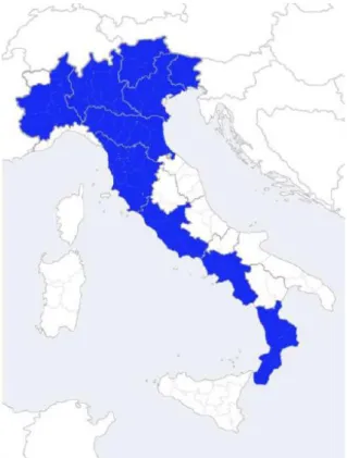
This disease is, currently, widespread in most Italian (Fig. 3) and world Kiwifruit areas (Fig. 4) and has assumed pandemic dimensions with an incidence that represents a serious risk for the future of the Actinidia cultivation and its whole business.
12
Fig. 3 – Current distribution of Psa in Italy
Fig. 4 – Current distribution of Psa in the world
13
Psa is active in the orchard at temperatures between 10 and 20 °C, in any case not exceeding 25 °C, at which temperature the bacterium remains latent within the host even if the external climatic conditions are adverse (Renzi et al., 2009).
During the autumn, the high relative humidity and temperatures that are stillmild, in association with the entry ways determined by harvest, leaf fall and high activity of stomata and lenticels, represent an opportunity of Psa establishment that can survive within the host (Renzi et al., 2012). Through winter pruning, Psa can penetrate and colonize the branches and begin its infectious cycle. In addition, frosts, hailstorms and heavy storms typical of this season are further risk factors for infections and reinfections (Mazzaglia et al., 2010).
At the beginning of the vegetative season, mild temperatures and relative humidity are very suitable for the multiplication of Psa, therefore, the pathogen is able to penetrate into the host through natural openings such as lenticels, stomata, natural wounds caused by emission of new shoots and leaves, or accidental wounds by wind and other meteorological events (Renzi et al., 2012).
During the hottest periods, with lower relative humidity, Psa reduces its biological activity, but hailstorms and heavy storms may contribute to the spread of the pathogen from infected plants to healthy ones, facilitating new infections.
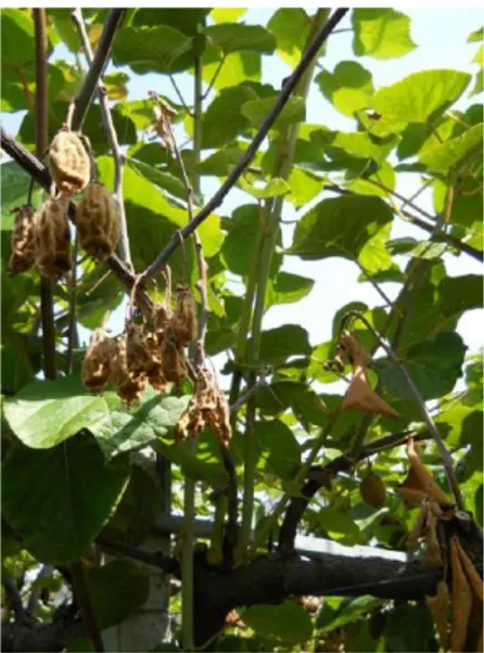
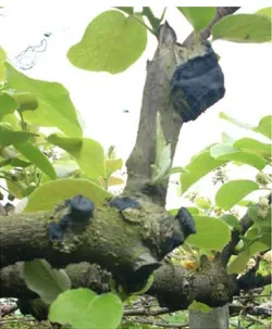
Spring and early summer infections are mainly characterized by necrotic angular leaf spots, surrounded by a yellowish halo (Fig. 5), which over time converge, resulting in a general wilting of the tissues and by cankers on trunks and branches almost always accompanied by discharge of a typical dark red or matt white exudate (Fig. 6), wilting of the shoots (Fig. 7) and browning of buds and flowers (Fig. 8). As far as the fruits are concerned, they are greatly wilted and often do not develop fully (Fig. 9).
Fig. 5 – Necrotic kiwifruit leaf spots with yellow halos caused by Psa.
14
Fig. 6 – Matt white (left) and with dark red exudates (right).
Fig. 7 – Wilting of kiwifruit shoots.
15
Fig. 9 – Wilting of kiwi fruits.
Differently, the autumn-winter infections present themselves with desiccation of leader canes and trunks, sometimes associated with discharge of exudate and cankers development (Rossetti et al., 2009; Renzi et al., 2009).
Moreover, Psa is able to colonize the root systems (Fig. 10) of plants affected by Bacterial Canker, proving the pathogen’s ability to infect organs that are far from entry sites (Mazzaglia et al., 2010; Renzi et al., 2012).
16 2.3 - Control of bacterial diseases
The conventional control means are not always sufficient to fight bacterial plant diseases because, to date, there are no effective chemical molecules commercially available. Therefore, the approach is preventive rather than curative.
In the establishment of ex-novo orchard is important to ensure that the propagation material purchased is certified, i.e. showing the conforming label to the technical standards of the Central Plant Protection Service in accordance with Article 49 paragraph 2 of Legislative Decree N° 214/2005, issued by the same institution, after carrying out the necessary inspections and made any necessary laboratory tests.
These measures, undertaken by Italy with the MD of February 7th 2011 and updated by the EU Commission Decision of December 5th2012, are necessary in order to prevent, control and eradicate the Bacterial canker of Actinidia caused by Psa.
The rules concerning the marketing of nursery productions are essential to prevent various diseases from short and long distance spread.
In young orchards (1-2 years) it is appropriate to proceed at eradication and immediate destruction of infected plants.
At present, the fight against Kiwifruit bacterial diseases and especially the Bacterial Canker is based on appropriate technical-agronomic practices such as frequent inspections, cleaning the orchard of infected portions, continuous disinfection of all tools and closing cuts larger than 2 cm in diameter with tar or ecological adhesive (Renzi et al., 2009) (Fig. 11).
17
The emergency measures/control for the Kiwifruit Bacterial Canker were also outlined by MIPAAF DM of February 7th2013, which indicates to cut the plant or remove its affected parts (the cut must be made at least 70 cm from the infected portion), to uproot the affected plants and to promptly proceed with their burning or deep burial.
Together with a good conduction of the orchard, other control means can be used.
The most important phytosanitary treatment is the constant use of copper (e.g. after harvest, leaf fall, winter pruning and before the new season).
Besides from being constant, the copper treatments must be timely especially after important meteorological events such as frosts and hailstorms (Fratarcangeli et al., 2010).
The copper ion (Cu++), adsorbed by bacteria in high doses, can modify the permeability of cell membranes substituting different metal ions (K+, H+, Ca++, Mg++), blocking the biosynthesis of enzymes and co-enzymes, interfering with breathing processes, blocking the redox processes and interfering with the membrane transport (Martelli, 1984; Pertot et al., 2005).
Moreover, due to its mechanism of multisite action, it is able to decreasing the risk to develop resistance phenomena (Brunelli & Palla, 2005).
However, for different populations of Pseudomonas spp., it was reported a potential development of copper resistance due to the inappropriate use of copper compounds (concentrations, nmb.and time of treatments, formulations used) (Vanneste et al., 2008). Copper could also determine phytotoxic effects on vegetal tissue, for this reason it is not used during the flowering phase. Because of its potential accumulation in soil, the European Union has imposed strict limitations for the use of copper (in organic farming) placing a maximum threshold of 6 Kg/ha per year or a total of 30 Kg/ha of copper in a period of five years (EU Regulation 473/2002) (Varvaro et al., 2001; Asinelli et al., 2006).
Another strategy to control bacterial pathogens is based on the use of antibiotics. Although it could be an efficient approach, the EU has banned their use in agriculture because they easily induce forms of resistance, with serious risks of cross-resistance between phytopathogenic bacteria and human pathogens (Vanneste et al., 2008; Ghosh et al., 2008). Resistance phenomena to antibiotics used for the control of PSA have been already recorded (Nakajima et al., 2002).
A further control strategy, which is under evaluation, is represented by a group of molecules known as resistance inducers. These are able to stimulate the plant to implement a series of actions that cause the so-called Systemic Acquired Resistance (SAR) and Local Acquired Resistance (LAR) phenomena. The best known of these molecules is the salicylic acid, a molecule involved in induced resistance that, interacting with catalase and ascorbate peroxidase, slows the catalysis of hydrogen peroxide, ROS (Reactive Oxygen Substance), key in the occurrence of hypersensitivity reaction.
18
Elicitors are compounds that stimulate different types of mechanisms, but in many instances with high levels of disease they proved to be much less effective. This lack of uniformity in results, together with some problems for plants development, limits their use (Walters et al., 2005).
Last, but not least, is the use of natural substances and natural antagonists. Both allow a good control of plant diseases in a preventive manner (Slusarenko et al., 2008). Appropriate technical-agronomic practices can minimize their critical aspects. In fact, while natural substances are subject to wash-out, natural antagonists may not remain sufficient in number because of their inadequate ability to substantially multiply on the plant, because of unsuitable environmental conditions, or because of the lack of nutrients.
The control adopted by these “organic strategies” is aimed at developing a biological barrier (antagonism) and to supply plants with a high level of natural resistance to infections; because of this, it is important the role played by environmental factors and the different strategies adopted by the phytobacteria (Mukerji & Garg, 1998; Pietrarelli et al., 2006; Slusarenko et al., 2008).
19
3 – Aim of work
The cultivation of Actinidia plants/cvs and related kiwi fruit production is of remarkable economical importance in Italy asin many Countries worldwide so, the Bacterial Canker of
Actinidia causes serious economic losses in many areas. For this reason, it seemed extremely
important to verify among qualitatively and quantitatively bacterial populations present into the sap of Actinidia spp. plants affected or not by Kiwifruit Bacterial Canker, how make a characterization of these microrganisms, verify different mechanisms of antagonism against Psa and, finally, check the spread of these potential BCA's inside Actinidia tissue.
20
4. - Bacterial populations in the sap of Actinidia spp.
4.1 - Materials and Methods
In this study two orchards of Actinidia spp. were investigated in Viterbo provinces in Lazio, a orchard situated in Montefiascone district, where the plant show typical symptoms of Psa, while in orchard situated in Viterbo (Azienda Agraria “Nello Lupori” of University of Tuscia) the plants did not show symptoms of disease. During the vegetative season, in March, April and May, was taken the sap from 10 plants of A. chinensis cv. Jintao and 10 plant of A. deliciosa cv. Hayward in symptomatic orchard and 5 plants of A. chinensis cv. Jintao and 5 plant of A. deliciosa cv. Hayward in asymptomatic orchard.
4.1.1 - Sap collection
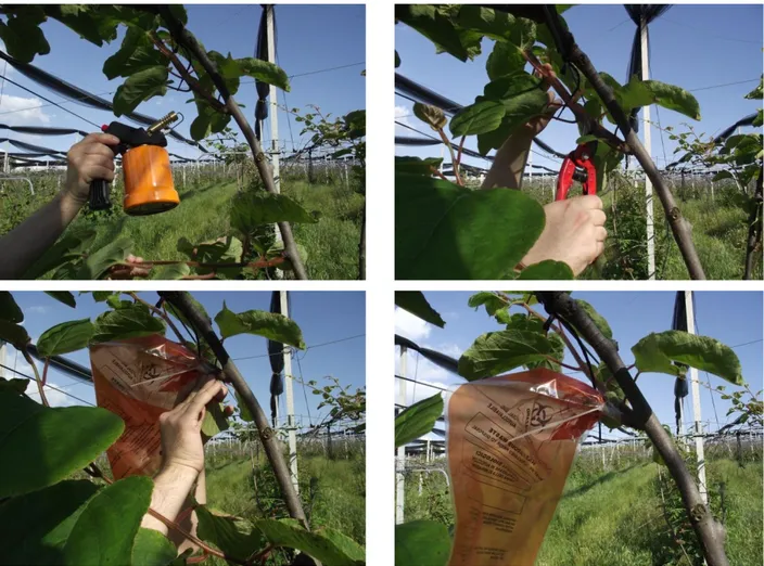
The sampling was performed by minimizing the external contamination, using an alcoholic solution and heat. The cutting area was previously cleaned with an alcohol solution and with flame, the cutting was performed with shears previously sterilized, always with alcoholic solution and flame. Once cut the branch was immediately inserted a sterile bag that was tied to the branch cut, closed with a string of plastic and parafilm. The bagging operation has always been held in the late afternoon, while taking in the early morning hours (Fig. 12).
21
Fig. 12 – different steps of the kiwifruit sap collection in the field.
4.1.2 - Bacterial populations into the sap
The sap taken in the field was plated on Petri dishes of two different culture media, NSA and semi-selective medium for Pseudomonadaceae.
Below shows the compositions of the two medium used
NSA (1 liter)
Lab-Lemco broth OXOID 8 g
Sucrose VWR 50 g
22
Semi-selective medium (1 liter)
Glycerol Bi-distilled 99.5% VWR 9 g
Pseudomonas Agar f MERCK 34.2 g
Technical Agar № 3 OXOID 8.10 g
The 1.5% solution of boric acid 100 ml
Solution of cycloheximide in ethanol at 2.5% 8 ml
Solution of cephalexin in water at 1% 8 ml
Deionized water 900 ml
The medium, was sterilized by autoclaving at 121 °C for 20 minutes.
In the meantime were added, by sterile filters, 4 ml of cycloheximide solution and 4 ml cephalexin solution, respectively in 50 ml of boric acid solution. The latter was then added to the medium previously cooled up to 50 °C.
Under sterile conditions, 18 ml of medium were poured per Petri dish. At occurred solidification the plates were placed for 24 h, at a temperature of 26±1 °C and then stored at 4 °C in sterile bags.
100 µl of sap were plates on both culture media, subsequently incubated at a temperature of 26±1 °C, after 24 and 48 hours were counted colonies divided by morphotypes.
4.1.3 - Molecular identification
Extraction of DNA
The genomic DNA of each isolate was extracted from individual colonies grown for 48 hours at 28 °C on KB medium. Approximately 2x10⁹cells were used and their DNA was extracted using the PureLinkTM Genomic DNA Kit (Invitrogen, USA) following the manufacturer’s specifications for the Gram- and Gram+ bacteria. DNA was then resuspended in TE-buffer (10 mM Tris-HCl, 1 mM EDTA pH 8.0). DNA was then stored at 4 °C (or -20 °C for longer storage times) until required. - Amplification by PCR (16S and ITS rDNA):
Amplification of the 16S and ITS ribosomal DNA gene sequence was performed by PCR (Polymerase Chain Reaction) using universal primers specific for bacteria designed on the sequence of interest:
23 16S 27F (forward) 5’-AGAGTTTGATCCTGGCTCAG-3’; 1492R (reverse) 5’-GGTTACCTTGTTACGACTT-3’. ITS (forward) 5’-AGCCGTAGGGGAACCTGCGG-3’; (reverse) 5’-TGACTGCCAAGGCATCCACC-3’.
In order to optimize the working times and to standardize the experiment, the PCR analysis was conducted using the GoTaq Colorless MasterMix (Promega Corporation, WI).
For each isolate a reaction solution was prepared with: 2X Go Taq MasterMix (2X Reaction Buffer, 1.25 units of Taq DNA Polymerase, 30 mM Tris-HCl pH 8.3, 125 mM KCl, 3.75 mM MgCl2, 500 μM for each dNTP), 1 μl of each primer (200 nM), between 25-35 ng of genomic DNA (template DNA) and SDW for a final volume of 25 μl. For each experiment a negative control was used, i.e. in the reaction solution the template DNA was replaced with SDW.
The conditions of amplification, which was performed in a C1000 thermal cycler (Bio-Rad Laboratories srl, Milan, Italy), was characterized by an initial stage of 12 min. at 94 °C (initial denaturation of template DNA and activation of Taq polymerase), a 30 cycles intermediate stage with a denaturation step at 94 °C for 30s, annealing at 50 °C for 45s and extension at 72 °C for 1.5 min., and a final extension stage at 72 °C for 12 min.
Amplicon quantification
The concentration of the amplified DNA, and its quality, were determined for the subsequent sequencing. For each sample, the amplified product was separated by horizontal electrophoresis on agarose gel 1% (10 g/l) in 1X TBE (89 mM Tris, 89 mM boric acid and 2 mM EDTA). The UV detection was carried out, after electrophoresis, by staining with GelRedTM (Bioticum Inc., CA). Each experiment was repeated at least twice to confirm the reproducibility of the profiles obtained. The concentration was estimated with the help of an optical fluorimeter (QubitTM, Invitrogen, Life Tecknologies Italy, Monza), suitably calibrated and eventually adjusted to a final concentration of 25 ng/μl.
Sequencing and sequence analysis
24
In order to determine their approximate taxonomic placement, the sequences, in the form of electropherograms, were edited manually and subsequently compared with those available in the GenBank database using BLAST (Basic Local Alignment Search Tool) in the NCBI website (Natiolal Center for Biotechnology Information). In cases of homology greater than 99% it was possible to attribute to the isolated its taxonomic allocation.
4.2 - Results
4.2.1 - Bacterial populations into the sap
Select them in the laboratory have been counted and the different bacterial colonies from the two different culture media used (NSA and Semi-selective), then were put in purity of King'B where it was possible to distinguish them according to morphology. In 2013 were isolated 34 morphotypes, while in 2014 27 morphotypes.
4.2.2 - Molecular identification
The comparative analysis in GenBank of the profiles obtained from the sequencing of the bacteria of interest showed that by 34 morphotypes of 2013 we moved to 18 bacterial isolates and 27 morphotypes of 2014 the number grew to 12 bacterial isolates, belong to the following species:
2013 2014
1 Bacillus megaterium 1 Paenibacillus sp.
2 Duganella zoogloeoides 2 Bacillus megaterium
3 Erwinina billingiae 3 Pseudomonas fluorescens
4 Leuconostoc mesenteroides 4 Stenotrophomonas sp.
5 Paenibacillus sp. Pa16 5 Pseudomonas sp. P 12
6 Paenibacillus sp. Pa 8 6 Pseudomonas NBRC 14077
7 Pseudomonas fluorescens 7 Agrobacterium tumefaciens
8 Pseudomonas sp. P 11 8 Pseudomonas sp. P 20
9 Pseudomonas sp. P 18 9 P. syringae pv. syringae
10 Pseudomonas sp. P 19 10 Pseudomonas sp. P 25
11 Pseudomonas sp. P 26, 28, 30, 31 11 Stenotrophomonas rhizophila
12 Pseudomonas syringae NBRC 14077 12 Pseudomonas syringae pv. actinidiae
13 Pseudomonas syringae pv actinidiae
14 Pseudomonas syringae pv syringae UB246
15 Pseudomonas syringae pv. syringae CC 457
16 Rahnella aquatilis
17 Sphingomonas sp
25
Being there a high homology of the sequences (99-100%) with those present in the NCBI international database, it has been possible to assign isolates to different species with reasonable certainty.
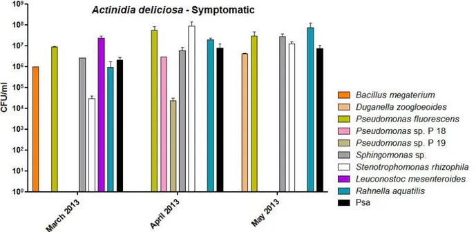
Below are the graphs of bacterial populations present in the sap of A. deliciosa and A. chinensis of 2013 (Fig. 13, 14, 15 and 16) and 2014 (Fig. 17, 18, 19 and 20).
Fig. 13 – Bacterial population into the sap of A. deliciosa symptomatic plants in 2013.
26
Fig. 15 – Bacterial population into the sap of A. chinensis symptomatic plants during 2013.
27
Fig. 17 – Bacterial population into the sap of A. deliciosa plants symptomatic in 2014
28
Fig. 19 – Bacterial population into the sap of A. chinensis symptomatic plants during 2014.
29
5. - Characterization of bacterial populations
5.1 - Materials and methods.
5.1.1 - Production of fluorescent pigments on King’s B
Fluorescein is a fluorescent pigment (from green to blue colours) visible under ultraviolet rays (λ365 nm). To perform this test, it is necessary to allow the growth of bacteria on agar medium King’s B (KB) (King et al., 1954), so composed per litre of distilled water:
Peptone 20,00 g
Glycerol Bidistilled 99.5% VWR 12.6 g
Potassium Phosphate 1,50 g
Magnesium Sulphate 1,50 g
Bacteriological Agar №1 OXOID 18,00 g
The medium was prepared with the same methodology previously described (see § 5.2.1).
A strain of Agrobacterium tumefaciens (AT 628) and a strain of Pseudomonas fluorescens (NCPPB 3008) were used as negative and positive control respectively., The plates were observed 24, 48 and 72 hours after being placed in the incubator at 26±1 °C, using a UV lamp at 365 nm (Sylvania Blacklite Blue Tubes F 15T8-BLB).
5.1.2 - Levan production
Due to the action of the enzyme Levan Saccharase, many bacterial species are able to split sucrose into two simple sugars that constitute it: glucose and fructose. While Glucose is used as carbon source, Fructose is polymerized in an extracellular polysaccharidic form, called Levan, which is a polymer of β-D-Fructose. Levan colonies are characterized by whitish dome-shaped mucous (Klement et al., 1990).
To verify this capacity for the selected natural bacterial antagonists aliquots of their culture were streaked, through a sterile loop for bacteriology, in Petri dishes with nutrient agar medium containing 5% Sucrose (NAS) and having the following formula per litre of distilled water:
30
Lab-Lemco broth OXOID 8 g
Sucrose VWR 50 g
Tecnical Agar n.3 OXOID 15 g
The NAS medium was prepared with the same methodology previously described. The plates were observed after 24 hours of incubation at 26±1 °C.
5.1.3 - Solubility assay in KOH
A good alternative to Gram staining is represented by the KOH solubility assay; for which a drop of 3% KOH solution in sterile distilled water is placed on a glass slide.
Then, with a sterile loop, an aliquot of the bacterial culture is streaked on the glass slide to obtain a homogeneous mixture. If the latter appears to be viscous and filamentous, bacterium under investigation are Gram-, otherwise they are Gram+. This is due to the ability of KOH to degrade only the cell wall of Gram- bacteria but not the one of Gram+ bacteria (Goszczynska et al., 2000).
5.1.4 - Tobacco hypersensitivity
The immediate response of a plant to a pathogenic infection with the targeted death of the invaded tissues is called hypersensitivity reaction and it is an active defence mechanism by which the infective agent is confined to a limited necrotic area (Klement, 1982).
A bacterial suspension 1x10⁸ CFU/ml of each selected isolate, was injected into the leaf parenchyma of tobacco plants cv. Virginia Bright at the fourth-fifth true leaf stage (Klement, 1963) using a sterile insulin syringe having needle 0,3 x 13 mm. After 24 hours it was possible to observe, in the inoculated portions of the leaf, the presence or absence of necrotic areas. The negative control used was sterile water, while the positive one was Psa (CFPB 7286).
31 5.2 - Results
The following table shows the results of the various tests carried out in 2013 and 2014:
2013
Bacterial strains Fluor. Levan Gram HR
Bacillus megaterium - + + - Duganella zoogloeoides - + - - Erwinina billingiae - - - - Leuconostoc mesenteroides - + + - Paenibacillus sp.Pa16 - - + - Paenibacillus sp.Pa8 - + + - Pseudomonas fluorescens I + +/- - - Pseudomonas sp. P11 - - - - Pseudomonas sp. P18 - + - - Pseudomonas sp. P19 - + - - Pseudomonas syringae + - - - Pseudomonas syringae NBRC 14077 + - - -
Pseudomonas syringae pv. syringae CC 457 + - - -
Pseudomonas syringae pv. syringae UB246 - - - -
Rahnella aquatilis - + - -
Sphingomonas sp. - - - -
Stenotrophomonas rhizophyla - - - -
Pseudomonas syringae pv. actinidiae - - - +
2014
Bacterial strains Fluor. Levan Gram HR
Paenibacillus sp. - - + - Bacillus megaterium - + + - Pseudomonas fluorescens II + +/- - - Stenotrophomonas sp. - - - - Pseudomonas sp. P11 + + - - Pseudomonas NBRC 14077 + - - - Agrobacterium tumefaciens - + + - Pseudomonas sp. P20 + + - - P. syringae pv. syringae + - - - Pseudomonas sp. P25 + + - - Stenotrophomonas rhizophila - - - -
32
6. - Antagonism: spot tests
6.1 - Materials and methods
6.1.1 - Antagonism: spot tests
For this test it was used the medium King B. The composition of the medium per liter of sterile distilled water was as follow:
Peptone 20,00 g
Glycerol Bidistilled 99.5% VWR 12.6 g
Potassium Phosphate 1,50 g
Magnesium Sulphate 1,50 g
Bacteriological Agar №1 OXOID 18,00 g
The medium was sterilized in the autoclave at 121 °C for 20 minutes. After cooling down the medium to 50 °C, and under sterile conditions, 18 ml of it were poured in each Petri dish.
A fresh culture (24 h old) of CFPB 7286 strain was diluted in 6 ml of sterile distilled water (SDW) and centrifuged at 12,000 rpm for 10 minutes at 8 °C (Vidaver & Buckner, 1977).
Subsequently, once the supernatant was removed, the centrifuged was resuspended in 5 ml of SDW. The optical density at 600 nm was read using a turbidimeter (21 Spetronic Mylton Roy Company®), and through appropriate dilutions it was adjusted to a value of 0.13, corresponding to a concentration of 1x10⁸colony forming units per millilitre (CFU/ml) (Varvaro & Surico, 1978). The concentration of 1x10⁶ CFU/ml was obtained through decimal dilutions. 100 µl of bacterial suspension 1x10⁶CFU/ml were plated on Petri dish.
Subsequently were added four spots of bacterial suspension (10 µl) of isolates to be tested at the concentration of 1x10⁸CFU/ml.
After adding the antagonist Petri dishes were left to incubate in an incubator at 26±1 °C for 24-48 hours.
6.2 - Results
For a better evaluation of the antagonistic capacity were assigned classes that represent the range of zones of inhibition of the pathogen and the culture of the antagonist (Fig. 21):
33 Classes of antagonism - 0 mm + between 0 and 2 mm ++ between 2 and 3 mm +++ more than 3 mm
The following table shows the results of spot test:
Bacterial strains Ant. Bacterial strains Ant.
Bacillus megaterium - Rahnella aquatilis -
Duganella zoogloeoides - Sphingomonas sp. -
Erwinina billingiae - Stenotrophomonas rhizophyla -
Leuconostoc mesenteroides - Pseudomonas fluorescens II ++
Paenibacillus sp. Pa16 - Stenotrophomonas sp. -
Paenibacillus sp. Pa8 - Pseudomonas sp. P11 -
Pseudomonas fluorescens I + Pseudomonas NBRC 14077 -
Pseudomonas sp. P11 - Pseudomonas sp. P13 -
Pseudomonas sp. P18 - Agrobacterium tumefaciens -
Pseudomonas sp. P19 - Pseudomonas sp. P20 -
Pseudomonas syringae + P. syringae pv. syringae -
Pseudomonas syringae NBRC 14077 - Pseudomonas sp. P25 +++
P. syringae pv. syringae CC 457 + Stenotrophomonas rhizophila -
P. syringae pv. syringae UB246 - P. s. pv. actinidiae -
Fig. 21 – Left, an example of assay of the spots, right, particular of the inhibition’ halos which is measured to determine the degree of antagonism.
34
In particular, tin the table below are reported the measurements of the inhibition’ halosof the isolates that showed antagonistic activity.
Bacterial strains active against Psa Inhibition’ halo in mm
Pseudomonas fluorescens I 1,92 ±
Pseudomonas syringae 1,52 ±
Pseudomonas syringae pv. syringae CC 457 1,51 ±
Pseudomonas fluorescens II 2,87 ±
35
7. Antagonism: broth Minimal Medium test
Seen the results obtained from the test spot we proceeded with the select isolates Pseudomonas
fluorescens II and Pseudomonas sp. P 25 to deepen the antagonistic activity.
7.1 - Materials and methods
7.1.1 - Minimal medium
All of the minimal media developed contained the following components:
K2HPO4 4.25 ml (1M) KH2PO4 45.75 ml (1M) (NH4)2SO4 1 g MgCl2 Hexahydrate 0.35 g NaCl 0.1 g Fructose OXOID 1.8 g Mannitol OXOID 1.8 g
The salts were dissolved in 900 ml of sterile deionized water and autoclaved at 120 ° C for 20 minutes. Once sterilized the mixture was put in an incubator thermostated at 50 °C. While the two sugars were dissolved in 100 ml of sterile deionized water and filtered with filters 0,22 µm, and built-in salt, previously autoclaved.
7.1.2 - Inoculum preparation
A fresh culture (24 h old) of two isolated, Pseudomonas fluorescens and Pseudomonas sp. P 25 that showed increased antagonistic activity and of CFPB 7286 strain was diluted in 6 ml of sterile distilled water (SDW) and centrifuged at 12,000 rpm for 10 minutes at 8 °C (Vidaver & Buckner, 1977).
Subsequently, once the supernatant was removed, the centrifuged was resuspended in 5 ml of SDW. The optical density at 600 nm was read using a turbidimeter (21 Spetronic Mylton Roy Company®), and through appropriate dilutions it was adjusted to a value of 0.13, corresponding to a concentration of 1x10⁸colony forming units per millilitere (CFU/ml) (Varvaro & Surico, 1978). In the end the sample was centrifuged and resuspended in 5 ml of minimal medium.
36
Antagonists and pathogen isolates were grown alone and in combination (Pseudomonas fluorescens II Vs CFPB 7286 and Pseudomonas sp. P 25 Vs CFPB 7286) in minimal medium broth.
The reading of the data was performed at 0, 6, 12, 18, 24, 48 and 72 hours.
7.2 - Results
Shown below are the graphs of the growth of individual bacterial isolates in purity (Fig. 22, 23 and 24):
37
Fig. 23 – Bacterial growth of isolate Pseudomonas sp. P 25.
Fig. 24 – Bacterial growth of isolate Psa II.
Simultaneous a reading was performed on bacterial growth of the antagonists with the pathogen. Discrimination between the two different isolates was carried out by colony morphology (Fig. 25, 26 and 27).
Fig. 25 – Left, entire plate with the presence of colonies of colonies of the pathogen and antagonist; right, particular of two bacterial colonies with different morphology (A PSA; B Antagonist).
A B
38
Fig. 26 – Bacterial growth of isolate Pseudomonas fluorescens II Vs Psa II.
39
8. - Antagonism: filtration spot
8.1 – Materials and methods
8.1.1 - Inoculum preparation
A fresh culture (24 h old) of two isolted, Pseudomonas fluorescens and Pseudomonas sp. P 25 that showed increased antagonistic activity and of CFPB 7286 strain was diluted in 6 ml of sterile distilled water (SDW) and centrifuged at 12,000 rpm for 10 minutes at 8 °C (Vidaver & Buckner, 1977).
Subsequently, once the supernatant was removed, the centrifuged was resuspended in 5 ml of SDW. The optical density at 600 nm was read using a turbidimeter (21 Spetronic Mylton Roy Company®), and through appropriate dilutions it was adjusted to a value of 0.13, corresponding to a concentration of 1x10⁸colony forming units per millilitre (CFU/ml) (Varvaro & Surico, 1978). Antagonists and pathogen isolates were grown alone and in combination (Pseudomonas fluorescens II Vs CFPB 7286 and Pseudomonas sp. P 25 Vs CFPB 7286) in minimal medium broth.
Aliquot of broth minimal medium was taken each 0, 6, 12, 18, 24, 48 and 72 hours and filtered to 0.22 nm to completely eliminate the bacterial cells.
8.1.2 – Biological activity of Pseudomonas fluorescens II and Pseudomonas sp. P25 culture filtrate
To test the antimicrobial activity of Pseudomonas fluorescens and Pseudomonas sp. P 25 filtrates, bacterial strains were grown overnight in minimal medium broth at different time (0, 6, 12, 18, 24, 48 and 72 hours). A 300 μl aliquot of the filtrate culture was spread onto the surface of a King B plate (100 × 15 mm, containing 25 mL of medium). After spreading the bacterial lawn, central wells were punched in the agar with a no. 9 cork borer, and a 300-μL aliquot of each, Pseudomonas
fluorescens and Pseudomonas sp. P 25 culture filtrates was dispensed into the well. The plates were
incubated for 48 h at 28°C, examined, and scored.
8.1.3 - Ethanol extraction of cultur filtrates
Measured volumes of Pseudomonas fluorescens II and Pseudomonas sp. P25 culture filtrates to which it was added 95% ethanol with 1: 4 ratio and centrifuged. The pellet obtained was resuspended in SDW, so as to obtain a concentration 20x compared to the initial. The supernatant was taken to dryness in vacuo at a temperature ≤ 45°C. After evaporation, the dry solids were
40
extracted three times (5 min per extraction) with 90% or 85% (v/v) ethanol as indicated. Each of the three extractions was performed by swirling the solids with a volume of ethanol solution equal to one-third of the original volume of culture filtrate. The three extracts prepared in this manner were combined, taken to dryness in vacuo as described above, dissolved volume of 76% (v/v) ethanol equal to one-twentieth of the original volume of culture filtrate (20× concentration), and stored at 4°C for later use.
The filtrate of bacterial growth which has been centrifuged led to separation of protein fractions from non-protein. These two fractions were tested again according to the assay of the spread in the culture medium. also in this case it was used the King B.
8.2 - Results
8.2.1 – Biological activity of Pseudomonas fluorescens II and Pseudomonas sp. P25 culture filtrate
The antimicrobial proprieties of Pseudomonas fluorescens II and Pseudomonas sp. P25 culture filtrates were tested against Psa cultured. The strains tested in our standard agar diffusion assay to the filtrate as evidenced by a large zone of clearing about the central well containing the
Pseudomonas fluorescens II Vs Psa and Pseudomonas sp. P25 Vs Psa culture filtrate.
Strains Presence of the inhibition halo
Pseudomonas fluorescens II No
Pseudomonas sp. P 25 No
Psa (CFPB 7286) No
Pseudomonas fluorescens II Vs Psa (CFPB 7286) Yes
Pseudomonas sp. P 25 Vs Psa (CFPB 7286) Yes
Here are pictures of the plates with different growth times where it was observed the zone of inhibition as a result of spreading the culture medium (Fig. 28 and 29).
41
0 h 6 h 12 h 18 h
24 h 48 h 72 h
Fig. 28 - Inoculated with PSA and wise filtrates of Pseudomonas fluorescens II and especially the halo of inhibition near the well.
0 h 6 h 12 h 18 h
24 h 48 h 72 h
Fig. 29 - Inoculated with PSA and wise filtrates of Pseudomonas sp. P25 and especially the halo of inhibition near the well.
8.2.2 - Ethanol extraction of culture filtrates
The diffusion test of culture medium of the villages (protein and non-protein) showed the following results (Fig. 30 and 31):
42
Protein fraction Non protein fraction Fig. 30 - Diffusion test with protein fraction and protein isolate of
Pseudomonas fluorescens II Vs Psa.
Protein fraction Non protein fraction Fig. 31 - Diffusion test with protein fraction and protein isolate of
43
9. – Spread on Actinidia spp. leaves of Antagonists
9.1 - Materials and methods
A fresh cultures (24 h old) of Pseudomonas fluorescens II and Pseudomonas sp. P25 strains were diluted in 6 ml of sterile distilled water (SDW) and centrifuged at 12,000 rpm for 10 minutes at 8 °C (Vidaver & Buckner, 1977).
Subsequently, once the supernatant was removed, the centrifuged was resuspended in 5 ml of SDW. The optical density at 600 nm was read using a turbidimeter (21 Spetronic Mylton Roy Company®), and through appropriate dilutions it was adjusted to a value of 0.13, corresponding to a concentration of 1x10⁸colony forming units per millilitre (CFU/ml) (Varvaro & Surico, 1978). The bacterial suspension was placed in glass tubes of 10 ml and used as liquid growth for leaves of
Actinidia spp.
The plant material of Actinidia spp. was collected from plants cultivated in the farm of the University of Tuscia, as potted seedlings of about a year old.
When taken the leaves with the stalk from the plant has been planted in the tubes with the bacterial suspension. Detection of the spread of the isolates in the plant tissue was performed after 7 days of planting the test. from the leaf surface they were identified six different points (Fig. 32). The leaves were sterilized with sodium hypochlorite (2.5%) for 30 seconds and thoroughly rinsed in sterile deionized water. At each point it was performed a hole 10 mm in diameter, which was homogenized and an aliquot of 100 µl was plating onto culture medium King B.
Fig. 32 - Six sampling points of the plant tissue that has been analyzed.
1
2
3
4
44 9.2 - Results
Record (CFU/cm2) of bacterial antagonists recorded in different leaf areas. samples
CFU/cm2
Pseudomonas fluorescens II Pseudomonas sp. P25
1 3.0 x 10³ 4.5 x 10² 2 0 0 3 0 0 4 0 0 5 0 0 6 0 0
45
10. Discussion
The control of plant pathogenic bacteria is particularly complex due to present absence of curative pesticides as well as the progressively EU restrictions on the use of copper salts and the prohibition of the use of antibiotics. .
The lack of effective alternatives constituted the trigger to expand on topics concerning the study and to research potential natural antagonists.
These appear to be very interesting in the control of bacterial diseases, however, further investigation is required, especially regarding the control by sustainable strategies of Psa.
A key aspect to understand the ecology of the plant pathogen Psa is to know the relationships with the bacterial populations with which it shares the living space. This study shows that most of the bacterial populations living within the sap of Actinidia spp. are environmental bacteria, naturally present in rainwater and soil.
Is it important to remark that by the present study, in the sap samples taken from symptomatic kiwifruit plants, was always possible to isolate the pathogen while, Psa cells were never isolated in the sap samples from asymptomatic plants of different Actinidia spp./cvs.
Since the different bacterial populations isolated share the same space is crucial to have a characterization of different strains to understand how it is possible to live together in the same biological niche.
The identification of possible natural antagonists/biocontrol agents (BCA’s) present in the same niche growth of the pathogen is of great importance given that it is of appropriate microorganisms to live in the same environments of the pathogen.
To understand and to evaluate the effectiveness of a natural antagonist, the potential biocontrol agents, characterized by the necessary safety features for its use in relation to human health and to the environment, was tested.
In consideration of the fact that the effectiveness of a biocontrol agent is measured, not only by its ability to produce antibiotic substances, but also by its degree of competition for nutrient sources and spaces, the isolation of BCAs in the same ecological niche of Psa seemed to be the best choice (Völksch & May, 2001); in this study that there are two antagonistic strains present in the sap:
Pseudomonas fluorescens II and Pseudomonas sp. P25.
It is known, from the bibliography, which bacteria use a polysaccharide matrix, the biofilm, for various reasons among which the defense, communication and the production of substances secondary (se è known qui devi mettere dei rifi biblio !!!). Investigating the production of this
46
matrix it is seen that the pathogen Psa does not produce biofilm, while the strains Pseudomonas
fluorescens II and Pseudomonas sp. P25 are strong producers of biofilm (idem rif biblo !!!).
Screening initial endowment has selected the bulk of five bacterial strains that showed antagonistic activity but, only two of them showed proper activities so that they were selected to be furtherly studied.
The two strains selected, Pseudomonas fluorescens II and Pseudomonas sp. P25, showed both an excellent antagonistic activity vs Psa. Among different times in broth minimal medium
Pseudomonas fluorescens II strain shown its action after 48 hours while, Pseudomonas sp. P25,has
cleared the growth of the pathogen after 12 hours.
Strains antagonists exert their function through the production of antibiotic substances, this is confirmed by the fact that the filtrate of bacterial growth also has antagonistic activity against Psa. In the specific case, the substances are produced only in the case in which the bacterial strain is in the same environment of the pathogen, seen that the filtrate of each BCA’s strain grown in purity do not show antagonistic activity.
Testing extraction with ethanol shows that the antagonistic activity is attributable to the non-protein fraction of the filtered BCA’s.. This production could be linked and supported, as suggested by previous studies (James et al., 1986), in relation to the glucose regulation.
Moreover, it was tested the ability of BCA’s to spread in the leaf tissue of Actinidia;the present results shown how the leaf proximal area (petiole) is the only one to be colonized. It should be noted that the stalk dipped in a bacterial suspension and SDW is unable to keep them vital more than seven days, and this implies a limited time to allow to these bacterial strains to spread. Then, in natural conditions (open field) the spread might be greater than that reported here. In addition to these aspects, the development of appropriate formulations by these BCA’s require further studies to exalt their properties as the capacity to effectively contrast Psa inside Actinidia plants.
The potentiality discovered of the selected BCA’s point out a great capacity to utilize them in a quite short time. The need to develop sustainable and also organic plant protection strategies against Psa/kiwifruit bacterial canker is a worldwide request and the present study offer new and original opportunity to continue inside this research line.
Different BCA’s formulations and related field trials have been already planned. Morevoer, taking into account what in the world was already applied until now against this bacterial plant pathogen for kiwifruit plants and the results also recently discovered (in NZ after the authorization during 2011 for the use of the antibiotics against Psa, were now isolated in kiwifruit orchards affected by bacterial canker disease, strains of Psa antibiotic-resistant.
http://phys.org/news/2013-02-chinese-47
nz-psa-outbreak.html), result of particolar importance to develop sustainable plant protection strategies to control Psa.
48
11. - References
Asinelli A., Mazzocchi C. Tellarini S. (2005). “I prodotti a base di rame in agricoltura biologica. Aspetti normativi relativi all’uso dei composti rameici: la situazione comunitaria ed italiana”. www.agrimoderna.it/biblioteca.
Balestra G.M., Mazzaglia A., Quartucci A., Spinelli R., Graziani S., Rossetti A. (2008). “Cancro Batterico su Actinidia chinensis”. Informatore Agrario, N° 38, 75-78.
Balestra G.M., Renzi M., Mazzaglia A. (2010). “First report of bacterial canker of Actinidia
deliciosa caused by Pseudomonas syringae pv. actinidiae in Portugal”. New Disease Report, N° 22,
10.
Balestra G.M., Renzi M., Mazzaglia A. (2011). “First report of Pseudomonas syringae pv.
actinidiae on kiwifruit plants in Spain”. New Disease Report, N° 24, 10.
Balestra G.M., Varvaro L. (1997). “Pseudomonas syringae pv. syringae causal agent of disease on floral buds of Actinidia deliciosa (A. Chev)”. Liang et Ferguson in Italy. Journal of
Phytopathology, Vol. 145, 375-378.
Bastas K.K., Karakaya A. (2012). “First report of bacterial canker of kiwifruit caused by Pseudomonas syringae pv actinidiae in Turkey”. Plant Disease, N° 96, 452.
Biosecurity Austalia (2011). “Final Pest Risk Analysis Report for: Pseudomonas syringae pv.
actinidiae Associated whit Actinidia (kiwifruit) Propagative Material”. Department of Agriculture,
Fisheries and Foresty. Canberra, Australia.
Brunelli A. Palla O. (2005). “Evoluzioni dei fungicidi rameici ed aspetti fitoiatrici”. www.agrimoderna.it/biblioteca.
Bucci V., Costa G. (2006). “Kiwi, è divenuto più ampio il panorama varietale”. Agricoltura