Biological Characterization and in Vivo Assessment of the Activity of
a New Synthetic Macrocyclic Antifungal Compound
Davide Deodato,
†,×Giorgio Maccari,
†,×Filomena De Luca,
‡Stefania Sanfilippo,
†Alexandru Casian,
†Riccardo Martini,
†Silvia D’Arezzo,
§Carlo Bonchi,
∥Francesca Bugli,
⊥Brunella Posteraro,
#Patrick Vandeputte,
∇Dominique Sanglard,
∇Jean-Denis Docquier,
‡,×Maurizio Sanguinetti,
⊥,#Paolo Visca,
∥and Maurizio Botta
*
,†,⊗,׆
Department of Biotechnology Chemistry and Pharmacy, University of Siena, I-53100 Siena, Italy
‡Department of Medical Biotechnology, University of Siena, I-53100 Siena, Italy
§
Istituto Nazionale per le Malattie Infettive
“Lazzaro Spallanzani”, I-00149 Roma, Italy
∥Dipartimento di Scienze, Universita
̀ Roma Tre, I-00154 Roma, Italy
⊥
Institute of Microbiology, Universita
̀ Cattolica del Sacro Cuore, I-00168 Roma, Italy
#Institute of Public Health, Universita
̀ Cattolica del Sacro Cuore, I-00168 Roma, Italy
∇
Institute of Microbiology, University of Lausanne and University Hospital Center, CH-1011 Lausanne, Switzerland
⊗
Sbarro Institute for Cancer Research and Molecular Medicine, Temple University, BioLife Science Building, Suite 333, 1900 North
12th Street, Philadelphia, Pennsylvania 19122, United States,
×
Lead Discovery Siena s.r.l., Via Vittorio Al
fieri 31, I-53019 Castelnuovo Berardenga, Italy
*
S Supporting InformationABSTRACT:
We recently identi
fied a novel family of macrocyclic amidinoureas showing potent antifungal activity against
Candida spp. In this study, we demonstrate the fungicidal e
ffect of these compounds as well as their killing activity in a
dose-dependent manner. Transcriptional analysis data indicate that our molecules induce a signi
ficant change in the transcriptome
involving ATP binding cassette (ABC) transporter genes. Notably, experiments against Candida albicans mutants lacking those
genes showed resistance to the compound, suggesting the involvement of ABC transporters in the uptake or intracellular
accumulation of the molecule. To probe the mode of action, we performed
fluorescence microscopy experiments on fungal cells
treated with an ad-hoc synthesized
fluorescent derivative. Fluorescence microscopy images confirm the ability of the compound
to cross the membrane and show a consistent accumulation within the cytoplasm. Finally, we provide data supporting the in vivo
e
fficacy in a systemic infection murine model setup with a drug-resistant strain of C. albicans.
■
INTRODUCTION
In the public opinion, mycoses are often associated with the
development of external infections involving the skin or the
nails. Less known, but more dangerous diseases, are those
caused by systemic fungal infections. In those cases, fungi are
systemically spread through the bloodstream and invade
internal organs.
1The large use of antifungal agents, most of
them launched in the market more than 20 years ago, has led to
the development of drug-resistant or even multi-drug-resistant
fungi.
2−6Multi-drug-resistant strains are responsible for
life-threatening acquired infections, which are especially relevant in
immunocompromised patients, such as AIDS patients, organ
and bone marrow transplant recipients under
immuno-suppressive therapy, or cancer patients treated with
anti-proliferative drugs.
7Among fungal pathogens, Candida species
represent one of the most common causes of nosocomial
bloodstream infections. The mortality rate associated with
candidemia is signi
ficantly high, ranging between 25% and 50%.
Furthermore, therapies for systemic candidosis commonly
Received: January 5, 2016
Published: April 5, 2016
treated with
fluconazole and amphotericin B might be
unsuccessful.
8This situation is further aggravated by the
emergence of drug-resistant Candida strains of both albicans
and non-albicans species, whose treatment represents an
increasingly worrisome challenge to clinicians.
9,10We recently
reported data on the in vitro e
fficacy of a novel class of
macrocyclic amidinoureas,
11which proved highly active against
various Candida species, including drug-resistant strains, and
showed a low cytotoxicity in vitro.
12−15Further efforts to
optimize the antifungal activity of the compounds allowed us to
develop a new generation of compounds showing extremely
promising biological activities and pharmacological
proper-ties.
16,17One representative of such optimized molecules is
compound 1 (
Figure 1
), whose synthesis and chemical
characterization have been described elsewhere.
13,18,19In this
work, we provide an in-depth biological characterization of this
compound, its impact on the fungal cell physiology, and a clear
assessment of its in vivo e
fficacy against drug-resistant Candida
clinical strains, using a murine model of systemic infection.
■
RESULTS AND DISCUSSION
In Vitro Antifungal Activity. In vitro experiments were
conducted to investigate the biological activity of compound 1.
A total of 14 clinical isolates belonging to clinically relevant
yeast species were tested, including strains of Candida albicans,
Candida parapsilosis, Candida krusei, Candida glabrata, Candida
tropicalis, Cryptococcus neoformans, Geotrichum silvicola, and
Geotrichum capitatum. Strains were previously identi
fied by
standard morphological, cultural, and biochemical tests.
20The
minimum inhibitory concentration (MIC) of 1 ranged between
20 and 2.5
μM (
Figure 1
A). At 24 h, 1 was very active in
inhibiting the growth of all strains of C. albicans, C. krusei, C.
parapsilosis, and C. tropicalis (MIC = 2.5
−20 μM). It also
showed activity against isolates of Cry. neoformans (MIC = 20
μM) and Geotrichum spp. (MIC = 10 μM), though MICs for
these species could only be determined at 72 and 48 h,
respectively, when growth in the control row could be seen.
The lowest activity was observed toward C. glabrata (MIC > 80
μM). We also investigated the effect of incubation time on
antifungal activity. Compound 1 showed a species-dependent
increase of MICs during time, most strikingly for C. krusei and
C. parapsilosis. At 48 h, the MIC increased 8-fold for C. krusei
(ATCC 14243 and 193
T) and C. parapsilosis ATCC 34136, and
2-fold for C. parapsilosis (64E and 81E) and C. tropicalis 86E.
No signi
ficant effect of the incubation time was observed for C.
albicans, Cry. neoformans, and Geotrichum spp., with unchanged
MICs for up to 96 h. It is possible that extended incubation
times allow resistant organisms to overgrow the initially
susceptible subpopulation, or that the whole population
requires an adaptation phase after exposure to the drug,
resulting in higher MICs after prolonged incubation. On the
other hand, during prolonged incubation, degradation of the
antifungal compound may also occur, possibly by enzymes
expressed only by certain species or strains. Indeed, the stability
of 1 in RMPI-1640 medium was assessed in order to exclude
spontaneous degradation of the compound during the
antifungal susceptibility assay. These experiments con
firmed
the stability of 1 in RMPI-1640 for up to 72 h of incubation at
30
°C (Figure S1,
Supporting Information
).
To evaluate the fungicidal activity of 1, yeast viability was
assessed after 24 h of incubation at 37
°C by colony-forming
unit (CFU) counts on Sabouraud dextrose agar. It was
considered to have fungicidal activity if a decrease greater
than or equal to 3
·Log10 CFU/mL (99.9%) of the initial
inoculum was observed at 24 h (
Experimental Section
).
Figure
1
B shows that 1 exerted the strongest fungicidal activity on C.
albicans, resulting in 99.9% killing within 24 h at 5
μM
(corresponding to the MIC value) of the drug. Compound 1
was fungistatic for C. parapsilosis at the MIC value and
fungicidal at 2 times the MIC. The killing activity was also
Figure 1.In vitro assays on compound 1. (A) Table of antifungal activity of 1 against 14 yeast strains. (B) Time-kill activity of 1 on representative fungal species. Concentrations of 1 are expressed as multiples of the minimum inhibitory concentration (MIC). Fungal viability was determined by plate counts after different times of exposure to the drug.observed on Cry. neoformans and G. capitatum at 2-fold and
4-fold the MICs determined at 72 and 48 h, respectively. C. krusei
and C. tropicalis exhibited 99.9% CFU reduction for 8-fold the
MICs of 1 determined at 24 h. The overall fungicidal e
ffect
observed has relevant clinical implications, since in vivo killing
of fungi would greatly facilitate the immune-mediated
eradication of the infection.
Transcriptional Analysis of
C. albicans Exposed to
Compound 1. To better understand the antifungal activity of
1
on C. albicans, whole genome transcriptional pro
file
experiments were performed in the presence and absence of
sub-inhibitory concentrations (3
μM) of this compound at two
di
fferent time points (15 and 45 min). These conditions were
chosen to identify the primary e
ffects of the drug without
inducing extensive cell damage. Transcriptional pro
files were
obtained with high-density microarrays and single-labeled
cRNA. Data were analyzed with biological triplicates.
Figure 2
shows Venn diagrams of up- and down-regulated genes (by at
least 1.5-fold) at both time points. The diagram reveals that 48
and 27 genes were commonly up- and down-regulated between
the two time points.
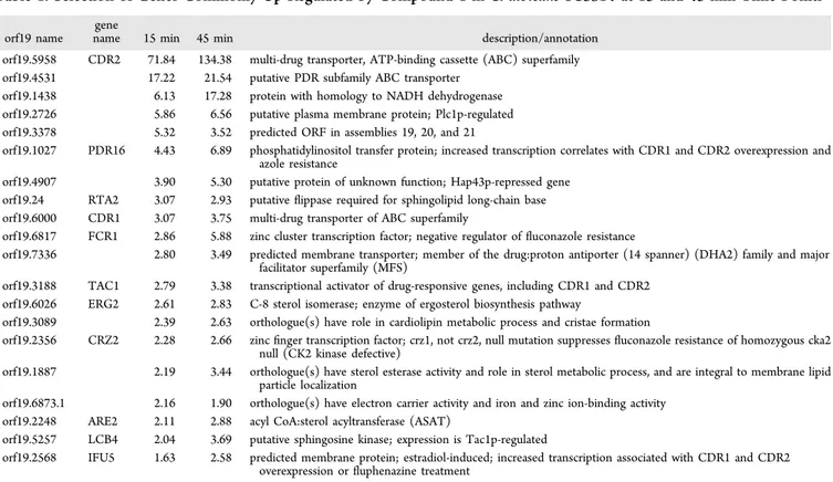
Details on these commonly expressed genes are given in
Tables 1
(up-regulated genes) and
S2
(down-regulated genes,
Supporting Information
). Strikingly, up-regulated genes
ex-hibited a typical signature for TAC1-regulated genes (
Table 1
).
TAC1 is a transcription factor regulating several genes involved
in drug resistance and particularly azole resistance. As
summarized in
Table 1
, TAC1 is itself up-regulated as well as
its target genes such as CDR1, CDR2, RTA3, LCB4, and IFU5.
CDR1 and CDR2 up-regulation was veri
fied by separate
real-time quantitative polymerase chain reaction (RT-qPCR)
analysis. Results are given in
Figure 3
, and show that genes
identi
fied to be up-regulated by microarrays are regulated in a
similar way by RT-qPCR analysis. CDR1 and CDR2 are ATP
binding cassette (ABC) transporters and contribute to drug
e
fflux and azole resistance.
21Susceptibility of
C. albicans CDR1/CDR2 Mutants to
Compound 1. We showed above that CDR1 and CDR2 were
regulated by 1. These genes have been reported to be
up-regulated by a large number of drugs, including antifungal
agents.
22In the majority of cases, inducers themselves serve as
substrates for transporters. In order to test this hypothesis, we
used C. albicans mutants lacking both CDR1 and CDR2
(DSY448, DSY654)
23and addressed their susceptibility to 1
(
Figure 4
). Hypersusceptibility to 1 of the mutants as
compared to a wild-type control (CAF2-1) would strongly
suggest that 1 is a substrate for these ABC transporters.
Surprisingly, as shown in
Figure 4
, the mutants became
unexpectedly more resistant to 1 as compared to the wild type.
The validity of the assay was con
firmed, since the C. albicans
CDR1/CDR2 mutants were susceptible to
fluphenazine, which
is consistent with previously published results. The resistance of
the CDR1/CDR2 mutants to 1 indicates a unique feature of this
antifungal substance, which probably requires the presence of
ABC transporters in order to be active. A similar behavior has
been recently observed by Sun and co-workers, in the case of
the antifungal compound BMQ,
24which proved to be more
active in C. albicans isolates overexpressing multi-drug-resistant
transporter Mdrp1. Those authors demonstrated that the
transporter facilitates the uptake and increases the intracellular
accumulation of the compound, rather than acting as an e
fflux
pump. It is thus rational to assume that, in our case, the
intracellular accumulation of 1 can be favored by the expression
of ABC transporters. The reversed biological pro
file observed
for this compound, compared to current antifungal drugs,
enhances the potential of our class of macrocyclic amidinoureas
for development as new antifungal agents to treat drug-resistant
infections.
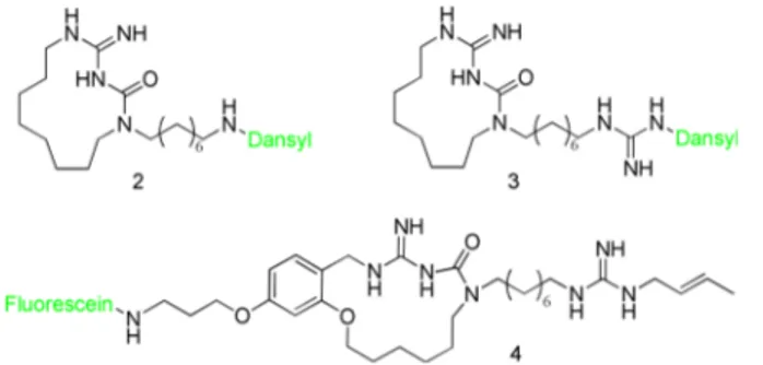
Fluorescence Microscopy Experiments. To test the
aforementioned hypothesis of an intracellular accumulation of
the compound, we performed
fluorescence microscopy assays.
The aim of our study is to investigate the interaction of
macrocyclic amidinoureas with the fungal cell and to verify if
the compounds are accumulated in the cytoplasm and/or they
interact with other structures (membrane, nucleus). In order to
perform
fluorescence experiments, three different fluorescent
probes were designed and synthesized. Two of them, namely
compounds 2 and 3 (
Figure 5
), derived from 1, with the
fluorophore (dansyl group) directly attached on the side chain.
In particular, 2 is linked to the
fluorophore directly on the
terminal amino group, while 3 has the dansyl group attached to
the guanidine moiety. We used the dansyl group as
first choice
for two reasons: it is easily introducted by nucleophilic
substitution of commercial dansyl chloride, and the small
dimensions give good chances to retain the biological activity.
Unfortunately, the introduction of the
fluorophore on the
guanidinium moiety in 2 and 3 resulted in a loss of antifungal
activity in vitro (MIC > 125
μM), probably because the
terminal guanidine does not allow for a bulky substituent.
Fluorescence microscopy experiments were conducted on
samples of C. albicans and Cry. neoformans cultures, but,
although both compounds showed a clear cell-association, the
resolution used did not allow for the discrimination between
cell wall association and internalization (Figure S2,
Supporting
Information
). In order to obtain better results, a new
fluorescent probe was thus synthesized (compound 4,
Figure
5
), bearing the
fluorophore group far away from the
guanidinium moiety. In this case an aromatic derivative (from
Sanguinetti et al.
16) was chosen as the parent compound, since
it possess antifungal activity comparable to that of 1, but the
presence of a benzene ring offers a useful attachment point for
fluorophores (fluorescein). Despite its chemical structure being
bulkier than the dansyl group, we chose
fluorescein as
fluorophore because it has better performance in confocal
microscopy and the distance from the active guanidine as well
Figure 2. Venn diagram showing the results of the comparativetranscriptome analysis and the impact of 1 on gene expression. Genes were at least 1.5-fold commonly up- and down-regulated between 15 and 45 min exposure to 1. The diagram was obtained using Genespring Software Version 12 (Agilent).
as the C3 spacer should avoid a pronounced loss of activity. As
a result, 4 showed a retention of antifungal activity, even though
with a reduced potency (MIC = 80
μM), and it was used in
confocal microscopy experiments.
Confocal microscopy examination of C. albicans ATCC
60193 and Cry. neoformans exposed for 48 h to 4 showed strong
cell-associated
fluorescence which was absent in untreated cells
(
Figure 6
). Fluorescent images were visually compared, and no
signi
ficant difference was observed between the two fungal
species treated with 4. Fluorescence was mostly associated with
the cytoplasm, con
firming the intracellular accumulation of the
compound, in accordance with the hypothesis of an ABC
transporters-mediated accumulation. Moreover,
fluorescence
showed a patchy distribution in some cells, suggesting that the
compound can interact with multiple and/or di
fferently located
targets within the fungal cell. No
fluorescent signal was
observed in cells presenting a damaged cell wall, suggesting
release of the
fluorescent compound due to cell lysis, in
accordance with the previously demonstrated fungicidal e
ffect
of this class of compounds.
Chemistry. The synthesis of compounds 2 and 3 is depicted
in
Scheme 1
, starting from primary amine 5, which was
prepared according to a procedure already described in a
previous paper.
19For the synthesis of 2, amine 5 was reacted
with dansyl chloride (6) in CH
3CN, furnishing the sulfonamide
derivative 7, which was deprotected with freshly distilled 10%
tri
fluoroacetic acid (TFA) in anhydrous dichloromethane
(DCM) solution, to give the desired
fluorescent probe 2. For
the preparation of
fluorescent probe 3, it was necessary to
synthesize the opportune guanylating agent 9, which was
obtained by reacting dansyl chloride (6) with
N-Boc-1H-Table 1. Selection of Genes Commonly Up-Regulated by Compound 1 in
C. albicans SC5314 at 15 and 45 min Time Points
aorf19 name gene
name 15 min 45 min description/annotation orf19.5958 CDR2 71.84 134.38 multi-drug transporter, ATP-binding cassette (ABC) superfamily orf19.4531 17.22 21.54 putative PDR subfamily ABC transporter
orf19.1438 6.13 17.28 protein with homology to NADH dehydrogenase orf19.2726 5.86 6.56 putative plasma membrane protein; Plc1p-regulated orf19.3378 5.32 3.52 predicted ORF in assemblies 19, 20, and 21
orf19.1027 PDR16 4.43 6.89 phosphatidylinositol transfer protein; increased transcription correlates with CDR1 and CDR2 overexpression and azole resistance
orf19.4907 3.90 5.30 putative protein of unknown function; Hap43p-repressed gene orf19.24 RTA2 3.07 2.93 putativeflippase required for sphingolipid long-chain base orf19.6000 CDR1 3.07 3.75 multi-drug transporter of ABC superfamily
orf19.6817 FCR1 2.86 5.88 zinc cluster transcription factor; negative regulator offluconazole resistance
orf19.7336 2.80 3.49 predicted membrane transporter; member of the drug:proton antiporter (14 spanner) (DHA2) family and major facilitator superfamily (MFS)
orf19.3188 TAC1 2.79 3.38 transcriptional activator of drug-responsive genes, including CDR1 and CDR2 orf19.6026 ERG2 2.61 2.83 C-8 sterol isomerase; enzyme of ergosterol biosynthesis pathway
orf19.3089 2.39 2.63 orthologue(s) have role in cardiolipin metabolic process and cristae formation
orf19.2356 CRZ2 2.28 2.66 zincfinger transcription factor; crz1, not crz2, null mutation suppresses fluconazole resistance of homozygous cka2 null (CK2 kinase defective)
orf19.1887 2.19 3.44 orthologue(s) have sterol esterase activity and role in sterol metabolic process, and are integral to membrane lipid particle localization
orf19.6873.1 2.16 1.90 orthologue(s) have electron carrier activity and iron and zinc ion-binding activity orf19.2248 ARE2 2.11 2.88 acyl CoA:sterol acyltransferase (ASAT)
orf19.5257 LCB4 2.04 3.69 putative sphingosine kinase; expression is Tac1p-regulated
orf19.2568 IFU5 1.63 2.58 predicted membrane protein; estradiol-induced; increased transcription associated with CDR1 and CDR2 overexpression orfluphenazine treatment
aComplete data are given in Table S1 (Supporting Information).
Figure 3. Validation of microarray results with RT-qPCR on C. albicans SC5314.
Figure 4. Serial dilution assay of C. albicans with drug inhibitory substances. C. albicans isolates were 10-fold serially diluted starting from a density of 2× 10⁷cells/mL. An aliquot (5μL) was spotted
pyrazolecarboxamidine (8) in the presence of NaH as a base.
The
final guanylation reaction of amine 5 with 9 furnished, after
Boc-cleavage, the desired
fluorescent probe 3 (
Scheme 1
).
The synthesis of 4 is described herein. Starting from
2,4-dihydroxybenzaldehyde (11), we
first functionalized the 4-OH
position with N-(3-bromopropyl)phthalimide to a
fford
inter-mediate 12, which was then etheri
fied with 5-bromo-1-pentene
to give 13. The aromatic aldehyde was converted to aldoxime
14
and then reduced to benzyl amine 15. During this reaction
one carbonyl group of the phthalimide moiety was reduced,
giving a 3-hydroxyisoindolinone moiety. Intermediate 16 was
o b t a i n e d
b y
r e a c t i n g
1 5
w i t h
1 , 3 d i B o c 2
-(tri
fluoromethylsulfonyl)guanidine. The coupling reaction
between the secondary amine 17
16and 16, followed by
ring-closing metathesis reaction, furnished the amidinoureic
macro-cycle 19 in good yields (
Scheme 2
).
While trying to remove the carboxybenzyl (Cbz) protecting
group by hydrogenation over Pd/C, we observed the
simultaneous reduction of the 3-hydroxyisoindolinone ring to
isoindolin-1-ol; despite several attemps, we were not able to
obtain selective Cbz removal. For this reason we decided to
change the protecting group on the C3 lateral chain of 19. To
do so, we
first oxidized the alcohol group of the
3-hydroxyisoindolinone to a carbonyl group with MnO
2, and
then cleaved the phthalimide moiety with hydrazine
mono-hydrate to a
fford the corresponding primary amine 20, which
was then protected using methyl tri
fluoroacetate, furnishing 21.
Hydrogenation over Pd/C allowed simultaneous reduction of
the endocyclic double bond and removal of the Cbz group,
a
ffording derivative 22. The following guanylation with
N-crotylpyrazoleguanidine (23)
16furnished 24. The C3 spacer of
24
was deprotected to give amine 25, which was reacted with
fluorescein isothiocyanate (FITC) in dimethylformamide
(DMF) to form the desired conjugate 26. Finally,
butylox-ycarbonyl (Boc) deprotection performed in 20% TFA in dry
DCM a
fforded the final product 4 in quantitative yield as a
yellow powder (
Scheme 3
).
In Vivo Animal Studies. In order to test the potential
antifungal therapeutic e
ffect of 1 in vivo, experimental
treatments of invasive candidiasis were carried out in
immunocompetent mice. Two di
fferent drug regimens, i.e.,
20 and 40 mg/kg/day, were applied, whereas
fluconazole was
used as a control at a dosage of 8 mg/kg/day.
Figure 7
A shows
results of tissue burdens obtained after 7 days of experiments in
spleen and kidneys with both 20 and 40 mg/kg/day regimens
of 1. Experiments showed that 1 signi
ficantly reduced the CFU
counts of the organs (kidneys and spleen) of mice infected
intravenously with C. albicans ATCC 90028 (a
fluconazole-susceptible control strain). A similar e
ffect was noticed in mice
treated with
fluconazole, as expected. Furthermore,
exper-imental mouse infections were also done with two isogenic
strains of C. albicans: DSY294 that is susceptible to azole drugs
(
fluconazole MIC = 0.25 μg/mL) and DSY296 that is
azole-Figure 5. Structures of the fluorescent probes synthesized.Fluorophores are represented in green.
Figure 6.Confocal microscope images of C. albicans ATCC 60193 and Cry. neoformans cells treated with compound 4 at 40 μM concentration. Panels A, B, and C show the visible,fluorescent, and merged images, respectively. Scale bar = 10μm.
Scheme 1
aaReagents and conditions: (a) TEA, CH
3CN, rt, 10 min; (b) TFA,
DCM, rt, 3 h; (c) 6, NaH, THF, rt, 3 h; (d) DIPEA, CH3CN/MeOH,
resistant (
fluconazole MIC = 64 μg/mL).
25In these
experi-ments, only the 40 mg/kg/day regimen of 1 was applied,
whereas
fluconazole was used at the same dosage (8 mg/kg/
day) (
Figure 7
B). By contrast to
fluconazole treatment, which
was e
ffective only against DSY294, 1 significantly reduced CFU
counts of the selected organs of mice infected with both
DSY294 and DSY296, and it is worth noting that the in vivo
e
fficacy of 1 was thus achieved also with the azole-resistant
isolate strain. With regard to DSY296, the reductions in CFU
counts by 1 treatment were markedly higher than those
observed in mice treated with
fluconazole. Remarkably, the
resistance to
fluconazole of the isolate DSY296 is due to the
overexpression of multi-drug transporters, triggered by an
up-regulation of CDR1 and CDR2 genes.
25The in vivo e
fficacy of
1
against DSY296 infection thus corroborates the assumption
of the ABC transporters-mediated accumulation of the
molecule inside the fungal cell.
Conclusions. To date, only three classes of antifungal
agents are used for the treatment of systemic candidiasis, and
the market calls for new drugs that would address a growing
medical need, determined by the emergence of drug-resistant
clinical isolates. In this work, we present a set of in vitro and in
vivo data supporting the promising potential of the herein
described novel antifungal agent, a macrocyclic amidinourea
derivative. Our research highlights that the molecular target is
new and not shared by any other antifungal compound on the
market, since our compound preserves a potent activity also
against drug-resistant strains. Indeed, compound 1 was proved
particularly active on resistant isolates overexpressing
multi-drug transporters of the ABC superfamily, a mutation usually
associated with resistance to therapeutic antifungals. The
involvement of ABC transporters in the mode of action of
the compound was demonstrated with in vitro experiments on
mutant strains and con
firmed with fluorescence microscopy
and in vivo assays. Our
findings could represent the first step
toward the development of a new therapeutic class of antifungal
agents, potentially useful to treat acquired infections, and might
represent a suitable strategy to address the growing emergence
of antifungal-resistant isolates in the clinical setting.
■
EXPERIMENTAL SECTION
Antifungal Activity in Vitro. C. albicans ATCC 60193, C. krusei ATCC 14243, and C. parapsilosis ATCC 34136 were purchased from the American Type Culture Collection (Manassas, VA, USA). The EUCAST susceptibility testing protocol, including the update for testing of non-fermenting yeasts (Cryptococcus spp. and Geotrichum spp.), was used for the determination of the minimal inhibitory concentrations (MICs) of antifungal agents.26,27 Results were read after 24, 48, and 72 h of incubation at 37°C. The MIC was defined as the lowest compound concentration which prevented visible growth. Aliquots of 10μL were removed from the wells corresponding to the MIC, 2MIC, 4MIC, and 8MIC and serially 10-fold diluted in sterile saline, to minimize the drug carry-over. An aliquot of 10μL of each dilution was plated on Sabouraud dextrose agar plates. The plates were incubated at 37°C for 24/48 h, and the number of CFUs was counted. For Cryptococcus spp. and Geotrichum spp., incubation was for 48 h to facilitate colony counting.28 When less than 100 CFU/mL was expected, 10 μL of sample was plated without dilution. The experiment was performed in duplicate. The minimum detection limit of this method was 10 CFU/mL. The Log10 CFU/mL was plotted on a graph as a function of time and used to compare the rate and extent of antifungal activity in the presence various concentrations of 1 and in its absence. Activity was considered fungicidal when there was a decrease greater than or equal to 3·Log10 CFU/mL (99.9%) of the initial inoculum in 24 h. Activity lower than 3·Log10 reduction in the number of CFU/mL of the initial inoculum was considered fungistatic.29C. glabrata was not tested since it is poorly susceptible to 1.
RPMI Medium Stability Assay. To verify the chemical stability of 1in RPMI-1640 medium, 0.4 mL of a stock solution in MeOH (10 mM) of the compound and 1.6 mL of RPMI-1640 (final concentration 2 mM) were mixed in a test tube. The solution wasfiltered through a 0.45μm nylon filter (Acrodisc) before LC-UV-MS analysis. The tube
Scheme 2
aaReagents and conditions: (a) N-(3-bromopropyl)phthalimide, K
2CO3, CH3CN, reflux, 6 h; (b) 5-bromo-1-pentene, K2CO3, CH3CN, reflux, 6 h;
(c) NH2OH−HCl, pyridine, EtOH, reflux, 3.5 h; (d) Zn, HCl, THF, reflux, 2 h; (e) 1,3-di-Boc-2-(trifluoromethylsulfonyl)guanidine, TEA, DCM, rt,
was maintained at 30 °C, and four aliquots (20 μL each) were removed and analyzed by HPLC at predetermined time points (1, 24, 48, and 72 h). Each aliquot was analyzed in triplicate. The stability was followed by HPLC with an UV-MS detection method (Supporting Information). Quantitative analysis was performed using an appro-priate calibration curve obtained by solubilizing 1 in MeOH.
Transcriptional Analysis. Sample preparation was performed on three biological triplicates. Total RNA was extracted from log phase cultures in liquid yeast extract peptone dextrose (YEPD) as previously described.30Compound 1 was added to an end concentration of 3μM, and incubation points of 15 and 45 min were chosen. Control cells were taken at the same time points but without 1. Briefly, after centrifugation of 5 mL of culture (corresponding to 108 cells), the yeast cell pellets were mixed with 0.3 g of glass beads, 300μL of RNA extraction buffer (0.1 M Tris-HCl at pH 7.5, 0.1 M LiCl, 10 mM EDTA, 0.5% SDS), and 300 μL of phenol−chloroform−isoamyl alcohol (24:24:1). After 1 min of vortexing in a bead beater (Fastprep-24 Instrument, MP Biomedicals Switzerland, Zürich), the aqueous phase was re-extracted with phenol−chloroform−isoamyl alcohol, and RNA was precipitated with 600μL of ethanol at −20 °C for 1 h. The RNA pellet was resuspended in 50μL of diethyl pyrocarbonate-treated H2O. The integrity of the input template RNA was determined prior
to labeling/amplification, using an Agilent RNA 6000 Nano LabChip kit and 2100 bioanalyzer (Agilent Technologies). Agilent’s One-Color Quick Amp Labeling Kit (Agilent Technologies) was used to generate fluorescent cRNA according to the manufacturer’s instructions. Briefly, 1μg of total RNA from each sample was used, to which a spike mix and T7 promoter primers were added, both of which are provided by the manufacturer. cDNA synthesis was promoted by MMLV-RT
(Moloney Murine Leukemia Virus Reverse Transcriptase) in the presence of dNTPs and RNaseOUT. Next, cRNA was produced from this first reaction with T7 RNA polymerase, which simultaneously amplifies target material and incorporates cyanine 3-labeled CTP. The labeled cRNAs were purified with RNeasy Mini Kit (Qiagen) and quantified using a NanoDrop ND-1000 UV−vis spectrophotometer. Next, 600 ng of Cy3-labeled cRNAs was fragmented and hybridized for 17 h at 65 °C to each array using the Gene Expression Hybridization Kit (Agilent Technologies) and a gasket slide with an 8 microarrays/slide format for sample hybridization to separate each sample in specific sub-arrays of the 8 × 15K format. The C. albicans microarray format was published by Synnott et al. (design ID 017942).31
Slides were washed and processed according to the Agilent 60-mer Oligo Microarray Processing protocol and scanned on a Agilent microarray scanner G2565BA (Agilent Technologies). Data were extracted from the images with Feature Extraction (FE) software (Agilent Technologies). FE softwareflags outlier features, and detects and removes spatial gradients and local backgrounds. Data were normalized using a combined rank consistencyfiltering with LOWESS intensity normalization. The gene expression values obtained from FE software were imported into GeneSpring 12 software (Agilent Technologies) for preprocessing and data analysis. For inter-array comparisons, a linear scaling of the data was performed using the 75th percentile signal value of all of non-control probes on the microarray to normalize one-color signal values. Probe sets with a signal intensity value below the 20th percentile were considered as absent and discarded from subsequent analysis. The expression of each gene was normalized by its median expression across all samples. Genes were
Scheme 3
aaReagents and conditions: (a) MnO
2, DCM, rt, 18 h; (b) hydrazine monohydrate, MeOH sol., rt, 12 h; (c) methyl trifluoroacetate, TEA, THF, 0 °C
to rt, 16 h; (d) H2, Pd/C, cat. HCl, i-PrOH, rt, 3 h; (e) N-crotylpyrazoleguanidine (23), DIPEA, CH3CN/MeOH, 60°C, 16 h; (f) K2CO3, MeOH,
included in thefinal data set if their expression changed by at least 1.5-fold as compared to 1-unexposed cells. Corrected P-value <0.05 was chosen as the cutoff for significance. Microarray data have been uploaded to the NCBI GEO microarray repository.
Real-Time Quantitative Polymerase Chain Reaction (RT-qPCR). Total RNA was extracted from log phase cultures with an RNeasy Protect Mini kit (Qiagen) by a process involving mechanical disruption of the cells with glass beads and an RNase-free DNase treatment step as previously described.32 To remove contaminating DNA, 10μg of each RNA preparation was treated with DNase using a DNA-Free DNA removal kit (Ambion). For the reverse transcription reactions, a Transcriptor High Fidelity cDNA Synthesis Kit (Roche) was used on 1 μg of DNA-free RNA of each sample, and the manufacturer’s instructions were followed. RNA samples were stored at−80 °C, and cDNA samples were stored at −20 °C. Quantitative expression of CDR1 and CDR2 was performed with the StepOne Real-time PCR System (Life Technologies). RT-qPCR was carried out in a 10 μL volume containing the following reagents: 5 μL of iTaq Supermix with Rox (BioRad), each primer pair, the Taqman probe at a final concentration of 200 nM for the primers (CDR2-ORF-F: TAGATATTTGAGCCACATG and CDR2-ORF-R: TTGGCA-TTGAAATTTTCG; CDR1-ORF-F: ATGACTCGAGATATTTTG-ATA; and CDR1-ORF-R: TTAACAGCAATGGTCTTTA) and 100 nM for the probes (CDR2-P2: TTAGTCCATTCAACGGCA-ACATTAG; CDR1-P2: CATTATGAGACCTGGTGAACTTACT), and 1% of the total cDNA sample produced as described above. Each reaction was run in triplicate, and data were analyzed with the StepOne software. For relative quantification of the target genes, each
set of primer pairs and the Taqman probe were used in combination with the ACT1 primers (ACT1-RT-F: ATAACGGTTCTGGTATGT; ACT1-RT-R: CCTTGATGTCTTGGTCTA) and an ACT1 probe (ACT1-RT-P: CGGTGACGACGCTCCAAG).
For data analysis and fold change calculations, the comparative CT method was used. For each experiment, a standard curve for the reference gene and the studied gene was included, and the amplification efficiency was determined for all genes. The CT values of the target genes were normalized to the endogenous reference. Fold changes were obtained from the mean normalized expression of the samples relative to the mean normalized expression of a selected control.
Confocal Fluorescence Microscopy Assays. Aliquots of 100μL of C. albicans and Cry. neoformans treated for 48 h with a 40μM DMSO solution of 4 were washed twice in phosphate-buffered saline (PBS) and fixed in 4% formaldehyde for 30 min at 4 °C. After a second PBS wash, the cells were suspended in 100μL of PBS, and 3 μL of the suspension was spotted onto a glass slide, dried, and mounted with 6μL of Vectashield mounting medium. Images were acquired with a Leica TCS SP5 inverted confocal microscope equipped with a 63×/1.40 OIL objective (Zeiss), set for fluorescein (FITC) (excitation 488 nm, emission window from 500 to 540 nm). General Chemistry Directions. Reagents were obtained from commercial suppliers and used without further purification. DCM and CH3CN were dried over sodium hydride. THF was dried over Na/
benzophenone prior to use. Anhydrous reactions were run under a positive pressure of dry N2or argon. Degassed DCM was prepared by
using the freeze−pump−thaw method. Silica gel 60 was used for flash Figure 7.In vivo animal studies. The regimens for 1 andfluconazole were 40 and 8 mg/kg/day, respectively. Each data point corresponds to one animal, and the origin of tissue (kidney or spleen) is shown for each diagram. (A) Fungal tissue burdens of mice infected with C. albicans and treated withfluconazole (FLC) and 1. (B) Fungal tissue burdens of mice infected with C. albicans DSY294 and DSY296, treated with fluconazole (Flu) and 1.
chromatography (23−400 mesh). 1H NMR and 13C NMR are
reported in parts per million (δ scale) and internally referenced to the CDCl3 or CD3OD signal, respectively at δ 7.24 and 3.31 ppm.
Chemical shifts for carbon are reported in parts per million (δ scale) and referenced to the carbon resonances of the solvent (CDCl3at δ
77.00 and CD3OD atδ 49.00 ppm). Data are presented as follows:
chemical shift, multiplicity (s = singlet, d = doublet, t = triplet, m = multiplet and/or multiplet resonances, br = broad), coupling constant in hertz (Hz), and integration. Mass spectrometry (MS) data were acquired on an Agilent 1100 LC/MSD VL system (G1946C) with a 0.4 mL/minflow rate using a binary solvent system of 95:5 methanol/ water. UV detection was monitored at 254 nm. Mass spectra were acquired in positive mode, scanning over the mass range. Purity offinal products was assessed by HPLC/MS analysis, conducted using a Polaris C18 column (150−4.6 mm, 5 μm particle size) at a flow rate of 0.8 mL min−1, with a mobile phase composed of 50% CH3CN/50%
H2O−formic acid 0.1%. The purity of all final compounds was above
95%.
5-(Dimethylamino)-N-(8-(2-oxo-4-(tert-butoxycarbonylimino)-1,3,5-triazacyclotridecan-1-yl)octyl)naphthalene-1-sulfonamide (7). A solution of macrocyclic amine 5 (20 mg, 1 equiv) and TEA (0.01 mL, 1.5 equiv) in CH3CN at room temperature under argon
atmosphere was treated with a solution of dansyl chloride 6 (17 mg, 1.5 equiv) in CH3CN. The mixture was stirred at room temperature
for 10 min. Solvent was then evaporated, and the crude residue was purified with flash chromatography on silica gel, eluting with 10% AcOEt/Hex to give 7 (isolated yield 79%). 1H NMR (400 MHz
CDCl3) 12.00 (1H, s); 8.50−8.48 (d, J = 8.4 Hz, 1H), 8.24−8.22 (d, J = 8.2 Hz, 1H), 8.19−8.17 (d, J = 8.1 Hz, 1H), 8.02 (t, J = 4.0 Hz, 1H), 7.52−7.44 (m, 2H), 7.14−7.12 (d, J = 8.2 Hz, 1H), 4.50 (s, 1H), 3.36 (m, 2H), 3.30 (m, 2H), 3.13−3.11(m, 2H), 2.81(s, 6H), 2.78 (m, 2H), 1.56 (m, 8H), 1.40 (s, 9H), 1.25−1.19 (m, 16H) ppm.13C NMR (100 MHz CD3OD) 163.9; 154.0; 153.5;134.7; 130.2, 129.6; 128.2; 123.2; 118.8; 115.1; 81.9; 46.9; 46.2; 45.4; 43.2; 39.5; 31.8; 29.6; 29.3; 29.1; 28.8; 28.3, 28.0; 27.7; 26.1; 25.8; 25.2; 23.6; 23.4 ppm. LRMS m/z (ESI) = 695.0 [M + Na]+, 673.0 [M + H]+. tert-Butyl ((5-(Dimethylamino)naphthalene-1-sulfonamido)(1H-pyrazol-1-yl)methylene)carbamate (9). To a suspension of NaH (27 mg, 3 equiv) in 2 mL of THF was added dropwise a solution of N-Boc-1H-pyrazole-1-carboxamidine 8 (78 mg, 1 equiv) in 0.5 mL of THF at 0°C. The mixture was stirred at room temperature for 30 min, and then a solution of dansyl chloride 6 (300 mg, 3 equiv) in 1 mL of THF was added. After further stirring for 2 h at room temperature, water (5 mL) and DCM (10 mL) were added, and the layers were separated. The aqueous layer was extracted with DCM (2× 10 mL), and the combined organic phases were washed with NaHCO3, water,
and brine, dried over Na2SO4, filtered, and concentrated under
reduced pressure. The crude residue was purified with flash chromatography on silica gel, eluting with 20% AcOEt/Hex to give 9(isolated yield 90%).1H NMR (400 MHz CDCl 3):δ 8.87 (s, 1H), 8.50−8.48 (d, J = 8.4 Hz, 1H), 8.37−8.35 (d, J = 8.4 Hz, 1H), 8.27− 8.25 (d, J = 8.4 Hz, 1H), 7.85 (m, 1H), 7.62 (m, 1H), 7.52−7.44 (m, 2H), 7.13−7.11 (d, J = 8.3 Hz, 1H), 6.27 (m, 1H), 2.84 (s, 6H), 1.40 (s, 9H) ppm. LRMS m/z (ESI) = 466.2 [M + Na]+, 443.2 [M + H]+. 5-(Dimethylamino)-N-(N ′-tert-butoxycarbonyl-N-(8-(4-(tert-bu- toxycarbonylimino)-2-oxo-1,3,5-triazacyclotridecan-1-yl)octyl)-carbamimidoyl)naphthalene-1-sulfonamide (10). To a stirred solution of guanylating agent 9 (21 mg, 1.1 equiv) in 5% MeOH/ CH3CN (1 mL) was added a solution of the macrocyclic amine 5 (20
mg, 1 equiv) in 5% MeOH/CH3CN (1 mL) dropwise, followed by the
addition of catalytic diisopropylethylamine (DIPEA) (2 drops). The mixture was stirred at 50 °C overnight. After cooling, solvent was evaporated under reduced pressure, and the crude residue was purified withflash chromatography on silica gel, eluting with 10% AcOEt/Hex to give 10 (isolated yield 93%).1H NMR (400 MHz CDCl
3):δ 12.02 (s, 1H), 9.79 (s, 1H), 8.50−8.48 (d, J = 8.4 Hz, 1H), 8.37−8.35 (d, J = 8.4 Hz, 1H), 8.27−8.25 (d, J = 8.4 Hz, 1H), 7.49−7.41 (m, 2H), 7.13−7.11 (d, J = 8.2 Hz, 1H), 3.38 (m, 2H), 3.29 (m, 2H), 3.18 (m, 2H), 2.82 (s, 6H), 1.48(m, 8H), 1.40 (s, 9H), 1.37 (s, 9H), 1.25−1.19 (m, 16H) ppm.13C NMR (100 MHz CD 3OD) 163.9; 154.0; 153.5; 152.5; 150.6; 138.9; 129.7; 129.1; 127.2; 126.2; 123.3; 115.1; 84.1; 81.9; 46.9; 46.2; 45.5; 41.3; 39.5; 31.8; 30.7; 29.5; 29.3; 29.0; 28.7; 28.3, 28.0; 27.7; 26.1; 25.8; 25.2; 23.6; 23.4 ppm. LRMS m/z (ESI) = 837.5 [M + Na]+, 814.5 [M + H]+.
General Procedure for Boc Deprotection. The appropriate protected compound 7 or 10 (0.025 mmol) was dissolved in a 10% solution of freshly distilled TFA in dry DCM (1 mL). The resulting solution was stirred at room temperature under argon for 24 h. The reaction mixture was then concentrated under reduced pressure, affording the desired compound as the trifluoroacetate salt in quantitative yield. 5-(Dimethylamino)-N-(8-(4-imino-2-oxo-1,3,5-triazacyclotride-can-1-yl)octyl)naphthalene-1-sulfonamide (2).1H NMR (400 MHz CDCl3):δ 9.79 (s, 1H), 8.50−8.48 (d, J = 8.4 Hz, 1H), 8.37−8.35 (d, J = 8.2 Hz, 1H), 8.27−8.25 (d, J = 8.4 Hz, 1H), 7.49−7.41 (m, 2H), 7.13−7.11 (d, J = 8.2 Hz, 1H), 3.56 (m, 2H), 3.46 (m, 2H), 3.23 (m, 2H), 3.05 (m, 2H), 2.80 (s, 6H), 1.48(m, 8H), 1.25−1.19 (m, 16H) ppm. 13C NMR (100 MHz CDCl3): δ 163.6, 153.7, 153.2, 135.1, 130.2, 129.7, 129.2, 124.0, 121.4, 81.6, 46.7, 46.0, 41.9, 39.3, 33.5, 29.2, 28.2, 27.8, 26.8, 25.8, 25.5, 24.9, 23.3 ppm. LRMS m/z (ESI) = 595.0 [M + Na]+, 573.0 [M + H]+. 5-(Dimethylamino)-N-(N-(8-(4-imino-2-oxo-1,3,5-triazacyclotri-decan-1-yl)octyl)carbamimidoyl)naphthalene-1-sulfonamide (3). 1H NMR (400 MHz CD 3OD): δ 12.00 (s, 1H); 8.44−8.42 (d, J = 8.4 Hz, 1H), 8.22−8.20 (d, J = 8.4 Hz, 1H), 8.19−8.17 (d, J = 8.4 Hz, 1H), 8.03 (m, 1H), 7.49−7.43 (m, 2H), 7.13−7.11 (d, J = 8.4 Hz, 1H), 3.38 (m, 2H), 3.31 (m, 2H), 3.12 (m, 2H), 2.80 (s, 6H), 2.77 (m, 2H), 1.56 (m, 8H), 1.25−1.19 (m, 16H) ppm.13C NMR (100 MHz CDCl3): δ 206.8, 163.9, 154.05, 153.0, 152.5, 150.6, 138.9, 129.8, 129.1, 127.2, 126.2, 123.3, 116.2, 84.1, 82.0, 46.9, 46.2, 45.7, 41.3, 39.6, 31.8, 30.8, 29.5, 29.2, 29.0, 28.4, 27.8, 25.8, 25.2, 23.5, 22.6, 14.0 ppm. LRMS m/z (ESI) = 637.5 [M + Na]+, 614.5 [M + H]+. 4-(3-(Phthalimido)propoxy)-2-hydroxybenzaldehyde (12). A sol-ution of 2,4-dihydroxybenzaldehyde 11 (0.500 g, 1 equiv), N-(3-bromopropyl) phthalimide (0.970 g, 1 equiv), and K2CO3(0.500 g, 1
equiv) was refluxed in CH3CN (20 mL) for 6 h. The reaction mixture
was concentrated and extracted with DCM and water. The organic phase was dried over Na2SO4,filtered, and concentrated. After the
purification of the crude by flash chromatography (SiO2) using DCM
as eluent, product 12 was obtained a white solid (isolated yield 34%).
1H NMR (400 MHz CDCl 3):δ 11.42 (s, 1H, 2-OH); 9.69 (s, 1H); 7.84−7.81 (m, 2H); 7.74−7.71 (m, 2H); 7.37 (d, J = 8.8 Hz, 1H); 6.41 (d, J = 8.5 Hz, 1H); 6.32 (d, J = 1.2 Hz, 1H); 4.07 (t, J = 6.8 Hz, 2H); 3.90 (t, J = 6.8 Hz, 2H); 2.22 (m, 2H) ppm.13C NMR (100 MHz, CDCl3): 194.3; 168.2; 165.8; 164.3; 135.1; 133.9; 132.0; 123.2; 115.2; 108.5; 101.0; 66.1; 35.1; 27.9 ppm. LRMS m/z (ESI) = 324 [M + H]+. 4-(3-(Phthalimido)propoxy)-2-(pent-4-en-1-yloxy)benzaldehyde (13). A solution of 12 (0.200 g, 1 equiv), K2CO3(0.255 g, 3 equiv),
and 5-bromo-1-pentene (0.160 mL, 2.2 equiv) was refluxed in CH3CN
(10 mL) for 6 h. The reaction mixture was concentrated under vacuum and extracted with DCM. The organic phase was dried over Na2SO4,filtered, and concentrated. The reaction crude was purified by
flash chromatography (SiO2) using DCM as eluent to obtain 13 as a
white solid (isolated yield 78%).1H NMR (400 MHz CDCl
3):δ 10.27 (s, 1H); 7.81−7.79 (m, 2H); 7.72−7.68 (m, 4H); 6.38 (d, J = 8.4 Hz, 1H); 6.27 (s, 1H); 5.87−5.76 (m, 1H); 5.04 (d, J = 17.2 Hz, 1H); 4.99 (d, J = 10 Hz, 1H); 4.06 (t, J = 6 Hz, 2H); 3.95 (t, J = 6.4 Hz, 2H); 3.88 (t, J = 6.8 Hz, 2H); 2.25−2.14 (m, 4H); 1.89 (m, 2H) ppm.13C NMR (100 MHz, CDCl3): 188.1; 183.7; 177.5; 168.2; 165.1; 163.0; 137.3; 133.9; 132.0; 130.1; 123.2; 119.1; 115.4; 106.0; 98.9; 67.5; 66.0; 35.2; 29.9; 28.05; 28.00 ppm. LRMS m/z (ESI) = 394.1 [M + H]+; 416.1 [M + Na]+. 4-(3-(Phthalimido)propoxy)-2-(pent-4-en-1-yloxy)benzaldehyde oxime (14). A stirred solution of aldehyde 13 (0.120 g, 1 equiv) in EtOH (10 mL), hydroxylamine hydrochloride (0.053 g, 2.5 equiv), and pyridine (0.028 g, 1.2 equiv) was heated to 80°C for 3.5 h. The reaction mixture was concentrated in vacuo and used with no further purification.1H NMR (400 MHz CDCl
3):δ 8.86 (br, 1H); 8.40 (s,
(d, J = 8.6 Hz, 1H); 5.88−5.77 (m, 1H); 5.05 (d, J = 17.6 Hz, 1H); 4.99 (d, J = 10.4 Hz, 1H); 4.03 (t, J = 5.6 Hz, 2H); 3.91−3.87 (m, 4H); 2.24−2.14 (m, 4H); 1.86 (quint, J = 6.8 Hz, 2H) ppm.13C NMR (100 MHz, CDCl3): 165.1; 163.2; 146.2; 137.3; 133.8; 132.1; 127.3; 123.2; 119.1; 115.3; 105.8; 98.9; 67.5; 66.0; 44.0; 35.4; 29.9; 28.1 ppm. LRMS m/z (ESI) = 409.0 [M + H]+;431.0 [M + Na]+. 2-(3-(4-(Aminomethyl)-3-(pent-4-en-1-yloxy)phenoxy)propyl)-3-hydroxyisoindolin-1-one (15). To a stirred solution of aldoxime 14 (0.124 g, 1 equiv) in THF (10 mL) were added Zn0granules (0.198 g, 10 equiv) and aqueous 2 M HCl solution (1.51 mL, 10 equiv), and the resulting mixture was heated to 80°C for 2 h. After the solution was cooled to room temperature, it wasfiltered to remove the residual Zn and extracted in DCM and 1 M NaOH solution. The organic layer was washed with brine, dried over Na2SO4,filtered, and concentrated to
yield 15 as a colorless oil that was used with no further purification (isolated yield 66%).1H NMR (400 MHz CDCl 3):δ 7.65 (d, J = 7.2 Hz, 1H); 7.53−7.48 (m, 2H); 7.42−7.39 (m, 2H); 6.88 (d, J = 8.4 Hz, 1H); 6.29 (s, 1H); 5.85−5.73 (m, 1H); 5.74 (s, 1H); 5.03 (d, J = 17.6 Hz, 1H); 4.98 (d, J = 11.2 Hz, 1H); 4.00−3.80 (m, 6H); 3.72−3.67 (m, 1H); 3.62−3.55 (m, 1H); 3.48 (s, 1H); 2.17−2.10 (m, 4H); 1.81 (m, 2H) ppm. LRMS m/z (ESI) = 419.1 [M + Na]+. 1-(4-(3-(1-Hydroxy-3-oxoisoindolin-2-yl)propoxy)-2-(pent-4-en-1-yloxy)benzyl)-2,3-bis(tert-butoxycarbonyl)guanidine (16). To a stirred solution of 15 (0.277 g, 1 equiv) in DCM (15 mL) Et3N (0.486
mL, 5 equiv) was added 1,3-di-Boc-2-(tri fluoromethylsulfonyl)-guanidine (0.300 g, 1.1 equiv), and the resulting mixture was stirred for 18 h at room temperature. Water was added, and after 30 min of stirring, the reaction mixture was extracted with DCM. The organic layer was washed with brine, dried over Na2SO4, filtered, and
concentrated in vacuo. The crude was purified by flash chromatog-raphy (SiO2) (petroleum ether:EtOAc:MeOH = 7:2:0.5) to give the
desired product 16 as an oil (isolated yield 46%).1H NMR (400 MHz CDCl3):δ 11.44 (s, 1H); 8.64 (m, 1H); 7.52−7.44 (m, 3H); 7.34 (t, J = 7.2 Hz, 1H); 7.08 (d, J = 8.0 Hz, 1H); 6.28 (s, 1H); 6.26 (s, 1H); 5.82−5.76 (m, 1H); 5.73 (s,1H); 5.01 (d, J = 17.6 Hz, 1H); 4.94 (d, J = 10.0 Hz, 1H); 4.67 (br, 1H); 4.47 (d, J = 4.8 Hz, 2H); 3.93−3.87 (m, 2H); 3.84 (t, J = 6.4 Hz, 2H); 3.57−3.44 (m, 2H); 2.18 (q, J = 7.2 Hz, 2H); 2.03 (m, 2H); 1.85 (m, 2H); 1.47 (s, 9H); 1.42 (s, 9H) ppm. 13C NMR (100 MHz, CDCl 3): 167.6; 163.5; 159.6; 158.0; 155.7; 152.8; 144.0; 137.7; 132.0; 131.4; 130.4; 129.4; 123.2; 122.9; 117.9; 115.1; 104.3; 99.6; 82.6; 81.9; 79.0; 67.1; 65.8; 40.6; 36.7; 30.0; 28.2; 28.1; 27.9 ppm. LRMS m/z (ESI) = 639.2 [M + H]+; 661.2 [M + Na]+. N-(N ′-(4-(3-(1-Hydroxy-3-oxoisoindolin-2-yl)propoxy)-2-(pent-4-en-1-yloxy)benzyl)-tert-butoxycarbonylcarbamimidoyl)-N ′-prop-2-enyl-N′-(8-(benzyloxycarbonylamino)octyl)urea (18). To a stirring solution of 16 (0.276 g, 1 equiv) in dry THF (20 mL) was added amine 17 (0.207 g, 1.5 equiv)19as a dry THF solution (5 mL). To the resulting mixture was added Et3N (0.060 mL, 1 equiv). After 12 h of
heating at 80°C, the reaction mixture was cooled to room temperature and concentrated in vacuo. The reaction crude was purified by flash chromatography (SiO2) (petroleum ether:EtOAc MeOH = 7:3:1) to
yield the desired compound 18 as an oil (isolated yield 60%). 1H
NMR (400 MHz CDCl3):δ 12.21 (d, J = 4 Hz, 1H); 8.42 (m, 1H); 7.58 (t, J = 6.4 Hz, 1H); 7.53 (d, J = 7.6 Hz, 1H); 7.48 (t, J = 7.2 Hz, 1H); 7.37 (t, J = 7.6 Hz, 1H); 7.28 (m, 5H); 7.05 (d, J = 8.8 Hz, 1H); 6.31 (d, J = 7.6 Hz, 2H); 5.83−5.78 (m, 1H); 5.75 (s, 1H); 5.12−4.96 (m, 6H); 4.42 (dd, J1 = 5.6 Hz, J2 = 16 Hz, 2H); 4.12 (d, J = 5.2 Hz, 1H); 3.95−3.68 (m, 6H); 3.67−3.54 (m, 2H), 3.42 (t, J = 7.6 Hz, 1H); 3.21 (t, J = 7.2 Hz, 1H); 3.10−3.06 (m, 2H); 2.22 (q, J = 6.8 Hz, 2H); 2.09 (m, 2H); 1.89 m, 2H); 1.48 (m, 2H); 1.41 (s, 9H); 1.24 (m, 8H) ppm.13C NMR (100 MHz, CDCl3): 167.5; 163.8; 163.7; 159.4; 157.9; 157.8; 156.4; 153.8; 153.6; 153.1; 144.1; 137.1; 136.5; 135.3; 134.7; 131.9; 131.6; 130.1; 129.6; 129.4; 128.4; 127.9; 123.1; 122.9; 118.9; 115.5; 115.1; 104.3; 99.6; 82.0; 81.9; 81.8; 67.1; 66.4; 65.8; 50.44; 48.4; 47.5; 45.6; 41.0; 36.9; 30.0; 29.8; 29.6; 29.4; 29.3; 29.1; 28.5; 28.2 ppm. LRMS m/z (ESI) = 883.3 [M + H]+. tert-Butyl (10-(8-(Benzyloxycarbonylamino)octyl)-11-oxo-18-(3- (1-Hydroxy-3-oxoisoindolin-2-yl)propoxy)-2,3,4,5,8,9,10,11,12,15-
decahydrobenzo[b][1,5,7,9]oxatriazacycloheptadecin-13-yl)-carbamate (19). In a 250 mL round-bottomed flask equipped with a stirring bar and a condenser, 18 (0.231 g, 1 equiv) was dissolved in 175 mL of dry DCM, and the resulting mixture was degassed three times. After the third degassing cycle, Grubbs’s second-generation (0.024 g, 0.1 equiv) catalyst was added, and the mixture was degassed another time. The reaction mixture was refluxed for 3 h. The solvent was removed, and the reaction crude was purified by flash chromatography (SiO2) (petroleum ether:EtOAc: = 7:3) to yield the
desired compound as a mixture of two isomers (E/Z) (isolated yield 75%).1H NMR (400 MHz CDCl 3):δ 12.21 (s, 1H), 12.03 (s, 1H), 8.21 (s, 1H), 7.61 (d, J = 7.3 Hz, 3H), 7.57−7.47 (m, 6H), 7.39 (t, J = 7.1 Hz, 3H), 7.31 (s, 7H), 7.06 (d, J = 8.1 Hz, 3H), 6.33 (t, J = 7.6 Hz, 3H), 6.27 (s, 2H), 6.05−5.95 (m, 1H), 5.76 (s, 3H), 5.63−5.48 (m, 2H), 5.41−5.31 (m, 2H), 5.03 (s, 4H), 4.85 (s, 2H), 4.56 (s, 4H), 4.22 (d, J = 5.8 Hz, 2H), 3.95 (s, 9H), 3.87 (d, J = 3.3 Hz, 6H), 3.64 (m, 6H), 3.32−3.22 (m, 4H), 3.12 (s, 6H), 2.40 (d, J = 6.7 Hz, 2H), 2.13 (m, 10H), 1.52 (s, 8H), 1.41 (m, 14H), 1.38 (m, 12H), 1.24 (m, 28H) ppm.13C NMR (100 MHz, CDCl 3):δ 167.48, 164.05, 163.39, 159.81, 159.26, 158.29, 157.38, 156.36, 153.41, 153.21, 152.42, 144.01, 134.30, 132.13, 131.96, 131.57, 131.13, 129.51, 128.38, 127.93, 125.98, 125.03, 123.15, 123.03, 119.52, 118.06, 104.80, 104.29, 99.80, 99.43, 82.00, 81.82, 69.70, 66.42, 65.85, 60.29, 50.36, 47.11, 44.58, 40.98, 39.61, 36.85, 32.25, 29.77, 29.56, 29.31, 29.11, 28.66, 27.99, 27.68, 26.98, 26.90, 26.55, 14.06 ppm. LRMS m/z (ESI) = 855.3 [M + H]+. tert-Butyl (10-(8-(Benzyloxycarbonylamino)octyl)-11-oxo-18-(3- (amino)propoxy)-2,3,4,5,8,9,10,11,12,15-decahydrobenzo[b]-[1,5,7,9]oxatriazacycloheptadecin-13-yl)carbamate (20). To a stirred solution of 19 (0.160 g, 1 equiv) in 30 mL of DCM was added MnO2 (0.244 g, 15 equiv) in one portion, and the resulting
mixture was stirred for 18 h at room temperature. The reaction mixture wasfiltered through a pad of Celite and concentrated. The crude was purified by flash chromatography (SiO2) (petroleum
ether:EtOAc:MeOH = 7:2:1) to yield the desired phthalimido derivative (tert-butyl (10-(8-(benzyloxycarbonylamino)octyl)-11-oxo- 18-(3-(1,3-dioxoisoindolin-2-yl)propoxy)-2,3,4,5,8,9,10,11,12,15- decahydrobenzo[b][1,5,7,9]oxatriazacycloheptadecin-13-yl)-carbamate) as a mixture of two isomers (E/Z) (isolated yield 85%).1H
NMR (400 MHz CDCl3):δ 12.28 (s, 1H), 12.11 (s, 1H), 8.21 (t, J = 5.3 Hz, 1H), 7.83−7.77 (m, 4H), 7.75 (t, J = 4.4 Hz, 1H), 7.67 (m, 4H), 7.31 (d, J = 3.8 Hz, 5H), 7.07 (d, J = 8.2 Hz, 2H), 6.32 (t, J = 7.8 Hz, 3H), 6.26 (s, 2H), 6.01 (m,1H), 5.64−5.48 (m, 2H), 5.06 (s, 4H), 4.86 (s, 2H), 4.57 (s, 4H), 4.22 (d, J = 6.6 Hz, 2H), 3.97 (m, 8H), 3.88 (dd, J = 8.1, 5.1 Hz, 10H), 3.35−3.22 (m, 4H), 3.15 (d, J = 5.7 Hz, 5H), 2.41 (dd, J = 14.3, 7.1 Hz, 2H), 2.15 (dd, J = 5.6, 3.5 Hz, 8H), 1.54 (s, 6H), 1.47−1.44 (m, 4H), 1.41 (s, 9H), 1.39 (s, 9H), 1.27 (m, 18H) ppm.13C NMR (100 MHz, CDCl 3):δ 168.22, 164.07, 163.40, 159.83, 159.29, 158.26, 157.35, 156.31, 153.43, 153.23, 152.47, 136.65, 134.26, 133.79, 132.08, 131.11, 129.46, 128.37, 127.95, 127.90, 126.03, 125.08, 123.12, 119.54, 118.08, 104.67, 104.18, 99.85, 99.47, 81.88, 81.73, 69.68, 66.58, 66.38, 65.68, 50.33, 47.09, 44.58, 42.49, 40.99, 39.61, 38.23, 35.40, 32.27, 29.81, 29.58, 29.32, 29.29, 29.12, 28.68, 28.14, 28.00, 27.70, 27.00, 26.91, 26.56, 24.30 ppm. LRMS m/z (ESI) = 853.3 [M + H]+; 875.3 [M + Na]+.
The protected intermediate (0.137 g, 1 equiv) was dissolved in 50 mL of 0.2 M hydrazine monohydrate methanol solution. The resulting mixture was stirred overnight at room temperature. The solvent was removed and the crude purified by flash chromatography (SiO2)
(petroleum ether:EtOAc:MeOH:Et3N = 7:2:1:0.25) to yield the
primary amine 20 as a mixture of two isomers (E/Z) as an oil (isolated yield 80%).1H NMR (400 MHz, CDCl 3):δ 12.36 (s, 1H), 12.28 (s, 1H), 8.21 (s, 1H), 7.98 (t, J = 5.1 Hz, 2H), 7.75 (s, 1H), 7.35−7.31 (m, 6H), 7.08 (dd, J = 8.2, 3.7 Hz, 2H), 6.39 (s, 4H), 6.36 (s, 1H), 6.01 (dd, J = 14.8, 7.4 Hz, 1H), 5.64−5.48 (m, 1H), 5.42−5.33 (m, 1H), 5.07 (s, 4H), 4.83 (s, 4H), 4.77 (s, 4H), 4.57 (d, J = 5.0 Hz, 4H), 4.23 (d, J = 6.4 Hz, 1H), 3.99 (d, J = 5.3 Hz, 4H), 3.95 (d, J = 5.2 Hz, 2H), 3.90 (s, 4H), 3.65 (t, J = 6.2 Hz, 2H), 3.35−3.28 (m, 1H), 3.28− 3.20 (m, 4H), 3.15 (d, J = 6.0 Hz, 4H), 3.02 (s, 4H), 2.07−1.99 (m, 4H), 1.65 (s, 4H), 1.59−1.49 (m, J = 4.8 Hz, 6H), 1.47−1.42 (m, 6H), 1.41 (s, 5H), 1.40 (s, 9H), 1.38 (s, 4H), 1.27 (s, 18H) ppm.13C NMR (100 MHz, CDCl3):δ 164.21, 163.40, 159.66, 158.09, 153.44, 153.39,
153.26, 138.00, 136.62, 134.27, 130.37, 128.38, 127.96, 127.93, 118.90, 104.55, 99.94, 81.93, 81.83, 67.99, 66.42, 65.59, 44.43, 40.99, 39.03, 38.44, 30.36, 29.80, 29.58, 29.31, 29.13, 28.29, 28.01, 27.85, 27.03, 26.56, 26.17, 24.49, 24.24 ppm. LRMS m/z (ESI) = 723.4 [M + H]+. tert-Butyl (10-(8-(Benzyloxycarbonylamino)octyl)-11-oxo-18-(3-(t r i fluoroacetylamino)propoxy)-2,3,4,5,8,9,10,11,12,15- decahydrobenzo[b][1,5,7,9]oxatriazacycloheptadecin-13-yl)-carbamate (21). Primary amine 20 (0.100 g, 1 equiv) was dissolved in dry THF (15 mL), and Et3N was added (20 μL). To the resulting
mixture, cooled to 0°C, was added methyl trifluoroacetate dropwise, and the ice bath was removed. The reaction mixture was stirred for 16 h at room temperature. The reaction was diluted with MeOH, concentrated in vacuo, and purified by flash chromatography (SiO2)
(petroleum ether:EtOAc:MeOH = 7:2:1) to yield the desired product 21as a mixture of two isomers (E/Z) as an amorphous white solid (isolated yield 90%). 1H NMR (400 MHz, CDCl 3):δ 12.35−12.31 (m, 1H), 12.27−12.22 (m, 1H), 12.09−12.05 (m, 1H), 8.23 (t, J = 5.3 Hz, 1H), 7.99 (t, J = 5.1 Hz, 1H), 7.77 (t, J = 4.3 Hz, 1H), 7.40 (s, 2H), 7.32 (d, J = 3.9 Hz, 4H), 7.12 (dd, J = 7.9, 4.3 Hz, 2H), 6.42− 6.33 (m, 4H), 6.07−5.97 (m, J = 14.7, 7.2 Hz, 1H), 5.64−5.48 (m, 1H), 5.42−5.33 (m, 1H), 5.06 (s, 3H), 4.86 (s, 1H), 4.62−4.57 (m, J = 5.1 Hz, 4H), 4.23 (d, J = 6.5 Hz, 1H), 4.04 (t, J = 5.2 Hz, 4H), 3.98− 3.88 (m, J = 18.4, 8.2 Hz, 4H), 3.66 (t, J = 6.5 Hz, 2H), 3.54 (q, J = 5.7 Hz, 4H), 3.35−3.20 (m, 4H), 3.14 (d, J = 6.2 Hz, 4H), 2.07−1.99 (m, 4H), 1.67 (s, 4H), 1.54 (s, 6H), 1.48−1.43 (m, 5H), 1.42 (s, 4H), 1.40 (s, 9H), 1.39 (s, 3H), 1.30−1.23 (m, 17H) ppm.13C NMR (100 MHz, CDCl3): δ 164.20, 164.06, 163.39, 159.39, 159.21, 158.82, 158.47, 158.44, 158.17, 157.54, 157.31, 156.94, 156.35, 153.45, 153.40, 153.26, 153.23, 152.42, 136.61, 134.23, 132.06, 131.27, 130.47, 129.63, 128.38, 127.92, 126.02, 125.11, 120.11, 119.33, 118.64, 117.30, 114.44, 104.55, 104.33, 104.05, 99.71, 99.56, 99.25, 82.02, 81.93, 81.88, 69.76, 67.95, 66.43, 60.28, 50.35, 47.10, 44.46, 42.52, 40.98, 39.03, 38.22, 32.23, 29.79, 29.57, 29.30, 29.11, 28.66, 28.26, 27.98, 27.84, 27.68, 27.01, 26.90, 26.55, 26.18, 24.48, 24.23 ppm. LRMS m/z (ESI) = 819.4 [M + H]+. t e r t - B u t y l ( 1 0 ( 8 ( A m i n o ) o c t y l ) 1 1 o x o 1 8 ( 3 -(tri fluoroacetylamino)propoxy)-2,3,4,5,6,7,8,9,10,11,12,15- dodecahydrobenzo[b][1,5,7,9]oxatriazacycloheptadecin-13-yl)-carbamate (22). To a solution of 21 (0.100 g, 1 equiv) in 2-propanol (20 mL) were added 10μL of 36% HCl and a catalytic amount of 10% Pd/C (0.032 g, 0.2 equiv). The resulting mixture was stirred under H2
atmosphere for 3 h. The reaction mixture wasfiltered through a pad of Celite and thefiltrate concentrated in vacuo. The crude was used for the next reaction without any further purification. 1H NMR (400
MHz, CD3OD):δ 9.27 (s, 1H), 7.20 (s, 1H), 6.51 (s, 2H), 4.57 (s, 2H), 4.01 (m, 4H), 3.65 (s, 2H), 3.47 (s, 2H), 2.91 (s, 2H), 2.02 (s, 2H), 1.77−1.52 (m, 14H), 1.46 (s, 9H), 1.43−1.25 (m, 12H) ppm. 13C NMR (100 MHz, CDCl 3/ CD3OD): δ 160.47, 158.37, 131.40, 104.92, 99.78, 68.03, 66.28, 45.22, 37.73, 28.58, 28.46, 28.42, 28.13, 27.72, 27.02, 26.01, 25.87, 25.78, 24.39 ppm. LRMS m/z (ESI) = 687.2 [M + H]+; 344.1 [M+2H]2+. tert-Butyl (10-(8-(((N ′tertButoxycarbonyl)Ntertbutoxycarbony l N c r o t ′tertButoxycarbonyl)Ntertbutoxycarbony l c a r b a m i m i d o ′tertButoxycarbonyl)Ntertbutoxycarbony l ) a m i n o ) o c t ′tertButoxycarbonyl)Ntertbutoxycarbony l ) 1 1 o x o 1 8 ( 3 -(tri fluoroacetylamino)propoxy)-2,3,4,5,6,7,8,9,10,11,12,15- dodecahydrobenzo[b][1,5,7,9]oxatriazacycloheptadecin-13-yl)-carbamate (24). To a stirred solution of amine 22 (0.082 g, 1 equiv) in 20 mL of CH3CN and MeOH (9:1) were added DIPEA (0.068 mL,
3 equiv) and N,N′-di-Boc-N-crotyl-1H-pyrazole-1-carboxamidine (23) (0.071 g, 1.5 equiv).16The reaction mixture was heated to 60°C for 16 h. The solvent was removed in vacuo and the crude purified by flash chromatography (SiO2) (petroleum ether:EtOAc:MeOH =
7:2:1) to yield the desired product 24 as an oil (isolated yield 70%).
1H NMR (400 MHz, CDCl 3):δ 12.34 (s, 1H), 7.99 (t, J = 5.3 Hz, 1H), 7.25 (s, 1H), 7.12 (d, J = 8.1 Hz, 1H), 6.39 (d, J = 1.9 Hz, 1H), 6.36 (s, 2H), 5.67−5.41 (m, 2H), 4.59 (d, J = 5.3 Hz, 2H), 4.13 (d, J = 6.3 Hz, 2H), 4.05 (t, J = 5.5 Hz, 2H), 3.91 (s, 2H), 3.65 (t, J = 6.6 Hz, 2H), 3.55 (q, J = 6.0 Hz, 2H), 3.26−3.21 (m, 2H), 3.18 (t, J = 7.1 Hz, 2H), 2.09−2.02 (m, 2H), 1.71−1.62 (m, 8H), 1.58−1.50 (m, J = 5.5 Hz, 6H), 1.47 (s, 9H), 1.43 (s, 9H), 1.40 (s, 9H), 1.29 (s, 8H) ppm. 13C NMR (100 MHz, CDCl 3): δ 164.20, 159.12, 158.18, 153.40, 153.27, 130.49, 126.08, 119.42, 104.30, 99.68, 81.89, 78.97, 67.94, 66.62, 44.46, 43.72, 39.01, 38.37, 29.28, 29.23, 29.12, 28.25, 28.13, 28.08, 27.99, 27.91, 27.88, 27.04, 26.75, 26.17, 24.46. tert-Butyl (10-(8-(((N ′-tert-Butoxycarbonyl)-N-tert-butoxycarbon- yl-N-crotylcarbamimidoyl)amino)octyl)-11-oxo-18-(3-(amino)- propoxy)-2,3,4,5,6,7,8,9,10,11,12,15-dodecahydrobenzo[b][1,5,7,9]-oxatriazacycloheptadecin-13-yl)carbamate (25). K2CO3(0.045 g, 5
equiv) was added to a solution (10 mL) of 24 (0.064 g, 1 equiv) in MeOH:H2O (9:1). The resulting mixture was heated to 70°C for 2 h,
concentrated, extracted with DCM (3× 10 mL), dried over Na2SO4,
and concentrated. The crude was used without any further purification.
1H NMR (400 MHz, CDCl 3):δ 12.36 (s, 1H), 7.99 (t, J = 4.9 Hz, 1H), 7.11 (d, J = 8.8 Hz, 1H), 6.40 (d, J = 6.3 Hz, 2H), 5.72−5.41 (m, 2H), 4.59 (d, J = 5.3 Hz, 2H), 4.14 (d, J = 6.3 Hz, 2H), 4.02 (t, J = 6.0 Hz, 2H), 3.93 (s, 2H), 3.66 (t, J = 6.5 Hz, 2H), 3.27−3.21 (m, 2H), 3.18 (t, J = 7.1 Hz, 2H), 2.91 (t, J = 6.2 Hz, 2H), 1.96−1.89 (m, 2H), 1.71−1.62 (m, 8H), 1.55 (d, J = 5.7 Hz, 8H), 1.47 (s, 9H), 1.44 (s, 9H), 1.41 (s, 9H), 1.29 (s, 12H), 1.23 (s, 6H) ppm.13C NMR (100 MHz, CDCl3): δ 164.23, 159.90, 158.11, 153.38, 153.28, 130.40, 126.13, 118.71, 104.56, 99.90, 81.81, 67.96, 65.85, 44.43, 43.74, 39.05, 32.48, 29.57, 29.28, 29.13, 28.39, 28.15, 28.10, 28.01, 27.89, 27.06, 26.77, 26.18, 24.50, 24.24, 17.59 ppm. LRMS m/z (ESI) = 887.5 [M + H]+; 909.5 [M + Na]+. tert-Butyl (10-(8-(((N ′-tert-Butoxycarbonyl)-N-tert-butoxycarbon-yl-N-crotylcarbamimidoyl)amino)octyl)-11-oxo-18-(3-(5- fluoresc e i n a m i n o ) t h i o fluoresc a r b o n y l a m i n o p r o p o x y ) - 2,3,4,5,6,7,8,9,10,11,12,15-dodecahydrobenzo[b][1,5,7,9]-oxatriazacycloheptadecin-13-yl)carbamate (26). To a stirred solution of 25 (0.035 g, 1 equiv) in 1.5 mL of dry DMF were added sequentially DIPEA (0.047 mL, 7 equiv) and FITC isomer I (0.022 g, 1.5 equiv). The resulting mixture was stirred for 24 h at room temperature. The solvent was removed and the crude purified by flash chromatography (SiO2) (DCM:MeOH = 9:1) to yield the desired
product a yellow powder (isolated yield 52%).1H NMR (400 MHz,
CDCl3/ CD3OD):δ 7.83 (d, J = 11.3 Hz, 1H), 7.03 (t, J = 7.1 Hz, 2H), 6.61 (s, 2H), 6.54 (d, J = 8.3 Hz, 2H), 6.43 (t, J = 10.1 Hz, 1H), 5.64−5.35 (m, 1H), 4.50 (s, 2H), 4.10−4.02 (m, 2H), 4.02−3.94 (m, 2H), 3.85 (s, 2H), 3.77 (s, 2H), 3.58 (s, 2H), 3.46−3.37 (m, 4H), 3.31 (d, J = 15.0 Hz, 4H), 3.13 (m, 6H), 2.13−2.04 (m, 2H), 1.58 (d, J = 4.7 Hz, 6H), 1.47 (d, J = 4.2 Hz, 6H), 1.40 (s, 9H), 1.38 (s, 9H), 1.34 (s, 9H), 1.23 (s, 10H) ppm. LRMS m/z (ESI) = 1276.6 [M + H]+. (10(8((NCrotylcarbamimidoyl)amino)octyl)11oxo18(3(5fl u o r e s c e i n a m i n o ) t h i o c a r b o n y l a m i n o p r o p o x y ) - 2,3,4,5,6,7,8,9,10,11,12,15-dodecahydrobenzo[b][1,5,7,9]-oxatriazacycloheptadecin-13-yl)amine (4). Boc-protected 26 (0.012 g, 1 equiv) was treated with a 20% CF3COOH solution in DCM. The
reaction mixture was stirred at room temperature for 8 h to yield the final product 4 as a yellow solid in quantitative yield.1H NMR (400
MHz, CD3OD):δ 7.95 (s, 1H), 7.10−7.01 (m, 2H), 6.64 (m, 4H), 6.51 (m, 3H), 5.69 (s, 1H), 5.46 (s, 1H), 4.14−3.75 (m, 5H), 3.70 (s, 3H), 3.20 (s, 4H), 3.11 (s, 3H), 2.97 (s, 2H), 2.83 (s, 2H), 1.67 (s, 4H), 1.51 (s, 9H), 1.34 (s, 7H), 1.26 (s,15H) ppm.13C NMR (100 MHz, CDCl3/ CD3OD): δ 180.77, 164.09, 159.54, 158.00, 153.12, 152.82, 152.74, 140.00, 130.27, 128.98, 125.69, 124.76, 118.90, 112.58, 110.03, 105.07, 102.75, 99.80, 82.34, 82.22, 79.34, 67.99, 66.37, 44.69, 44.52, 43.67, 38.96, 31.77, 29.54, 29.21, 29.08, 28.02, 27.88, 26.96, 26.72, 22.52, 17.50, 13.91 ppm. LRMS m/z (ESI) = 976.5 [M + H]+; 488.5 [M+2H]2+; 326.4 [M+3H]3+.
Animal Experiments. Mouse experiments were performed under the approval of the Institutional Animal Use and Care Committee at the “Università Cattolica del Sacro Cuore”, Rome, Italy, and authorized by the Italian Ministry of Health, according to the Legislative Decree 116/92, which implemented the European Directive 86/609/EEC on laboratory animal protection in Italy. Animal welfare was routinely checked by veterinarians of the Service for Animal Welfare.
Experimental animal infections were carried out according to protocols described previously.33Briefly, cell suspensions of C. albicans strains, namely the reference ATCC 90028 and the clinical isolates DSY294 (azole-susceptible) and DSY296 (azole-resistant), were prepared in sterile saline and used separately (each in a volume of 200μL) to inject immunocompetent BALB/c mice into their lateral
vein. Groups of 10 mice were established for each strain and used in tissue burden experiments. Seven days after injection with 7× 10⁴ CFU of yeast cells, mice were sacrificed, and their target organs (spleen and kidneys) were excised aseptically, weighted individually, and homogenized in sterile saline. Organ homogenates were then diluted and plated onto yeast peptone dextrose agar, and plates were incubated for 2 days at 30 °C. After growth, yeast colonies were counted, and the numbers of CFU per gram of organ were calculated. CFU counts were analyzed with nonparametric Wilcoxon rank-sum tests. Statistical significance was set at a P-value of <0.05. An injectable solution of 1 was prepared by adding 1 volume of PEG 400 to 2 volumes of a DMSO solution of 1 (0.2 mg/mL) in order to obtain a 0.1 mg/mL solution with DMSO/PEG 400 ratio of 2:1. The DMSO/ PEG solution was injected in a 100μL volume intraperitoneally each day. Fluconazole was injected intraperitoneally at dosages of 8 mg/kg/ day.
■
ASSOCIATED CONTENT
*
S Supporting InformationThe Supporting Information is available free of charge on the
ACS Publications website
at DOI:
10.1021/acs.jmed-chem.6b00018
.
Molecular formula strings (
CSV
)
Figure S1, showing the result of RMPI stability assays;
Tables S1 and S2, showing common up- and
down-regulated genes resulting from transcriptional analysis;
Figure S2, showing
fluorescence microscopy images of 2
and 3; and
1H NMR and
13C NMR spectra of selected
compounds (
)
■
AUTHOR INFORMATION
Corresponding Author
*Tel.: +39 0577 234306. E-mail:
[email protected]
.
Notes
The authors declare no competing
financial interest.
■
ACKNOWLEDGMENTS
This work was partially supported by Bakker Medical s.r.l. D.S.
was supported by a Swiss Research National Foundation grant
(31003A 146936/1).
■
ABBREVIATIONS USED
ABC, ATP binding cassette; ATCC, American type culture
collection; CFU, colony-forming unit; cRNA, complementary
RNA; DCM, dichloromethane; DIPEA, diisopropylethylamine;
DMF, dimethylformamide; FITC,
fluorescein isothiocyanate;
LRMS, low-resolution mass spectrometry; Mdrp1,
multi-drug-resistant protein; RT-qPCR, real-time quantitative polymerase
chain reaction; TEA, triethylamine; TFA, tri
fluoroacetic acid;
YEPD, yeast extract peptone dextrose
■
REFERENCES
(1) Arendrup, M. C. Candida and Candidaemia. Susceptibility and Epidemiology. Dan. Med. J. 2013, 60, 4698.
(2) Morschhäuser, J. Regulation of Multidrug Resistance in Pathogenic Fungi. Fungal Genet. Biol. 2010, 47, 94−106.
(3) Pfaller, M. A.; Messer, S. A.; Moet, G. J.; Jones, R. N.; Castanheira, M. Candida Bloodstream Infections: Comparison of Species Distribution and Resistance to Echinocandin and Azole Antifungal Agents in Intensive Care Unit (ICU) and Non-ICU Settings in the SENTRY Antimicrobial Surveillance Program (2008− 2009). Int. J. Antimicrob. Agents 2011, 38, 65−69.
(4) Groll, A. H.; Lumb, J. New Developments in Invasive Fungal Disease. Future Microbiol. 2012, 7, 179−184.
(5) Alexander, B. D.; Johnson, M. D.; Pfeiffer, C. D.; Jiménez-Ortigosa, C.; Catania, J.; Booker, R.; Castanheira, M.; Messer, S. A.; Perlin, D. S.; Pfaller, M. A. Increasing Echinocandin Resistance in Candida Glabrata: Clinical Failure Correlates with Presence of FKS Mutations and Elevated Minimum Inhibitory Concentrations. Clin. Infect. Dis. 2013, 56, 1724−1732.
(6) Bizerra, F. C.; Jimenez-Ortigosa, C.; Souza, A. C.; Breda, G.; Queiroz-Telles, F.; Perlin, D. S.; Colombo, A. L. Breakthrough Candidemia Due to Multidrug-Resistant Candida Glabrata during Prophylaxis with a Low Dose of Micafungin. Antimicrob. Agents Chemother. 2014, 58, 2438−2440.
(7) Sternberg, S. The Emerging Fungal Threat. Science 1994, 266, 1632−1634.
(8) Pappas, P. G.; Rex, J. H.; Lee, J.; Hamill, R. J.; Larsen, R. A.; Powderly, W.; Kauffman, C. A.; Hyslop, N.; Mangino, J. E.; Chapman, S.; Horowitz, H. W.; Edwards, J. E.; Dismukes, W. E. NIAID Mycoses Study Group. A Prospective Observational Study of Candidemia: Epidemiology, Therapy, and Influences on Mortality in Hospitalized Adult and Pediatric Patients. Clin. Infect. Dis. 2003, 37, 634−643.
(9) Klepser, M. E. Candida Resistance and its Clinical Relevance. Pharmacotherapy 2006, 26, 68−75.
(10) Ahmad, A.; Khan, A.; Manzoor, N. Reversal of Efflux Mediated Antifungal Resistance Underlies Synergistic Activity of two Mono-terpenes with Fluconazole. Eur. J. Pharm. Sci. 2013, 48, 80−86.
(11) Castagnolo, D.; Raffi, F.; Giorgi, G.; Botta, M. Macrocyclization of Di-Boc-guanidino-alkylamines Related to Guazatine Components: Discovery and Synthesis of Innovative Macrocyclic Amidinoureas. Eur. J. Org. Chem. 2009, 3, 334−337.
(12) Dreassi, E.; Zizzari, A. T.; D’Arezzo, S.; Visca, P.; Botta, M. Analysis of Guazatine Mixture by LC and LC-MS and Antimycotic Activity Determination of Principal Components. J. Pharm. Biomed. Anal. 2007, 43, 1499−1506.
(13) Raffi, F.; Corelli, F.; Botta, M. Efficient Synthesis of Iminoctadine, a Potent Antifungal Agent and Polyamine Oxidase Inhibitor (PAO). Synthesis 2007, 19, 3013−3016.
(14) Manetti, F.; Castagnolo, D.; Raffi, F.; Zizzari, A. T.; Rajamäki, S.; D’Arezzo, S.; Visca, P.; Cona, A.; Fracasso, M. A.; Doria, D.; Posteraro, B.; Sanguinetti, M.; Fadda, G.; Botta, M. Synthesis of New Linear Guanidines and Macrocyclic Amidinourea Derivatives Endowed with High Antifungal Activity against Candida spp. and Aspergillus spp. J. Med. Chem. 2009, 52, 7376−7379.
(15) Castagnolo, D.; Schenone, S.; Botta, M. Guanylated Diamines, Triamines, and Polyamines: Chemistry and Biological Properties. Chem. Rev. 2011, 111, 5247−5300.
(16) Sanguinetti, M.; Sanfilippo, S.; Castagnolo, D.; Sanglard, D.; Posteraro, B.; Donzellini, G.; Botta, M. Novel Macrocyclic Amidinoureas: Potent Non-Azole Antifungals Active against Wild-Type and Resistant Candida Species. ACS Med. Chem. Lett. 2013, 4, 852−857.
(17) Sanfilippo, S.; Posteraro, B.; Sanguinetti, M.; Botta, M.; Maccari, G.; De Luca, F.; Docquier, J. D.; Deodato, D. New Macrocyclic Amidinourea, Methods of Preparation and Uses Thereof as Chitinase Inhibitors. Int. Patent Appl. WO/2014/202697A1, 2014.
(18) Botta, M.; Raffi, F.; Visca, P. Linear and Cyclic Guanidine Derivatives, Method of Preparation and Uses Thereof. Int. Patent Appl. WO/2009/113033A2, 2009.
(19) Maccari, G.; Sanfilippo, S.; De Luca, F.; Deodato, D.; Casian, A.; Dasso Lang, M. C.; Zamperini, C.; Dreassi, E.; Rossolini, G. M.; Docquier, J. D.; Botta, M. Synthesis of Linear and Cyclic Guazatine Derivatives Endowed with Antibacterial Activity. Bioorg. Med. Chem. Lett. 2014, 24, 5525−5529.
(20) Putignani, L.; Paglia, M. G.; Bordi, E.; Nebuloso, E.; Pucillo, L. P.; Visca, P. Identification of Clinically Relevant Yeast Species by DNA Sequence Analysis of the D2 Variable Region of the 25−28S rRNA Gene. Mycoses 2008, 51, 209−227.
(21) Coste, A. T.; Karababa, M.; Ischer, F.; Bille, J.; Sanglard, D. TAC1, Transcriptional Activator of CDR Genes, is a New Tran-scription Factor Involved in the Regulation of Candida Albicans ABC Transporters CDR1 and CDR2. Eukaryotic Cell 2004, 3, 1639−1652.