Abstract
Exon skipping using antisense oligonucleotides (AONs) has successfully been used to reframe the mRNA in various DMD (Duchenne muscular dystrophy) patients carrying deletions and in the mdx mouse model. This study can be devided in two parts: in the first part we have tested the feasibility of the exon skipping approach for patients with small mutations in in-frame exons, while in the second part a quantitative comparison of exon skipping revealing techniques is addressed.
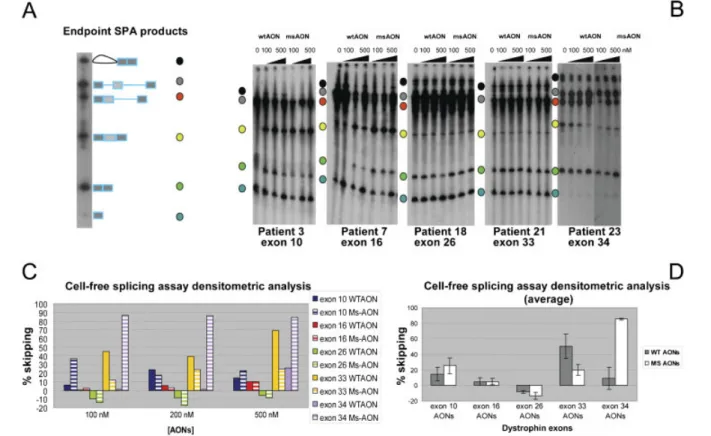
We first identified 55 novel disease-causing point mutations. We selected 5 patients with nonsense or frameshifting mutations in exons 10, 16, 26, 33 and 34. Wild type and mutation specific 2‟OMePS AONs were tested in cell-free splicing assays and in cultured cells derived from the selected patients. The results obtained confirm cell-free splicing assay as an alternative system to test exon skipping propensity when patients‟ cells are unavailable. In myogenic cells, similar levels of exon skipping were observed for wild type and mutation specific AONs for exons 16, 26 and 33, while for exon 10 and exon 34 the efficiency of the AONs was significantly different. Interestingly, in some cases skipping efficiencies for mutated exons were quite dissimilar compared to what previously reported for the respective wild type exons. This behaviour may be related to effect of the mutations on exon skipping propensity and highlights the complexity of identifying optimal AONs for skipping exons with small mutations.
In the second part we compared different techniques to reveal the exon skipping levels in the muscles of 7 different mdx mice. An absolute quantification of the dystrophin transcript amount was possible using a digital array. Results underline the low expression of the dytrophin gene and the amount needed to correctly quantify the exon skipping percentage.
II
List of Publications
I Spitali P, Rimessi P, Fabris M, Perrone D, Falzarano S, Bovolenta M, Trabanelli C,
Mari L, Bassi E, Tuffery S, Gualandi F, Maraldi NM, Sabatelli-Giraud P, Medici A, Merlini L, Ferlini A. Exon skipping-mediated dystrophin reading frame restoration for small mutations. Hum Mutat. 2009 30:1527-34.
II Rimessi P, Sabatelli P, Fabris M, Braghetta P, Bassi E, Spitali P, Vattemi G, Tomelleri G, Mari L, Perrone D, Medici A, Neri M, Bovolenta M, Martoni E, Maraldi NM, Gualandi F, Merlini L, Ballestri M, Tondelli L, Sparnacci K, Bonaldo P, Caputo A, Laus
M, Ferlini A. Cationic PMMA nanoparticles bind and deliver antisense
oligoribonucleotides allowing restoration of dystrophin expression in the mdx mouse. Mol Ther. 2009 7:820-7.
III Spitali P, Heemskerk JA, Vossen R.H.A.M, Ferlini A, den Dunnen J.T., ‟t Hoen P.A.C. and Aartsma-Rus A. Accurate quantification of dystrophin mRNA and exon skipping levels in Duchenne Muscular Dystrophy. Laboratory Investigation. Manuscipt in Press.
III
Contents
1. Phenotype page 3
2. Positional Cloning of the DMD gene 4
3. The DMD gene 7
4. Dystrophin and the dystrophin associated glycoprotein complex (DGC) 10
5. Dystrophin isoforms and homologs 13
6. Mutations 17
7. Revertant fibers 21
8. Animal models 24
9. Current therapies: 27
Pharmacological treatment with corticosteroids 27
10. Potential therapies: 28 Pharmacological treatments: 28 Utrophin Upregulation 28 Myostatin Inhibition 29 Deacetylase Inhibitors 29 Idebenone 30 PTC-124 30 Cell Therapy 31 Gene Replacement 34 Splicing Modulation: 37 Splicing mechanism 37
Antisense-mediated restoration of normal splicing – Rational for exon
skipping 40
Antisense oligonucleotides (AONs) design 42
AONs modification 43
Multi exon skipping 46
Single exon skipping 46
In vitro experiments 46
Restoration of the reading frame in vivo 48
In vivo studies 48
IV
AON delivery without delivery compounds 52
Clinical Trials 54
RNA quantification studies 55
List of abbreviations
2OMe 2'-O-methyl modified
AAV Adeno-associated virus
ABD Actin binding domain
AON Antisense oligonucleotide
BMD Becker muscular dystrophy
DGC Dystrophin associated glycoprotein complex
DGGE Denaturant gradient gel electrophoresis\
DMD Duchenne muscular dystrophy
DNA Deoxiribose nucleic acid
DRP2 Dystrophin related protein 2
ESE Exonic splicing enhancer
ESS Exonic splicing silencer
GRMD Golden retriever muscular dystrophy
HFMD Hypertrophic feline muscular dystrophy
hnRNP Heterogeneous nuclear ribonucleoprotein
LGMD Limb-girdle muscular dystrophy
LNA Locked nucleic acid
nNOS Neuronal nitric oxide synthase
PCR Polymerase chain reaction
PEG Polyethyleneglycol
PEI Polyethylenimine
PMMA polymethylmethacrylate
PMO morpholino oligonucleotides
PNA Peptide nucleic acid
PPMO peptide-conjugated morpholino oligonucleoides
PPNA peptide-conjugated peptide nucleic acid
PS Phosphorothioate
PTT Protein truncation test
RNA Ribose nucleic acid
2
snRNA Small nuclear ribonucleoprotein
SCAIP single-condition amplification/internal primer
SSCA Single strand conformation polymorphism analysis
U2AF U2 auxillary factor
UTR Untranslated region
3 1. Phenotype
Duchenne muscular dystrophy (DMD) is a severe, progressive, muscle-wasting neuromuscular disorder (Emery 2002). The worldwide incidence of DMD is estimated to be 1 in 3500 newborn boys (Moser 1984). The first symptoms involve the lower limbs and appear between the third and fifth year. Patients have hypertrophic calves, show difficulty in running and climbing stairs, run on their tiptoes and frequently fall. Due to weakness of the knee and hip extensors, patients rise from a sitting position using the Gower's maneuver (patients rest on their hands and toes, and then use their hands to push down on their thighs to get into an upright position). Muscle weakness progresses to the shoulder girdle-, upper armand trunk-muscles and patients loose ambulance before the age of 12 (a detailed description of the clinical features can be found in (Emery 1993)).
Histological changes are readily apparent with light microscopy analysis of cross -sections from patient muscle biopsies (Figure 1). They involve variation in fiber size with atrophic and hypertrophic fibers, degeneration and regeneration of the muscle fibers, infiltration of inflammatory cells and fibrosis (Dubowitz 2000). The fiber necrosis results in leakage of the enzyme creatine kinase (CK), resulting in very high serum CK levels in DMD patients (20,000 to 50,000 U/L compared to 80 to 250 U/L in unaffected individuals). These levels decline as patients get older and the overall muscle mass decreases progressively.
Until the early nineties of the last century, patients used to die from respiratory insufficiency in the second decade of their life. Nowadays, patients live into their third decade, due to improved respiratory care and assisted ventilation, and often die as a result of congestive cardio-myopathy (Emery 2002; Simonds 1998). A substantial proportion, however, still dies before their twenties from pneumonia or other respiratory infections.
One third of all affected boys are also mentally impaired. About 20% of DMD patients have an IQ of less than 70 (Emery 2002) and learning difficulties are commonly associated with the disease (Polakoff 1998). In contrast to the muscle wasting, the mental impairment is not progressive.
4 2. Positionl cloning of the DMD gene
Since in general only boys are affected by DMD, it was obvious to geneticists that the gene mutated in DMD patients, had to be located on the X-chromosome. A few, rare, cases of female DMD patients have been described. In the majority of these females, the disease was associated with translocations in the Xp21 band, indicating that the DMD locus was located in this region (Greenstein 1977; Jacobs 1981). Besides this cytogenetical evidence, genetic linkage analysis also localized the DMD locus to the Xp21 band (Davies 1983). Using probes located in and around this band prenatal diagnosis and carrier detection was performed as early as 1985 (Bakker 1985).
Surprisingly, linkage analysis localized Becker muscular dystrophy (BMD) to the same locus as DMD (Kingston 1984). BMD affects 1 in 20,000 men, and patients also suffer from muscle weakness. However, the course of the disease is more benign than for DMD (Emery 2002). Patients have a phenotype that varies from very mild to moderately severe. The age of onset is around 12 years, but some patients remain asymptomatic until later in life. Most patients lose ambulance around 20-30 years after diagnosis (Emery 1993), although some remain ambulant for much longer (England 1990;
5 Mirabella 1998; Yazaki 1999). About 50% of all BMD patients suffer from cardiomyopathy, which sometimes is the main symptom (de Visser 1990; Emery 1993). Severely affected patients die between 40 and 50 years, whereas mildly affected patients have near normal life expectancies. The pERT87 probe, which is deleted in some DMD patients, was shown to be deleted in several BMD patients as well (Hart 1987), suggesting that DMD and BMD are either allelic or that the genes mutated in these diseases are close together. Using different probes the first partial DMD cDNAs were identified by Monaco and collegues in 1986 (Monaco 1986), whereupon the complete 14 kb DMD cDNA could be cloned in 1987 (Koenig 1987). The gene (described in more detail in Chapter 3) was found to contain at least 60 exons and to be dispersed over at least 2000 kb. Deletions in the DMD gene were found in 50% of all patients using cDNA probes (den Dunnen 1987; Koenig 1987). Following studies found similar or even higher percentages of deletions in DMD patients as well as in BMD patients (Davies 1988; Den Dunnen 1989; Forrest 1988). The protein product of the DMD gene was identified shortly after the DMD cDNA. It was named 'dystrophin', because of its identification via the isolation of the Duchenne muscular dystrophy locus (Hoffman 1987). The protein was shown to be approximately 400 kD and to represent 0.002% of total striated muscle protein (Hoffman 1987). Dystrophin could not be detected in muscle tissue isolated from DMD patients. Microscopic analysis of cross -sections revealed that dystrophin is located in the sarcolemma in unaffected muscle tissue, and confirmed that it is absent in muscles from DMD patients (Arahata 1988; Bonilla 1988; Watkins 1988; Zubrzycka-Gaarn 1988) (Figure 1C-F). In contrast to DMD patients, dystrophin could be detected in BMD patients (Figure 1E). Western blot analysis revealed that these dystrophins were of an abnormal size, but present in near normal levels in mildly affected BMD patients, and in low amounts for more severely affected patients (Hoffman 1988). These findings implied that the amount of the protein was more important than the size of the protein. In 1988 Monaco and colleagues proposed the open reading frame hypothesis, by which the phenotypic differences between DMD and BMD patients could be explained (Monaco 1988). This model was based on the breakpoints of intragenic DMD deletions and their effect on the translation of triplet codons into amino acids of the dystrophin protein. They hypothesized that deletions in DMD patients lead to a shift in the open reading frame, resulting in premature stop codons and truncated non-functional, instable proteins, associated with
6 the severe DMD phenoptype. In BMD patients the deletions do not disrupt the open reading frame and internally deleted, but semi-functional dystrophins can be produced, leading to a milder phenotype (See Figure 2 for a schematic depiction). Some studies detected mutations that contradicted this 'open reading frame rule', and suggested that the location of the mutation determined the outcome of the disease (Malhotra 1988; Medori 1989). However, the vast majority of these exceptions consisted of BMD patients carrying an out-of-frame deletion of exon 3-7 (discussed in more detail in Chapter 6). An extensive study revealed that for mutations detected on DNA level the reading frame rule holds true for over 90% of all BMD and DMD patients (Baumbach 1989; Gillard 1989; Koenig 1989), and this rule is generally used as a differential diagnotic and prognostic tool for BMD versus DMD patients.
7 3. The DMD gene
The DMD gene is the largest known gene and consists of no less than 2,2 Mb, making it 80 times as big as the average human gene (estimated to be 30 kb (Nebert 2002)). The gene contributes to approximately 0.1 % of the human genome (Lander 2001). When compared to the average gene (spanning ~6 kb and containing ~10 exons (Lewin 2000)), the DMD mRNA is larger (14 kb) and contains more exons (79) (Roberts 1993). However, there are genes that contain even more exons and have larger mRNAs than the DMD gene, but which are nevertheless shorter. This can be explained by the fact that the coding sequence of the DMD gene accounts for only 0.6% of the gene and the rest of the gene is occupied by huge non-coding introns (Ahn 1993) (Figure 3). The dystrophin gene is also unusually large in other organisms, such as the mouse (Mus musculus; 2.2 Mb where the average murine gene is 27 kb and the pufferfish (Fugu rubripes; 165kb vs. 4,8 kb for an average gene); and much larger than the average gene in the fruitfly (Drosophila melanogaster; 131 kb vs. 11,3 kb for an average gene).
Figure 3. Exons and introns size in the DMD gene. Exons (blue bars) an introns (red bars) are indicated.
8 Since the extreme size of the gene makes it very vulnerable for pathological rearrangements, the question arises why this gene is so large. One hypothesis is, that it prevents the dystrophin protein from being present too early during myogenesis and brain formation. Full-length dystrophins are mainly transcribed in post-mitotic cells in muscle and brain. During mitosis, there is a decrease in transcription and it would be almost impossible to express a gene that takes an estimated 16 hours to transcribe (Tennyson 1995) in early embryonic cells that divide every 24 hours. Another hypothesis is that the introns contain regulatory elements for (alternative) splicing events. This theory is supported by the fact that in-frame exons, which can be skipped without serious consequences, are on average flanked by smaller introns than out-of-frame exons, which will result in the generation of a truncated, non- functional dystrophin when abusively skipped (Pozzoli 2003).
The huge size of the introns requires an enormous effort of the transcription and splicing machinery of the cell. Furthermore, the predicted splice sites of the DMD exons are not significantly stronger than those of other genes (Sironi 2001), yet the splicing machinery manages to correctly splice the 14 kb mRNA from the 2.4 Mb pre-mRNA for the majority of the transcripts. This is even more astonishing if you appreciate that for instance intron 44 is almost 250 kb long, and that exon 2 is flanked by 360 kb of intron sequence (i.e. 190 kb for intron 1 and 170 kb for intron 2), in which many other potential exons are located, with equally good or better splice sites than the exon itself. It may be that the genuine exons are recognized due to additional signals present in the introns, or due to the secondary structure of the pre-mRNA.
The dystrophin pre-mRNA is subjected to alternative splicing throughout its coding sequence (Sironi 2002). In the C-terminal part the in-frame exon 71 and the out-of-frame exon 78 are omitted in 50% of dystrophin transcripts (Feener 1989). The absence of exon 78 results in an alternative C-terminus, which may have a function in development (see Chapter 5). Exons 71-74 code for a syntrophin binding domain and the functional meaning of the loss of this site is not yet known. Several transcripts lacking up to 17 exons in the 5' part have been described (Surono 1997 and 1999). Since most of these skips result in an in-frame mRNA, they are thought to generate internally truncated dystrophins that lack the actin-binding domains. The function of these proteins is as yet unknown, and it may be that they are unstable and/or non
9 functional. Besides the exclusion of exons, one exon has been described that is alternatively inserted into the transcript. Exon 2a is situated in intron 2 and is inserted either between exon 2 and 8 or between exon 2 and 18. These transcripts have been detected in skeletal muscle, small intestine and colon tissue (Pramono 2000). Exon 2a creates a shift in the reading frame. Therefore, the insertion of these exons is more likely to be a naturally occurring artefact (i.e. mistake of the splicing machinery) or an experimental artefact (i.e. cold-shock effect on splicing) than the genuine insertion of an alternative exon. Alternative splicing allows expression of the dystrophins isoforms (see Chapter 5) driven by at least 7 promoters. Three are located before exon 2 (the cortical, muscle and purkinje promoters), whereas the other four are located in intron 29, 44, 5 5 and 62 (Dp260, Dp 140, Dp116 and Dp71/40, respectively) (Byers 1993; Chelly 1990b; D'Souza 1995; Gorecki 1992; Lederfein 1992; Lidov 1995) (Figure 4 A).
10 Another mysterious aspect of the DMD gene is the final exon, which is 2307 bp long, but only codes for the last 3 amino acids of the dystrophin protein, and the last 31 amino acids for the alternatively spliced isoforms that lack exon 78. Several parts of this large 3' UTR are unusually well-conserved in human, mouse and chicken dystrophin (Greener 2002; Lemaire 1988) and a deletion within the 3' UTR has been described in one BMD patient (Love 1990) and a DMD patient (Spitali 2009). This implies that the UTR may have a regulatory function, although one that as yet remains unknown.
4. Dystrophin and the dystrophin associated Glycoprotein Complex (DGC)
Dystrophin consists of 3685 amino acids and is a 427 kD protein (Koenig 1988). The protein is hydrophilic (31% of the amino acids are charged) and does not have stretches of hydrophobic amino acids, indicating that it is not a transmembrane protein. Dystrophin contains four distinct domains (Figure 4 B). The first 240 N-terminal amino acids define the actin-binding domain, which contains two actin-binding sites (Jarrett 1995; Koenig 1990). This domain is followed by a central rod shaped domain, which consists of 24 spectrin-like repeat units interrupted by 4 proline-rich hinge regions (Koenig 1990). It has recently been shown that repeat units 11-17 contain an additional actin-binding domain (Rybakova 1996). The cysteine-rich domain encompasses amino acids 3080 to 3360 and includes 15 cysteines, two EF hand motifs and a ZZ domain (Koenig 1988). The C-terminal domain consists of the final 325 amino acids, and contains two stretches that are predicted to form α-helical coiled coil domains, which are involved in protein-protein interactions (Koenig 1988). Dystrophin is part of the dystrophin-associated glycoprotein complex (DGC) (Figure 5). The cysteine-rich and C-terminal domains of dystrophin bind to several parts of the DGC (Figure 5 A), which can be divided into the dystroglycan complex, the sarcoglycan-sarcospan complex and the cytoplasmatic, dystrophin containing complex (Blake 2002; Yoshida 1994). In skeletal muscle the dystroglycan complex consists of α-dystroglycan and β-dystroglycan, which are both heavily glycosylated (Ibraghimov-Beskrovnaya 1992). Dystrophin binds to β-dystroglycan, a transmembrane protein that binds to the extra-cellular α-dystroglycan. Alpha-dystroglycan on its part binds to the extracellular matrix component laminin-2 (Hohenester 1999; Rentschler 1999; Suzuki 1994). In this way dystrophin provides a mechanical link between the actin cytoskeleton and the extra-cellular matrix of the
11 connective tissue. The sarcoglycan-sarcospan complex includes α-, β-, γ- and δ- sarcoglycan and sarcospan (Blake 2002). These transmembrane proteins are hypothesized to have a function in the stabilisation of the dystroglycan-dystrophin complex (Araishi 1999). The cytoplasmatic part of the DGC includes dystrophin itself, α-dystrobrevin and syntrophin. Alpha-dystrobrevin binds to both dystrophin and α-syntrophin (Ahn 1996). Alpha-α-syntrophin also binds to dystrophin and, additionally, it recruits the enzyme nNOS to the sarcolemma (Ahn 1995; Brenman 1996; Brenman 1995; Yoshida 1995).
In DMD patients the absence of dystrophin results in the complete loss or decrease of other DGC proteins, and in the loss of nNOS at the sarcolemma (Brenman 1995; Ervasti 1990; Ohlendieck 1991a).
In BMD patients, on the other hand, the internally truncated dystrophins are able to bind to the dystrophin associated proteins, since the C-terminal part of the protein is intact. For most BMD patients the DGC proteins can be detected at the membrane, although in some cases at decreased levels (Matsumura 1994a; Matsumura 1993; Mirabella 1998).
12 The function of the DGC is still largely unknown. However, since the complex forms a mechanical link between the cytoskeleton and the extracellular matrix, it is assumed that the DGC has a function in maintaining sarcolemma stability during contraction (Matsumura 1994b). Recent studies suggest that the DGC may also have a function in signal transduction, because some DGC proteins can bind to proteins, which are involved in signalling transduction, such as calmodulin, Grb2 and nNOS (reviewed in (Blake 2002; Rando 2001)). Additionally the DGC seems to be required for the functionality of the blood brain barrier, as its permeability is increased and its integrity is lost in mdx mice (the mouse homologue of DMD, see Chapter 8) (Nico 2003; Nico 2004; Vajda 2002). The loss of blood brain barrier function may underlie the mental retardation observed in a portion of DMD patients. The loss of other DGC proteins can also result in muscular dystrophies. Mutations in the α-sarcoglycan gene found in Limb-girdle muscular dystrophy type 2D (LGMD) and in severe childhood autosomal recessive muscular dystrophy (SCARMD) patients (Piccolo 1995; Roberds 1994). Mutations in the other sarcoglycan genes are found in patients suffering from other types of LGMD (Bonnemann 1995; Lim 1995; Nigro 1996; Noguchi 1995). As yet no mutations in the sarcospan or syntrophin genes have been found in patients suffering from muscular dystrophy or other diseases. Moreover, transgenic sarcospan and syntrophin knockout mice do not develop a muscular dystrophy phenotype (Kameya 1999; Lebakken 2000). Dystroglycan knockout mice, on the other hand, are embryonic lethal (Williamson 1997). Recently, mutations in genes coding for fukutin and fukutin related protein have been identified in congenital muscular dystrophy (CMD) and LMGD-2I patients (Brockington 2001b; Kobayashi 1998). These proteins are thought to be glycosyl-transferases, and are likely to be involved in the glycosylation of α-dystroglycan. Indeed, the expression of α-dystroglycan is decreased in CMD and LMGC-2I patients (Brockington 2001a; Hayashi 2001). The exact pathophysiology of DMD is not yet known, but a hypothetical flowchart describes the most important mechanisms thought to give rise to the muscle wasting that is found in DMD patients. Due to the lack of dystrophin and the DGC complex in DMD patients, the muscle membrane is leaky and more vulnerable for mechanical damage (Blake 2002). This results in an influx of calcium into the muscle fiber, leading to a calcium overload of the mitochondrial matrix and the down-regulation of the transcription of mitochondrial genes, resulting in a metabolic crisis and eventually necrosis (Chen 2000). The calcium
13 influx also over stimulates calcium dependent signal transduction pathways, which eventually causes a downregulation of calcium regulating signal molecules through a negative feedback, enhancing the metabolic crisis (Chen 2000). Finally, the calcium influx activates calcium dependent proteases in general, and the calpaines in particular (Alderton 2000; Spencer 1995). The proteases break down the muscle membrane and modify calcium leak channels, thus augmenting the influx of calcium into the cell (Alderton 2000). Due to the muscle damage the fiber becomes "leaky" and muscle enzymes such as creatin kinase will leak into the bloodstream. Additionally, since nNOS synthesizes the molecule NO that induces vaso-dilatation, the absence of nNOS can cause ischemia after severe mechanical stress. This can lead to necrosis (Sander 2000, Thomas, 1998), which attracts inflammatory cells, such as dendritical cells, mast cells and T-cells, which eventually trigger fibrosis (McDouall 1990; Nahirney 1997).
5. Dystrophin isoforms and homologs
Besides the dystrophin initially detected in skeletal muscle and cardiomyocytes, at least seven promoters drive the expression of other dystrophin isoforms from the DMD gene (Figure 4 B, Table 1). In addition to the muscle dystrophin – named Dp427m, because of its molecular weight (427 kD) and its main site of expression (muscle) – two other full-length dystrophins are produced, i.e. Dp427c and Dp427p (Boyce 1991; Chelly 1990b; Gorecki 1992; Holder 1996). Both isoforms have their own unique first exon, which is spliced to exon two of the Dp427m transcript (Boyce 1991; Gorecki 1992). Dp427c is mainly expressed in the cortical neurons – hence the postfix 'c' – and the hippocampus in the brain (Barnea 1990; Gorecki 1992), whereas Dp427p is expressed in the cerebellar Purkinje cells and at lower levels in skeletal muscle (Gorecki 1992; Holder 1996). There are also four internal promoters from which shorter isoforms are produced; (Byers 1993; D'Souza 1995; Hugnot 1992; Lidov 1995) named after their respective molecular weights. Dp260 is expressed in the retina and is derived from a unique first exon, located in intron 29, which is spliced to exon 30 of Dp427m (D'Souza 1995). Two Dp260 isoforms (Dp260-1 and Dp260-2) are formed by alternative splicing of the first exon and a different translation initiation site. Dp260 lacks the N-terminal domain, two hinge regions and nine repeats, when compared to the fulllength dystrophins. The promoter of Dp140 is located in intron 44 (Lidov 1995). The methionine translation
14 initiation codon, however, is located in exon 51, and as a consequence this protein does contain a large 5' untranslated region (UTR), but no isoform specific amino acids. Dp140 has retained only the last two hinge regions, five of the spectrin-like repeats, the cysteine-rich and C-terminal domain. The protein is found throughout the central nervous system (Lidov 1995). Dp116 has a unique N-terminal part, which is coded by an exon located in intron 55. This exon is spliced to exon 56 of Dp427m, which codes for the distal part of the 21st repeat unit (Byers 1993). This isoform is expressed in Schwann cells. Dp71 and Dp40 derive from a unique first exon in intron 62 that is spliced to exon 63 (Blake 1992; Feener 1989; Hugnot 1992; Lederfein 1992; Tinsley 1993). Both proteins have an alternative C-terminal domain when compared to the other dystrophin isoforms. In Dp71 exon 78 is often omitted, resulting in a shifted reading frame and the insertion of 31 new amino acids instead of the original final 13 amino acids (Lederfein 1992). These amino acids are mainly hydrophobic, whereas the original C-terminal part is hydrophilic. Dp40 uses an alternative poly adenylation site located in intron 70, which results in a shortened C-terminal domain (Feener 1989; Tinsley 1993). Both isoforms are ubiquitously expressed in non-muscle cells (Hugnot 1992; Tinsley 1993). The diversity of dystrophin isoforms is further increased by alternative splicing of the 3' part of the gene (Feener 1989) (see Chapter 3). As yet the functional significance of the different dystrophins is unknown. It is assumed that the full-length isoforms will have a comparable function to Dp427m, i.e. providing a mechanical link between the cytoskeleton and the extracellular compartments. Since the shorter isoforms do not contain an actin-binding domain, these dystrophins can only bind to the DGC proteins and are thought to be involved in the stabilisation and function of non-muscle DGC-like protein complexes (Blake 2002). Dp260 is thought to be required for normal retina function, since this isoform is expressed mainly in the retina and some DMD patients have abnormal electro-retinograms (D'Souza 1995; Pillers 1993). The hydrophobic Dp71 has been detected in embryonic tissues and in developing muscles (Howard 1999; Sarig 1999). It is replac ed by the hydrophilic Dp427 when the muscle cells mature, indicating that it may have a function in development (Howard 1999). The dystrophin isoforms may also have a function in signal transduction, either by binding to signalling molecules themselves or by recruiting proteins that have a function in signalling transduction (reviewed in (Rando 2000)). Dp427c, Dp427p, Dp140 and Dp116 are expressed in brain and the central nervous
15 system. As a result of the mutation one or more isoforms may be absent in addition to the Dp427m isoform, and cause the mental impairment sometimes found in DMD and BMD patients (Byers 1993; Comi 1995; Gorecki 1992; Lidov 1995). In some studies a correlation has been found between the absence of Dp140 (due to mutations spanning the Dp140 promoter) and the presence of mental abnormalities in DMD and BMD patients (Bardoni 2000; Bushby 1995; Felisari 2000). A very recent study shows the correlation between the Full Scale Intelligence Quotients (FSIQ) results with the location of the dystrophin gene mutation suggesting that the risk of cognitive deficit is a result of the cumulative loss of central nervous system (CNS) expressed dystrophin isoforms, and that correct classification of isoform involvement results in improved estimates of risk (Taylor 2010).
In addition to the isoforms derived from the DMD gene, there are also three known dystrophin homologues, which are derived from other genes: utrophin, dystrophin related protein 2 (DRP2) and dystrobrevin. Utrophin (also called dystrophin related protein 1 (DRP1)) shares considerable homology with dystrophin along the entire length on both DNA and protein level (Tinsley 1992).
The utrophin gene is located on chromosome 6 and consists of 74 exons dispersed over 0.9 Mb (compared to 2.4 Mb for dystrophin) (Pozzoli 2002). The 13 kb mRNA gives rise to a 395 kDa protein (Tinsley 1992), which contains an N-terminal actin binding
16 domain, a central rod domain, a cysteine-rich and C-terminal domain (Tinsley 1992). When compared to dystrophin, utrophin lacks repeat units 15 and 19 and hinge regions 1 and 3 (Winder 1995a), and the N-terminal domain of utrophin has a higher affinity for actin (Winder 1995b). Utrophin is ubiquitously expressed (hence the name: ubiquitously expressed dystrophin-like protein (Blake 1992)). In muscle fibers utrophin is expressed in the neuromuscular synapses and the myotendinous junctions, where it participates in post-synaptic membrane maintenance and acetylecholine receptor clustering (Nguyen 1991). In vitro and in vivo studies have shown that utrophin can bind to β-dystroglycan, α-dystrobrevin, syntrophin and the sarcoglycans and it is assumed that utrophin forms a DGC-like complex (Matsumura 1992; Peters 1998). In developing and regenerating muscle, utrophin is upregulated and present along the entire sarcolemma ( Pons 1993). This is also observed in DMD patients (Galvagni 2002; Karpati 1993; Mizuno 1993; Mizuno 1994), suggesting that there is some functional redundancy between dystrophin and utrophin (Blake 2002). However, the utrophin overexpression found in DMD patients is apparently too low to fully prevent muscle degeneration (upregulation of the utrophin gene as a therapeutic strategy for DMD is discussed in Chapter 10).
The dystrophin related protein 2 (DRP2) shows similarity to Dp116. It has a unique proline-rich N-terminal part, followed by two spectrin-like repeats, a cysteine rich domain and a C-terminal domain, which are homologous to those of dystrophin (Roberts 1996). DRP2 is not expressed in muscle, but is expressed in brain where it is associated with the postsynaptic densities and cholinergic neurons (Roberts 2000). The α- and β-dystrobrevin proteins are derived from two different genes and both show homology to the C-terminal part of dystrophin (Blake 1998; Sadoulet-Puccio 1996; Wagner 1993). At least five different α-dystrobrevin isoforms are generated through the use of alternative promoters and alternative splicing (Blake 1996; Sadoulet-Puccio 1996). These isoforms can bind to dystrophin and are located at the neuromuscular junction (Nawrotzki 1998; Peters 1998). Beta-dystrobrevin is expressed in non-muscle cells, and is complexed with utrophin and Dp71 (Blake 1999; Loh 2000). Dystrobrevins contain a domain that can be phosphorylated by tyrosine kinases, and can associate with several proteins involved in signalling transduction (Blake 2002).
17 6. Mutations
The mutation rate for the DMD coding sequence is estimated at 1 x 10⁻⁴ genes per generation (Blake 2002). This is high compared to an estimated average mutation rate of 1 x 10⁻⁵ – 10-6 for human genes (Lewin 2000). As a consequence of this relatively high mutation rate, one third of all mutations are de novo (Laing 1993), and there is a broad variation of different mutations. Intragenic deletions of one or more exons are found in ~65% of all patients (Koenig 1989; White 2002), while duplications are found in 6-8% of all patients (Galvagni 1994; White 2002). Nonsense or frame-shifting mutations in the DMD gene lead to Duchenne muscular dystrophy (DMD) (MIM# 310200), by contrast, Becker muscular dystrophy (BMD)(MIM# 310376) is caused by in-frame mutations that give rise to a smaller but functional protein (Hoffman 1987; Monaco 1988). The interruption or maintenance of the dystrophin reading frame by the gene mutations explains the phenotypic differences observed in approximately 92% of the BMD/DMD cases (Aartsma-Rus 2006a; Koenig 1989). The majority of the deletions and duplications cluster into two hotspot regions: the minor hotspot spans exons 2-20 and the major hotspot region exons 45-53 (Beggs 1990; Liechti-Gallati 1989). The highest numbers of breakpoints are found in the extremely large introns 1, 2 and 7 (major duplication and minor deletion hotspots) and 44 (major deletion hotspot) (Tuffery-Giraud 2009; Fokkema 2005). However, if you correct for the length of the introns (i.e. calculate the number of breakpoints per kb intron), relatively high numbers of breakpoints are located in introns 45 trough 53. There is as yet no explanation for the preferential occurrence of breakpoints in these introns. Extensive analysis of 22 breakpoints in intron 49 revealed that the breakpoints were evenly spread throughout the intron, and a sequence motive near or at the breakpoints could not be identified (Nobile 2002). Further research of breakpoints in introns 47 and 48 showed that most breakpoints cluster around intronic motifs that could predispose for double-stranded DNA breaks (Sironi 2003; Toffolatti 2002). However, these motives are present throughout the gene and not only in intron 47 and 48, so there must be another underlying mechanism for the predisposition of breakpoints in the two hotspots (Sironi 2003). Another explanation is that the breakpoints are in fact evenly spread throughout the gene, but that we observe the hot spots due to the fact that only breakpoints occurring in the hot spots result in a phenotype. This may hold true for breakpoints in introns 22 through 40. A deletion of this region (that contains only in-frame exons) is usually associated with a
18 very mild phenotype. However, even in-frame deletions in the proximal introns can result in a DMD phenotype (Flanigan 2009).
In some cases a mutation detected in affected siblings, cannot be found in the somatic cells of their mother, suggesting that the mutation is only present in part of the germ-line cells of the mother (Bakker 1987). The estimated frequency of this so-called germ-line mosaicism within a family varies between 12 and 20% (Emery 1995). Unexpectedly, it has been shown that deletions found in children from germline mosaic carriers involve the proximal part of the gene in 79% of all cases, while mutations in the distal part were found in only 21% (Passos-Bueno 1992). The reason for this phenomenon is as yet unknown.
For diagnostic purposes the high frequency of deletions clustering in the hotspots is an advantage; 98% of all DMD/BMD deletions can be detected using multiplex PCR of only 8 exons (Beggs 1990; Chamberlain 1988). Southern blot analysis, multiplex amplifiable probe hybridisation (MAPH), Multiplex ligation-dependent probe amplification (MLPA), high resolution melting curve analysis (MCA) and comparative genomic hybridization array (CGH) can be used to detect the precise range of both deletions and duplications (Hodgson 1992; White 2002; Janssen 2005; Almomani 2009; Hegde 2008; Saillour 2008; del Gaudio 2008; Bovolenta 2008). In about one third of the patients small deletions and insertions or point mutations are found (Deburgrave 2007). These mutations are evenly spread throughout the gene and are therefore harder to find (Flanigan 2009). They can be detected by denaturant gradient gel electrophoresis (DGGE), single strand conformation polymorphism analysis (SSCA), direct sequencing or chemical cleavage of each individual exon, the protein trunction test (PTT), or by single-condition amplification/internal primer (SCAIP) (Dolinsky 2002; Flanigan 2003; Hofstra 2004; Roest 1993, Kilimann, 1992; Tuffery 1993; Tuffery-Giraud 1999; Spitali 2009; Flanigan 2009). The majority of the detected mutations results in-frameshifts, non-sense codons, the abolishment of splice sites, or the introduction of cryptic splice sites (Kilimann 1992; Tuffery-Giraud 1999; Flanigan 2009). DMD causing missense mutations account only for 0,18% of all mutations (Flanigan 2009) and were located in an actin-binding domain or in the part of the cysteine rich region that binds to β-dystroglycan (Goldberg 1998; Lenk 1996; Prior 1995; Flanigan 2009).
19 The reading frame rule holds true for over 93% of all BMD and DMD patients (Aartsma-Rus 2006a), however exception to the rules are found expecially for 5‟ gene deletions occurring in BMD patients, a region known to be hotspot for exceptions (Kesari 2008). For in-frame mutations the phenotype is also to some extent determined by the location and the size of the deletion (Arikawa-Hirasawa 1995; Beggs 1991; Fanin 1996). Huge in-frame deletions removing up to 35 exons in the central rod domain have been described for relatively mild Becker patients (England 1990; Fanin 1996; Mirabella 1998). However, larger deletions are always associated with a DMD phenotype, suggesting that at least a small part of the rod domain is required for proper dystrophin function (Fanin 1996). Deletions removing only the first part of the central rod domain have been found in a-symptomatic individuals, in persons with elevated CK levels and in patients suffering from muscle cramps upon exercise (Angelini 1994a; Gospe 1989; Ishigaki 1996). In-frame deletions in the major hotspot generally cause typical BMD, while deletions in the actin-binding domain cause severe BMD (Beggs 1991). Deletions removing both the actin-binding domain and part of the central rod domain usually cause DMD (Arikawa-Hirasawa 1995; Fanin 1996; Vainzof 1993). This may be explained by the fact that a third actin-binding site is present in the central rod domain (Rybakova 1996). The removal of the first two actin-binding domains (located in the actin-binding domain) may partially be compensated for by this central rod actin-binding domain and therefore cause severe BMD, whereas the deletion of all three domains results in DMD. Deletions in the C-terminal domain are usually associated with DMD (Bies 1992). In addition to the location of the deletion, the amount of dystrophin produced in the patients also correlates with the phenotype: the more dystrophin a patient produces, the milder the phenotype (Angelini 1994a; Hoffman 1988). No dystrophin or up to 3% of normal levels are usually found in DMD patients, whereas levels of over 30% of dystrophin (of an abnormal weight) are found in BMD patients (Neri 2007).
Certain in-frame mutations are always associated with a DMD phenotype, whereas a number of out-of-frame mutations lead to a BMD phenotype (Fokkema 2005). In-frame mutations may be too large to result in a functional dystrophin or they may be located in areas of the protein, which are indispensable (see above). However, for a significant part of in-frame DMD patients no dystrophin protein can be detected. This may be because additional exons are be spliced out as a consequence of the deletion, resulting
20 in out-of-frame transcripts. Alternatively, the in-frame deletion could lead to an unstable dystrophin. Out-of-frame mutations on DNA level associated with a BMD phenotype do not necessarily have to be inconsistent with the reading frame rule on RNA level. For instance, two nonsense mutations in the in-frame exon 29 disrupt a sequence that is required for the proper inclusion of this exon (Ginjaar 2000; Franz 2000). As a result, exon 29 (and thus the truncating mutation) is not always included in the transcript by the splicing machinery. Dystrophin lacking the amino acids coded by exon 29 can be detected in these patients, explaining why they have a BMD and not a DMD phenotype. Unfortunately, for the majority of all patients mutations are determined on DNA level only, and not confirmed on RNA level. Therefore, it is not known whether apparent inconsistencies with the reading frame rule are also present on RNA level. There are also a number of deletions (detected on DNA level), which are found in both DMD and BMD patients. The majority of these mutations start either in intron 3 or in intron 44. Exon 44 skipping in patients with a deletion of exon 45 (Prior 1997; Roberts 1991), or a duplication of exon 44 (Aartsma-Rus personal communication) has been described, and this might explain the discrepancy: high levels of alternative splicing of exon 44 will generate in-frame transcripts and internally deleted dystrophins. These will therefore be associated with a BMD phenotype, while no or low levels of exon 44 skipping will result a DMD phenotype. The levels of alternative splicing are influenced by the different splicing regulation patterns of each individual and will additionally be influenced by the extent of the deletions and the location of the breakpoints in intron 44. Even though the deletions may seem identical on DNA levels, in reality the exact location of the breakpoints differs between individuals of different families. However, as yet no intronic regulating sequences that influence the inclusion or exclusion of exon 44 have been identified. The deletion of exon 3-7 is the most commonly found and most extensively investigated deletion that is associated with both phenotypes. Alternative splicing of exon 2 would restore the reading frame for these patients and has been described for two BMD patients albeit at levels of 1-2% (Chelly 1990a). Despite extensive research exon 2 skipping has not been described elsewhere for patients with this deletion (Gangopadhyay 1992; Winnard 1993; Winnard 1995). An explanation for this phenomenon may be the use of an alternative translation initiation codon (Winnard 1995). This hypothesis is supported by the fact that there are three in-frame ATG codons present in exon 8. Furthermore, the dystrophins detected in these BMD patients
21 where shown to lack the exon 1 and 2 encoded sequence, but did contain the exon 8 encoded sequence (Winnard 1995). In addition to BMD and DMD, mutations in the DMD gene have also been found in X-linked dilated cardiomyopathy (XLDC) patients (Muntoni 1993). These patients have no signs of skeletal myopathy but suffer from rapidly progressive congestive heart failure (Milasin 1996). Without heart transplantation premature death usually occurs within 2 years after diagnosis (often in their twenties). Most mutations are present in the first exon of the muscle specific dystrophin isoform and completely abolish dystrophin expression in the heart (Ferlini 1999, Neri 2007). In skeletal muscle, on the other hand, upregulation of the purkinje and brain isoforms rescues the phenotype (Bastianutto 2001). This upregulation is controlled by the dystrophin muscle enhancer 1 that is located downstream of the muscle specific first exon, and which increases the expression of the Dp427c and Dp427p isoforms exclusively in skeletal muscle cells. In-frame deletions involving exon 48 and exon 49-51 have been detected in less severely affected XLCD patients, and in BMD patients (with or without XLDC) (Ferlini 1999 and Ferlini personal communication).
7. Revertant Fibres
Individual dystrophin-positive fibers have been found in DMD patients and in otherwise dystrophin-negative mdx mice (see Chapter 8) (Thanh 1995; van Deutekom 2007; Rimessi 2009) (Figure 6). These 'revertant fibers' grow in clusters and comprise 1% to 10% of all fibers. The frequency of these fibers increases with age in both DMD patients (Fanin 1995) and mdx mice (Lu 2000), and after mutagene doses of X-rays in mdx mice (Hoffman 1990). The revertant dystrophins are located at the membrane and may be functional. The correlation between the amount of revertant fibers and the severity of the DMD phenotype is still controversial (Fanin 1995; Nicholson 1993; Thanh 1995). Revertant dystrophins were shown to lack domains coded by the exons adjacent to exons deleted in the DMD patients (Fanin 1995; Thanh 1995). By omitting these exons the reading frame was restored, explaining why BMD-like, probably semi-functional dystrophins could be produced. Revertant fibers have been studied more extensively in mdx mice (Lu 2000). A few isolated dystrophin positive fibers were detected in newborn mice and the amount of revertant fibers, the number of fibers per cluster and the length of the fibers increased with age (2 weeks to 18 months). Not only was the dystrophin
22 synthesis restored in revertant fibers, dystrophin and the DGC-complex were also correctly located at the membrane, providing more evidence that the revertant dystrophin is at least partly functional. Different inframe transcripts lacking multiple exons were identified in different clusters. In most dystrophins only domains immediately surrounding the mutated exon 23 were missing.
However, dystrophins lacking up to 35 exons were also detected. Potential mechanisms responsible for the occurrence of revertant fibers are somatic reversion and alternative splicing (Klein 1992; Lu 2000). There are arguments in favour of, and against, both mechanisms. Somatic reversion, i.e. the occurrence of secondary, frame-restoring mutations in muscle or muscle precursor cells, fits with the clonal nature of the revertant fibers, their increase with age and their appearance after exposure to mutagenic doses of X-rays. Conversely, mutations are generally proliferation-related events and would be
23 more likely to occur during the many cell divisions in the embryogenesis than in differentiated muscles. However, only very low levels of revertant fibers are found in newborn mice. Furthermore, in situ hybridisation experiments were not sensitive enough to detect deletions on DNA level with a probe for intron 22 in mdx revertants (the probe detected 80% positive nuclei in both dystrophin positive and dystrophin negative fibers). This means that there is either no deletion present or that the deletion is present in too low a number of nuclei to detect a significant difference. This finding, however, does not exclude the possibility of a point mutation in a splice site. In fact, it was recently shown that the substitution of one base pair in the acceptor splice site of exon 23 induces several different alternative splicing patterns that involve 12 or more exons (Bertoni 2003). The other potential mechanism, alternative splicing, has been described for the DMD gene throughout the coding sequence in both patients and unaffected individuals (Sironi 2002). Furthermore, in a substantial part of revertant fibers investigated in mdx mice, two non-consecutive domains were absent in the revertant dystrophins, located around exon 23 and around exon 40-46 (Lu 2000). Intuitively one would think this to be more likely the result of two independent alternative splicing events than of two different new mutations. It should be noted, however, that deletions of two non-consecutive parts of the DMD gene have been reported for DMD patients (Zatz 1998). Finally, one should also not overlook the possibility of a combination of the two mechanisms. Mutations frequently occur in the mutation hotspot located between exon 45-55 and alternative splicing around exon 23 has been observed both in normal and mdx mice (personal observation).
Alternative splicing seems to be at odds with the clonal nature of the fibers. Lu and colleagues proposed that the splicing pattern is changed during degeneration and regeneration and that a pattern of frame-restoring splicing is preserved by functional selection (Lu 2000). The revertant dystrophin would protect the fiber from degeneration and the "splicing imprinting" would expand to adjacent fibers, thus explaining the clonal nature of revertant clusters. However, this hypothetical mechanism has never been demonstrated.
24 8. Animal models
Several animal models carrying mutations in the DMD gene were reported, among these the mdx mouse, the double knockout mdx/utrn-/- mice and the golden retriever muscular dystrophy (GRMD) dog are used most.
The mdx mouse is a naturally occurring dystrophin-deficient mutant, which was first described in 1984 in a colony of C57BL/10 mice (C57BL/10ScSnJ) (Bulfield 1984) and it is now called C57BL/10ScSn-Dmdmdx/J; it is readily available from commercial breeders and thus widely used in basic and translational research. It carries a point mutation in exon 23 of the mouse dystrophin gene introducing a premature stop codon, which leads to the absence of full-length dystrophin. Mdx mice have a slightly shorter life span as compared to wildtype controls (Chamberlain 2007). The muscle degeneration in mdx mice differs from DMD patients in that it appears in waves and is not a continuum. At first, a marked degeneration and regeneration of muscle fibers is observed at a young age (2–4 weeks (McGeachie 1993)), which results in an increase in the number of newly differentiating myofibers characterized by centralized nuclei and an increased heterogeneity in myofiber size. In parallel, necrosis is observed in muscles at this early stage (Grounds 2004; Messina 2006) while it decreases again after 60 days (Spencer 1996). Subsequently, loss of muscle tissue is slow and general muscle weakness in cage-reared animals is not evident until later in life (Lefaucheur 1995). Fibrosis in most mdx mouse muscles is less pronounced than in DMD patients with the exception of the diaphragm muscle (Stedman 1991; Connolly 2001; Rolland 2006). Although absolute muscle force of limb muscles remains comparable to unaffected mice, the relative force normalized to body weight decreases by approximately 20% and 50% at the age of 8– 10 weeks and 4 months, respectively (Messina 2006; Connolly 2001; Raymackers 2003). However, others could not confirm this finding (Burdi 2006; Frysse 2004), which is probably based on variations in the mice strains (e.g. body weight, cooperation of the animals in force measurement assays) and on different experimental procedures. Similarly, a reduced force of contraction has also been reported in isolated muscle preparations although the lack of standardized conditions makes it again difficult to compare the results obtained by different groups (Deconinck 1998; Lynch 2001; Louis 2004; Laws 2004). These discrepancies clearly show that the availability of standardized experimental protocols would be of advantage both to elucidate basic
25 aspects of the pathology and to test emerging therapeutic strategies. Respiratory pathology is evident in 16-month-old mice but not in 4-month-old mice, showing a worsening of respiratory capacity with age, in line with the increased histological damage of respiratory muscles (Gayraud 2007). Abnormal cardiac function is also seen in the mdx mouse sharing several characteristics of DMD-associated cardiomyopathy (Quinlan 2004). For example, changes in the electrocardiogram (ECG) and echocardiogram have been reported in mice that are older than 6 months (Bia 1999; Wehling-Henricks 2005; Spurney 2008). This change in cardiac function is paralleled by an increase in myocardial fibrosis and the occurrence of foci of myocardial necrosis and inflammation (Quinlan 2004; Van Erp 2006; Cohn 2007; Buyse 2009). Mdx mice have served as a model for several studies on calcium homeostasis and alterations thereof. Basal cytosolic calcium levels were reported to be identical in mdx and wild-type mice (De Backer 2002) showing a very robust calcium homeostasis, possibly due to elevated expression of the calcium-buffer parvalbumin (Gailly 1993; Sano 1990). Nevertheless, [Ca2+] in the subsarcolemmal compartment (Mallouk 2000) and in the sarcoplasmic reticulum (Robert 2001) was increased and levels of inositol 1,4,5-triphosphate (IP3) and of the IP3 receptor were higher in mouse and human dystrophic cell lines (Liberona 1998). Muscle pathology in the mdx mouse can be aggravated by forced exercise, a method introduced to allow a better evaluation of the efficacy of experimental therapies. Several exercise paradigms have been described (Granchelli 2000; Vilquin 1998; Hodgetts 2006). Muscle damage of exercised mdx animals is increased as reflected by an increased number of muscle fibers with centralized nuclei and increased fibrosis (Burdi 2006; De Luca 2005). Plasma creatine kinase levels are further elevated, calcium homeostasis is more severely impaired and fore-limb strength is generally reduced (Rolland 2006; Burdi 2006; Fraysse 2004; De Luca 2005) while body weight remains unchanged. Cardiomyopathy has been described for the exercised mdx mouse (Nakamura 2002), however, a systematic analysis of the impact of exercise on cardiac pathology is still lacking.
In an attempt to aggravate the pathology of the mdx model and to study utrophin-based gene therapy strategies, utrophin mutant mice (on C57B5/129J background) were crossed with C57BL/10 mdx mice to obtain double knock-out mice (Deconinck 1997; Grady 1997). Mice lacking both dystrophin and utrophin share many phenotypical hallmarks with DMD. The mice have a markedly reduced life span of 4–20 weeks,
26 severe muscle weakness with joint contractures, kyphosis and a pronounced growth retardation that starts around weaning. Necrotic fibers and connective tissue are visible in the diaphragm already 6 days after birth and centrally nucleated fibers are evident at 2 weeks. Interestingly, the diaphragm is not more affected than that of the mdx mice, suggesting that utrophin does not compensate for the lack of dystrophin in this muscle (Deconinck 1997; Grady 1997). The skeletal muscles appear normal at birth but show first signs of degeneration/regeneration already after 2 weeks. Although the dystrophic pathology is similar to mdx mice at 4–5 weeks of age, it then remains severe in contrast to the improvements seen in mdx mice. By 10 weeks of age, fibrosis is evident in tibialis anterior muscle. Occasionally, larger necrotic areas are observed in the heart (Grady 1997). The neuromuscular and the myotendinous junctions in mdx/utr-/- mice show a marked reduction in membrane folds, presumably as a result of the lack of utrophin (Deconinck 1997; Grady 1997). Therefore, both the disease progression and the phenotype of these double mutant mice resemble the progression of muscle dystrophy in DMD patients and the interstitial fibrosis of skeletal muscle lends itself to assess the efficacy of anti-fibrotic treatments.
In the golden retriever, the best characterized dog model, the disease results from a single base pair change in the 30 consensus splice site of intron 6, leading to skipping of exon 7 and alteration of the reading frame in exon 8, which creates a premature stop codon (Sharp 1992). Unlike the dystrophin-deficient mdx mouse, GRMD dogs suffer from a rapidly progressing, fatal disease similar to DMD. There is, however, a large variation in disease severity as some pups survive only for a few days, while others are ambulant for months or even years (Ambrosio 2008). To account for this variation, it is possible to distinguish between the severe and the less severe pathology (Ruegg 2009). Incomplete muscle repair in GRMD results in progressive weakness and gait abnormalities at the age of 6–9 weeks leading to muscle atrophy, fibrosis and contractures by 6 months (Ruegg 2009). The growth of affected pups is delayed and their walking ability is impaired. The dogs become less active than non-affected littermates at about 9 months of age. At this time, their gait is still uncoordinated and their limbs are abducted (Valentine 1988). Severely affected dogs have difficulties to rise and can walk only a few steps. At the age of 12 months, pharyngeal and esophageal dysfunctions start to develop and respiratory capacity is decreased. Kyphosis develops by the 6th month of age. Moreover, as seen in DMD patients, GRMD
27 dogs display selective muscle involvement although the most affected muscles in dogs (i.e. tongue, masticatory and trunk muscles) are different to those of humans (Kornegay 2003). Myocardial involvement is much more evident in the golden retriever than in other animal models, matching very closely the cardiac complications encountered in DMD patients. Atrophy already appears during the neonatal period (Valentine 1988), although histological changes are generally not present up to 3 months of age (Moise 1991). Systolic dysfunction and left ventricular dilation becomes apparent after several months or years (Moise 1991), although Tissue Doppler Imaging has allowed the detection of severely impaired systolic and diastolic indices in young dogs (25 ± 11 weeks) (Chetboul 2004). Recently, NMR techniques have been applied to assess differences between dystrophic and healthy muscles in the dog, a method that may be useful to monitor a potential therapeutic response (Thibaud 2007).
Other animal models are the HFMD cat (Gaschen 1999), the mdx 2CV-5CV mice (Chapman 1989), the mdx52, mdx/myoD-/-, mdx/adbn-/-, mdx/a7 integrin-/-, mdx/PV-/-, the Beagle (CXMDJ) (Ruegg 2009) and the Cavalier King Charles Spaniels dogs (Walmsley 2010).
Finally a mouse carrying the entire human DMD gene on a yeast artificial chromosome (YAC) was generated (hDMD mouse) („t Hoen 2008). RT-PCR, Western blot and immuno-histochemical analyses confirmed the expression of the human dystrophin in muscle, heart, kidney and testis. In the mdx mouse, the human dystrophin was able to rescue the dystrophic phenotype („t Hoen 2008). This system allows for the testing of genetic therapies affecting the human gene in a mouse background.
9. Current therapies
Pharmacological Treatment with Corticosteroids
Glucocorticoid administration is currently the only drug treatment known to offer real clinical benefit to patients suffering from Duchenne muscular dystrophy (DMD). Glucocorticoids used to treat DMD include prednisone (Manzur 2004) and its oxazoline derivative, deflazacort (de Groot 2006). DMD patients treated with glucocorticoids exhibit delayed progression of muscle weakness (Mendell 1989) and remain ambulatory for a greater period of their lives (Griggs 1991). Different doses as well as different
28 intermittent schemes of administration have been tried in clinical trials with the aim of reducing side effects (Angelini 1994b; Merlini 2003; Biggar 2004; Connolly 2002; Beenakker 2005). However, there is as yet no internationally accepted consensus on which corticosteroid treatment scheme is the best for patients with DMD. In addition, there has yet to be a randomized study involving a head-to-head comparison. The mechanism by which patients benefit from glucocorticoid treatment is not fully understood, although it is thought that the clinical benefits arise in part from the anti-inflammatory and immunosuppressive effects of these drugs (Tidball 2004). St Pierre and colleagues have shown that deflazacort treatment of the mdx mouse can alleviate symptoms of the dystrophic pathology and results in the stimulation of utrophin A expression in skeletal muscle fibers (St Pierre 2004) via an IRES element located at the utorphin 5‟UTR (Miura 2008).
10. Potential Treatments
Pharmacological Treatments
Utrophin Upregulation
The utrophin gene spans 1 Mb, giving rise to a 13 kb cDNA and a 400 kD protein (Pozzoli 2002). Dystrophin and utrophin show over 80% similarity in the actin-binding, cysteine-rich and C-terminal domains, and 35% similarity in the central rod domain (Pearce 1993; Tinsley 1992). Utrophin is ubiquitoussly expressed, but in adult muscle utrophin expression is restricted to intramuscular nerves, blood vessels and myofibers, where it is mainly located in acetylcholine receptor rich crests at the neuromuscular junctions, and at the myotendinious junctions (Khurana 1991; Nguyen 1991; Ohlendieck 1991b). Here it recruits DGC proteins into a DGC-like complex and is probably involved in maintaining the structural integrity of the postsynaptic cytoskeleton (Love 1993; Matsumura 1992). In developing and regenerating skeletal muscle utrophin is located along the sarcolemma, where it is later replaced by dystrophin (Helliwell 1992; Lin 2000; Pons 1993).
In DMD patients and mdx mice utrophin expression is upregulated and the protein is located along the entire sarcolemma, where it probably compensates to some extent for
29 the lack of dystrophin (Galvagni 2002; Karpati 1993; Khurana 1991; Mizuno 1993; Mizuno 1994). This theory is supported by the finding that dystrophin-utrophin double knockout mice have a very severe phenotype (Deconinck 1997). It is obvious that the upregulated levels of utrophin in mdx mice and DMD patients cannot fully compensate for the loss of dystrophin. However, transgenic studies with mice carrying utrophin overexpressing cDNA constructs have shown that a 2-3 fold increase of utrophin expression in the muscle could improve the phenotype of mdx mice significantly, and that a 10-fold increase could prevent the dystrophic phenotype entirely (Tinsley 1998). Thus the upregulation of utrophin gene expression is a promising approach that may be therapeutically applicable to DMD patients. Drugs to enhance utrophin expression in cultured cells and animal models have been identified by Summit PLC (John Tinsley and Kay Davies, UK), BioMarin Pharmaceutical Inc. and PTC Therapeutics. Clinical trials are under preparation.
Myostatin inhibition
Myostatin inhibits muscle growth. Knock-out animals for myostatin leads to increased muscle formation in animals (Belgium blue cattle, Texel sheep, greyhounds, mice) and humans. This could compensate for the loss of muscle tissue in Duchenne patients and can be achieved by antibodies for myostatin. Myostatin antibodies have been tested in
healthy volunteers and were deemed safe
(http://www.clinicaltrials.gov/ct2/show/NCT00563810?term=myo-029&rank=1). They
were consecutively tested in adult patients with muscle diseases
(http://www.clinicaltrials.gov/ct2/show/NCT00104078?term=myo-029&rank=2). While treatment was safe, it did not result in an increase in muscle mass in the patients. However, patients were only treated for 28 days, which might not have been long enough.
Deacetylase Inhibitors
Pharmacological interventions that increase myofiber size counter the functional decline of dystrophic muscles as for myostatin inhibitors (Zammit 2002). Minetti and coworkers showed that deacetylase inhibitors increase the size of myofibers in dystrophin-deficient
30 (mdx) and a-sarcoglycan (a-SG)–deficient mice by inducing the expression of the myostatin antagonist follistatin3 in satellite cells. Deacetylase inhibitor treatment conferred on dystrophic muscles resistance to contraction-coupled degeneration and alleviated both morphological and functional consequences of the primary genetic defect (Minetti 2006). Colussi and colleagues elucidated the role of HDAC2 in the pathogenesis of Duchenne muscular dystrophy and indicate that HDAC2 inhibition by NO-dependent S-nitrosylation is important for the therapeutic response to NO donors in mdx mice for using deacetylase inhibitors in the pharmacological therapy of muscular dystrophies (Colussi 2008).
Idebenone
Buyse and coworkers tested SNT-MC17/idebenone in the mdx mouse, based on the drug‟s potential to improve mitochondrial respiratory chain function and reduce oxidative stress which is themajor cause of heart failure and death inDMD patients (Buyse 2009). In this study, 200 mg/kg bodyweight of either SNT-MC17/idebenone or placebo was given from age 4 weeks until 10 months in mdx and wild-type mice. Idebenone treatment significantly corrected cardiac diastolic dysfunction and prevented mortality from cardiac pump failure induced by dobutamine stress testing in vivo, significantly reduced cardiac inflammation and fibrosis, and significantly improved voluntary running performance in mdx mice. A Phase II extension trial where the effect of longer treatment was tested is now fully recruited and ongoing whilst a Phase III study has also been initiated and is currently recruiting patients.
PTC-124
PTC124 {3-[5-(2-fluorophenyl)-1,2,4-oxadiazol-3-yl]benzoic acid} is a chemical entity that selectively induces ribosomal readthrough of premature but not normal termination codons. Only patients with nonsense mutations would benefit from PTC124. However, 40% of the small mutations cause a frameshift (e.g., through a one or two nucleotide deletion or insertion, or by interfering with normal splicing), and here PTC would not be applicable (Aartsma-Rus 2009a). PTC124 activity, optimized using nonsense-containing reporters, promoted dystrophin production in primary muscle cells from humans and