U
NIVERSITÀD
EGLIS
TUDID
IM
ESSINAT
ESI DID
OTTORATO DIR
ICERCA INB
IOLOGIAA
PPLICATAE
M
EDICINAS
PERIMENTALEC
URRICULUM INM
EDICINAS
PERIMENTALEXXIX CICLO
SSD BIO/14
The role of central PRCP in the metabolism
regulation
Candidato:
D
OTT.
G
IUSEPPEB
RUSCHETTA
Correlatore: Relatore:
Ch.ma Prof.ssa Ch.ma Prof.ssa
S
ABRINAD
IANOE
MANUELAE
SPOSITOCoordinatore:
Ch.mo Prof. SALVATORE CUZZOCREA
INTRODUCTION ... 4
1.OBESITY EPIDEMIC ... 4
1.1CONSEQUENCES OF OBESITY ... 9
1.2FINANCIAL EFFECTS OF OBESITY ... 9
1.3EFFECTS OF OBESITY ON EMPLOYMENT AND WAGES ... 10
1.4ENVIRONMENTAL FACTORS ... 10
2.HYPOTHALAMIC CIRCUITRY REGULATING FOOD INTAKE ... 11
2.1HYPOTHALAMIC MELANOCORTIN SYSTEM AND CONTROL OF ENERGY METABOLISM ... 15
2.1.1THE NPY/AGRP NEURONS AND THE OREXIGENIC PATHWAY ... 17
2.1.2THE POMC NEURONS AND THE ANOREXIGENIC PATHWAY ... 18
3.HORMONAL REGULATION OF THE HYPOTHALAMIC MELANOCORTIN SYSTEM ... 36
3.1LEPTIN ... 36
3.2INSULIN ... 37
3.3GHRELIN ... 38
3.4CIRCULATING NUTRIENTS ... 41
6. AIMS ... 44
AIM1:TOELUCIDATETHEROLEOFPRCPINNYP/AGRPEXPRESSINGNEURONS OFTHEARCUATENUCLEUS. ... 44
AIM2:TOELUCIDATETHEROLEOFPRCPINMC4R-EXPRESSINGNEURONS. ... 44
7. MATERIAL AND METHODS ... 45
7.1ANIMALS ... 45
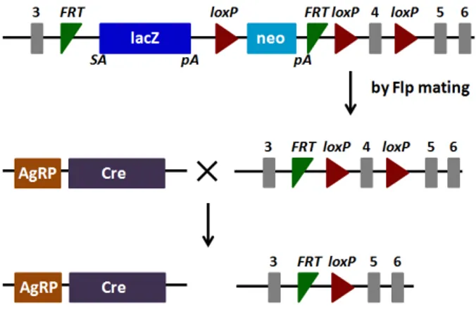
7.2GENERATION OF MICE WITH SELECTIVE DELETION OF PRCP IN AGRP NEURONS ... 45
1.3GENOTYPING ... 46
7.4METABOLIC CHAMBER RECORDINGS ... 47
7.5REAL-TIME PCR ... 48
7.6GLUCOSE AND INSULIN TOLERANCE TESTS ... 48
7.7 CFOS IMMUNOSTAINING ... 49
7.8ELECTRON MICROSCOPY ANALYSIS ... 49
7.9STATISTICAL ANALYSIS ... 50
8. RESULTS ... 52
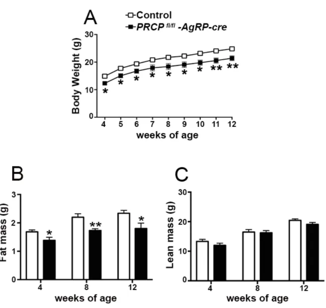
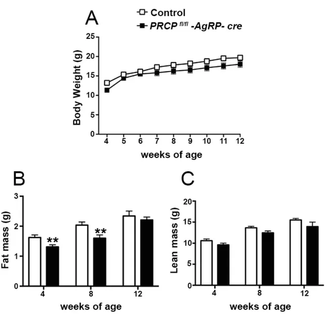
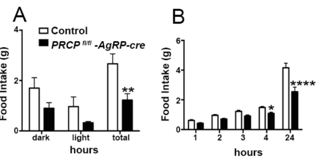
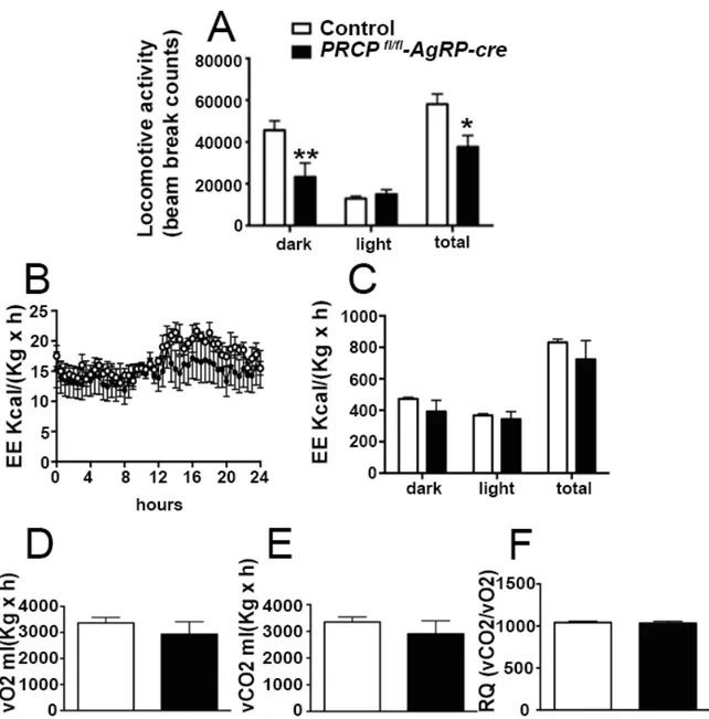
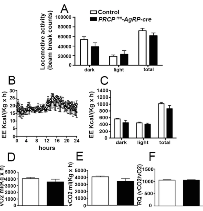
8.1SELECTIVE DELETION OF PRCP IN AGRP NEURONS ALTERS ENERGY BALANCE IN A GENDER-SPECIFIC MANNER ... 52
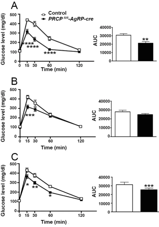
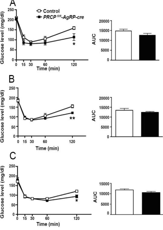
8.2GENDER-SPECIFIC EFFECT OF SELECTIVE DELETION OF PRCP IN AGRP NEURONS ON INSULIN SENSITIVITY ... 57
8.3SELECTIVE DELETION OF PRCP IN AGRP NEURONS INDUCES DECREASED AGRP NEURONAL ACTIVATION IN MALE MICE ... 61
8.4PRCP IN AGRP NEURONS MEDIATES GHRELIN-INDUCED FOOD INTAKE ... 63
8.5ABLATION OF PRCP IN AGRP NEURONS PREVENTS HYPERPHAGIA AND ALTERS ENERGY BALANCE DURING HIGH FAT DIET (HFD) FEEDING IN BOTH MALE AND FEMALE MICE ... 64
8.6INSULIN SENSITIVITY IN PRCPFL/FL-AGRP-CREMICE ... 68
8.7MITOCHONDRIAL SIZE IN ACTIVATED AGRP NEURONS OF FASTED ANIMALS ... 70
8.8INCREASED INTERACTIONS BETWEEN ER AND MITOCHONDRIA IN AGRP NEURONS OF FASTED ANIMALS ... 71
9.2GENOTYPING ... 73
9.3METABOLIC PHENOTYPE ANALYSES ... 74
9.4BLOOD PRESSURE MEASUREMENT ... 74
10. RESULTS ... 75
10.1SELECTIVE DELETION OF PRCP IN MC4R-EXPRESSING NEURONS ALTERS ENERGY BALANCE ... 75
10.2GLUCOSE HOMEOSTASIS ... 77
10.3ROLE OF PRCP IN THE REGULATION OF BLOOD PRESSURE ... 78
11. DISCUSSION ... 80
Introduction
The role of the central melanocortin system in the regulation of energy metabolism has received much attention during the past decade since gene mutations of key components in melanocortin signaling cause monogenic forms of obesity in animals and humans. In the arcuate nucleus of the hypothalamus the prohormone proopiomelanocortin (POMC) is posttranslationally cleaved to produce α-melanocyte stimulating hormone (α-MSH), a peptide with anorexigenic effects upon activation of the melanocortin receptors (MCRs). αMSH undergoes extensive posttranslational processing and its in vivo activity is short -lived due to rapid degradation. The enzymatic process that controls α-MSH inactivation is not completely understood. Recent evidence suggests that prolyl carboxypeptidase (PRCP) is an enzyme responsible for α-MSH degradation. PRCP is widely expressed in the body in organs such as the liver, lung, kidney and brain, with a variety of known substrates such as plasma prekallikrein, bradykinin, angiotensins II and III, suggesting its role in the processing of tissue- specific substrates. As with many key melanocortin peptides, gene mutation of PRCP causes a change in the metabolic phenotype of rodents. The purpose of our study is to evaluate the role of PRCP in energy metabolism.
1. Obesity epidemic
The obesity epidemic has received considerable attention worldwide. Numerous studies have documented that the instances of obesity are climbing around the world. Obesity rates have been rising in virtually all population groups, regardless of the demographic or socio-economic status [1-3]. The growth in the population fraction with unhealthy body weight
was particularly high in the U.S., but it has reached worrisome proportions in other developed and developing countries as well. The prevalence of obesity in U.S. adults more than doubled between the mid-1970s and late 1990s with the most dramatic growth occurring in the 1980s [2]. It increased more than three-fold among children and adolescents over that same period [4]. As a result obesity now is a major public health problem in the United States [5]. Approximately 30.4% of adults (≥ 20 years) are obese and 16% of children (6–19 years) are overweight [6] (Figure 1). Adult obesity is associated with excess mortality and a myriad of health problems and overweight children are at greater risk of becoming overweight adults [7]. Worldwide, the estimated number of overweight adults exceeds one billion, of which at least 300 million qualify as obese.
Figure 1: Percent of obese adults in the U.S.A (Body Mass index of 30+)
The terms overweight and obesity are defined as a process characterised by excessive accumulation of body fat with multiple organ specific consequences that could impair health. The most common method of assessing the degree of overweight/obesity in adults
is Body Mass Index (BMI). This is a simple index of weight-height that is calculated by dividing the weight of a person in kilograms by the square of their height in metres (kg/m2 ). The international classification of overweight/obesity according to BMI is presented below in Figure 2
National obesity prevalence is monitored using the National Health and Nutrition Examination Survey (NHANES), a database of measured heights and weights from a representative sample of the US population. Because NHANES is limited by changes in survey methodology over time and by the fact that some people, including, possibly, the super-obese, are less likely to come for the examinations, this data source is preferable to other databases that rely on self-reported data.
There is evidence going back a number of decades that show an increase in the prevalence of overweight and obesity instances in higher income countries, to the point that almost half of the adult population is now overweight (BMI ≥ 25-29.9 Kg/m2) or obese (BMI ≥ 30 Kg/m2)s. More recently, reports indicate that this scenario is also occurring in some middle income countries [8, 9] such as Central and South America (Mexico, Peru and Bolivia), the Gulf and Middle East (Egypt, Saudi Arabia), as well as Africa and Asia [9] to include a few (Figure 3). This is associated with an increase in the cost of treatment of obesity-related diseases.
Figure 3: Prevalence of obesity, ages 20+, ages standardized both sexes, 2008
In epidemiology, two related but different measures describe the distribution of a disease in a particular population: prevalence and incidence. Prevalence is a measure of the proportion of the total population affected by the disease in question at a certain point in time [10]. The incidence of the disease is the number of new cases occurring in a population over a defined time interval (refers to new cases of disease occurring among previously unaffected individuals). Prevalence depends on both the incidence and the duration of the problem [10]. A problem with long duration is that it will have a high probability of being encountered at that designated time when prevalence is estimated [10].The prevalence of obesity in a general population may be growing because of both the higher incidence and the longer survival of subjects with obesity.
The reported continuous occurrence of obesity in all age groups and in many countries of the world is most likely due to the lack of priority intervention strategies to prevent or reverse obesity at the population level, thus causing the upsurge of obesity cases year after year leading with time to increased prevalence.
Complex and interrelated factors are thought to be responsible for the current obesity epidemic, which is hastened by economic growth, modernization, urbanization and globalization of the food market. While the genetic background of populations is comparatively stable, serious societal changes and universal nutrition transitions have paved the way to more diverse diets with higher proportions of fats and sugars, accompanied with a decline in physical activity [11, 12].
Over time, stronger associations have been made between socioeconomic status (SES) and obesity, but inconsistent relationships have been reported between these two factors, depending on the degree of economic development of the country [13]. In higher-income countries, SES tends to be inversely related to obesity, particularly among females [14, 15]. Conversely, in lower-income countries, obesity was once viewed as a problem only among the prosperous population segments in men, women and children [15, 16]. However, a growing body of literature suggests that the SES-obesity gradient is shifting inversely as the Gross Development Product (GDP) Per Capita increases in a variety of developing countries with low and middle income, occurring in women first [17].
The trend in low and middle income countries is starting to resemble that in their high-income counterparts [17, 18]. This change is attributed to several transitions brought about with the increase in the GDP resulting from the country‘s economic advancement. These transitions are demographic (younger to older population distribution, rural to urban), epidemiological (infectious diseases to non-communicable diseases), technological (low to high automation and motorization), and nutritional (traditional foods to more processed energy-dense alternatives) [19].
1.1 Consequences of obesity
The spread in obesity is troubling because obesity is associated with increased morbidity and mortality. Its adverse health consequences may exceed those of smoking or alcohol abuse [20]. Obesity has become one of the major contributors to the global burden of disease and disability. It is one of the main risk factors for a number of life threatening non-communicable disease such as heart disease, type 2 diabetes, hypertension, stroke and certain types of cancers (endometrial, breast and colon) [21]. Cardiovascular diseases (heart disease and stroke) kill 17 million people globally each year and deaths due to diabetes is predicted to increase by more than 50% worldwide in the next ten years. Other medical conditions related to overweight and obesity include gastrointestinal diseases, sleep apnea and respiratory problems, osteoarthritis, low back pain, metabolic syndrome, reproductive hormone abnormalities, and various psychological consequences such as poor self-esteem, depression, body dissatisfaction, and psychological discomfort [22, 23].
1.2 Financial Effects of Obesity
Through the increased risk of adverse health conditions and work disability, obesity expands the financial burden of public transfer programs and private health plans. The costs are considerable. At the individual level, obesity is associated with health care expenditures that are on average about one-third above medical costs of otherwise similar individuals with normal weight [24]. At the aggregate level, obesity-related direct and indirect economic costs exceed $100 billion per year [25], and this number is expected to grow. In relative terms, obesity accounts for about 6-10% of U.S. health care spending [26] and 2.0-3.5% in other Western countries [27]. In the U.S., the burden of
obesity-attributable costs falls disproportionately large on public health care, draining out resources of public programs like Medicare and Medicaid [27]. The dynamic estimates describe the cost implications of obesity in a similar fashion, suggesting that obesity accounts for 27% of the growth in real U.S. health care spending over 1987-2001 [28].
1.3 Effects of Obesity on Employment and Wages
A recent review in 2009 on the stigma of obesity reported that obese people face discrimination in employment settings, health care facilities and educational institutions [29]. Discrimination against obese people among Americans rose by 66% between 1995 and 2004 [30] on a par with racial discrimination [30]. This discrimination has psychological consequences that can lead to depression, body dissatisfaction and low self-esteem among obese people [29].
Increase in obesity levels not only has grave consequences for individuals with increased risk of many co-morbidities, but it also has cost implications.
1.4 Environmental factors
Environment strongly influences obesity. This includes lifestyle behaviors such as what and how much a person eats, as well as his or her level of physical activity. People tend to eat high-fat foods and put taste and convenience ahead of nutrition. Lack of exercise is an important factor. Sedentary Lifestyle Syndrome, or ‘SeDS’, is the term developed by more than 200 of the nation's leading physiologists to diagnose the growing epidemic of physical inactivity and its relationship to chronic, preventable diseases. ‘Sedentary’ is defined as engaging in less than 30 minutes of moderate physical activity, equivalent to brisk
walking, each day. More than 60% of US adults do not engage in the recommended amount of physical activity. Approximately 25% of US adults are not active at all. Inactivity increases with age and is more common among women than men and among those with lower income and less education.In the US, only 22% of adults are regularly physically active five times per week for at least 30 minutes, 15% get the recommended amount of vigorous activity at least three times per week for at least 20 minutes, and 25% do no physical activity at all in their leisure time.Among young people aged 12−21, almost 50% are not vigorously active on a regular basis. Female adolescents are much less physically active than male adolescents. Physical activity declines dramatically with age during adolescence.
2. Hypothalamic circuitry regulating food intake
The amount of stored fuel in the body is regulated in a very accurate manner. To accomplish this, energy intake must be matched to energy expenditure over time. Energy homeostasis is a tightly regulated process and an imbalance between its components, food intake and energy expenditure causes metabolic dysfunctions including obesity, a major risk factor for comorbidities including type 2 diabetes, hypertension and stroke, and cardiovascular diseases. Compelling evidence has been gathered during the last 100 years to suggest that a major player in the regulation of energy homeostasis as well as the underlying cause for fat accumulation in peripheral tissues, arises from the Central Nervous System (CNS) [31, 32]. From the dawn of modern neurobiology at the end of the 19th century, both intellectual as well as experimental attempts have been made to identify and isolate the brain’s role in energy metabolism regulation. For example, based on the
emerging experimental observations on brain stem regulation of respiration at the time, Sherrington suggested that a similar blood link between the periphery and brain should exist for the regulation of food intake. Conclusions on signals from the periphery to the brain regarding feeding regulation were also drawn by others implicating gut-born humoral signals for appropriate brain responses to changing metabolic needs [33]. It is still remarkable to realize that these minds, and undoubtedly many more in or before the late 19th and early 20th centuries without the luxury of modern technology and widespread availability of knowledge, had the intellectual capability to predict basic principles that, 100 years later, proved to be correct. These past 100 years, however, have not elapsed without other advancements in the understanding of the brain-obesity relationship.
The involvement of various hypothalamic regions in the regulation of energy homeostasis stems from degeneration studies in the 1940s and 1950s: destruction of the hypothalamic ventromedial (VMH), paraventricular (PVH), and dorsomedial (DMH) nuclei induced hyperphagia [34]. In contrast, discrete lesions placed in the lateral hypothalamus (LH) [34] reduced food intake. While these approaches may be considered crude by today’s standards of research, they were pivotal in pinpointing the hypothalamus as a major regulator of energy homeostasis. In fact, in retrospect to our current knowledge of specific neurotransmitters and neuromodulator-containing hypothalamic neuronal populations, these lesion studies were strikingly precise in distinguishing between sub-regions of the hypothalamus that house circuits that either promote or suppress feeding. The degeneration studies [35] confirmed the proposition that humoral signals arising from the periphery may exist to inform brain sites about overall energy needs.
A fundamental breakthrough came in 1994 and 1995 when the gene encoding the adipose signal leptin was discovered and its product tested on leptin deficient obese ob/ob mice [36]. Subsequently, receptors for leptin were cloned and localized to structures of the hypothalamus [37], some of which had been implicated in metabolism regulation by lesion studies. However, leptin binding and its receptors accumulated predominantly in a ventromedial hypothalamic structure, the arcuate nucleus [37], an area that was not specifically suggested by the lesion studies.
Figure 4: Regulation of leptin on hypothalamic nucleus
The appreciation of this small hypothalamic nucleus gained momentum regarding energy homeostasis when, due partially to serendipity, the melanocortin system was discovered as a player in obesity. The melanocortin system is comprised of neurons expressing pro-opiomelanocortin peptide (POMC), the Neuropeptide Y and Agouti-related peptide (NPY/AgRP) and neurons expressing melanocortin receptors (MCRs). In addition to data obtained from lesioning experiments, there has been considerable information obtained on the mechanisms of hypothalamic regulation of obesity by examining genetic models of obesity. The discovery that the cause of obesity in the ob/ob mouse was leptin deficiency, and that leptin receptor mutantions were the cause of obesity in fa/fa rats and db/db mice
enable investigation of levels of hypothalamic peptides in hyperphagic, hypometabolic obese animals (Figure 5). The level of NPY mRNA and protein was increased in the hypothalamus of several obese animal models and the role of hypothalamic NPY in energy expenditure has been a subject of intense study for many years. Another spontaneously occurring obesity mutation, the agouti yellow mouse, produced evidence for another neuropeptide system being involved in regulating food intake and energy expenditure. The agouti protein binds to melanocortin receptors in the hypothalamus and blocks the anorectic effect of the normal agonist of this receptor α-MSH, a peptide derived from POMC.
In a quest to better understand the role of various melanocortin receptors in the determination of skin color [38], the striking revelation was made that when melanocortin receptor 4 (MC4R) was eliminated, animals became morbidly obese [39]. Since the melanocortin system had long been known to exist in neurons of the arcuate nucleus, it was reasonable to test whether leptin exerts its effect on metabolism by the mediation of the arcuate nucleus melanocortin system, and this was found to be the case [40]. Further analysis of components of the central melanocortin system delivered an attractive model, which today is considered to be the primun movens of metabolism regulation.
Figure 5: The central melanocortin system is defined as arcuate nucleus POMC neurons, arcuate nucleus AGRP/NPY neurons, and their downstream target sites expressing melanocortin 3 and/or melanocortin 4 receptors, such as the various hypothalamic nuclei indicated. AGRP, agouti-‐ related protein; ARC, arcuate nucleus; DMN, dorsomedial nucleus; LHA, lateral hypothalamic area; NPY, neuropeptide Y; PVN,
2.1 Hypothalamic melanocortin system and control of energy metabolism
The critical role that the arcuate nucleus melanocortin system (consisting of POMC and NPY/AgRP cells [41]) plays in mediating leptin’s effect on metabolism provided an exceptionally attractive and simple model of the central feeding center. In this model, activation of the POMC neurons by leptin [42] triggers release of α-MSH from POMC axon terminals, which in turn, activates MCRs leading to suppressed food intake and increased energy expenditure (Figure 6). Simultaneously, leptin suppresses the activity of arcuate nucleus NPY/AgRP neurons [42], which otherwise, through the release of AgRP, antagonize the effect of α- MSH on MCRs [43]. The NPY/AgRP system not only antagonizes anorexigenic melanocortin cells at their target sites where MCRs are located, but it also very robustly and directly inhibits POMC perikarya [44]. Both NPY, as well as the small inhibitory amino acid neurotransmitter, GABA, are involved in this inhibition, which occurs through basket-like synaptic innervations of POMC cells by NPY/AgRP terminals [44]. This unidirectional interaction between the NPY/AgRP and POMC perikarya is of critical significance, because it provides a tonic inhibition of the melanocortin cells whenever the NPY/AgRP neurons are active. Because there is no constitutive direct feedback mechanism from the POMC cells to disengage NPY/AgRP neurons, this simple anatomical constellation may provide the easiest explanation as to why the baseline blueprint of feeding circuits is more likely to promote feeding then satiety (Figure 6). While this bias towards positive energy balance is a necessity from an evolutionary perspective, it is also a likely contributor to the etiology of metabolic disorders, such as obesity.
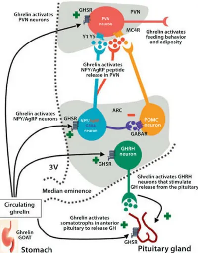
Figure 6: The primary neurons of the melanocortin system are the orexigenic NPY–AgRP neurons and the anorexigenic POMC neurons, which both send projections to the paraventricular nucleus. NPY–AgRP neurons also send inhibitory projections to neighbouring POMC cells. Activation of POMC cells by leptin triggers the release of α-‐MSH which binds to MC4R and promotes satiety. AgRP neurons are inhibited by leptin but stimulated by ghrelin, which promotes feeding and silences firing of POMC neurons. The effect of the NPY–AgRP cells is mediated by release of GABA, NPY and AgRP. Abbreviations: α-‐MSH, α-‐melanocyte stimulating hormone; AgRP,
agouti-‐related protein; GABA, γ-‐aminobutyric acid; GABAAR, GABA receptor; GHSR, growth hormone secretagogue
receptor (also known as ghrelin receptor); MC4R, melanocortin receptor 4; NPY, neuropeptide Y; POMC, proopiomelanocortin; PVN, paraventricular nucleus.
The projections of the two components of the melanocortin system, POMC and NPY/AgRP neurons, grossly overlap both within and outside of the hypothalamus, although they are rarely very long. The common targets of the NPY/AgRP and POMC projections are the hypothalamic parvicellular paraventricular nucleus (PVN), and the lateral hypothalamic and perifornical areas, all of which express melanocortin receptors MC3/4R [45]. The interplay between AgRP and α-MSH fibers on melanocortin receptor-expressing neurons in the PVN is suggested to be critical for metabolism regulation [45]. The signals that target the PVN can then be distributed in various directions as the PVN projects to brain stem nuclei, the spinal cord, cortex (via various routes), and thalamus, as well as to the portal vessels connecting the hypothalamus to the anterior pituitary.
Besides the PVN, other areas to which melanocortin neurons project include the medial preoptic area (MPA), the periacquedoctal gray (PAG), the nucleus of the solitary tract
An important target area of the mediobasal hypothalamic POMC neurons is the preganglionic sympathetic system in the thoracic segment of the spinal cord [46]. These POMC projections are unopposed by NPY/AgRP efferents [46] and are thought to play an important role in melanocortin regulation of the sympathetic nervous system [46]. Interestingly, POMC neurons that give rise to this spinal cord projection are not homogenously distributed within the mediobasal hypothalamus. They are mainly located in the retrochiasmatic area of the hypothalamus and in an area lateral from the arcuate nucleus [46]. In fact, when analyzed closely, the appearance of POMC neurons is not homogenous throughout the mediobasal hypothalamus; within the arcuate nucleus, and specifically in its more medial aspects, POMC neurons are smaller compared to the laterally located, larger melanocortin cells. This enlarged soma size is consistent with neurons that have long projections, such as those POMC cells that project to the spinal cord.
2.1.1 The NPY/AgRP neurons and the orexigenic pathway
The NPY/AgRP neurons within the arcuate nucleus (ARC) are probably the most established orexigenic population in the CNS. However, earlier studies reported that mice with NPY and/or AgRP deficiency have normal food intake and body weight [47]. Similarly, ablation of AgRP neurons during development resulted in normal body weight [48]. More recent studies used toxins to ablate the NPY/AgRP neurons in adults and confirmed the orexigenic role of these neurons [49]. Acute activation of NPY/AgRP neurons using optogenetic or pharmacogenetic stimulation methods also resulted a robust increase in food intake [50]. These series of experiments have established the NPY/AgRP neurons as the major orexigenic population in the CNS.
The NPY/AgRP neurons release the neuropeptide NPY (agonist at the Y receptors), AgRP (inverse agonist at the MC4Rs), and the inhibitory neurotransmitter GABA. The metabolic effects of NPY on Y receptors are somewhat complicated, and many are not consistent with the orexigenic role of NPY/AgRP neurons [51]. The ability of AgRP to antagonize the central melanocortin pathways received much attention, since this was considered to be the underlying mechanism of orexigenic effects. However, the orexigenic effects of NPY/AgRP neurons are not affected by the deletion of MC4Rs [52], and it is now regarded that the NPY/AgRP neurons increase food intake independent of the central melanocortin system. Currently, GABA release is believed to mediate most of the orexigenic effects of the NPY/AgRP neurons. For instance, deletion of vesicular GABA transporter (vgat) genes specifically in the AgRP neurons leads to lean phenotype [53], which confirms the importance of GABAergic neurotransmission in the orexigenic effects.
Multiple “anorexigenic” neuronal populations within the CNS receive the “inhibitory” GABAergic input from the NPY/AgRP neurons. The ARC POMC neurons receive GABAergic input from the NPY/AgRP neurons, which form a local circuit of appetite regulation within the ARC [42]. Indeed, when the vgat gene is deleted in the NPY/AgRP neurons, the frequency of inhibitory postsynaptic current (IPSC) is not increased by ghrelin, a hormone produced by the stomach, which excites the NPY/AgRP neurons [53].
2.1.2 The POMC neurons and the anorexigenic pathway
The POMC gene is expressed in the brain as well as in the periphery such as in the pituitary gland, immune system and skin. Within the brain, POMC is expressed in the hypothalamus and in the NTS of the brainstem [54]. The critical role of POMC in the
individuals with POMC gene mutations display early-onset obesity [55, 56]. A similar obese phenotype occurs also in POMC-deficient mice [57]. However, while the acute ablation of POMC neurons in adult mice results in an obese phenotype with hyperphagia [58], postnatal ablation of POMC neurons leads to an obese phenotype with reduced energy expenditure but no hyperphagia [59]. On the other hand, reactivation of central POMC at different stages of development in neural specific POMC deficient mice reduces food intake and weight gain, and attenuates comorbidities such as hyperglycemia and hyperinsulinemia [60].
The POMC gene encodes several peptides that are the products of a complex post-translational process. The bioactive peptides of the POMC gene include adrenocorticotrophin (ACTH), β-endorphin, , β- and γ-MSH. Among these peptides, α-MSH is the most well-known anorexigenic peptide which mediates the effects on food intake and energy expenditure through its binding and activation of MCRs [43]. The biosynthesis of POMC-derived peptides, as the majority of mammalian neuropeptide hormones, meets the criteria of the “prohormone theory”, which begins with the mRNA translation process into a large, inactive precursor polypeptide, followed by posttranslational proteolysis to release different products [61]. In the case of POMC, this is achieved through differential processing by the action of different prohormone convertases (PCs), which results in biological and functional diversity within the central nervous system. Once encoded from the corresponding mRNA, POMC follows the intracellular trafficking of a secreted protein through the Golgi complex, ultimately reaching secretory granules in which the end products of processing are stored before being secreted by exocytosis. During this trafficking, POMC is proteolytically processed into a number of physiologically important peptides, including endorphins, ACTH, and α-MSH.
2.1.3 The melanocortin receptor-expressing neurons
Receptors of melanocortins are G-protein-coupled receptors with characteristic seven transmembrane domains. So far, five melanocortin receptors have been identified in humans – MC1R–MC5R [62].The encoded receptors bind four ligands: α-, β- and γ-MSH and the ACTH. The melanocortin receptors are Gs-coupled and signal via the adenylate cyclase-cAMP-protein kinase A second messenger pathway (Figure 7). However, depending on the cell type and the melanocortin receptor expression, signal transduction pathways other than cAMP may be activated which include the inositol triphosphate-diacyl glycerol- protein kinase C (IP3-DAG-PKC) pathway, extracellular Ca2+ influx, MAP kinase pathway, and the JAK/STAT pathway [63].
Figure 7: Melanocortin receptors and their endogenus ligands
Expression of the MCR genes is tissue-specific; While MC1R, MC2R and MC5R are mainly expressed in peripheral tissues, MC3R and MC4R are expressed in the Central Nervous System and have been shown to play a role in metabolism regulation.
2.1.3a MC4R
The melanocortin receptor type 4 was first cloned by Gantz et al in 1993 [62]. MC4R is a 332 amino acid protein encoded by a single gene, localized to chromosome 18q21.3. Using Northern blot analysis and in situ hybridization, MC4R was originally found to be expressed primarily in the brain. MC4R expression was notably absent in the adrenal cortex, melanocytes, and placenta [62]. MC4R is widely distributed in the central nervous system, especially in the cortex, hippocampus, amygdala, septum, corpus striatum, nucleus accumbens, hypothalamus, nucleus tractus solitarius, visual and motor nuclei of the brainstem, and the dorsal horn of the spinal cord. α- MSH, β-MSH, and ACTH are the known agonists and AgRP is the known reverse agonist of the MC4R.
Since MC4R is highly expressed in the hypothalamus and has a strong affinity for α-MSH it is believed to be a strong candidate for energy balance, appetite control, and body weight regulation. In rodents and in humans, mutations of the MC4R gene causes obesity [55]. Humans with MC4R mutations, showing severe, early onset childhood obesity, represent 4–6% of the obese population [64]. Mc4r knockout mice have been shown to also develop obesity syndrome characterized by hyperphagia, hyperglycemia and hyperinsulinemia [65]. The restoration of the MC4R gene selectively in the PVN of MC4R knockout mice by viral or genetic manipulation produced mice displaying lower body weight and reduced food intake compared to the MC4R knockout controls [66]. However, the restoration of the
receptor is not sufficient to bring energy expenditure to the level of wild type controls. Thus, this suggests that melanocortin regulation of energy expenditure occurs elsewhere in the hypothalamus or through receptors other than MC4R [66]. In addition, MC4R neurons also express Single-minded 1 (SIM1), a transcription factor that is critical for PVN development and one of only six genes implicated in human monogenic obesity. Haploinsufficiency of SIM1 is associated with hyperphagic obesity and increased linear growth in both humans and mice. Additionally, SIM1 heterozygous mice also show hyperphagia and obesity in response to a high-fat diet [67]. Thus, the phenotype of SIM1 haploinsufficiency is similar to that of agouti yellow (Ay), and MC4R knockout mice, both of which are defective in hypothalamic melanocortin signaling.
It has been shown that MC4R mRNA is expressed in autonomic control regions projecting to postganglionic neurons innervating peripheral metabolic tissues [68, 69]. Through sympathetic nerve innervation, CNS-MC4R has been shown to increase lipolysis in white adipose tissue (WAT) [70], insulin sensitivity in muscle, thermogenesis in brown adipose tissue (BAT), fatty acid oxidation in liver and decrease insulin release in pancreatic β-cells [71].
2.1.3b MC3R
The MC3R gene, similarly to the MC4R gene, plays a crucial role in energy homeostasis [72], but associations of its polymorphisms/mutations with human obesity are not as evident as in case of the MC4R gene [73]. MC3R is encoded by a single gene localized to the q13.2–q13.3 region of chromosome 20. Since this locus is associated with type 2 noninsulin-dependent diabetes mellitus (NIDDM), Mc3r could represent a candidate gene for NIDDM. MC3R is unique in that it binds to α-, β- and γ-MSH with similar affinities. Using Northern blot hybridization and polymerase chain reaction, Gantz et al first showed that MC3R is expressed in brain, placental, and gut tissues but not in melanoma cells or in adrenal glands [43]. MC3R expression in the brain is highest in the hypothalamus especially in the arcuate nucleus, ventromedial nucleus, preoptic nucleus, lateral hypothalamic area, and posterior hypothalamic area. Agouti- related protein (AgRP), which is normally expressed in the hypothalamus, is a potent reverse agonist of both MC3R and MC4R [43]. However, POMC and AgRP neurons in the arcuate nucleus of hypothalamus selectively express MC3R, and not MC4R, suggesting that MC3R might function as a presynaptic autoreceptor regulating the release of melanocortins. Physiological support for this hypothesis was provided by Cowley et al, [42] who demonstrated that the selective MC3R agonist, D-Trp-γ-MSH inhibited firing of GFP- labeled POMC neurons in the PVN. Also, Marks et al showed that peripheral administration of the MC3R agonist stimulates feeding via MC3R-mediated inhibition of the ARC POMC neurons [74].
An association between MC3R and human obesity has been identified by linkage studies. Several sequence variants have been found in the Mc3r coding region and in 5′ flanking sequences. Mc3r variants are associated with subtle changes in weight, leptin levels, and
insulin-glucose ratios, but none of these explain human morbid obesity [75]. A novel heterozygous mutation I183N in Mc3r was identified in two obese patients of the same family [76]. Functional characterization of I183N showed that this mutation completely abolished the activity of the mutated receptor to stimulate intracellular cAMP production, suggesting that I183N might play an important role in obesity. Similarly, Tao et al showed that a novel mutation I335S in Mc3r results in complete loss of ligand binding and signaling suggesting that this mutation might contribute to obesity [77]. However, a recent study evaluating the functional consequences of all mutations found in Mc3r and Mc4r in severely obese North American adults concluded that Mc4r, but not Mc3r mutations are associated with severe obesity in this population [78]. Thus, the significance of Mc3r mutations in human obesity is still not conclusively established to date.
2.2 POMC processing
POMC, a prohormone with molecular weight of 31 kDa, is ubiquitously expressed in various tissues of mammals [79]. POMC expression in the central nervous system, however, is limited to the ARC, NTS, and corticotrophs and melanotrophs of the anterior pituitary. The 1200 base pair POMC transcript encodes for the 267 amino acid prohormone with an N-terminal signal peptide of 26 residues [80]. As this precursor peptide passes through the Golgi stacks, it is targeted, via a specific signal peptide, into regulated secretory granules. POMC undergoes extensive posttranslational modification within these secretory granules mediated by a family of serine proteases, the prohormone convertases (PCs).
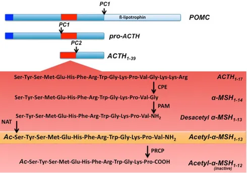
POMC is cleaved by prohormone convertase 1 (PC1) to produce 22 kDa pro-ACTH and β- lipoprotein hormone (β-LPH). Pro-ACTH is further cleaved by PC1 to produce the N-terminus of POMC-joining peptide and 4.5 kDa ACTH. Prohormone convertase 2 (PC2) cleaves ACTH to ACTH 1–17 and corticotropin-like intermediate lobe peptide (CLIP) and β-LPH to γ-lipoprotein hormone (γ-LPH) and β-endorphin. γ-LPH and the N-terminus of POMC are further modified to produce β-MSH and γ-MSH, respectively (Figure 8). α-MSH is a tridecapeptide derived from POMC. The synthesis of α-MSH from POMC involves several specific enzymes in addition to PC1 and PC2. First, carboxypeptidase E cleaves the C-terminal basic amino acid residues of ACTH 1–17. The peptide is then amidated by peptidyl α-amidating monooxygenase (PAM) to produce desacetyl α-MSH (Des-α-MSH). Finally, Des-α-MSH is acetylated by N-acetyltransferase (NAT) to produce acetylated α-MSH (Act-α-MSH). Act-α-MSH is more potent than Des-α-MSH in activating melanocortin receptor signaling and in reducing food intake, effects that are likely due to rapid degradation of Des-α-MSH. Guo et al have shown that total hypothalamic α-MSH levels are decreased in leptin-deficient ob/ob mice and increased in leptin-treated ob/ob and C57BL/6J mice. Leptin specifically enhances hypothalamic levels of Act-α-MSH without significantly affecting the amounts of Des-α-MSH, possibly by activating NAT in the POMC neurons [81].
While the maturation process of α-MSH has been largely studied, little is known on its fast degradation process. Only recently, it has been reported that PRCP, also known as lysosomal Pro- X carboxypeptidase, is a key enzyme responsible for the degradation and inactivation of α- MSH in the brain, [82] (Figure 8). Evidence was provided that hypothalamic PRCP is in an anatomical position to determine the efficacy of released α-MSH, thereby controlling the output and thus the efficacy of the melanocortin system. In
support of this idea, mice lacking PRCP display a lean phenotype when exposed to either a regular diet or high fat diet. Furthermore, in support of its function, PRCP has been previously associated with metabolic syndrome in human males [83].
Figure 8: Schematic illustration of hypothalamic POMC processing. Prohormone convertase 1 (PC1) cleaves the 32 kDa preprotein into proACTH and β-‐lipotrophin . ProACTH is then further converted by PC1 to form ACTH1– 39. Prohormone convertase 2 (PC2) will then cleave ACTH1–39, to form ACTH1–17. ACTH1–17 will be first processed by carboxypeptidase E (CPE) and then α-‐amidating mono-‐oxygenase (PAM) to form desacetyl α-‐MSH1– 13. An n-‐ actelyltransferase (NAT) will then convert desacetyl-‐α-‐MSH1–13 into the more active form acetyl-‐α-‐ MSH1–13. Finally acetyl-‐ α-‐MSH1–13 will be degraded by prolyl carboxypeptidase (PRCP) into the inactive product acetyl-‐ α-‐MSH1–12.
2.2.1a Enzymatic properties of PRCP
PRCP is an enzyme in the family of the carboxypeptidase (CPs) that is a highly conserved enzyme in eukaryotes with high amino acid homology between mouse, rat, and human. PRCP is a serine protease with a unique protease structure that includes a novel helical structural domain, which caps the catalytic active site [84]. The enzyme cleaves the last amino acid at the C-terminal part of the target proteins if the substrate contains a proline as a penultimate amino acid [85]. Furthermore, substrate specificity studies have shown that
PRCP has preferences for the amino acid sequences Xxx-Pro- Phe-OH and Xxx-Pro-Val-OH, where Xxx can be any amino acid [85].
PRCP was first identified over 40 years ago during studies of bradykinin metabolism in kidney. It is expressed in many other tissues including lung, liver, heart, and adipose as well as brain, suggesting possible multiple tissue- and/or cellular specific roles, with a variety of substrates. Within tissues, PRCP has been found in lysosomes and extracellularly either as a membrane-bound or as a soluble form [85]. PRCP is a single chain protein of approximately 58 KDa. The cDNA for human PRCP was cloned from a human kidney library and based on that, the molecular weight of the mature protein should be around 51 kDa, indicating the presence of about 12% carbohydrate by weight. This is consistent with the existence of six potential N-linked glycosylation sites. The deduced protein sequence contains 496 amino acids and includes a 30 residue signal peptide and a 15 amino acid propeptide [86]. The C-terminal half of the sequence contains an interesting “serine repeat” in which Ser is repeated as the 26th residue 6 out of 9 times [86]. In the other three repeats, Ser is replaced by Thr, Gly or Glu. When the Ser residues are aligned to yield 26 residue repeats, the positions of some of the other residues also fall into a pattern. The significance of this repeat region is not clear, but it might be involved in the maintenance of a secondary or tertiary structural motif or in the formation of the homodimer. The gene encoding human PRCP is on chromosome 11, while in the mouse it is on chromosome 7. PRCP is inhibited by compounds that react with the active site serine residue. However, this action is not true for all serine protease inhibitors, such as aprotinin, or by chelating agents or inhibitors that react specifically with the active site cysteine in sulfhydryl proteases. Particularly, PRCP is not inhibited by non-specific SH-reactive
agents, but is relatively sensitive to the prolylendopeptidase inhibitor, Cbz-Pro-prolinal, because of its specificity for peptides with penultimate proline residue [86].
2.1.2b Function
The PRCP-catalyzed reaction was initially found to be part of the pathway for angiotensin II (Ang II) metabolism in renal tissues, whereas PRCP appeared to control the total amount of Ang II. Odya and others demonstrated that PRCP metabolizes Ang II to angiotensin 1–7 (Ang 1–7) [87]. The activation of Ang 1–7 receptor Mas (a G-protein-coupled protein) by Ang 1–7 results in the generation of nitric oxide (NO) and prostaglandins. Thus, Ang 1–7 counteracts Ang II function, providing evidence that PRCP regulates the negative effects of Ang II such as high blood pressure and heart failure. In addition, the activation of the Ang 1–7 receptor Mas may also lead to diminished cell proliferation through down- regulation of the phosphorylation and activation of Erk1 and Erk2 in the Erk1/Erk2 Map kinase signaling pathway [88]. In theory, the PRCP inhibitors target the production of pro-inflammatory prostaglandins and promote proliferation through the Ang 1–7 receptor Mas-dependent pathway. This represents a novel approach to suppress unwanted inflammation- causing prostaglandins.
Later, it was shown that PRCP is one of several enzymes that convert Ang II to a unique bioactive molecule. In vitro studies showed that angiotensin-converting enzyme 2 (ACE2) is an exopeptidase that converts Ang II to Ang 1–7 at a much faster rate than PRCP.12 This data suggests that Ang II is a poor substrate for PRCP. Clinical studies have provided reliable evidence that ACE2 is an essential regulator of angiotensin I (Ang I), Ang II, and angiotensin-induced cardiac hypertrophy.13 Recent studies clearly show increased
myocardial levels of Ang II and a significant decrease in Ang 1–7 in ACE2-deficient hearts, suggesting that the role of PRCP in metabolizing Ang II may be insignificant. Taken together, these observations suggest that PRCP is a redundant catalyst contributing to alternate pathways for Ang II metabolism.
While the well-established cardiovascular and renal actions of Ang II are attributed to the angiotensin type 1 receptor (AT1R), much less is known about angiotensin III and its cardiovascular effects. For more than 30 years, it was known that PRCP metabolizes Ang III to Ang 2–7 [87]. Soon after, studies demonstrated that Ang III, is a pressor agent whose response, like that of Ang II, is mediated by AT1 receptors [89]. Apparently, Ang III has multiple effects on renal function in the diseased kidney and can enhance renal disease through the overproduction of aldosterone, leading to arterial hypertension and/or atrial fibrillation. Aldosterone maintains blood volume, pressure, and electrolyte balance. Its production is known to be regulated by renin, an enzyme produced in the kidneys. Renin increases in response to low blood pressure, decreased blood flow to the kidneys, or sodium deficiency. The elevation of renin results in an increase in synthesis and secretion of aldosterone. Studies indicate that Ang III also activates the secretion of aldosterone [89]. Recently, we have demonstrated that recombinant PRCP (rPCRP) metabolizes Ang III to Ang 2–7, removing phenylalanine (Phe) [90]. It is tempting to speculate that PRCP might funnel the generation of angiotensin 3–4 (Ang 3–4) through Ang 2–7. If the over- secretion of aldosterone by Ang III is viewed as a trigger of arterial hypertension, then inactivation of Ang III by PRCP might lead to a decrease in blood pressure. Further studies are required to determine whether PRCP is critically important for regulating Ang III- induced hypertension and preserving renal structure and function. This is an important area of
research to pursue given the increasing prevalence of cardiovascular disease and stroke in the older population.
The possible actions of another substrate of PRCP, plasma prekallikrein (PK, Fletcher factor), have recently begun to receive much attention. When the complex of high molecular weight kininogen (HK) and PK binds to endothelial membrane, PK is rapidly converted to kallikrein by PRCP. The formed kallikrein then cleaves HK to liberate bradykinin (BK), which leads to NO and prostaglandin-I2 formation, as well as subsequent vasodilation, by activating constitutive bradykinin B2 and inducible bradykinin B1 receptors. The PRCP-dependent PK activation pathway might be considered an additional mechanism to preserve the availability of NO and prostacyclin as vasodilatory agents in vascular smooth muscle. We proposed that chronic PRCP inhibition might elevate blood pressure. In accordance, we have found that PRCPgt/gt mice have mild hypertension, suggesting a causative relationship between PRCP levels and signs of hypertension . Local skeletal muscle ischemia and acidosis are shown to increase the generation of BK and prostaglandins, the two circulating products of the PRCP-induced cell activation [91]. The increased acidotic response during exercise and inflammatory mediators such as BK and prostacyclin have been shown to cause abnormal exercise-related symptoms and autonomic responses in congestive heart failure syndrome [91]. Nonetheless, the long-term elevated concentrations of NO and prostacyclin through PRCP-dependent pathways may be detrimental and eventually responsible for cardiovascular diseases such as congestive heart disease. Since BK and Prostaglandins exacerbate the genesis of the symptoms of exercise intolerance in heart failure, the inhibitors of PRCP might be effective in ameliorating the exercise-limiting symptoms.
Clinical studies demonstrate that PRCP is involved in the pathogenesis of inflammatory conditions such as rheumatoid arthritis and infection [92]. Melanocortin peptides have numerous effects on the host such as the modulation of fever, inflammation and appetite [92].
In addition to its cardiovascular and proinflammatory functions, PRCP, as reported by a genome-wide association study (GWAS), is a risk factor for the metabolic syndrome because it was significantly associated with coronary heart disease in men [83]. This report supports recent data produced in rodents showing that central PRCP is an important regulator of energy metabolism because it is responsible for the degradation and thus inactivation of hypothalamic α-MSH [82]. In peripheral tissues such as in the kidney, PRCP has been shown to have an acidic pH optimum (5.0). However, PRCP retains significant enzymatic activity also at neutral pH [82]. Subsequently, it was shown that, at neutral pH, α- MSH is a better PRCP substrate than angiotensin II. α-MSH is a 13 amino acid neuropeptide in which a proline in position 12 precedes a valine in position 13. By removing this valine residue, PRCP produces α-MSH1–12 from α- MSH1–13. The removal of the last amino acid is of physiological importance.
α- MSH1–13 is a potent anti-inflammatory agent. In addition to the specificity of cleavage, the cellular release of pro-inflammatory mediators seemed to be critical to PRCP actions. In theory, agents that increase production and effects of α-MSH1–13 could be used to counteract the effects of pro-inflammatory mediators such as bradykinin and cytokines. Obesity is known to cause inflammation and insulin resistance in the vasculature and non- vascular tissues involved in glucose metabolism. Evidence suggests that hyperglycemia may contribute to defective NO-dependent vasodilation in diabetes [93]. The inducible NO synthase (iNOS) expression is elevated in adipose tissue of obese people compared to
those of lean people and is a mediator of inflammation and a key enzyme in insulin resistance [93]. The colocalization of α-MSH1–13 receptors (MC4R) with iNOS has been reported, suggesting a role for MSH1–13 in obese people [93]. The inactivation of α-MSH1–13 by PRCP provides a positive feedback loop for postprandial enhancement of food intake and inflammation by inhibiting α-MSH1–13 function. However is demonstrated by the inability of α-MSH1–12 to decrease food intake when administered centrally as well as by its inability to increase the action-potential frequency of hypothalamic MC4R-expressing neurons, compared to α-MSH1–13 peptide [82]. Further support for the role of PRCP in the degradation of α-MSH1–13 is provided by studies using the PRCPgt/gt null mice. These mice have been shown to be sensitive to peripheral administration of α-MSH and to have higher α-MSH1–13 levels in the hypothalamus compared to WT controls [82].
Although α-MSH is so far the only substrate involved in the regulation of food intake that has been identified as a PRCP substrate, we cannot exclude the possibility that more peptides with a proline as the penultimate amino acid and a function associated with body- weight regulation could be degraded by PRCP. However, data from human and rodent studies suggest that PRCP inhibition might provide a mechanism to increase central bioavailability of α-MSH, and thus reduce food intake.
2.1.2c A potential role for PRCP in leptin resistance
A postulated cause for the development of obesity is leptin resistance. As fat stores increase during the development of obesity, circulating leptin levels are continuously rising. However, these elevated leptin levels or administration of exogenous leptin to obese
subjects does not lead to decreased body weight. This discrepancy between leptin levels and the metabolic phenotype is attributed to leptin resistance. A primary site of leptin resistance is suggested to be the POMC neurons. Indeed, intracellular signaling cascades triggered by leptin receptor activation were found to be impaired in diet-induced obese animals [94]. Specifically, activation of the transcription factor STAT3 by leptin is diminished in these animals, an event that is attributed to the inhibitory action of SOCS3 on STAT3 phosphorylation [94]. Additionally, ex vivo studies revealed that leptin induced secretion of α-MSH1–13 is reduced from hypothalamic slices taken from diet- induced obese mice [95]. An alternative explanation for the development of leptin resistance was the decreased ability of leptin from the circulation to reach hypothalamic neurons. Despite of all this elegant and logical data, several parameters of POMC neurons in diet induced obesity show alterations completely consistent with elevated leptin levels. For example, the wiring of the melanocortin system and transcript levels of POMC and AgRP/NPY of obese animals in response to a hypercaloric environment reflects an adequate and possibly maximal responsiveness of these cells to leptin [96]. The synaptic organization triggered in these animals shows similar trends of changes to those that were described previously for highly leptin sensitive ob/ob mice in response to leptin administration. Although reactive gliosis was also detected in response to high fat diet specifically associated with arcuate nucleus capillaries [96], this gliosis does not appear to block the effect of leptin on the synaptic input organization of the POMC neurons and POMC, NPY and AgRP transcript levels, and hence, may not be the cause of leptin resistance as proposed earlier. On the other hand, it is conceivable that assays using hypothalamic slices to measure α -MSH release [95] may be skewed by gliosis, an event that could be a structural obstacle for the release of measurable α -MSH into the medium, thereby limiting the usefulness of these
assays for the study of α -MSH release kinetics. Thus, if neurobiological attributes of leptin action are present in the hypothalamus of diet- induced obese animals, it is a reasonable suggestion that the degradation of released α - MSH1–13 may interfere with the ability of MC4R activation in POMC target sites. As described above, PRCP location and function in the hypothalamus makes this carboxypeptidase, although previously unsuspected, a player in the emergence of systemic leptin resistance as it relates to the regulation of food intake, energy expenditure and glucose homeostasis.
2.1.2d Pharmacological aspects
Obesity and related metabolic dysfunctions, such as type 2 diabetes, have emerged as a leading cause of morbidity and mortality in developed societies. The development of obesity and type 2 diabetes may be triggered or abrogated by the mechanisms of the CNS. The hypothalamic melanocortin system and its major anorexigenic peptide α-MSH have an important role in such disorder, as revealed by the dramatic development of obesity after selective inactivation of the neuronal Pomc gene in mice, and the presence of POMC mutations in humans with severe early-onset obesity [97]. Thus, the α-MSH target MC4 receptor is an attractive candidate for a drug target to treat obesity, as it not only affects several aspects of feeding behavior, but the activation of the MC system also increases insulin sensitivity and energy expenditure [98].
Pharmaceutical development of MC4 receptor-specific drugs have been the object of many studies [99]. However, it has been challenging to design selective MC4 receptor agonists that would completely lack affinity for the MC3 receptor [99]. Nevertheless, mixed MC3/4 agonists might provide a therapeutic advantage since, as shown in MC3R knockout mice,
reduction in MC3R activity is associated with an increased adipogenesis. However, studies in rodents and humans have revealed some side effects of the use of MC receptor agonists. For example, administration of MC4 receptor agonists is associated with penile erections as well as flushing, which has resulted in a new application area for these drugs in erectile dysfunction. Also, preclinical studies have identified roles for the melanocortin system in blood pressure regulation (MC receptor agonists have a depressor effect most probably via the nucleus tractus solitarius in brainstem), in the inflammatory responses (where agonists limit thermogenic responses to pyrogens), and in pain processing (where antagonists suppress pain sensation) [100]. Thus, the development of MC4 receptor agonists in the treatment of obesity should be carefully monitored to avoid undesired side effects due to melanocortin function in other systems.
Because of these side effects, PRCP becomes a very promising drug target. It has been already reported that inhibition of PRCP activity by small molecule protease inhibitors administered peripherally or centrally decrease food intake in wild type, as well as obese animals [82]. The pharmacological manipulation of the melanocortin system accomplished by inhibiting α-MSH catabolism represents a highly innovative approach that may help to combat metabolic disorders such as obesity.
The local metabolism of neurotransmitters and neuro- modulators are generally considered an optimum drug target with a lower incidence of side/non-specific effects compared to direct agonists. This is the case, among others, of the actual pharmacotherapy for many pathologies of the CNS such as depression and Alzheimer’s disease. The discovery of PRCP as a pharmacological target may allow, indeed, to selectively focus on the benefits derived from the enhancement of melano- cortin activation in discrete brain areas by its
endogenous ligand, avoiding the whole set of possible central and systemic effects due to the use of MC receptor agonists.
In conclusion, further studies on PRCP will shed light on a novel central regulatory element in the regulation of energy metabolism. Differential activity levels of PRCP in specific brain areas in different metabolic condition by affecting α-MSH levels will, in turn, affect the efficacy of melanocortin signaling. Thus, understanding PRCP role in metabolism regulation is important to reveal whether PRCP can be used as new drug targets for metabolic disorders, including obesity and type 2 diabetes.
3. Hormonal regulation of the hypothalamic melanocortin system
3.1 Leptin
POMC is a regulator of appetite, energy expenditure and glucose metabolism [101]. Therefore, its own regulation must be important for the control of energy homeostasis. The hormone leptin is a major regulator of POMC. Leptin is an anorexigenic hormone produced and released by the white adipose tissue in the amount proportional to the mass of fat in the body [102]. Leptin interacts with six types of receptors (Ob-Ra, -Rb, -Rc, Rd,
Re, and Rf) encoded by a single leptin receptor gene (Ob-R) [103]. However, only the long
form of leptin receptors (Ob-Rb), consisting of an extracellular and an intact cytoplasmic domain, mediates the anorexigenic effect of leptin [104]. Deficiency in leptin or leptin receptors induces a morbid obese phenotype characterized by hyperphagia, hyperglycemia, hyperlipidemia, and reduced energy expenditure in both rodents and humans [105]. Neuron-specific Ob-R-deficient (db/db) mice displayed an obese phenotype while hepatocyte-specific db/db mice are normal, suggesting that the direct effect of leptin in the
brain is essential for metabolism regulation. Mice lacking Ob-Rb in POMC neurons, AgRP neurons or both POMC/AgRP neurons displayed increased body weight and fat mass [106], although their phenotypes are milder compared to those of whole brain neuron-specific Ob-Rb deficient mice, suggesting the involvement of other leptin receptor-expressing neurons in mediating the effect of leptin on energy homeostasis.
Leptin regulates POMC and NPY/AgRP neurons at different levels. For example, leptin increases POMC mRNA levels while decreasing NPY/AgRP mRNAs. Besides the transcriptional regulation, leptin directly depolarizes (activates) POMC neurons while simultaneously hyperpolarizing (inactivates) NPY/AgRP neurons. In addition, systemic leptin administration to leptin deficient (ob/ob) mice rapidly induced synaptic input reorganization onto NPY/AgRP and POMC neurons, suggesting that the synaptic rearrangement is an important event for leptin-induced behavioral changes [107]. A recent study demonstrated that AgRP neurons are critical in mediating metabolic syndrome in ob/ob mice since ablation of AgRP neurons in leptin deficient (ob/ob) mice showed reduced food intake and improved glucose tolerance [108].
3.2 Insulin
In addition to leptin, other hormones such as insulin are also regulators of POMC neurons. Insulin, produced from pancreatic β-cells, plays a fundamental role in the regulation of glucose homeostasis by modulating glucose uptake in peripheral organs. However, since insulin receptors (IR) are widely expressed in the brain, accumulated evidence has indicated a role for insulin in the CNS. Indeed, brain specific-IR deficient mice develop obesity with increased body weight, fat mass, and food intake [109]. However, mice with
selective deletion of IR in the arcuate melanocortin system, either POMC or AgRP neurons, did not display alteration in energy homeostasis, while only a defective suppression on hepatic glucose production was found in mice with selective deletion of IR in AgRP neurons [110]. In addition, re-expression of IR in either POMC or AgRP neurons of L1 mice, a genetic mouse model with significant reduction of IR in the ARC, suggested that insulin signaling in AgRP neurons negatively regulates hepatic glucose production while IR activation in POMC neurons positively regulates hepatic glucose production and energy expenditure [111]. Interestingly, when both leptin and IR were ablated in POMC neurons, systemic insulin resistance despite increased pancreatic insulin secretion was observed in these mice. In addition, the obese phenotype observed in POMC-specific ObR knockout mice was ameliorated when both ObR and IR were selectively deleted from POMC neurons, suggesting that insulin and leptin signaling in POMC neurons may have opposing effects in the regulation of body weight but additive effects on glucose homeostasis. In support of this, leptin and insulin responsive neurons are expressed in distinct subpopulations of POMC neurons [112]. Thus, the divergent effects of leptin and insulin on energy homeostasis may be due to the activation of different POMC subpopulations.
3.3 Ghrelin
Ghrelin, the hunger hormone, is predominantly secreted by specialized endocrine cells of the stomach when the stomach is empty. Ghrelin is synthesized as a preprohormone and several processes are required to generate its active form. After removing the signal peptide from the preprohormone, ghrelin precursor is acylated at the third serine with