Hereditary hyperhomocysteinemia associated with nephrotic syndrome
complicated by artery thrombosis and chronic thromboembolic
pulmonary hypertension: A case report
Paolo Ruggeri
1, Federica Lo Bello
1, Francesco Nucera
1, Michele Gaeta
2, Francesco Monaco
3,
Gaetano Caramori
1, Giuseppe Girbino
11
Pneumology Operating Unit, Department of Biomedical and Dental Sciences and of Morphological
and Functional Images, University of Messina
2
Diagnostic Imaging Unit, Department of Biomedical and Dental Sciences and of Morphological
and Functional Images, University of Messina
3
Thoracic Surgery Unit, Department of Biomedical and Dental Sciences and of Morphological
and Functional Images, Università di Messina, Italy
Abstract
We present here the case of a 30-year-old man with a long term history of nephrotic syndrome (NS) who developed an episode of acute left main pulmonary artery thrombosis complicated by a lung abscess. During the hospital admission was also identified a concomitant hyperhomocys-teinemia. After an atypical resection of the left upper pulmonary lobe and the starting of long term anticoagulation the patient was discharged but
did not attend the planned follow up visits until one year later when he was seen again for severe dyspnea and exercise intolerance. At this time chronic thromboembolic pulmonary hypertension (CTEPH) was diag-nosed by lung perfusion scintigraphy and right heart catheterization. He initially refused the surgical treatment but, after six months, for the pres-ence of worsening dyspnea was referred for bilateral pulmonary en-darterectomy followed by a cardio-thoracic rehabilitation program. After a follow-up of seven years the patient is alive and in stable conditions. NS and hyperhomocysteinemia are both known risk factors for pul-monary embolism (PE), but their association with CTEPH is extremely rare. We discuss here the possible mechanisms linking these condi-tions. CTEPH must be suspected in any patient with NS, with or without hyperhomocysteinemia, and unexplained dyspnea.
Introduction
Thromboembolism is one of the major complications of nephrotic syndrome (NS) [1]. In particular, the presence of pulmonary artery thrombosis is often fatal [2]. Hyperhomocysteinemia is another ac-cepted risk factor for pulmonary thromboembolism [3]. Chronic thromboembolic pulmonary hypertension (CTEPH) is a rare condition presenting in 2-4% of patients after acute pulmonary embolism but characterized by significant morbidity and mortality [4,5]. The associ-ation of NS and hyperhomocysteinemia with CTEPH is extremely rare.
Case Report
The case concerns a 30-year-old (height 160 cm, weight 60 Kg, body mass index 23 Kg/m2) Caucasian man, medical doctor, who presented
at the emergency department of our University Hospital of Messina, Italy (www.polime.it) for the presence by 30 days of persistent dry cough associated in the last 7 days with exercise-induced dyspnea and bilateral pitting edema of the ankles. He was a current smoker of 3 pack-years, with of a history of minimal change nephrotic syndrome di-agnosed (with a kidney biopsy) 20 years before the presentation and treated with low dose oral glucocorticoid (5 mg daily of prednisone equivalent) therapy from childhood. He was under regular treatment with diuretic (25 mg once daily furosemide) and proton pump inhibitor (20 mg once daily of omeprazole) for systemic arterial hypertension and gastroesophageal reflux diagnosed 5 years before the presentation.
Corresponding Author: Paolo Ruggeri, Unità Operativa Complessa di Pneumologia, Dipartimento di Scienze Biomediche, Odontoiatriche e delle Immagini Morfologiche e Funzionali (BIOMORF), Università di Messina, Italy. Tel. +39.090.2212002 - Fax: +39.090.2934157. E-mail: [email protected]
Key words: Chronic thromboembolic pulmonary hypertension; pulmonary embolism; nephrotic syndrome; hyperhomocysteinemia.
Acknowledgments: We thank Ms Franca Mollica and Mr Angelo Zodda for their administrative support.
Contributions: PR contributed substantially to conception, design and drafting of case report manuscript; FL, MG, involved in data acquisition; AB, MG, involved in imaging acquisition; GG, FM, GC, equally involved in critically revising the manuscript.
Consent for publication: Written informed consent was obtained from the patient for publication of this case report and any accompanying images. A copy of the written consent is available for review by the Editor of this journal.
Conflict of interest: The authors declare no conflict of interest.
Received for publication: 7 August 2017 Accepted for publication: 5 November 2017
©Copyright P. Ruggeri et al., 2017 Tipografia PI-ME Editrice, Italy
Monaldi Archives for Chest Disease 2017; 87: 880 doi: 10.4081/monaldi.2017.880
This article is distributed under the terms of the Creative Commons Attribution Noncommercial License (by-nc 4.0) which permits any noncommercial use, distribution, and reproduction in any medium, provided the original author(s) and source are credited.
Non
commercial
enoxaparin (1 mg/Kg). A duplex ultrasonography of the venous system of the lower legs was normal. The pathological routine laboratory tests are shown in Table 1. The most relevant abnormality was the presence of hyperhomocysteinemia with an homozygous T-variant of the common methylenetetrahydrofolate reductase (MTHFR) 677C→T poly-morphism in the MTHFR gene. The lung abscess did not improve de-spite 15 days of treatment with adequate empiric wide spectrum an-tibiotic treatment (piperacillin/tazobactam 3.375 g IV every 6 hours). For this reason, the thoracic surgeon performed an atypical resection of the left upper pulmonary lobe. At the hospital discharge, after 21 days from his admission, the patient was in stable conditions with only residual exercise dyspnea (Borg scale 2) but without cough, chest pain, fever and/or peripheral edema. At the discharge he was under regular treatment with warfarin (with a target INR of 2.0-3.0) together with his previous regular pharmacological treatment (oral prednisone 5 mg and furosemide 25 mg, both once daily). The patient did not attend our clinic for the planned three and six months follow-up visits but he pre-sented spontaneously at our clinic again ~12 months after his hospital discharge reporting worsening exercise dyspnea (Borg scale 5). The One month before the current admission he had been treated
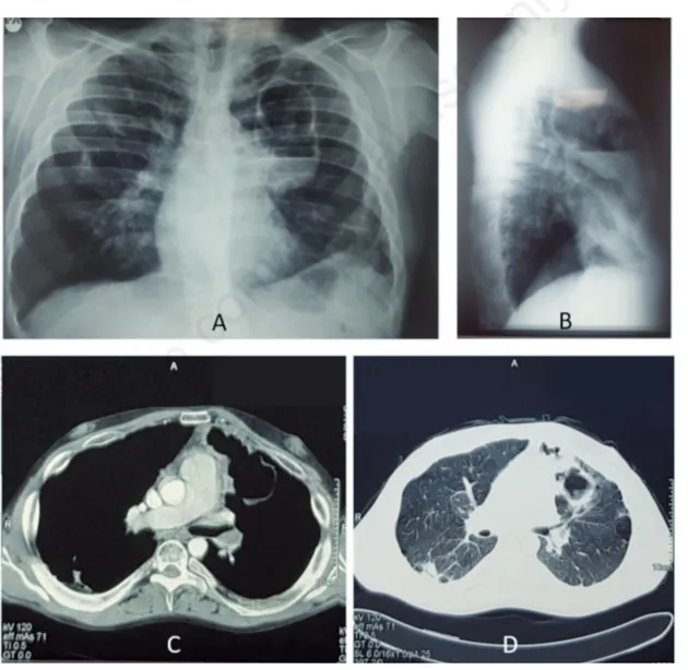
intra-muscularly for seven days at home by his General Practitioner with cef-triaxone (1 gram/once daily) for the presence of cough with yellow sputum, diffuse thoracic pain and fever (37.5°C). At the admission, the patient presented with dyspnea (Borg scale 4) and oxygen desaturation (92% when breathing room air). Blood gas test on room air was: pH 7.50, PaO2 60 mmHg; PaCO2 35 mmHg, HCO3- 28,5 mmol/L, BE + 4,6, Sat 93%; Physical examination of the chest revealed only the presence of an hypophonesis in the left upper lobe. No other physical signs out-side the chest were pathological. An x-ray of the chest showed the pres-ence of a cavitated mass (10 cm of diameter) located in the upper left lobe with a frank air-fluid level (Figure 1). A computed tomography (CT) scan of the chest, performed before and after the administration of contrast medium, confirmed the presence of an excavated lesion in the upper left lobe of the lungs associated to the partial occlusion of the left main pulmonary artery containing a thrombus inside the lumen (Figure 1). These lesions were interpreted as a pulmonary infarction with a superimposed lung abscess secondary to left main pulmonary ar-tery thrombosis. Immediately the patient was placed on twice daily
Figure 1. A,B) Posteroanterior and lateral chest x-ray at presentation showed the presence of a cavitated mass (10 cm of diameter) located in the upper left lobe with a frank air-fluid level. C,D) Computed tomography (CT) scan of the chest, performed before and after the administration of contrast medium, confirmed the presence of an excavated lesion in the upper left lobe of the lungs associated to the partial occlusion of the left main pulmonary artery containing a thrombus inside the lumen.
Non
commercial
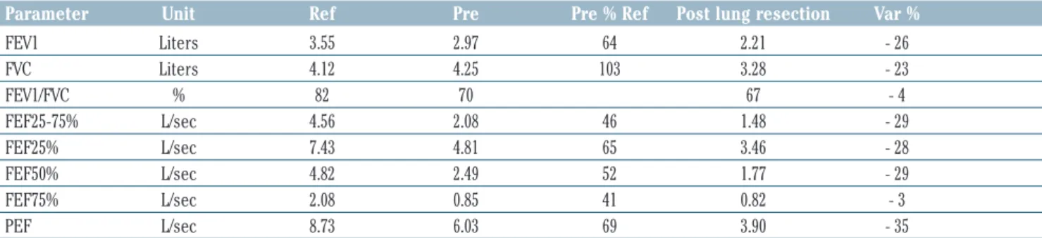
Table 2. Pre- and post-operative spirometry (after atypical resection of left upper pulmonary lobe).
Parameter Unit Ref Pre Pre % Ref Post lung resection Var %
FEV1 Liters 3.55 2.97 64 2.21 - 26 FVC Liters 4.12 4.25 103 3.28 - 23 FEV1/FVC % 82 70 67 - 4 FEF25-75% L/sec 4.56 2.08 46 1.48 - 29 FEF25% L/sec 7.43 4.81 65 3.46 - 28 FEF50% L/sec 4.82 2.49 52 1.77 - 29 FEF75% L/sec 2.08 0.85 41 0.82 - 3 PEF L/sec 8.73 6.03 69 3.90 - 35
Table 1. Summary of the clinical and laboratory parameters.
Clinical parameters
Parameters Before atypical pulmonary Before pulmonary After pulmonary endarterectomy
resection endarterectomy (three months follow-up)
Blood sistolic pressure 120 mmHg 100 mmHg 120 mmHg
Blood diastolic pressure 80 mmHg 70 mmHg 80 mmHg
Respiratory rate 23/min 25/min 20/min
Pulse rate 92/min 95/min 85/min
Oxigen saturation ° 92% (room air) 93% 95%
Axillary temperature 37.5C ° 36.5 C ° 36.2 C °
Arterial blood gases analysis, performed with the patient breathing room air
Parameters Before atypical pulmonary Before pulmonary After pulmonary endarterectomy
resection endarterectomy (three months follow-up)
pH (7.35-7.45) 7.50 7.49 7.38
PaCO2 (35-45 mmHg) 35 mmHg 30.7 mmHg 38 mmHg
PaO2 (80-100 mmHg) 60 mmHg 68.7 mmHg 75 mmHg
Serum bicarbonates (22-26 mmol/L) 28.5 mmol/L 25.3 mmol/L 23.4 mmol/L
SaO2 (94%-100%) 93.5% 94.5% 96%
Laboratory parameters
Parameters Before atypical pulmonary Before pulmonary After pulmonary endarterectomy
resection endarterectomy (three months follow-up)
Serum CRP (0-0.50) 7.10 mg/dl 2.5 mg/dl Nd
WBC (4500-9000) 10.400 mm3 8.500 mm3 Nd
Platelets (150000-350000) 429.000 mm3 380.000 mm3 Nd
Total plasma proteins (normal range) 4.6 g/dL 5.6 g/dL Nd
Albumin (53%-65%) 2.44 g/dL (40.59%) 3.5 g/dL Nd
Homocysteinemia 70 µmol/L
(5-9 µmol/L)
Cardiac function tests
Left ejection fraction (echocardiography) 55% 60% 62%
sPAP (echocardiography) 40-45 mmHg 70 mmHg 35 mmHg
(15-30 mmHg)
mPAP Nd 46 mmHg 20.7 mmHg
(9-18 mmHg)
Pulmonary resistance Nd 343 dyne*sec*cm-5 117 dyne*sec*cm-5
(80-240 dyne*sec*cm-5)
Cardiac output Nd 4.7 L/min 5.2 L/min
(4-8 L/min)
Cardiac index Nd 2.6 L/min/mq Nd
(2-4 L/min/mq)
Pulmonary wedge pressure Nd 16 mmHg Nd
(6-15 mmHg)
°Measured with a pulse oximeter; nd: not done; CRP, C reactive protein; WBC, white blood cells; sPAP, systolic pulmonary artery pressure; mPAP,mean pulmonary artery pressure.
Non
commercial
Discussion
An association between hypercoagulability and nephrotic syndrome (NS), which is characterized by heavy proteinuria, hypoalbuminemia, hyperlipidemia and edema, has been established many years ago [6,7]. This hypercoagulable state is likely caused by multiple factors, including platelet activation and aggregation, hyperfibrinogenemia, loss of an-tithrombin, hypoplasminogenemia and increased levels of plasminogen activator inhibitor [7]. The prevalence of pulmonary embolism in nephrotic patients has been greatly different among studies and can be asymptomatic or even lethal [6]. Although the MTHFR 677C->T poly-morphism increases the homocysteine levels, the association between clinical suspicion of chronic thromboembolic pulmonary hypertension
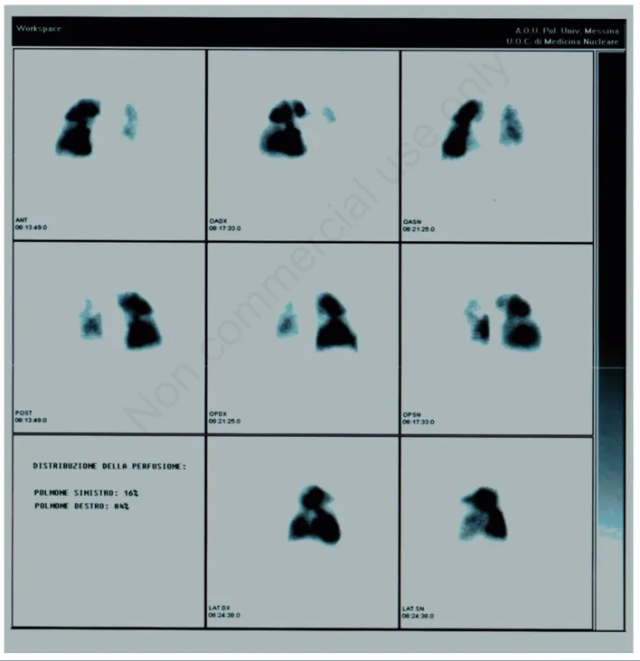
was confirmed using lung perfusion scintigraphy and the right hearth catheterization (Figure 2). The mean pulmonary artery pressure at rest was 46 mmHg (normal value 9-18 mmHg). The patient refused to per-form an evaluation at a cardiothoracic surgery referral center at the University Hospital of Pavia, Italy. However, after six months and a fur-ther deterioration of the dyspnea he accepted to be referred to this center for a bilateral pulmonary endarterectomy. The surgical interven-tion was uneventful and followed by clinical and funcinterven-tional improve-ment. After three months of a cardio-thoracic rehabilitation program the patient was discharged at home. The pre- and postoperative spirometry are shown in Figure 3. After a follow-up of seven years the patient is alive and in stable conditions.
Figure 2. Lung perfusion scintigraphy. There are many perfusion defects in both lungs, partly consistent with the previous atypical resec-tion of the upper left lobe and in other points strongly suggestive of their embolic nature.
Non
commercial
this polymorphism and an increased risk of pulmonary thromboem-bolism is controversial [3,8-10]. It is, possible that hyperhomocys-teinemia led to venous thrombosis, with secondary embolism in the pa-tient’s pulmonary circulation. Vascular endothelial necrosis caused by extremely high homocysteine levels might have caused localized thrombus formation in the vascular tree. Nevertheless, duplex ultra-sonography and a CT scan did not detect deep vein thrombosis in the pa-tient’s legs, abdomen or pelvis of this patient as previously demon-strated in another case report [11]. Chronic thromboembolic pulmonary hypertension (CTEPH) is a rare but life-threatening complication of pul-monary embolism and is defined as precapillary pulpul-monary hyperten-sion (mean pulmonary artery pressure ≥25 mmHg with a pulmonary capillary wedge pressure ≤15 mmHg) associated with mismatched per-fusion defects on ventilation-perper-fusion lung scan and signs of chronic thromboembolic disease on computed tomography pulmonary an-giogram and/or conventional pulmonary angiography, in a patient who received at least 3 months of therapeutic anticoagulation [5]. The diag-nostic work-up to detect or rule out CTEPH should include ventilation-perfusion scintigraphy, which has high sensitivity and a negative pre-dictive value of nearly 100%. Patients with suspected CTEPH should be referred to a specialist centre for right-heart catheterization and pul-monary angiography. Surgical pulpul-monary endarterectomy is still the treatment of choice for CTEPH and is associated with excellent long-term results and a high probability of cure [4].
Although several studies have shown that homocysteine levels can be re-duced by vitamin supplementation, it has yet to be proven that this reduc-tion leads to a reduced risk of cardiovascular morbidity and mortality [12].
Conclusions
In summary, we describe an unusual case of a 30-year-old man with nephrotic syndrome from childhood and concomitant occult hyperho-mocysteinemia who presented with pulmonary artery thrombosis and lung abscess that evolved in a chronic thromboembolic pulmonary hy-pertension successfully treated with bilateral pulmonary endarterec-tomy. CPEPH must be suspected in any patient with NS, with or without hyperhomocysteinemia, and unexplained dyspnea.
References
1. Kerlin BA, Ayoob R, Smoyer WE. Epidemiology and pathophysiology of nephrotic syndrome-associated thromboembolic disease. Clin J Am Soc Nephrol 2012;7:513-20.
2. Matsuda A, Tsuchiya K, Yabuki Y, et al. Fatal diffuse pulmonary ar-terial thrombosis as a complication of nephrotic syndrome. Clin Exp Nephrol 2007;11:316-20.
3. Mannucci PM, Franchini M. The real value of thrombophilia markers in identifying patients at high risk of venous thromboem-bolism. Expert Rev Hematol 2014;7:757-65.
4. Hoeper MM, Madani MM, Nakanishi N, et al. Chronic thromboem-bolic pulmonary hypertension. Lancet Respir Med 2014;2:573-82. 5. O’Connell C, Montani D, Savale L, et al. Chronic thromboembolic
pulmonary hypertension. Presse Med 2015;44:e409-16.
6. Kayali F, Najjar R, Aswad F, et al. Venous thromboembolism in patients hospitalized with nephrotic syndrome. Am J Med 2008;121:226-30.
7. Singhal R, Brimble KS. Thromboembolic complications in the nephrotic syndrome: pathophysiology and clinical management. Thromb Res 2006;118:397-407.
8. Bezemer ID, Doggen CJ, Vos HL, et al. No association between the common MTHFR 677C->T polymorphism and venous thrombosis: results from the MEGA study. Arch Intern Med 2007;167:497-501. 9. Den Heijer M, Lewington S, Clarke R. Homocysteine, MTHFR and risk of venous thrombosis: a meta-analysis of published epidemio-logical studies. J Thromb Haemost 2005;3:292-9.
10. Gao L, Kolanuvada B, Naik G, et al. Hyperhomocysteinemia-in-duced upper extremity deep vein thrombosis and pulmonary em-bolism in a patient with methyltetrahydrofolate reductase muta-tion: a case report and literature review. Blood Coagul Fibrinolysis 2016;27:720-3.
11. 11 Goette A, Hammwohner M, Dierkes J, et al. Aortic thrombus and pulmonary embolism in a patient with hyperhomocysteinemia. Nat Clin Pract Cardiovasc Med 2006;3:396-9; quiz following 399. 12. 12 Kumar T, Sharma GS, Singh LR. Homocystinuria: Therapeutic
approach. Clin Chim Acta 2016;458:55-62.
Figure 3. A) Preoperative spirometry documented a normal flow-volume curve. B) Spirometry performed after atypical resection of left upper pulmonary lobe documented a mild decrease of the vital capacity.