Altered surface mGluR5 dynamics provoke
synaptic NMDAR dysfunction and cognitive defects
in
Fmr1 knockout mice
Elisabetta Aloisi
1,2
, Katy Le Corf
1,2
, Julien Dupuis
3,4
, Pei Zhang
3,4
, Melanie Ginger
1,2
, Virginie Labrousse
1,2,3,4
,
Michela Spatuzza
5
, Matthias Georg Haberl
1,2
, Lara Costa
6
, Ryuichi Shigemoto
7
, Anke Tappe-Theodor
8
,
Filippo Drago
9
, Pier Vincenzo Piazza
1,2
, Christophe Mulle
3,4
, Laurent Groc
3,4
, Lucia Ciranna
9
,
Maria Vincenza Catania
5,10
& Andreas Frick
1,2
Metabotropic glutamate receptor subtype 5 (mGluR5) is crucially implicated in the
patho-physiology of Fragile X Syndrome (FXS); however, its dysfunction at the sub-cellular level,
and related synaptic and cognitive phenotypes are unexplored. Here, we probed the
con-sequences of mGluR5/Homer scaffold disruption for mGluR5 cell-surface mobility, synaptic
N-methyl-D-aspartate receptor (NMDAR) function, and behavioral phenotypes in the
second-generation
Fmr1 knockout (KO) mouse. Using single-molecule tracking, we found that
mGluR5 was signi
ficantly more mobile at synapses in hippocampal Fmr1 KO neurons, causing
an increased synaptic surface co-clustering of mGluR5 and NMDAR. This correlated with a
reduced amplitude of synaptic NMDAR currents, a lack of their mGluR5-activated long-term
depression, and NMDAR/hippocampus dependent cognitive deficits. These synaptic and
behavioral phenomena were reversed by knocking down Homer1a in
Fmr1 KO mice. Our study
provides a mechanistic link between changes of mGluR5 dynamics and pathological
phenotypes of FXS, unveiling novel targets for mGluR5-based therapeutics.
DOI: 10.1038/s41467-017-01191-2
OPEN
1INSERM, Neurocentre Magendie, Physiopathologie de la plasticité neuronale, U1215, 33077 Bordeaux, cedex, France.2University of Bordeaux, Neurocentre Magendie, Physiopathologie de la plasticité neuronale, U1215, 33077 Bordeaux, cedex, France.3Interdisciplinary Institute for Neuroscience, IINS-CNRS, UMR 5297, University of Bordeaux, 33077 Bordeaux, cedex, France.4University of Bordeaux, Interdisciplinary Institute for Neuroscience, UMR 5297, 33077 Bordeaux, cedex, France.5Institute of Neurological Sciences, National Research Council, ISN-CNR, 95126 Catania, Italy.6Department of Clinical and Experimental Medicine, University of Messina, 98125 Messina, Italy.7IST Austria, Klosterneuburg, 3400, Austria.8Institute for Pharmacology, University of Heidelberg, Im Neuenheimer Feld 366, 69120 Heidelberg, Germany.9Department of Biomedical and Biotechnological Sciences, University of Catania, 95123 Catania, Italy.10Oasi Maria SS Institute for Research on Mental Retardation and Brain Aging (IRCCS), 94018 Troina (EN), Italy. Katy Le Corf, Julien Dupuis, Pei Zhang and Melanie Ginger contributed equally to this work. Maria Vincenza Catania and Andreas Frick jointly supervised this work. Correspondence and requests for materials should be addressed to M.V.C. (email:[email protected]) or to A.F. (email:[email protected])
F
ragile X syndrome (FXS) is the most common form of
inherited intellectual disability and best-known cause of
autism
1. In most cases FXS is caused by transcriptional
silencing of the FMR1 gene and the ensuing lack of encoded
Fragile X Mental Retardation Protein (FMRP) (reviewed in ref.
2),
an RNA-binding protein that regulates translation and trafficking
of its interacting mRNAs in dendrites and axons (reviewed in
ref.
3). During the last decade numerous FMRP target mRNAs
have been identified
4–7. In contrast, how changes in the
expres-sion of their protein products contribute to different features of
FXS pathology remains to be elucidated in detail (reviewed in
refs.
8–11). Studies from the Fmr1 knockout (KO) mouse model of
FXS provide compelling evidence that an increased expression of
a subset of synaptic proteins—and subsequent alteration in
synaptic plasticity—contribute to numerous cognitive phenotypes
of this disorder (reviewed in ref.
12). In particular, exaggerated
group-I metabotropic glutamate receptor subtype 5 (mGluR5)/
protein synthesis-dependent hippocampal long-term depression
(LTD) of
α-amino-3-hydroxy-5-methyl-4-isoxazole propionic
acid receptor (AMPAR) currents is a hallmark feature of FXS
13.
This seminal
finding forms the basis of the mGluR theory of
FXS
14. In support of this theory, correction of the aberrant
mGluR5 signaling through either pharmacological or genetic
means, leads to the rescue of a number of disease phenotypes
(reviewed in ref.
15).
Although much work has focused on the protein
synthesis-dependent functional consequences of inappropriate mGluR5
activation, other
findings suggest that the intrinsic properties and
signal transduction mechanisms of mGluR5 might also be altered
in FXS (reviewed in ref.
16). Indeed, previous work has
demon-strated that the interaction between long Homer proteins and
mGluR5 is reduced in the absence of FMRP
17, likely contributing
to an altered mGluR5-mediated signaling in Fmr1 KO mice
18,19.
The consequences of altered protein interactions for receptor
dynamics at synapses, however, remain to be investigated.
The dynamic movement of synaptic components has emerged
as a key feature of synaptic transmission and plasticity (reviewed
in refs.
20, 21). Indeed, receptors on the neuronal surface
con-stantly switch between mobile and immobile states, driven by
thermal agitation and reversible binding to stable elements such
as scaffolding proteins, cytoskeletal anchoring slots or
extra-cellular anchors (reviewed in ref.
22). The mobility of receptors
within the membrane may promote their interactions with other
synaptic receptors (reviewed in ref.
22), and its alteration might
also correlate with pathophysiological states, as recently suggested
for neurodegenerative disorders
23,24. Thus, an understanding of
the dynamics of receptors at Fmr1 KO synapses may provide
novel insights into the mechanisms underlying the synaptic
pathology in FXS.
Homer proteins are a family of post-synaptic density (PSD)
scaffolding proteins responsible for the link between mGluR5 and
other PSD proteins
25. Both long (Homer1b/c, Homer2, and
Homer3, here collectively referred to as Homer) and short
(Homer1a) isoforms have been identified. The long Homer
iso-forms are constitutively expressed, multimerize, and link mGluR5
to signaling pathways within the PSD (reviewed in ref.
26).
Homer1a, on the other hand, is an immediate early gene
indu-cible by synaptic activity, which functions as a dominant negative
regulator of group-I mGluR signaling by disrupting the binding
between mGluR5 and Homer
27, 28. Interestingly, mGluR5 and
NMDA receptor (NMDAR) co-assemble in the same
Homer-containing PSD complex
25,29. In the presence of Homer1a, the
multimeric mGluR5/Homer complex is disrupted, permitting
direct physical and functional interactions between NMDAR and
mGluR5
and
promoting
mGluR5-mediated
inhibition
of
NMDAR currents
30,31.
Here we explored the dynamics of mGluR5 at hippocampal
synapses and the consequences of a disrupted interaction with
Homer proteins for NMDAR function and plasticity, as well as
for related cognitive deficits in Fmr1 KO mice. We addressed this
question using a powerful combination of high-resolution
single-molecule tracking, electrophysiological and knockdown
approa-ches in hippocampal neurons from wild type (WT) and Fmr1 KO
mice, together with behavioral analysis. The majority of these
experiments were performed using the second-generation Fmr1
KO mouse line, which lacks both Fmr1 mRNA and FMRP
32.
Certain electrophysiological experiments were performed both in
the second-generation and
first-generation
33mutants,
demon-strating good comparability between these models. We found that
the lateral mobility of mGluR5 was increased specifically at the
synaptic sites in Fmr1 KO hippocampal neurons and correlated
with an increased synaptic confinement and co-clustering of
mGluR5 and NMDAR, likely resulting from the mGluR5/Homer
disruption. This led us to investigate changes in synaptic
NMDAR currents and their long-term depression following
mGluR5 activation. These synaptic phenomena were
recapitu-lated in WT neurons by a peptide-based approach that disrupted
the mGluR5/Homer scaffold. Importantly, we found that
restor-ing this mGluR5/Homer interaction by reducrestor-ing the expression of
Homer1a in the hippocampus rescued abnormal NMDAR
func-tion and plasticity as well as cognitive deficits in Fmr1 KO mice.
Our data highlights the importance of altered mGluR5 dynamics
for the pathophysiology of FXS, corroborating the view that the
regulation of the interaction of mGluR5 with long Homer
iso-forms represents a promising therapeutic target for FXS.
Results
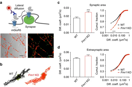
Exaggerated synaptic mobility of mGluR5 in
Fmr1 KO neurons.
In spite of its prominent role in the pathophysiology of FXS, the
dynamics of mGluR5 at synapses have not yet been studied in the
context of this disorder. Here we used a single nanoparticle
(quantum dot; QD) imaging approach to track surface mGluR5 in
live hippocampal neurons derived from second-generation Fmr1
KO and WT mouse embryos (12–15 days in vitro). This
tech-nique permitted us to examine the exploratory activity of
single-particle complexes within defined sub-cellular compartments.
Synapses were labeled using an active mitochondria marker
(MitoTracker) to distinguish them from extrasynaptic sites, as
previously described
34(Fig.
1
a). MitoTracker labeled synaptic
sites similarly in WT and KO neurons (Supplementary Fig.
1
). An
analysis of the trajectories of single mGluR5 molecules revealed
that their diffusion coefficient was significantly enhanced within
the synaptic compartment of Fmr1 KO as compared with WT
neurons (Fig.
1
b, c; 41.18%; P
< 0.001; WT mobility values were
similar to those reported previously
23). This result indicates an
increased mobility of mGluR5 within the synaptic membrane of
Fmr1 KO neurons. Accordingly, the fraction of mobile mGluR5
(diffusion coefficient >0.005 μm
2/s) at the synapse was higher in
Fmr1 KO neurons (+16.83%; P
< 0.001). In contrast to the
synaptic sites, lateral mobility of mGluR5 at extrasynaptic sites
was comparable between Fmr1 KO and WT neurons (Fig.
1
d;
diffusion coefficient, P = 0.106; mobile fraction, P = 0.833).
To determine whether the observed increase in membrane
mobility was specific for mGluR5, or a more general phenomenon
affecting other glutamate receptors as well, we also quantified the
mobility of individual AMPA-type (AMPAR) and NMDA-type
(NMDAR) glutamate receptors. To this end, we used antibodies
specific to the extracellular domains of the GluA2 and GluN1
receptor subunits comprising the AMPAR and NMDAR tetramer
complexes, respectively, in conjunction with the same QD
tracking approach. The dynamics of these receptor subunits have
been extensively characterized previously using QD-based
track-ing approaches
34(see also Supplementary refs.
1–4). We found no
differences in the lateral diffusion and the mobile fraction of
AMPAR within the synaptic compartment (diffusion coefficient,
P
= 0.732; mobile fraction, P = 0.913), whereas a small but
significant reduction was detected in the extrasynaptic
compart-ment (diffusion coefficient: –4.21%, P < 0.001; mobile fraction:
–4.53%, P < 0.001) of Fmr1 KO neurons (Supplementary
Fig.
2
a–c). Conversely, NMDAR showed a small yet statistically
significant increase in the lateral diffusion both in the synaptic
(diffusion coefficient: +13.33 %, P < 0.001) and the extrasynaptic
sites (diffusion coefficient: +13.60 %, P < 0.001) of Fmr1 KO
neurons, which was not sufficient to affect the fraction of mobile
receptors in both compartments (synaptic sites, P
= 0.517;
extrasynaptic sites, P
= 0.860; Supplementary Fig.
3
a–c). These
data suggest that the absence of FMRP differentially impacts the
mobility of mGluR5, AMPAR and NMDAR, with a major effect
for mGluR5 at synaptic sites.
Impaired mGluR5/Homer scaffold alters mGluR5 diffusion.
Since Homer isoforms function as anchoring molecules for
mGluR5 at synapses
25, we hypothesized that the previously
described reduction in mGluR5/Homer interaction
17might lead
to the exaggerated membrane mobility of mGluR5 in Fmr1 KO
neurons reported here. If our prediction was correct, then
dis-rupting the specific mGluR5/Homer interaction in WT neurons
should mimic the disease phenotype. We tested this hypothesis,
using a cell-permeable peptide containing the Homer binding
motif of mGluR5 (TAT-mGluR5ct; characterized previously
35,36;
Fig.
2
a). Indeed, pre-incubation of WT neurons with
TAT-mGluR5ct caused an increase in the lateral diffusion and mobile
fraction of mGluR5 in the synaptic compartment (Fig.
2
b–d;
diffusion coefficient: +62.5%; mobile fraction: +12.57%; WT
TAT-mGluR5ct vs. WT TAT-mGluR5mu; P
< 0.001 for both
parameters). Importantly, both parameters were now comparable
to those of Fmr1 KO neurons without the peptide (Fig.
2
c, d;
diffusion coefficient, P > 0.999; mobile fraction, P = 0.531). As
expected, pre-incubation of WT neurons with a peptide
con-taining a mutated Homer binding motif (TAT-mGluR5mu
35,36)
had no effect on the lateral diffusion of mGluR5 (Fig.
2
c, d;
diffusion coefficient, P > 0.999; mobile fraction, P = 0.795).
Moreover, neither TAT-mGluR5ct nor TAT-mGluR5mu
treat-ment had any effect on the mGluR5 mobility in Fmr1 KO
neu-rons (Fig.
2
c, d; diffusion coefficient, P = 0.966; mobile fraction, P
= 0.088; Fmr1 KO mGluR5ct vs. Fmr1 KO
TAT-mGluR5mu). Taken together, these experiments provide strong
correlative evidence that changes in the lateral diffusion of
mGluR5 detected in Fmr1 KO neurons are indeed due to a
dis-rupted link between the long Homer scaffolding proteins and
mGluR5.
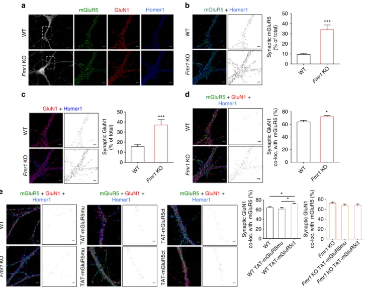
Enhanced mGluR5/NMDAR co-clustering in
Fmr1 KO neurons.
In addition to anchoring mGluR5 at synapses, Homer-containing
complexes also provide a physical link to NMDARs
29. We thus
explored whether the disrupted mGluR5–Homer scaffold might
also alter the interaction between mGluR5 and NMDAR in Fmr1
a
c
Synaptic area Extrasynaptic areab
WT Fmr1 KO mGluR5 Synapse Lateral diffusion WT Fmr1 KO 0.00 0.02 0.04 0.06 0.001 0.010 Diff. coeff. (µm2/s) Diff. coeff. ( µ m 2/s) Diff. coeff. ( µ m 2/s) 0.100 1 0.001 0.010 Diff. coeff. (µm2/s) 0.100 1 0.6 0.7 0.8 0.9 1.0 Cumul. fraction WT Fmr1 KO *** 0.0 0.3 0.5 0.8 1.0 Cumul. fraction WT Fmr1 KO WT Fmr1 KO 0.00 0.01 0.02 0.03 ***d
Fig. 1 Cell-surface mGluR5 displays an increased lateral diffusion rate within the synaptic compartment of hippocampalFmr1 KO neurons. (a) Experimental setup. Upper panel: schematic representation of endogenous mGluR5 in the dendritic membrane labeled with a QD-antibody complex targeting the extracellular domain of the receptor. Lower panel: representative images of the dendrites of hippocampal neurons shown in phase contrast (left), and their MitoTracker-labeled synaptic sites (right; green) overlaid with reconstructed trajectories of surface mGluR5-QD complexes (depicted in red) in the dendritic membrane of the same neurons. Scale bar= 5 μm. (b) Representative trajectories of single surface mGluR5-QD in WT and Fmr1 KO neurons. The synaptic sites are represented by the green areas. Scale bar= 1 μm. (c) Cumulative distribution (left panel) and cumulative frequency distribution (right panel) of the instantaneous diffusion coefficient of mGluR5-QDs in the synaptic compartment. The lateral diffusion is significantly higher in Fmr1 KO neurons (WT, 0.017± 0.001 µm2/s, n= 1632 trajectories from 16 dendritic fields of 3 different cultures); Fmr1 KO, 0.024 ± 0.001 µm2/s, n= 1451 trajectories (14 dendriticfields from 3 cultures); ***P < 0.001 by Mann–Whitney test on cumulative distribution; ***P < 0.001 by Kolmogorov–Smirnov test on cumulative frequency distribution). (d) Cumulative distribution (left panel) and cumulative frequency distribution (right panel) of the instantaneous diffusion coefficient of mGluR5-QDs in the extrasynaptic area of WT and Fmr1 KO neurons (WT, 0.045 ± 0.002 µm2/s,n = 1907 trajectories from 16 dendriticfields of 3 different cultures; Fmr1 KO, 0.048 ± 0.002 µm2/s,n = 1347 trajectories from 14 dendritic fields of 3 different cultures; P = 0.106 by Mann–Whitney test on cumulative distribution; P = 0.649 by Kolmogorov–Smirnov test on cumulative frequency distribution)
KO neurons. In a
first set of experiments, we took advantage of
the detection accuracy of single QDs (reviewed in ref.
20) by
measuring the synaptic fraction of mGluR5-QD and GluN1-QD
co-localized with MitoTracker (Fig.
3
a). We found that the
synaptic fraction of both mGluR5-QD and GluN1-QD was
increased in Fmr1 KO neurons (Fig.
3
b; mGluR5, P
< 0.001;
GluN1, P
< 0.01). This finding provides direct evidence that
mGluR5 and NMDAR are more confined within the synapse in
WT Fmr1 KO Fmr1 KO 0 5 10 15 20 mGluR5-QDs
Syn. fraction (% of total)
***
a
b
WT 0 3 6 9 12 GluN1-QDsSyn. fraction (% of total)
**
GluN1-QDs mGluR5-QDs
WT Fmr1 KO
Fig. 3 mGluR5 and GluN1 are more confined within the synaptic compartment in Fmr1 KO neurons. (a) Representative surface distribution of mGluR5-QD (upper panel) and GluN1-QD (lower panel) in a 500-frame stack (each dot represents the detection of a single receptor during 50-ms acquisition time), revealing the synaptic site as a trapping zone (green). (b) Relative fractions of synaptic mGluR5-QD (left panel) and GluN1-QD (right panel) particles. These values are increased inFmr1 KO neurons (mGluR5-QD: WT, 8.053 ± 0.504 %, n = 16 dendritic fields from 3 cultures; Fmr1 KO, 15.95 ± 0.685 %, n = 14 dendriticfields from 3 cultures; ***P < 0.001, t = 9.44, df = 28, unpaired Student’s t-test; GluN1-QD: WT, 8.318 ± 0.382 %, n = 63 dendritic fields from 6 cultures;Fmr1 KO, 10.02 ± 0.446 %, n = 51 dendritic fields from 6 cultures; **P < 0.01, t = 2.91, df = 112, unpaired Student’s t-test)
a
COOH Homer1a FXS COOH Long homer Control mGluR5 COOH TAT-mGluR5ct TAT-mGluR5ctExperimental condition mimicking FXS
WT WT TAT-mGluR5mu Fmr1 KO Fmr1 KO TAT-mGluR5ct Fmr1 KO TAT-mGluR5mu
b
WT TAT-mGluR5ctd
0.001 0.010 0.100 1 0.6 0.7 0.8 0.9 1.0Cumul. fraction Fmr1 KO TAT-mGluR5mu Fmr1 KO TAT-mGluR5ct
WT TAT-mGluR5mu WT TAT-mGluR5ct
***
Syn. diff. coeff. (µm2 /s) WT WT TAT-mGluR5muWT TAT-mGluR5ct Fmr1 KO Fmr1 KO TAT-mGluR5mu Fmr1 KO TAT-mGluR5ct 0 50 100 150 200 Normalized syn. diff. coeff. ( µ m 2/s) (% of WT) *** NS
c
Fig. 2 Disruption of the link between mGluR5 and Homer in WT neurons mimics theFmr1 KO phenotype. (a) Schematic illustration of the effect of the cell-permeable TAT-mGluR5ct peptide. This peptide disrupts the mGluR5/Homer link, mimicking the situation inFmr1 KO neurons. (b) Representative trajectories of single surface mGluR5-QD in WT andFmr1 KO neurons treated with TAT-mGluR5ct and its mutated TAT-mGluR5mu control peptide (both peptides 5µM, 1 h). The synaptic sites are represented by the green areas. Scale bar = 1 μm. (c) Normalized cumulative distribution of the instantaneous diffusion coefficient of mGluR5-QDs in the synaptic area of WT and Fmr1 KO neurons treated with TAT-mGluR5mu and TAT-mGluR5ct. The lateral diffusion rate of mGluR5-QDs in WT neurons treated with TAT-mGluR5ct peptide is comparable to that inFmr1 KO neurons under basal conditions (WT, 0.016± 0.002 µm2/s,n = 636 trajectories from 12 dendritic fields of 3 cultures; WT TAT-mGluR5mu, 0.016 ± 0.002 µm2/s,n = 1798 trajectories from 12 dendriticfields of 3 cultures; WT TAT-mGluR5ct, 0.026 ± 0.002 µm2/s,n = 1444 trajectories from 19 dendritic fields of 3 cultures; Fmr1 KO, 0.025 ± 0.002µm2/s,n = 797 trajectories from 16 dendritic fields of 3 cultures; Fmr1 KO TAT-mGluR5mu, 0.023 ± 0.001 µm2/s,n = 1419 trajectories from 13 dendriticfields of 3 cultures; Fmr1 KO TAT-mGluR5ct, 0.024 ± 0.002 µm2/s,n = 489 trajectories from 9 dendritic fields of 3 cultures; WT TAT-mGluR5mu vs. WT TAT-mGluR5ct***P < 0.001 by Kruskal–Wallis test with Dunn’s multiple comparison test; WT TAT-mGluR5ct vs. Fmr1 KO P > 0.999 by Kruskal–Wallis test with Dunn’s multiple comparison test). (d) Cumulative frequency distribution of the instantaneous diffusion coefficient of mGluR5-QDs within the synaptic area of WT andFmr1 KO neurons treated with TAT-mGluR5mu and TAT-mGluR5ct (***P < 0.001 by Kolmogorov–Smirnov test. For statistical analysis each condition was separately compared to WT TAT-mGluR5mu)
Fmr1 KO neurons, likely increasing the probability of physical
interactions between these receptors within a given time window.
To extend these
findings, we performed a triple
immuno-fluorescence labeling experiment for mGluR5, NMDAR and
Homer1 (the latter used as a synaptic marker), together with
confocal microscopy and post hoc image analysis (Fig.
4
a–d).
Quantitative analysis of the proportion of
mGluR5-/Homer1-positive or GluN1-/Homer1-mGluR5-/Homer1-positive
fluorescence intensity
(expressed as a function of total mGluR5- or GluN1 signal)
suggests an increased localization of both mGluR5 and NMDAR
at the synapse in Fmr1 KO neurons (Fig.
4
b, c, respectively;
mGluR5, P
< 0.001; NMDAR, P < 0.001). In addition, combined
analysis of all three markers points to a higher degree of
co-clustering of mGluR5 and NMDAR at synaptic sites (Fig.
4
d;
P
< 0.05) further supporting a tighter association of these
receptors in the absence of FMRP.
To further examine whether the disrupted mGluR5/Homer
scaffold might provide a causal mechanism for this increased
co-clustering of mGluR5 and NMDAR in Fmr1 KO neurons, we
again exploited the interfering peptide. As expected, the
pre-incubation of WT neurons with TAT-mGluR5ct resulted in a
significant increase in the mGluR5/NMDAR co-localization
(Fig.
4
e; P
< 0.05), reflecting the increased percentage of synaptic
mGluR5 and NMDAR (Supplementary Fig.
4
a–c). Pre-incubation
Fmr1 KO Fmr1 KO TAT-mGluR5mu Fmr1 KO TAT-mGluR5ct 0 20 40 60 80 Synaptic GluN1
co-loc. with mGluR5 (%)
WT WT TAT-mGluR5muWT TAT-mGluR5ct 0 20 40 60 80 Synaptic GluN1
co-loc. with mGluR5 (%)
* * WT Fmr1 KO 0 10 20 30 40 50
Synaptic GluN1 (% of total)
*** WT Fmr1 KO 0 10 20 30 40 50 Synaptic mGluR5 (% of total) *** WT Fmr1 KO 0 20 40 60 80 Synaptic GluN1
co-loc. with mGluR5 (%)
* WT mGluR5 Homer1 Fmr1 KO GluN1 WT mGluR5 + Homer1 Fmr1 KO GluN1 + Homer1 WT Fmr1 KO mGluR5 + GluN1 + Homer1 WT Fmr1 KO TAT-mGluR5mu WT mGluR5 + GluN1 + Homer1 mGluR5 + GluN1 + Homer1 mGluR5 + GluN1 + Homer1 Fmr1 KO TAT-mGluR5mu TAT-mGluR5ct TAT-mGluR5ct
e
a
b
c
d
Fig. 4 Disruption of the mGluR5/Homer scaffold increases surface mGluR5/NMDAR co-clustering. (a) Cultured WT andFmr1 KO hippocampal neurons were triple-labeled for mGluR5, GluN1 and Homer1. (b, c) Left: Representative image of Homer/mGluR5 and Homer/GluN1 co-localization; middle: Distribution of co-localized mGluR5/Homer1 or GluN-/Homer1 clusters; right: mGluR5/Homer1 and GluN1/Homer1 clusters as percentage of total mGluR5 or GluN1 signal (mGluR5: WT, 9.53± 0.959 %, n = 27; Fmr1 KO, 34.14 ± 4.598 %, n = 20; ***P < 0.001, t = 6, df = 45, unpaired Student’s t-test; GluN1: WT 15.75± 1.841 %, n = 29; Fmr1 KO 37.15 ± 5.324 %, n = 18; ***P < 0.001, t = 4.47, df = 45, unpaired Student’s t-test). (d) Left: Representative image showing mGluR5/GluN1/Homer1 colocalization; middle: Distribution of co-localized mGluR5/GluN1/Homer1 labeling; right: Co-localized mGluR5/GluN1/Homer1 clusters as percentage of synaptic GluN1 signal (WT, 63.97± 2.414 %, n = 26; Fmr1 KO, 72.14 ± 2.081 %, n = 23; *P < 0.05, t = 2.53, df = 47, unpaired Student’s t-test). (e) mGluR5ct peptide increased mGluR5/GluN1 co-clustering at synapses in WT neurons, whereas mGluR5mu or TAT-mGluR5ct (both 5µM, 1 h) had no effect in Fmr1 KO neurons; Left: Representative images and distribution of mGluR5/GluN1/Homer1-co-labeling signal in control and TAT-mGluR5mu or TAT-mGluR5ct treated WT andFmr1 KO neurons. Right: Co-localized mGluR5/GluN1/Homer1-positive signals as percentage of synaptic GluN1 signal (WT: 63.97± 2.414 %, n = 26; WT TAT-mGluR5mu: 61.19 ± 3.489 %, n = 14; WT TAT-mGluR5ct: 71.79 ± 1.528 %, n = 22; WT vs. WT TAT-mGluR5ct, *P = 0.043; WT TAT-mGluR5mu vs. WT TAT-mGluR5ct *P = 0.017, F (2, 59) = 4.87; Fmr1 KO: 72.14 ± 2.081 %, n = 23; Fmr1 KO TAT-mGluR5mu: 67.32 ± 2.832 %, n = 18; Fmr1 KO TAT-mGluR5ct: 67.58 ± 2.69 %, n = 26; Fmr1 KO vs. Fmr1 KO TAT-mGluR5ct P = 0.391; Fmr1 KO TAT-mGluR5mu vs. Fmr1 KO TAT-mGluR5ct P = 0.997, F (2, 64) = 1.13). P values by one-way ANOVA test with Tukey’s Multiple Comparison test.n = dendritic fields from 3 cultures. Scale bar = 10 μm (a) 2 μm (b, c, d, e)
with TAT-mGluR5mu had no effect on the co-localization
(Fig.
4
e; P
= 0.725; Supplementary Fig.
4
a–c). Thus, the
disruption of the mGluR5/Homer binding is likely responsible
for a tighter physical association between mGluR5 and NMDAR
at synapses in Fmr1 KO neurons.
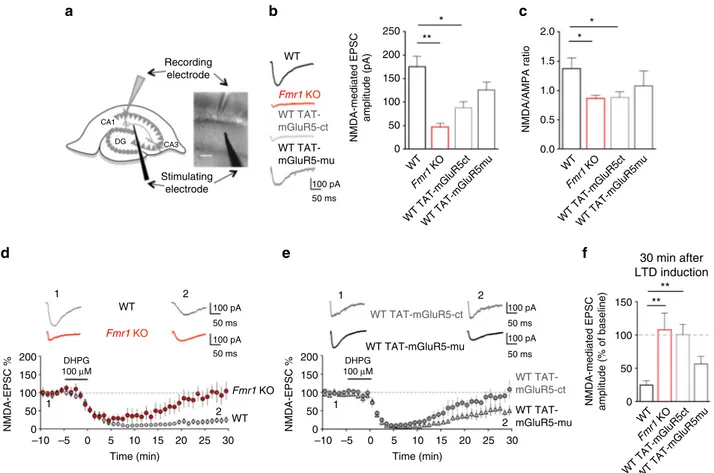
Reduced NMDAR function and plasticity in
Fmr1 KO neurons.
What are the consequences of this tighter mGluR5/NMDAR
association at synaptic sites for NMDAR function? To address
this question, we measured synaptic NMDAR-mediated
excita-tory postsynaptic currents (EPSCsNMDA) induced by Schaffer
collateral stimulation using whole-cell patch-clamp recordings
from CA1 pyramidal neurons in acute hippocampal slices
(Fig.
5
a). EPSCsNMDA
displayed lower amplitudes in Fmr1 KO
neurons when compared with WT neurons (Fig.
5
b; Fmr1 KO:
46.4
± 8.4 pA; WT: 175.7 ± 21.9 pA; P < 0.001). Consistently, the
NMDA/AMPA ratio was significantly lower in Fmr1 KO neurons
(Fig.
5
c; Fmr1 KO: 0.86
± 0.05; WT: 1.37 ± 0.29; P = 0.02). These
defects in NMDAR function were mimicked in WT neurons by
application of the interfering peptide TAT-mGluR5ct (Fig.
5
b;
EPSCsNMDA, TAT-mGluR5ct: 88.01
± 12.70 pA; P < 0.01
com-pared to untreated WT; Fig.
5
c; NMDA/AMPA ratio,
TAT-mGluR5ct: 0.88
± 0.10; P = 0.02 compared to untreated WT).
These data thus strongly support our hypothesis that alterations
in the membrane dynamics of mGluR5, and its tighter coupling
with NMDAR—in the synapses of both Fmr1 KO neurons as well
as in neurons treated with the disrupting peptide—contribute to
abnormal NMDAR function.
It is well established that the activation of group-I mGluR
induces an exaggerated LTD of excitatory postsynaptic AMPA
currents (EPSCAMPA) in hippocampal Fmr1 KO neurons
13. In
contrast, LTD of excitatory postsynaptic NMDA currents
(EPSCsNMDA) mediated by group-I mGluR activation
37–39has
not been investigated in Fmr1 KO mice. In WT hippocampal
CA1 neurons, application of the group-I mGluR orthosteric
agonist (S)-3,5-dihydroxyphenylglycine (DHPG, 100
µM, 5 min)
induced a strong and long-lasting reduction in EPSCsNMDA
(Fig.
5
d). Remarkably, this form of synaptic NMDAR plasticity
was largely absent in Fmr1 KO neurons (Fig.
5
d; P
= 0.0006). As
d
b
e
WT TAT-mGluR5-mu WT TAT-mGluR5-ct Fmr1 KO WTf
100 pA 50 ms Recording electrode Stimulating electrode CA3 DG CA1c
Fmr1 KO WT 1 2 1 2 100 pA 50 ms 50 ms 100 pA Time (min) 30 25 DHPG 100 µM DHPG 100 µM 20 15 10 5 0 –5 –10 Time (min) 30 25 20 15 10 5 0 –5 –10 200 150 100 50 0 NMDA-EPSC % 200 150 100 50 0 NMDA-EPSC % WT TAT-mGluR5-mu WT TAT-mGluR5-ct 1 2 50 ms 50 ms 100 pA 100 pA 1 2 WT TAT-mGluR5-mu WT TAT-mGluR5-ct ** ** ** WT Fmr1 KO 30 min after LTD induction 150 100NMDA-mediated EPSC amplitude (% of baseline)
NMDA-mediated EPSC amplitude (pA) 50 0 WT Fmr1 KO WT TAT-mGluR5ctWT TAT-mGluR5mu WT Fmr1 KO WT TAT-mGluR5ctWT TAT-mGluR5mu WT Fmr1 KO WT TAT-mGluR5ctWT TAT-mGluR5mu 2.0 * * * 1.5 1.0 0.5 NMDA/AMPA ratio 0.0 200 250 150 100 50 0
a
Fig. 5 Disruption of mGluR5/Homer coupling alters synaptic NMDAR function and plasticity. (a) NMDAR-mediated excitatory post-synaptic currents (EPSCsNMDA) were recorded from CA1 pyramidal neurons. Scale bar= 100 μm (b) Left: representative EPSCsNMDAtraces from WT,Fmr1 KO, WT treated
with either TAT-mGluR5ct or TAT-mGluR5mu (both 5µM, 4 h). Histograms: EPSCsNMDAamplitude (F(3, 35)= 6.26; **P = 0.0016; one-way ANOVA with
Tukey’s Multiple Comparison). Compared to WT (175.7 ± 21.9 pA, n = 11 neurons from 7 animals), EPSCsNMDAwere lower inFmr1 KO (46.4 ± 8.4 pA, n =
7 neurons from 4 animals, **P < 0.01) and in WT treated with TAT-mGluR5ct (88.1 ± 12.7 pA, n = 13 neurons from 7 animals, *P < 0.05) but not with TAT-mGluR5mu (125.9± 16.8 pA n = 8 neurons from 3 animals, P = 0.14). (c) NMDA/AMPA ratio in WT, Fmr1 KO, WT treated with TAT-mGluR5ct or TAT-mGluR5mu (F(3, 32)= 4.1; *P = 0.013; one-way ANOVA with Tukey’s Multiple Comparison). NMDA/AMPA ratio (in WT 1.37 ± 0.17, n = 9 neurons
from 4 animals) was reduced inFmr1 KO (0.86 ± 0.05, n = 8 neurons from 3 animals, *P = 0.02) and in WT treated with TAT-mGluR5ct (0.88 ± 0.10, n = 9 neurons from 3 animals, *P = 0.02). (d, f) The mGluR1/5 agonist DHPG (100 µM, 5 min) induced long-term depression (mGluR-LTD) of EPSCsNMDAin
WT (EPSCsNMDAamplitude: 24.9± 2 % of baseline, n = 8 neurons from 5 animals) but not in Fmr1 KO (EPSCsNMDA107.8± 25 %, n = 6 neurons from 4
animals, **P < 0.01, WT vs. Fmr1 KO). (e, f) mGluR-LTD of EPSCsNMDAwas abolished in WT treated with TAT-mGluR5ct (EPSCsNMDA100.6± 15 %, n = 8
neurons from 7 animals, **P < 0.01, WT vs. TAT-mGluR5ct) but not with TAT-mGluR5mu (EPSCsNMDA56.6± 11 %, n = 7 neurons from 3 animals). (f)
with the enhanced mGluR5/NMDAR co-clustering, this
pheno-type was also recapitulated in WT neurons by use of the peptide
mimicking approach, suggesting the defective mGluR5/Homer
interaction as the underlying cause (Fig.
5
e, f; P
< 0.01). As
expected, in Fmr1 KO slices pre-treated with TAT-mGluR5ct,
mGluR-LTD of EPSCsNMDA
was still largely absent (Fmr1 KO
TAT-mGluR5ct vs. WT, P
= 0.032), and comparable to untreated
Fmr1 KO slices (Supplementary Fig.
5
a, b; P
= 0.69). These data
suggest that the disruption of the mGluR5/Homer scaffold
compromises NMDAR function under both basal conditions, as
well as during synaptic plasticity, in Fmr1 KO neurons.
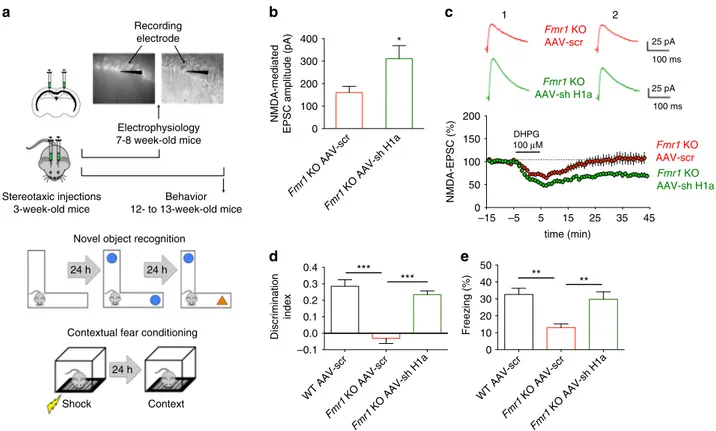
Homer1a knockdown rescues NMDAR function and plasticity.
Homer1a is known to antagonize the interaction between
mGluR5 and Homer. Thus, we asked whether knocking down
Homer1a—an approach that fosters the mGluR5/Homer
inter-action by decreasing the Homer1a/Homer balance
18—could
correct the dysfunction in NMDAR currents in Fmr1 KO
neu-rons. To address this question we exploited an AAV vector
expressing a small interfering hairpin RNA (shRNA) targeted
against the unique 3’-untranslated region of Homer1a mRNA and
GFP bicistronically
40,41. A similar vector expressing scrambled
shRNA and GFP served as control
40,41. AAV vectors were
ste-reotaxically injected into the hippocampal CA1 area of Fmr1 KO
mice and EPSCsNMDA
were measured from infected (i.e., GFP
expressing) CA1 pyramidal neurons in acute brain slices 4 weeks
later when transgene protein expression had peaked and
remained at stable levels (Fig.
6
a). EPSCsNMDA
displayed higher
amplitudes in Fmr1 KO neurons expressing the shRNA targeted
against Homer1a when compared to Fmr1 KO neurons
expres-sing the scrambled shRNA (Fig.
6
b; P
< 0.05). In addition, the
mGluR-dependent LTD of EPSCsNMDA, that was absent in Fmr1
KO neurons, was restored in Fmr1 KO neurons expressing the
shRNA for Homer1a (Fig.
6
c; P
< 0.001). Both findings confirm
our hypothesis that mGluR5/Homer disruption can cause
abnormal NMDAR function and plasticity in Fmr1 KO neurons.
Homer1a knockdown rescues cognitive defects in
Fmr1 KO
mice. Can the correction of the disrupted mGluR5/Homer
scaf-fold also rescue cognitive defects linked to NMDAR dysfunction?
To evaluate the effects of Homer1a reduction on hippocampus
dependent memory formation, we used two behavioral tasks—a
one-trial acquisition novel object-recognition task (NOR),
per-formed on an L-maze and which tests episodic memory
42, and
WT AAV-scr Fmr1 KO AAV-scr Fmr1 KO AAV-sh H1a 0 10 20 30 40 50 Freezing (%) ** ** Fmr1 KO AAV-scr Fmr1 KO AAV-sh H1a 0 100 200 300 400 NMDA-mediated
EPSC amplitude (pA)
* WT AAV-scr Fmr1 KO AAV-scr Fmr1 KO AAV-sh H1a –0.1 0.0 0.1 0.2 0.3 0.4 Discrimination index *** *** Stereotaxic injections 3-week-old mice Electrophysiology 7-8 week-old mice Behavior 12- to 13-week-old mice Recording electrode
Novel object recognition 24 h 24 h
24 h Shock Context
Contextual fear conditioning
–15 –5 5 15 25 35 45 0 50 100 150 200 time (min) NMDA-EPSC (%) DHPG 100 µM Fmr1 KO AAV-sh H1a Fmr1 KO AAV-scr 25 pA 100 ms 1 2 25 pA 100 ms Fmr1 KO AAV-scr Fmr1 KO AAV-sh H1a
a
b
c
d
e
Fig. 6 Homer1a knockdown rescues synaptic NMDAR dysfunction and cognitive defects inFmr1 KO mice. (a) Experimental procedure: electrophysiology (upper panel), novel object-recognition task (NOR) on L-maze (middle panel), contextual fear-conditioning task (CFC) (lower panel). WT andFmr1 KO mice (21 days) were bilaterally injected into the dorsal hippocampus with AAV-shH1a or AAV-Scr expressing GFP, and tested at 7–8 weeks
(electrophysiology;b, c) or 12–13 weeks of age (behavior; d, e). (b) The EPSCNMDAamplitude ofFmr1 KO AAV-sh H1a mice (311.3 ± 58 pA) was larger than
that ofFmr1 KO AAV-scr mice (160.4 ± 27.8 pA, n = 12 neurons from 4 animals for both conditions, *P < 0.05, t = 2.35, df = 22, unpaired Student’s t-test). (c) AAV-sh H1a infection inFmr1 KO mice rescues LTD of EPSCNMDAinduced by DHPG (100µM, 10 min) (EPSCNMDAamplitude 40 min after LTD
induction: 70.4± 6 % of control, n = 6 neurons from 5 animals). mGluR-LTD of EPSCNMDAwas absent inFmr1 KO AAV-scr mice (EPSCNMDAamplitude:
106.8± 7 % of control, n = 7 neurons from 6 animals, ***P < 0.001, t = 7.73, df = 117 by unpaired Student’s t-test). (d) Discrimination index of NOR on test day: WT AAV-scr, 0.285± 0.040, n = 8; Fmr1 KO AAV-scr, −0.030 ± 0.032, n = 9; Fmr1 KO AAV-sh H1a, 0.235 ± 0.022, n = 8; F (2, 22) = 29.02 (***P < 0.001, one-way ANOVA);Fmr1 KO AAV-sh H1a mice performed better than Fmr1 KO AAV-scr, while Fmr1 KO AAV-scr were impaired cf. WT AAV-scr (***P < 0.001, one-way ANOVA test with Tukey’s test). (e) Percentage of time freezing on test day in the CFC (WT AAV-scr, 32.623 ± 3.637 %, n = 9; Fmr1 KO AAV-scr, 13.038 ± 2.203 %, n = 8; Fmr1 KO AAV-sh H1a, 29.743 ± 4.371 %, n = 8, F (2, 22) = 8.836, **P < 0.01, one-way ANOVA). Fmr1 KO AAV-scr exhibited an impaired CFC memory cf. WT AAV-scr, whereasFmr1 KO AAV-sh H1a showed rescued CFC memory (**P < 0.01, one-way ANOVA test with Tukey’s test)
contextual fear conditioning (CFC)
43. Both procedures induce
robust hippocampus dependent learning with a single training
episode, and have previously been utilized for the investigation of
memory defects in the Fmr1
tm1Cgrmouse
42, 43. Since these
phe-notypes have not previously been investigated in the
second-generation Fmr1 KO mouse model, we initially performed a pilot
experiment with a separate batch of experimentally naïve animals
to determine whether we could recapitulate these phenotypes.
Consistent with the aforementioned studies employing the
first-generation Fmr1 KO mouse line, the second-first-generation model,
used here, exhibited similar decreases in the discrimination index
(DI) in the NOR task (Supplementary Fig.
6
, Fig.
7
, Fig.
8
and
Fig.
9
; P
< 0.001) and in the percentage of freezing compared with
WT mice following retrieval of CFC memory (Supplementary
Fig.
10
; P
< 0.001). We then tested whether mGluR5/Homer
crosstalk modulation could correct these defects in another batch
of behaviorally naive Fmr1 KO mice, stereotaxically injected with
the aforementioned AAV vectors into the CA1 area of the
hip-pocampus. Importantly, the selective Homer1a knockdown in the
hippocampus (Fig.
6
a) corrected defects in both the NOR
memory (Fig.
6
d; Fmr1 KO AAV-sh H1a vs. Fmr1 KO AAV-scr,
P
< 0.001; Supplementary Fig.
11
, Fig.
12
, Fig.
13
and Fig.
14
) and
CFC memory (Fig.
6
e; Fmr1 KO AAV-sh H1a vs. Fmr1 KO
AAV-scr, P
< 0.01) in adult Fmr1 KO mice. Taken together with
the aforementioned results, these data strongly support the idea
that disruption of mGluR5/Homer scaffold, leading to altered
NMDAR function in the hippocampus contributes to impairment
of hippocampal-dependent cognitive function in Fmr1 KO mice.
Discussion
In spite of the widespread acceptance of the mGluR theory of
FXS, the sub-cellular mechanisms underlying mGluR5-dependent
defects in synaptic function and plasticity, as well as their
asso-ciated cognitive phenotypes remain poorly understood. Most
studies, to date, have focused on the altered translational
pro-cesses arising from perturbations in mGluR signaling and their
ensuing effect on synaptic plasticity. Surprisingly scant attention,
however, has been paid to the behavior of the receptor itself and
its interactions with other membrane-bound receptors. Here we
present several novel aspects of mGluR5 pathophysiology in FXS.
Specifically, we show that not only are the dynamics of mGluR5
altered at the synapse of Fmr1 KO neurons (leading to a greater
lateral diffusion of the receptor), but also that the confinement of
the receptor at the synapse is increased. These changes are
accompanied by an enhanced co-clustering of mGluR5 and
NMDAR at synapses as well as altered NMDAR
function/plas-ticity in the hippocampal CA1 region of Fmr1 KO mice.
Importantly, our experiments point to a plasticity defect not
previously reported involving mGluR5-mediated LTD of NMDA
receptor currents in FXS. This NMDAR dysfunction and lack of
plasticity correlates with hippocampus dependent cognitive
deficits.
Previous studies have shown that interactions between
mGluR5 and postsynaptic density scaffolding proteins, in
parti-cular Homer, are critical for the correct functioning of mGluR5
(reviewed
in
ref.
26).
Conversely,
disruption
of
this
mGluR5–Homer association in the absence of FMRP
17has been
reported to play a key causal role in several FXS phenotypes such
as anxiety, susceptibility to seizures and changes in circuit level
hyperexcitability in the neocortex and hippocampus
18,19,44. To
explore whether alterations in mGluR5–Homer crosstalk might
contribute to the changes in mGluR5 mobility and NMDAR
function reported here, we made use of a peptide disruption
approach
35to perturb the normal interactions between mGluR5
and long Homer isoforms. Indeed, we found that application of
this peptide recapitulated changes in mGluR5 mobility detected
in Fmr1 KO mice. This
finding was in coherence with previous
studies demonstrating that interaction of mGluR5 with Homer
proteins at the postsynaptic site regulates lateral diffusion of
mGluR5
45. The fact that the exaggerated mGluR5 mobility is
restricted to the synapse is not surprising taking into
considera-tion that long Homer isoforms are enriched at synaptic sites
28.
Interestingly, these alterations in receptor dynamics appear to be
specific to the mGluR5–Homer interaction because the AMPAR
subunit, GluA2, which does not bind Homer proteins, did not
exhibit substantial alterations in lateral diffusion in the synaptic
area. Noteworthy, although these mobility values may be
influ-enced by a number of experimental variables, our measures for
WT neurons are consistent with those reported under similar
conditions (e.g., ref.
23; see also Supplementary Table
1
). In
addition to alterations in mGluR5 mobility, we also found that
application of the peptide led to an increased synaptic
confine-ment of mGluR5, the co-clustering of mGluR5 and NMDAR, and
ultimately, to alterations in NMDAR function and
NMDAR-dependent plasticity—features that we also observed in Fmr1 KO
neurons. Taken together, these
findings suggest a strong causative
role for the altered mGluR5–Homer crosstalk in the
aforemen-tioned novel changes in mGluR5 dynamics and NMDAR
func-tion in FXS (see model in Fig.
7
).
Although alterations in synaptic plasticity have been well
documented in FXS, this is the
first report of a defect in
mGluR5-mediated LTD of NMDAR-mGluR5-mediated EPSCs in Fmr1 KO mice.
This is surprising, given that this form of plasticity is well
char-acterized in non-disease models (e.g. refs.
37–39). Our results
suggest that a disruption of the link between mGluR5 and Homer
proteins in Fmr1 KO neurons plays a negative role in the
induction of LTD of synaptic EPSCsNMDA
in response to group-I
mGluR activation. In this scenario, mGluR5 once liberated from a
co-assembly with Homer1 partners would undergo an increased
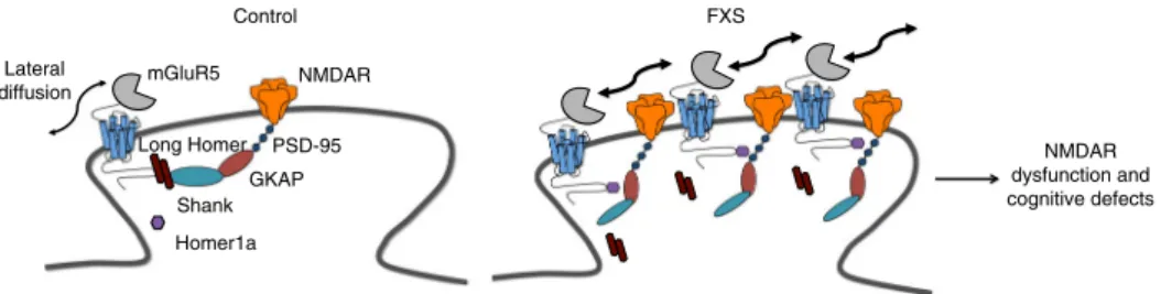
NMDAR dysfunction and cognitive defects Long Homer mGluR5 Control NMDAR Shank GKAP PSD-95 Lateral diffusion Homer1a FXS
Fig. 7 Model for dysfunction of the NMDAR/mGluR5 crosstalk inFmr1 KO neurons. In WT neurons, long Homer proteins anchor mGluR5 to a chain of PSD proteins in the synapse and prevent a direct interaction with NMDAR. Under those conditions, a co-clustering of mGluR5 and NMDAR is prevented. InFmr1 KO neurons, mGluR5 is less associated with the long Homer proteins and more associated with the short isoform, Homer1a. This disengagement from the long Homer protein–containing complex increases the lateral diffusion of mGluR5 and promotes its interaction with synaptic NMDAR. This configuration at the synapse prevents boosting of NMDAR currents under control conditions and their LTD following mGluR5 stimulation
physical and functional interaction with NMDAR, altering the
function of this receptor type
31. In line with previous results, we
found that EPSCsNMDA
evoked by Schaffer collateral stimulation
showed lower amplitudes in Fmr1 KO neurons. The altered
mGluR5/NMDAR partnership might also impact on NMDAR
removal from the synapse, a mechanism that is believed to be
responsible for depression of synaptic NMDAR currents
38. In
support of this notion, we found that NMDAR is more confined
at synapses in the Fmr1 KO neurons (and in WT neurons
fol-lowing incubation with the TAT-mGluR5ct peptide). One
pos-sible mechanism for the inhibition of NMDAR currents might be
a direct interaction of NMDAR with G-protein
βγ subunits
30—
facilitated by the physical constraint between mGluR5 and
NMDA. Similarly, steric hindrance caused by a closer association
between NMDAR and mGluR5 could prevent post-translational
modification of NMDAR, underlying their removal from
synap-ses. Whereas the induction of group-I mGluR mediated LTD of
AMPA currents involves both pre- and post-synaptic
mechan-isms
46–48, we suggest that the group-I mGluR mediated LTD of
NMDA currents reported here engages a post-synaptic
mechan-ism. This conclusion is based on the post-synaptic location of
Homer proteins, and also supported by previous studies into the
mechanism of this plasticity form
37.
How might the absence of FMRP cause mGluR5/Homer
dis-ruption and consequently the described changes in mGluR5 and
NMDAR function? The increased phosphorylation of Homer has
previously been demonstrated to reduce its affinity for mGluR5
49.
On the other hand, the increased phosphorylation of mGluR5
induces a higher affinity for Homer
50. This would provoke a
remodeling of mGluR5–Homer-NMDAR complexes, freeing
mGluR5 to form functional associations with NMDAR and
potentially leading to the observations reported here. Although
this provides a tantalizing explanation for our
findings, it may not
be the only mechanism underlying these changes. Moreover,
given that the loss of FMRP leads to the dysregulated translation
of a plethora of target mRNAs it is difficult to pinpoint one kinase
as the causative factor. While inhibition of CaMKII in acute slices
and cultured neurons from Fmr1 KO mice has been shown to
correct aberrant UP states and frequency of spontaneous
firing—
features that are indicative of altered circuit level activity
49—it is
conversely possible that altered circuit function can impinge on
mGluR5–Homer crosstalk. Altered circuit level activity has been
well documented in Fmr1 KO mice (e.g. refs.
51,52) and may play
a role in a number of central phenotypes of the disorder
53.
Increased circuit activity can be expected to influence immediate
early gene induction
27, 28, leading to increased Homer1a
expression, which exacerbates crosstalk disruption. This
disrup-tion, in turn, may drive further alterations in circuit level
func-tion
49. Interestingly, our experiments involving hippocampal
specific knockdown of Homer1a suggest discrete and
indepen-dent mechanisms for Homer1a in the pathology of FXS. In
par-ticular, we show here that hippocampal-dependent defects occur
through functional modification of NMDAR. Altered
mGluR-dependent NMDAR-LTD might be expected to profoundly
impact cognitive functions in FXS. In support of this notion, the
plasticity of NMDAR currents has recently been suggested to
provide a mechanism for metaplasticity
39, 54. In addition,
hippocampal-dependent cognitive impairment induced by social
defeat stress has been linked to a reduced Homer-mGluR5
interaction in mice
55. Likewise, a selective over-expression of
Homer1a in the dorsal hippocampus has been shown to impair
spatial working memory
56.
Here we show the rescue of two hippocampus- and
NMDAR-dependent cognitive phenotypes by knockdown of Homer1a
expression specifically in the hippocampus. Although previous
work has shown that generalized genetic ablation of Homer1a
rescued certain behavioral phenotypes of Fmr1 KO mice related
to anxiety and seizure susceptibility
18, these tests have no direct
correspondence with cognitive performance, which is one of the
prevailing features of FXS. Here we demonstrate rescue of two
phenotypes with direct relevance to cognitive function. Moreover,
we demonstrated that knockdown within the hippocampus alone
is sufficient to rescue these phenotypes. In addition, rescue of
cognitive function was correlated with rescue of a novel synaptic
plasticity defect. We are aware that recent literature has raised
concerns about the robustness of the cognitive phenotypes
reported in the Fmr1 KO model (reviewed in refs.
57,58). It is thus
imperative to establish robust tests that can be replicated between
laboratories to strengthen the use of the Fmr1 KO mouse as a
preclinical model of FXS
57. To this end, the use of the
non-classical object–recognition test (performed on an L-maze)
42,59,
and of a purely contextual fear memory paradigm
43rather than
auditory fear conditioning (that has typically been used for the
Fmr1 KO model e.g. refs.
60–63) may represent an important
refinement for cognitive testing of the Fmr1 KO mouse. Our
study, using the second-generation model, replicates
findings
reported for the
first-generation Fmr1
tm1Cgrmodel, suggesting
that these tests are robust and reproducible between independent
laboratories. With respect to contextual fear memory, the key
difference between our work (and the
findings of refs.
43,64) and
previous
findings in the field is that we paired the shock with the
presentation of the context. This paradigm leads to a different
relevance of the background vs. foreground sensorial cues and the
related recruitment of the hippocampus, compared with the
protocol used for auditory fear conditioning. This task could thus
be more suitable for the detection of behavioral deficits in the
Fmr1 KO mice.
These
finding can be expected to have wider implications for
future therapeutic approaches for the treatment of FXS and other
neurodevelopmental disorders. NMDAR hypofunction has been
detected in FXS models in other brain regions and has been
recently proposed to contribute to cognitive defects in FXS
(reviewed in ref.
65). In addition, it has been considered
sig-nificant in the context of autism and schizophrenia and its
cor-rection by means of mGluR5-positive allosteric modulators has
been proposed
66. Indeed, mice with reduced expression of the
NMDAR subunit, GluN1 exhibit a range of behavioral
pheno-types that are not only consistent with autism spectrum disorder,
but also which overlap with those observed in Fmr1 KO mice.
Given the known reciprocal modulation of mGluR5 and NMDA
responses, our results of an altered crosstalk of these two
recep-tors in FXS should be taken into consideration when predicting
the outcome of single or combined therapy with agents targeting
both receptors. Our
finding may also contribute to an improved
understanding of several factors impeding clinical trial design,
such as the marked heterogeneity present within the FXS
popu-lation
67. If increased circuit activity (as discussed above) indeed
leads to altered Homer1a levels, then this is likely to vary widely
amongst individuals based on their responses to external stimuli,
environment, etc. Here we propose that therapeutic approaches
aimed at restoring the normal mGluR5/Homer and mGluR5/
NMDAR interactions might provide a promising alternative for
the treatment of FXS. It is hoped that these
findings will
con-tribute to the development of alternative, targeted therapies for
this disorder and its co-morbidities, and provide mechanistic
links to other genetic causes of autism.
Methods
Animals. All experiments were conducted in strict compliance with the European Directive (2010/63/EU), and French and Italian law governing the use of laboratory animals and were approved by the Bordeaux Ethics Committee (C2EA50, authorization #5012024-A) and by the Ethics Committee of Catania University
(project # 181). Mice were housed in a SPF animal facility prior to experiments, kept on a 12 h–12 h light–dark cycle (lights on at 0:700 am) and had ad libitum access to food and water at all times. Second generation Fmr1 KO mice32were mostly used in our study. This model was generated by deletion of both the promoter and exon 1 of the Fmr1 gene, as described previously32. These mice are distinct from the original Fmr1 KO (Fmr1tm1Cgr) mouse line33, because they are deficient for both Fmr1 mRNA and FMRP protein. This mouse model has largely been used for physiological, molecular, and anatomical studies (e.g., ref.53, see also Supplementary refs.5–10), however limited behavioral characterization has also been performed (e.g., ref.60and Supplementary refs.11–14). Mice were backcrossed six generations into a C57BL/6 J (Charles River, L’Abresle, France) background and maintained in this mixed background for all experiments. The genotype of all progenitors, as well as experimental subjects, was determined by tail PCR as described previously32). For dissociated neuronal cultures WT and Fmr1 KO embryos were generated by crossing homozygous (Fmr1+/+X Fmr1+/yor Fmr1−/− X Fmr1−/y) progenitor mice. The genotype of the embryos was confirmed by tail PCR of the mother. For electrophysiology and behavioral experiments male WT and Fmr1 KO littermates were generated by crossing a heterozygous (Fmr1+/−) female mouse with a wild-type (Fmr1+/y) male mouse as described previously53. Mice were subsequently re-genotyped after the experiment by tail PCR as described previously53. Some electrophysiology experiments (see Fig.5)were carried out on first-generation (Fmr1tm1Cgr) Fmr1 KO mice (on an FVB background)33. Genotype was assessed by tail PCR as described previously68.
Primary cell cultures. Cultures of hippocampal neurons and glial cells were prepared from E18 WT and Fmr1 KO embryos. Pregnant mice were killed by decapitation after deep anesthesia with isoflurane and the uterine horn dissected. Hippocampi were subsequently dissected from the embryos in ice-cold dissection solution and then dissociated in trypsin (Sigma, 0.25%). Briefly, cells were plated at a density of 100 to 200 × 103cells per milliliter on poly-L-lysine (Sigma) precoated coverslips and kept at 37 °C in 5% CO2. The original plating Neurobasal culture
medium (Gibco) supplemented with B27 (Gibco, 2%) and complemented with 5% fetal bovine serum was replaced with a serum free medium on day in vitro (DIV) 2. Cytosine B-D-arabinofuranoside (Sigma, 5μM) was added on DIV 4. All the experiments were performed at DIV 12/15.
Pharmacological treatments. A cell-permeable (TAT-fused) peptide containing the proline-rich motif (PPXXF) of the mGluR5 C-terminal tail that binds the EVH1 domain of Homer, TAT-mGluR5ct (YGRKKRRQRRR-ALTPPSPFR), and a control peptide with a mutated Homer binding motif, mGluR5mu
(YGRKKRRQRRR-ALTPLSPRR), were synthesized at the UT Southwestern Pro-tein Chemistry Technology Center and kindly provided by Prof. K. Huber (Department of Neuroscience, University of Texas Southwestern Medical Center, Dallas, TX 75390, USA; see ref.35,36). The peptides were dissolved in H20 at a
concentration of 5 mM, and aliquots of this stock concentration were stored at −20 °C. Frozen aliquots of both TAT-fused peptides were used within 10 days and diluted to the desiredfinal concentration. Hippocampal cultures were treated with TAT-mGluR5ct or TAT-mGluR5mu for 1 h at afinal concentration of 5 µM in serum free culture medium at 37 °C. Slices were incubated during 4 h with either TAT-mGluR5ct or TAT-mGluR5mu (each at 5µM) in oxygenated ACSF at room temperature (21–22 °C). (S)-3,5-Dihydroxyphenylglycine (DHPG) (Abcam, # ab120007, 100µM) was dissolved in ACSF and applied by bath perfusion. Single-Particle Tracking and Surface Diffusion Calculation. For single-molecule tracking experiments, neurons werefirst exposed for 10 min to either mouse monoclonal anti-NH2mGluR5 antibody (1:20)69, mouse monoclonal anti-GluA2
AMPA receptor (AMPAR) subunit antibody (Millipore, #MAB397, 1:200), or rabbit polyclonal anti-GluN1 NMDA receptor (NMDAR) subunit antibody (Alomone Laboratories, #AGC-001, 1 : 20034and Supplementary refs.15–19) at 37 °C. Neurons were then incubated for 10 min in a solution containing quantum dots (QD) 655 coupled to goat anti-mouse IgG (Invitrogen, #Q11022MP) or coupled to goat anti-rabbit IgG (Invitrogen, #Q-11421MP) (final dilution 1:5000/ 1 : 10,000) at 37 °C. To label synaptic sites, neurons were incubated for 40 s at RT (~ 22 °C) in a solution containing the orange mitochondria marker MitoTracker (Invitrogen, #M-7510, 20 nM). A fraction of coverslips was also incubated for 1 h with TAT-mGluR5ct or TAT-mGluR5mu (5µM) in culture medium at 37 °C before and during the incubation with the primary antibodies. For QD 655 fluorescence imaging we used an EM-CCD camera (Evolve 512, Photometrics) with a 512 × 512 imaging array together with an HXP-120 light source (Zeiss) and the appropriatefilters for excitation and emission. Images were acquired at an integration time of 50 milliseconds for up to 500 consecutive frames (24 s) as described previously (Supplementary refs.15,18). QD movements were followed on randomly selected healthy looking dendritic regions for up to 20 min, and analyzed using Metamorph software (Universal Imaging Corporation, PA, USA). Briefly, the instantaneous diffusion coefficient, D, was calculated for each trajectory, from linearfits of the first 4 points of the mean-square-displacement vs. time function using the following equation: MSD (t)= < r2 > (t) = 4Dt. To assign synaptic localization, trajectories were sorted into extrasynaptic (i.e. MitoTracker-negative pixels) and synaptic regions (MitoTracker-positive pixels). To determine the
distribution and synaptic fraction of single QD complexes, frame stacks were obtained, and on each frame the receptor/QD complexes were precisely located in synaptic and extrasynaptic compartments. Then, those locations were projected on a single image, providing a high-resolution distribution of the receptor/QD complexes.
Immunocytochemistry and confocal analysis. The surface expression of mGluR5 was studied using an antibody against the NH2terminal of the mGluR5 in
non-permeabilized neurons (see above QD tracking experiments). After removing the medium, cell cultures were incubated with the antibody (1:10) for 30 min at 37 °C. Subsequently, cultures werefixed with a solution containing 4% paraformaldehyde (PFA) and 4% sucrose for 10 min at RT, permeabilized in PBS containing 0.1% Triton-X for 10 min, incubated with blocking solution containing 4% BSA for 45 min at RT, followed by incubation with the rabbit monoclonal anti-GluN1- NH2
antibody (Alomone Labs, #AGC-001, 1:200) and the Guinea pig polyclonal anti-Homer1 antibody (Synaptic Systems, #160 004, 1:500) for 1 h at RT. After washing, cultures were incubated for 45 min at RT with the appropriate secondary fluor-escent antibodies (Alexa Fluor 647 anti-mouse, Invitrogen, #A-21236, 1:750; Alexa Fluor 555 anti-rabbit, Invitrogen, #A-21428, 1:750; Alexa Fluor 488 anti-Guinea pig, Invitrogen, #A-11073, 1:750). Some coverslips were also incubated for 1 h with TAT-mGluR5ct or TAT-mGluR5mu (final concentration for each: 5 µM in culture medium) at 37 °C before the incubation with anti-mGluR5 antibodies. For immunohistochemistry onfixed brain tissue for the evaluation of transduction efficiency, free-floating 50 µm slices were permeabilized in blocking solution (3% BSA, 10 % normal goat serum in 1× PBS) containing 0.5% Triton-X for 90 min, then incubated overnight with anti-NeuN (Millipore, clone 60, #MAB377). After washing, slices were incubated for 2 h at RT with Alexa Fluor 594-conjugated goat anti-mouse, Invitrogen, #A-11032, 1 : 500) and counter-stained with DAPI.
Images were acquired to measure co-localization of mGluR5, GluN1 and Homer1, using a commercial Leica DMI6000 TCS SP5 confocal microscope with identical settings for all conditions. Ten individual confocal images per coverslip were acquired at 12-bit depth with a pixel size of 96.2 × 96.2 nm (×63 objective, 1.4 NA, 2.5 digital zoom, 1024 × 1024 pixel per image, 50 Hz scanning speed, 98.41 × 98.41µm field of view). Images were processed with AutoquantX software (MediaCybernetics) and ImageJ software. A minimum of eight randomly chosen cells per condition was acquired and analyzed. A 2D blind deconvolution algorithm wasfirst applied to each image in order to retrieve better data from our images. Then, analysis of the co-localization of mGluR5, GluN1 and Homer1 was performed using the“Co-localization” module of ImageJ (version 1.49; Scion Image, Frederick, MD). A custom-made macro was used to analyze the dendritic part of each image by measuring thefluorescence intensity of each label using fixed threshold intensities.
For the analysis of the ability of the colocalization of MitoTracker and synapses in WT and Fmr1 KO neurons, cultures (12–15 DIV) were incubated with MitoTracker (Invitrogen, #M-7510, 20 nM) for 40 min at 37 °C andfixed with 3.7% PFA for 10 min. Then, cultures were permeabilized in PBS containing 0.2% Triton-X for 10 min, and incubated with a blocking solution containing 4% BSA and 0.2% Triton-X for 20 min. Next, cultures were incubated for 90 min at RT with the following primary antibodies: Guinea pig polyclonal anti-Homer antibody (Synaptic System, #160 004, 1 : 500) or mouse monoclonal anti-PSD 95 (Thermoscientific # MA1-046, 1:750); rabbit polyclonal anti-Bassoon antibody (Synaptic System #141 003, 1 : 1000); after washing, cultures were incubated for 45 min with the secondaryfluorescent antibodies: Biotin-SB-conjugated affinity pure donkey anti-guinea pig (Jackson Immunoresearch, #706-065-148, 1:250) and Cy5 conjugated affinity pure goat anti-rabbit (Jackson Immunoresearch, #111-175-144, 1:500), or Cy5 conjugated affinity pure goat anti-mouse (Jackson Immunoresearch, #115-175-146, 1:500) and Alexa Fluor 488-conjugated affinity pure goat anti-rabbit (Jackson Immunoresearch, #111-545-144, 1:500). Cultures incubated with the anti-Homer antibody were then incubated for 30 min with Fluorescein Streptavidin (Vector #SA-5001, 1:250). Images were acquired using either a LSM-510 Meta confocal microscope (Zeiss) or Leica DMI6000 TCS SP5 and respecting Nyquist sampling parameters (voxel size: 75.2× 75.2× 209 nm; using 63 × 1.4 NA oil DIC, 1.6× digital zoom, 2048 × 2048 pixel size per image, 12 bit depth, 154 × 154µm field of view and 100 Hz scanning speed). To establish acquisition parameters, a negative control (without primary antibodies) was used and images from WT and Fmr1 KO were acquired using identical settings. Images were then thresholded and analysis of colocalization of MitoTracker-positive puncta with synapses was performed on randomly selected dendrites using the “Colocalization module” of ImageJ software in two steps. First, the synapses were identified by the colocalization of the post-synaptic markers Homer or PSD-95 with the pre- synaptic marker Bassoon, and then the colocalization between synapses and MitoTracker was calculated.
Production of recombinant adeno-associated virus. Homer1a mRNA was tar-geted by RNA interference-mediated silencing. The recombinant adeno-associated viruses (AAVs) were composed of biscistronic expression of short hairpin RNA, driven by the mouse RNA polymerase III U6 promoter (oligonucleotides corre-sponding to Homer1a-specific shRNA (shH1a; sense strand, 5′-GGAGCAUUGA GCUAAUUAUTT-3′; antisense strand, 5′-AUAAUUAGCUCAAUGCUCCTT-3′; Sigma Genosyses), and of control shRNA (sense strand, 5′-GUACUGCUUA