PHOTO QUIZ
A 38-year-old woman with zosteriform skin
lesions
Daniel Camprubı´1*, Ana Requena-Me´ndez1, Natalia Rodrı´guez1, M. Eugenia Valls2,
Adriana Garcı´a-Herrera3, Teresa Estrach4, Xavier Fustà4, Juan Garcı´a-Bernalt Diego5, Pedro Ferna´ndez-Soto5, Jose Muñoz1
1 ISGlobal, Barcelona Ctr. Int. Health Res. (CRESIB), Hospital Clı´nic-Universitat de Barcelona, Barcelona,
Spain, 2 Microbiology and Parasitology, Hospital Clı´nic, Barcelona, Spain, 3 Pathology Department, Hospital Clinic, Barcelona, Spain, 4 Dermatology Department, Hospital Clinic, Barcelona, Spain, 5 Infectious and Tropical Diseases Research Group (e-INTRO), Biomedical Research Institute of Salamanca-Research Centre for Tropical Diseases at the University of Salamanca (IBSAL-CIETUS), University of Salamanca, Salamanca, Spain
Question
A 38-year-old white woman with no previous medical history presented to our hospital with a 7-day clinical history of skin lesions on her shoulder. She had visited Uganda for 3 weeks, where she swam in Bunyonyi Lake at the end of the first week of the trip. She remained asymptomatic until 2 weeks after her return (approximately 5 weeks after freshwater exposure), when she developed pruritic painless erythematous papules grouped on the back part of her left shoulder following the C3-C4 dermatome (Fig 1). Under clinical suspicion of cutaneous herpes zoster, oral valganciclovir was prescribed. No clinical improvement was observed, and varicella zoster virus polymerase chain reaction from a skin lesion was negative. Routine blood tests showed eosinophilia (5,200 cells/mm3, normal count below 450 cells/mm3). No parasites, cysts, or eggs were observed in excreta. Two weeks after the clinical symptoms appeared and while waiting for the results of some serological tests, a skin biopsy (Fig 2) and a specific loop-mediated iso-thermal amplification (LAMP) assay of the sample were performed. What is your diagnosis?
Diagnosis
Early ectopic cutaneous schistosomiasis
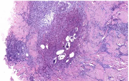
Pathological examination of the skin showed dermal inflammatory infiltrates composed of lymphocytes, eosinophils, and histiocytes including multiple eggs with lateral spine and mira-cidia inside (Fig 2), and Ziehl-Neelsen staining was positive [1]. An indirect hemagglutination test (Schistosomiasis Fumouze) detected high titers (1/640, titers above 1/80 are considered positive) ofSchistosoma antibodies. A diagnosis of ectopic cutaneous schistosomiasis was
made, and treatment with prednisone 0.5 mg/kg/day and a single dose of praziquantel (40 mg/ kg) was started. In the next few days, the patient’s eosinophil count decreased progressively, and cutaneous lesions partially improved. Given the particular chronology of the case, the patient was asked about previous travel to endemic areas of schistosomiasis and previous freshwater exposure during travel, but there were none. Finally, the suspicion ofS. mansoni as
the most likely etiological agent, considering the epidemiological history and the histological findings on the skin biopsy, was confirmed by a species-specific LAMP-based real-time assay.
A molecular LAMP assay previously developed for sensitive and specificS. mansoni
detec-tion (SmMIT-LAMP) [2] was performed using the patient’s skin tissue sample. Briefly, DNA
a1111111111 a1111111111 a1111111111 a1111111111 a1111111111 OPEN ACCESS
Citation: Camprubı´ D, Requena-Me´ndez A,
Rodrı´guez N, Valls ME, Garcı´a-Herrera A, Estrach T, et al. (2017) A 38-year-old woman with zosteriform skin lesions. PLoS Negl Trop Dis 11(11): e0005906.https://doi.org/10.1371/journal. pntd.0005906
Editor: David Joseph Diemert, George Washington
University School of Medicine and Health Sciences, UNITED STATES
Published: November 2, 2017
Copyright:© 2017 Camprubı´ et al. This is an open access article distributed under the terms of the Creative Commons Attribution License, which permits unrestricted use, distribution, and reproduction in any medium, provided the original author and source are credited.
Funding: The authors received no specific funding
for this work.
Competing interests: The authors have declared
from a formalin-fixed paraffin-embedded biopsy was isolated using the QIAamp DNA Mini Kit (QIAgen, Hilden, Germany) following manufacturer instructions. The isothermal SmMI-T-LAMP master mixtures—including the patient’s sample and controls—were, in this case, monitored using 2 fluorochromes to compare (SYBR Green I and EvaGreen) in real time at 65˚C for 60 minutes using the portable Genie III instrument (Optigene Ltd., Horsham, UK). Amplification results were observed in positive control samples (genomic DNA extracted fromS. mansoni) as well as in the patient’s tissue sample when using EvaGreen dye (0.5x).
There was 100% concordance with the results on agarose gel electrophoresis.
Discussion
Human schistosomiasis is a parasitic disease caused by several species ofSchistosoma, affecting
more than 200 million people worldwide [3]. After skin penetration of cercariae, adult worms infect human blood vessels for years, predominantly from mesenteric and perivesical plexus, Fig 1. Clinical image. Pruritic painless erythematous papules grouped on the back part of the left shoulder of
the patient following the C3-C4 dermatome. https://doi.org/10.1371/journal.pntd.0005906.g001
depending on theSchistosoma species. They can successfully evade the immune system while
excreting fertilized eggs that will leave the body in feces or urine to reach freshwater and infect snails to complete their cycle, or they can become trapped in nearby tissues [3].
Two different clinical pictures ofSchistosoma spp. infection have been described, depending
on the immunity of their hosts. Acute schistosomiasis—also called Katayama syndrome—is considered an immune reaction to theSchistosoma larvae and/or eggs that affects nonimmune
individuals and requires treatment with corticosteroids alone or in addition to praziquantel [4]. On the other hand, chronic schistosomiasis is the result of an immune-mediated granulo-matous response to trapped eggs in immune local patients that produces organ-specific mani-festations, such as hematuria, obstructive uropathology, hepatosplenism, periportal fibrosis with portal hypertension, and neurological symptoms [3].
Skin lesions caused by this trematode can be classified depending on their chronopathology as immediate, early, and chronic. The first one, also known as swimmer’s itch or cercarial der-matitis, is an immediate skin manifestation characterized by a generalized pruritic papular rash that appears as an immune reaction when cercariae penetrate the skin. Early skin lesions are allergic reactions (usually urticaria) produced in the context of Katayama syndrome [4]. Finally, classical chronic cutaneous schistosomiasis consists of papular, erythematous, and pruritic lesions located in the anogenital region, and only a few cases of extragenital skin lesions have been described [5–9]. Therefore, ectopic cutaneous schistosomiasis is character-ized by erythematous pruritic papules with a zosteriform distribution that can evolve to nod-ules and plaques. It mainly affects the thorax—particularly around the shoulders—and abdomen, and it has been reported more frequently in nonimmune travelers visiting endemic areas [7].
The differential diagnosis of chronic cutaneous schistosomiasis includes sexually transmit-ted diseases and neoplasms due to its usual perigenital distribution. In cases of ectopic schisto-somiasis alternative diagnoses like subcutaneous tuberculosis and tuberculoid leprosy, more frequently diagnosed in people from endemic areas, must be considered. Furthermore, in our case (given the distribution of the lesions), the differential diagnosis must include viral infec-tions like herpes zoster or zosteriform infecinfec-tions by herpes simplex virus, bacterial infecinfec-tions like erisipela or anthrax, contact dermatitis, phytodermatosis, and sarcoidosis.
Fig 2. Pathological image. Dermal inflammatory infiltrates composed of lymphocytes, eosinophils, and
histiocytes involving multiple eggs with lateral spine (asterisk) and miracidia inside (arrow). https://doi.org/10.1371/journal.pntd.0005906.g002
As in our case, eosinophilia is a usual laboratory finding, and histopathological examination of the lesions shows granulomas aroundSchistosoma eggs and inflammatory infiltrates of
eosinophils and lymphocytes. The mechanism of ectopic oviposition is unknown, but some theories have been proposed. Some authors suggested the unlikely existence of arteriovenous shunts like foramen ovale, but probably the most accepted hypothesis is the aberrant migration of adult worms against the venous blood stream.Schistosoma worms ascend from iliac and
cava veins through lumbar veins and valveless vertebral venous circulation to paraspinal and intercostals cutaneous locations. That could entail the deposition of eggs in the skin following a zosteriform distribution [8]. Although ectopic skin lesions usually occur independently, an association with some severe clinical coexisting manifestations such as myelitis has been reported in a few cases [9]. Therefore, treatment is important not only to eliminate the adult worms and halt the oviposition but also to avoid the evolution to severe forms [9].
A diagnosis of acute schistosomiasis is usually based on a combination of clinical and epide-miological factors. In our case, we used, for the first time, a specific real-time LAMP assay based on a skin biopsy to confirm the diagnosis and to determine theS. mansoni species
caus-ing the cutaneous lesions as a proof of concept of the usefulness of LAMP in the diagnosis of acute schistosomiasis.
Finally, although ectopic cutaneous schistosomiasis has been classified as a manifestation of chronic and/or late schistosomiasis—usually described as occurring 3 months after water exposure—physicians must be aware of early presentations, as in the reported case of our patient, who started having cutaneous manifestations only 5 weeks after her exposure to the infected freshwater. Despite differences in treatment, there is no clear consensus about defini-tions of acute and chronic schistosomiasis. At this point, we consider that new definidefini-tions of chronic and acute schistosomiasis should be established, based not only on the chronology but also on the physiopathological aspects. Furthermore, it is important to highlight the role of LAMP assays in the identification ofSchistosoma species and their potential importance in the
diagnosis of schistosomiasis.
Statement of agreement
The patient agreed to the publication of pictures and information related with the case. She signed an informed consent form.
Key learning points
• Cutaneous zosteriform lesions in a person with the appropriate epidemiological risk factors can be a clue for the diagnosis of schistosomiasis.
• Ectopic cutaneous lesions can appear in early stages of the disease.
• New molecular techniques such as LAMP can help in the diagnosis of schistosomiasis. • Treatment of cutaneous schistosomiasis is important to avoid progression to severe
forms.
• New definitions of chronic and acute schistosomiasis should be considered on the basis of the chronology and physiopathological aspects.
References
1. Vieira S, Belo S, Ha¨nscheid T. Ziehl-Neelsen in Schistosomiasis: much more than staining the shell and species identification. Am J Trop Med Hyg. 2016; 94(4): 699–700. https://doi.org/10.4269/ajtmh.15-0798PMID:27053189
2. Ferna´ndez-Soto P, Gandasegui Arahuetes J, Sa´nchez Herna´ndez A, Lo´pez Aba´n J, Vicente Santiago B, Muro A. A Loop-Mediated Isothermal Amplification (LAMP) Assay for Early Detection of Schistosoma
mansoni in Stool Samples: A Diagnostic Approach in a Murine Model. PLoS Negl Trop Dis. 2014; 8(9):
e3126.https://doi.org/10.1371/journal.pntd.0003126PMID:25187956
3. Colley D, Bustinduy A, Evan W, King C. Human schistosomiasis. Lancet. 2014; 383(9936): 2253– 2264.https://doi.org/10.1016/S0140-6736(13)61949-2PMID:24698483
4. Jaure´guiberry S, Paris L, Caumes E. Acute Schistosomiasis, a diagnostic and therapeutic challenge.
CMI. 2010; 16: 225–231.https://doi.org/10.1111/j.1469-0691.2009.03131.xPMID:20222897 5. Barros CRCR Maia DCC, Santos JB Medeiros CCQ, Arau´jo JG. Cutaneous ectopic schistosomiasis:
diagnostic challenge. An Bras Dermatol. 2016; 91(1): 109–110.https://doi.org/10.1590/abd1806-4841. 20164647PMID:26982792
6. Al-Kawari KS, Al-Amro Al-Akloby OM, Murgharbel RM. Ectopic cutaneous schistosomiasis. Int J
Der-matol. 2004; 43: 550–552.
7. Mota LS, Silva SF, Almeida FC, Mesquita LSU, Teixeira RDL, Soares AM. Ectopic cutaneous schisto-somiasis—Case report. An Bras Dermatol. 2014; 89 (4): 646–648. https://doi.org/10.1590/abd1806-4841.20142967PMID:25054754
8. Ramdial PK, Calonje E, Singh B, Sing Y, Govender P. Extra-anogenital bilharziasis cutanea tarda revis-ited. J Cutan Pathol. 2009; 36: 766–771.https://doi.org/10.1111/j.1600-0560.2008.01165.xPMID: 19519608
9. Wood MG, Srolovitz H, Schetman D. Schistosomiasis. Paraplegia and ectopic skin lesions as admis-sion symptoms. Arch Dermatol. 1976; 112(5): 690–695. PMID:1275524