FACULTY OF PHARMACY AND MEDICINE
Ph.D. Thesis in Pharmaceutical Sciences
XXXI cycle
Synthesis and biological evaluation of new
saccharin-based inhibitors of cancer-related carbonic anhydrase
IX and XII isoforms
&
Benzo[b]tiophen-3-ol derivatives as effective inhibitors
of hMAOs: design, synthesis and biological activity
Ph.D. student Supervisor
ii
To those who pointed me the direction,
giving me the freedom and opportunity
iii
Table of contents
Chapter 1
Carbonic anhydrases: functions and dark side
1.1 Introduction 7
1.2 Involvement of carbonic anhydrase IX and XII isoforms in tumors: the
dark side of the enzyme 13
1.3 Carbonic anhydrase inhibition 15
1.4 Development of new saccharin inhibitors 19
Chapter 2
Open saccharin-based secondary sulfonamides as potent and selective inhibitors of cancer-related carbonic anhydrase IX and XII isoforms
2.1 Open saccharin-based secondary sulfonamides: aim of the work 22
2.2 Chemistry 23
2.3 Biological evaluation 24
2.4 Results and discussion 25
2.4.1 Inhibition of hCA I, II, IX, and XII 25
2.4.2 Docking studies into the active site of hCA XII 28
2.5 Conclusions 29
2.6 Experimental section 30
Chapter 3
Design, synthesis and biological activity of saccharin/isoxazole and saccharin/isoxazoline derivatives as selective inhibitors of carbonic anhydrase IX and XII isoforms
3.1 Saccharin/isoxazoline and saccharin/isoxazoles derivatives:
aim of the work 62
iv 3.3 Two-dimensional nuclear Overhauser enhancement spectroscopy
(2D-NOESY NMR) 64
3.4 Biological evaluation 65
3.5 Results and discussion 65
3.5.1 Inhibition of hCA I, II, IX, and XII 65
3.5.2 2D-NOESY NMR of compound 3b 69
3.6 Conclusions 70
3.7 Experimental section 71
Chapter 4
Human monoamine oxidases (hMAOs)
4.1 Introduction 103
4.2 Structural properties and catalytic mechanism of hMAOs 105
4.3 Pathological roles and inhibitors of hMAOs 109
Chapter 5
Benzo[b]tiophen-3-ol derivatives as effective inhibitors of hMAOs: design, synthesis and biological activity
5.1 Development of new inhibitors of hMAOs: aim of the work 113
5.2 Chemistry 115
5.3 Biological assays 117
5.3.1 hMAO-A and hMAO-B inhibition studies 117
5.3.2 Evaluation of DOPAC/DA ratio and LDH activity 117
5.4 Results and discussion 117
5.4.1 In vitro MAO inhibition study 117
5.4.2 Evaluation of DOPAC/DA ratio and LDH activity 121
5.5 Conclusions 125
5.6 Experimental section 127
5
During my PhD studies, I have worked with two enzymatic targets having
diverse functions and so, different physiological/pathological features.
The first part focused on the design and synthesis of human carbonic anhydrase
inhibitors, which were tested against four hCA isoforms, the ubiquitous CA I and
CA II and the tumor-associated ones CA IX and CA XII, thanks to the precious
collaboration of Prof. Claudiu T. Supuran and colleagues.
I have spent the last part of my PhD for the discovery of human monoamine
oxidases inhibitors, which were tested against hMAOs and cortex synaptosomes
thank to the valuable collaboration of Prof. Jacobus P. Petzer and Prof. Claudio
Ferrante.
6
Chapter 1
7
1.1 Introduction
Carbonic anhydrases (CAs, EC 4.2.1.1) are ubiquitous enzymes found in numerous organisms across the tree of life, encoded by seven genetically distinct CA families: α-, β-α-, γ-α-, δ-α-, η-α-, ζ- and the last discovered family θ-CAs [1–9]. Carbonic anhydrases are metalloenzymes and they are catalytically effective only with one metal ion bound within the active site cavity, the apoenzymes being devoid of any catalytic action [10– 16]. The active centre contains three amino acid residues which act as ligands, coordinating in a tetrahedral geometry the central divalent ion M(II). All the seven genetic families of CA may contain Zn(II) as metal ion, although it is substitutable with Cd(II) in the ζ-CAs [10]. γ-CAs seem to be endowed with Fe(II) ion, at least in anaerobic conditions [4,17], whereas Co(II) may substitute the zinc ion in many α-CAs without significant loss of the catalytic activity [1,18–20].
Carbonic anhydrases catalyse reversible hydration of CO2 (eq.1) transforming two
neutral molecules, CO2 and water, into a weak base (bicarbonate) and a strong acid (H+
ions). This very simple reaction is particularly slow at the physiological pH, while it becomes very effective at higher pH values, being instantaneous at pH > 12 [12,19,21– 23].
Carbonic anhydrases make faster this reaction in physiological condition that is important for pH regulation, as well as for other crucial physiological processes like respiration, photosynthesis, pH homeostasis, CO2 transport and electrolyte secretion,
virtually in all tissues in organisms [5,6,9–11,13,24–34].
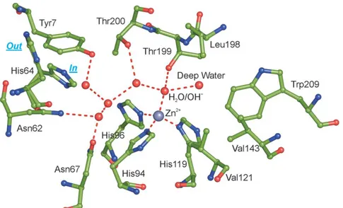
To date, fifteen isoforms of human carbonic anhydrase (hCAs) have been discovered (hCAI-XIV). They belong to the α-class and share a common organization of the active site, located in a deep cleft and containing a central zinc ion (Zn2+), coordinated by
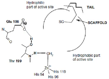
8 three histidine residues (His94, His96 and His119) and a water molecule/hydroxide ion (Figure 1.1) [5,19,32,35].
Figure 1.1 Active site of hCA II, showing the network of interactions in the active site. Water
molecules are indicated as red circles. The side chain of His64 is shown in both the “in” and “out” conformations [19].
The zinc-bound water molecule/hydroxide ion establishes hydrogen bond network with the hydroxyl group of Thr residue (Thr 199), and with two water molecules positioned on two opposite side: the first called “deep water” is located in a hydrophobic cavity, while the second is in a hydrophilic environment toward the entrance of the active site (Figure 1.1) [19,36]. All these interactions are able to increase zinc bound water molecule nucleophilicity. As a consequence, the proton transfer occurs leading to the production of the catalytically active form of the enzyme containing zinc-bound hydroxide ion. The hydration of carbon dioxide proceeds through a two-step catalytic mechanism (eq.2 and 3).
The first step (eq.2) is the nucleophilic zinc-bound hydroxide ion attack on the CO2
molecule located in a hydrophobic binding pocket, to obtain metal coordinated bicarbonate. The interaction between bicarbonate and central zinc ion is fairly weak; so, it is displaced by a water molecule and released in solution forming water
9 coordinated Zn2+, which is the acidic and inactive form of the enzyme (catalytically
inactive enzyme, eq.2).
The second step is the rate limiting one in which enzyme’s active form (eq.3) is regenerated by the proton transfer directly to the buffer or assisted by His 64, which serves as a proton shuttle between the metal center and buffer molecules of the medium [37]. The presence of His 64 that works as a “proton-shuttle” affects positively the catalytic activity. Indeed the absence of this system in enzymes as CAIII, impaires activity about 500-fold [29,38–40].
This working machinery based on a “ping-pong” mechanism, makes some of the members of the CA superfamily among the most effective enzymes known in nature, with kcat/KM values close to the limit of the diffusion-controlled processes [25]. How
anticipated, human carbonic anhydrases exist in fifteen isoforms which differ by molecular features, oligomeric arrangement, cellular localization, distribution in organs and tissues, expression levels, kinetic properties and response to different classes of inhibitors [5,34,35,41].
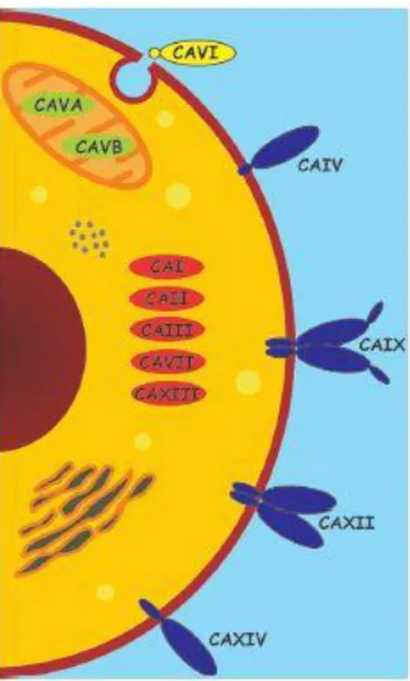
Five of these enzymes are cytosolic (hCA I-III, hCA VII and hCA XIII), two are mitochondrial (CA VA and VB), four are membrane bound (hCA IV, hCA IX, hCA XII and hCA XIV), one is secreted (hCA VI) and three are acatalytic hCA-related proteins (CARPs, hCA VIII, X and XI) (Figure 1.2). Even if CARPs are acatalytic isozymes because of the absence of metal ion (these proteins not possess histidine residues required for the coordination of the zinc atom) [42], they still possess significant functions in physiologic and pathologic processes as well as the other hCA isoforms (Table 1.1) [43].
10
Figure 1.2. Domain composition and subcellular localization of catalytically active human
α-Cas [19].
CA I is the major isozyme found in human erythrocytes with a concentration of about 6-fold higher than CA II, also found in erythrocytes. However, the specific activity of CA I in erythrocytes is 2 x 10⁵ s-1, whereas the specific activity of CA II in erythrocytes
is 106 s-1. Although CA I has been found to be expressed in a variety of tissues, its
physiological role is not completely clear [19]. Feeener’s group demonstrated that this enzyme is involved in retinal and cerebral edema, and its inhibition may be a valuable tool for fighting these conditions [44]. CA II is the high activity or “rapid” isozyme in order to distinguish it from the low activity or “slow” one, hCA I. It is expressed in the cytoplasm of many cell types and it is involved in processes spanning from bone resorption to respiration and pH regulation. Other functions include urine formation and bicarbonate reabsorption in the kidney tubules, biosynthetic reactions such as gluconeogenesis, lipogenesis and ureagenesis, bone resorption and calcification, and probably many other less well understood physiologic/pathologic processes [19,45,46]. Deficiency of hCA II is characterized by renal tubular acidosis, osteopetrosis, cerebral calcification, and growth retardation [47]. hCA III is known as muscle isoform because
11 it is found in high levels in red skeletal muscle, even if this isoform is present also in adipocytes. The activity of hCA III is only 3% that of hCA II and is thought that its physiological importance is beyond its catalytical activity. In fact, this enzyme possesses two reactive sulfhydryl groups able to reversibly bind glutathione, protecting cells from irreversible protein oxidation [48–50]. hCA IV is found in heart [51], brain [52], capillary bed of the eye [53], and erythrocytes [54] and is a possible drug target for several pathologies, including glaucoma (together with CA II and XII), retinitis pigmentosa and stroke [55,56]. The two mitochondrial isoforms hCA VA and hCA VB are implicated in metabolism. hCA VA is found in the mitochondria of the kidney, heart, lung, spleen and intestines; it provides bicarbonate for gluconeogenesis and fatty acids for pyrimidine base synthesis [57]. On the other hand, hCA VB is found in pancreas, kidney and salivary glands mitochondria, showing intermediate role in metabolism [58]. These two isoforms could be useful targets for antiobesity agents [59]. CA VI, the secreted isoform, is implicated in cariogenesis [60,61]. hCA VII has been implicated in neuronal excitation, contributing to epileptiform activity together with CA II and XIV [60–62]. Although hCAVIII not possess catalytic activity, its role has been documented in various pathologies. Indeed, has been associated with neurodegenerative diseases with studies conducted in mice and then in human, showing how mutations in CA8 gene cause mental retardation and ataxia. CARP VIII is known to be also involved in tumors, being upregulated in colorectal, lung and several other cancers [63]. The precise physiological/pathological roles of the remaining two acatalytic isoforms CA X and XI are poorly known, albeit ongoing studies are trying to shed light on these proteins.
12
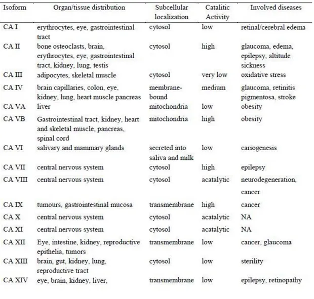
Table 1.1. hCA isoforms, their organ/tissue distribution, subcellular localization, relative CO2
hydratase activity and diseases in which they are involved.
hCA XIII was localized in several tissues as the thymus, kidney, submandibular gland, small intestine, and notably in reproductive organs, accounting for its involvement in the sperm motility processes (probably together with CA XIV) [64]. hCA XIV is involved in epileptogenesis and has been localized to the apical and basal membranes of the retinal pigment epithelium. This implies that CA XIV have specific and unique functions in the context of acid-based balance in the retina.
13
1.2 Involvement of carbonic anhydrase IX and XII isoforms in tumors: the dark side of the enzyme
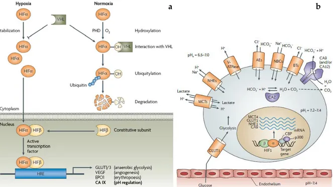
The two remaining isoforms are the established tumor-related enzymes IX and XII [58,65,66]. These membrane-bound isoforms show a limited expression in normal tissues, unlike hCA I and hCA II [67]. hCA IX is a dimeric transmembrane glycoprotein possessing the catalytic domain oriented toward the extracellular milieu, working at the outer side of the cells [65,68–70]. It is overexpressed mainly in hypoxic tumours, being regulated by hypoxia and facilitate also the metastatic spread of solid tumors [69]. The extracellular acidosis is known to negatively affect drug uptake and radiation damage, so the overexpression of hCA IX is associated to radio- and chemotherapy resistance [71]. hCA XII shares common features with hCA IX, as the secondary structure, orientation and hypoxia induced expression; however, it is a monomer and lacks the proteoglycan-like domain [72]. It has a wider tissue distribution compared with isoform IX, including kidney, lung, prostate, ovaries, uterine endometrium, breast. Unlike isoform hCA IX, hCA XII is regulated by estrogens and in breast cancer patients, hCA XII expression correlates with positive prognosis [73,74]. hCA XII possesses catalytic activity lower than hCA IX [75], and is generally associated with less-aggressive, well-differentiated tumor phenotypes, compared to the hCA IX- expressing tumors [67,76,77]. hCA IX mainly, but probably also hCA XII, regulates intra- and extracellular pH variations which take place in cellular metabolism stimulated by hypoxia. In fact, hypoxia is able to affect genes regulation through hypoxia-inducible factor 1, (HIF-1) favouring cellular adaptation to an anaerobic metabolism [78,79]. HIF-1 is a transcriptional factor protein constituted by two subunits: HIF-1α and HIF-1β. HIF-1β subunit is constitutively expressed and is located inside the nucleus. HIF-1α is an oxygen-regulated subunit working as “O2 sensor”
system. During normoxia condition its concentration is influenced by degradation mechanism controlled by oxygen availability. Prolyl-4-hydroxylase (PHD), is able to hydroxylates the P564 on HIF-1α. This modification works as a signal for the von
14 Hippel-Lindau protein that binds HIF-1α and targets it for degradation by the ubiquitin–proteasome system (Figure 1.3, a) [80–88].
Figure 1.3 (a) Mechanism of hypoxia-induced gene expression mediated by the HIF
transcription factor [88]; (b) Proteins involved in pH regulation within a tumour cell [23].
On the other hand, the lack of oxygen under hypoxia inhibits the PHD activity leading to the absence of HIF-1α hydroxylation which cannot be recognized by the VHL protein. So, HIF-1α translocates to the nucleus where dimerizes with the HIF-1β, constituting the active form of the transcription factor that binds the hypoxia response element (HRE) in target genes and activate their transcription. Target genes include glucose transporters (GLUT1 and GLUT3) that increase glucose caption and take part in glucose metabolism, vascular endothelial growth factor (VEGF) that triggers neoangiogenesis, erythropoietin (EPO1) involved in erythropoiesis, carbonic anhydrase (CA) IX involved in pH regulation and tumorigenesis, and additional genes with functions in cell survival, proliferation, metabolism and other processes. The final effect of this transformation is a cell ready to face hypoxic condition switching the metabolism to anaerobic route. In normal tissues glucose follows the normal aerobic
b a
15 processes (glycolysis, Krebs-cycle and oxidative phosphorylation) until its complete oxidation to carbon dioxide and water. Cancer cells in hypoxic conditions produce energy from glycolysis and lactic acid fermentation in the cytosol, completely changing its metabolism (the so-called Warburg effect). In this condition cells organize a complex machinery system constituted by transporters, pumps and carbonic anhydrases, able to cope the new state (Figure 1.3 b). The new system encourages the extracellular acidification and the maintaining of a weakly alkaline pH, which is optimal for cell proliferation and tumour survival as well as metastatic behaviour [66,89,90].
1.3 Carbonic anhydrase inhibition
Since the discovery of Mann and Keilinin in 1940 about the capability of sulfanilamide to inhibit carbonic anhydrase, the researchers have discovered a huge number of molecules which effectively inhibit hCAs isoforms.
Up to day, five inhibition mechanism have been discovered [91]:
1. CAIs anchoring to the central zinc ion: this kind of inhibition mechanism belong to most of the inhibitors discovered up to day. Compounds exercising this mechanism have a scaffold endowed with an anchoring group (AG) which works as zinc binder group (ZBG) [92–95]. The most used ZBG is the primary sulfonamide moiety, although other groups are able to coordinate central zinc ion (e.g. sulfamates, and sulfamides, which are in fact sulfonamide isosters) [96]. Although these inhibitors are very effective, inhibiting hCAs in the low nanomolar range, they lack of selectivity due to the common active site organization of the fifteen isoforms. With the aim to increase selectivity, these inhibitors are designed using the “tail approach” [97], which takes advantage from the insertion of functional groups able to interact fairly away from the Zn2+ ion, where more
16
Figure 1.4. CAIs anchoring to the central zinc ion.
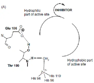
2. CAIs anchoring to the zinc-coordinated water/hydroxide ion: belong to this class compounds able to anchor the zinc-coordinated water molecule/hydroxide ion. This mechanism was observed for the first time with phenol, but other compounds showing this kind of inhibition contain primary amine, carboxylic acids or esters, and sulfonic acids [98–100]. The scaffold of these compounds can be aromatic, aliphatic, heterocyclic, or sugar-based type and could be designed keeping in mind the “tail approach” (Figure 1.5).
17 3. CA inhibition by occlusion of the active site entrance: the first compound with this interesting CA inhibition mechanism was a natural compound with coumarin structure [101]. The de facto inhibitor is in this case the hydrolysed form of coumarin, produced because of the esterase activity of hCAs. Other compounds endowed with “sticky group” showed similar activity [102]. The most notable aspect of this inhibition mechanism is that the inhibitors bind the active site region, which is the most variable between the various isoforms, i.e. the entrance to the cavity (Figure 1.6).
Figure 1.6. CA inhibition by occlusion of the active site entrance
4. Out of the active site binding as a CA inhibition mechanism: this is the last mechanism discovered with crystal structure obtained between hCA II and 2-(benzylsulfonyl)-benzoic acid [103]. The electronic density of the inhibitor was not observed within the active site but in a binding pocket next to to the active site. The compound inhibits hCA blocking the proton-shuttling residue His64 in the “out” conformation, interfering with the transfer of a proton from the zinc-coordinated water molecule to the environment. In this way it prevents the formation of zinc-coordinated hydroxide ion, the active form of the enzyme (Figure 1.7).
18
Figure 1.7. Out of the active site binding CA inhibition mechanism.
5. Compounds acting as CAIs with an unknown mechanism of action: finally, there are a series of compounds whose inhibition mechanism has not been discovered, due to the absence of their co-crystal with hCA enzymes. Compounds based on saccharin scaffold, belong to this class.
19
1.4 Development of new saccharin inhibitors
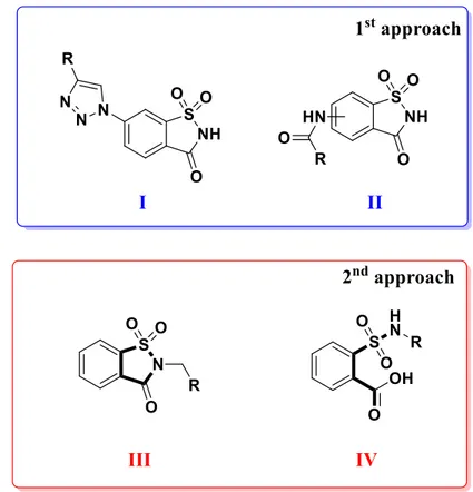
Saccharin derivatives have been documented as good inhibitors of hCA by various research groups, that in the last years focused their attention towards the development of new molecules based on this scaffold [104–109]. By analysing the structure and substitution pattern of published compounds possessing saccharin scaffold, it is possible to find that for the design of new molecules two general approach have been exploited (Figure 1.5). The first is relative to compounds obtained through the substitution of benzene ring of saccharin, with various substituents, maintaining the secondary sulfonamide functional group free (Figure 1.5, I and II). Compounds containing secondary sulfonamides, have been extensively studied for their ability to inhibit hCAs [110–112]. In fact, this approach produced molecules endowed with activity in the nanomolar range against isoforms IX and XII, although residual activity against the off-targets hCA I and hCA II was unfortunately observed [107,113].
Figure 1.5 The two different approaches for the synthesis of hCAs inhibitors based on
20 The second approach was based on the substitution of saccharin nitrogen with different groups, in order to obtain N-substituted saccharins (Figure 1.5, III). Tertiary sulfonamides have been investigated for the atypical mechanism that these substances must possess, because they are not able to coordinate zinc ion due to the absence of deprotonable nitrogen atom [114–117]. N-substituted saccharins were effective inhibitors of hCA IX and XII, even if some of them retained activity against off-targets [106,109].
Here I report two approaches used to increase activity and selectivity of saccharin-based inhibitors of the two tumor-related isoforms hCA IX and XII. The outcomes of these strategies were the open saccharin-based secondary sulfonamides and the saccharin/isoxazole - saccharin/isoxazoline derivatives.
21
Chapter 2
Open saccharin-based secondary sulfonamides as potent and selective
inhibitors of cancer-related carbonic anhydrase IX and XII isoforms
22
2.1 Open saccharin-based secondary sulfonamides: aim of the work
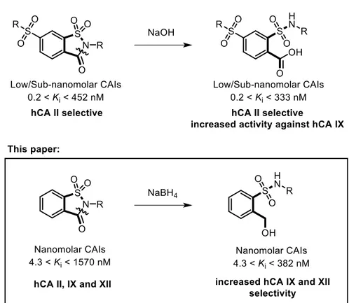
In a recent publication, Ivanova et al. reported on the spontaneous ring opening of cyclic tertiary sulfonamides under basic (pH= 9) crystallization conditions [118]. Since hCAs do not possess any peptidase activity, they concluded that the base-catalysed hydrolysis of the saccharin isothiazolone ring happened before the inhibitor entered the active site, and proved their hypothesis by testing both open and closed analogues against a panel of hCAs (hCA I, hCA II, hCA IX and hCA XII) [119]. In particular, these open saccharin derivatives determined an increased inhibition of hCA IX, while retaining high activity against hCA II. I used a reductive ring opening approach to induce the 5-membered isothiazolone ring of saccharin to collapse into its corresponding secondary sulfonamide and benzyl alcohol (Figure 2.1) [120].
23 The rationale behind this choice could be found in the opportunity of generating two new potential anchoring points for the zinc ion, while introducing several degrees of freedom to the bonds connecting the two hydrophobic phenyl substituents to the polar core of the molecule. In general, the newly synthesized compounds (1-21) proved to be as potent as or slightly less potent than parent inhibitors (I-XXI), while the selectivity for the cancer-related isoforms (hCA IX and XII) over the off-target hCA I and II improved dramatically. In fact, none of the reported compounds inhibited hCA I and II isoforms at concentrations lower than 10 nM, while Ki values spanned from 20
to 298 nM against hCA IX and from 4.3 to 382 nM against hCA XII.
2.2 Chemistry
Saccharin (1.0 eq.) was activated using freshly ground anhydrous potassium carbonate and the corresponding salt was then directly reacted with a proper electrophile (2 eq. of substituted benzyl halide or α-haloacetophenone) by stirring the reaction mixture in N,N-dimethylformamide at 80 °C overnight (Scheme 1). Following these optimized conditions (polar aprotic solvent), we strictly obtained only the more stable regioisomers (N-substituted saccharin derivatives) limiting the Chapman-Mumm thermal rearrangement to the less stable O-substituted counterparts [121]. N-substituted saccharin (the parent drugs) derivatives were then subjected to reductive ring opening with an excess of NaBH4 in dry methanol at room temperature for 2-8 hrs
to give the corresponding secondary sulfonamide compounds 1-21 in discrete yields following a previously reported procedure with slight modifications [120].
24
Scheme 2.1. Synthesis and structure of compounds I-XXI and 1-21. For R substituents see Table 2.1 and 2.2.
The choice of NaBH4 as a reducing agent was influenced by preliminary experiments
which suggested that (i) the sulfone group was not affected by these mild reducing conditions leading to the carbonyl group reduction to the alcohol level only with saccharin ring cleavage and that (ii) similar results were obtained for the preparation of o-hydroxymethyl-N-alkyl-benzamides starting from N-alkyl-phthalimides [120]. In their IR spectra, I usually registered for the open saccharin derivatives new but expected signals for the OH and NH stretching at 3240 and 3470 cm-1, respectively, and
the disappearance of the C=O stretching at 1735 cm-1.
2.3 Biological evaluation
All the synthesized compounds were tested to evaluate their inhibitory activity towards the ubiquitous off-target isoforms, hCA I and II, and the cancer-related ones, hCA IX and XII, by a stopped-flow, CO2 hydrase assay method and their CA inhibition
25
2.4 Results and discussion
2.4.1 Inhibition of hCA I, II, IX, and XII.
The analysis of the biological data was accomplished comparing the open saccharin-based derivatives (Table 1) with their corresponding parent compounds (activities reported in Table 2) in order to evaluate if the ring opening enhanced or reduced their biological activity. All the tested compounds had no affinity for the common off-target hCA I and II isoforms (Kis> 10000 nM) compared to their corresponding parent drugs.
Moreover, our molecules with a benzyl alcohol group, instead of a carboxylic acid one [118], abolished completely this inhibitory activity improving the biological profile of this scaffold. The inhibition profile of the open saccharin-based derivatives against the two tumor-related hCA IX and XII isoforms also displayed some important changes compared to parent drugs. Among the new open saccharin derivatives reported here, the best activity was obtained toward hCA XII isoform by compounds 1, 5, 6, 10 and
11, all provided of CH3 or CF3 groups. These molecules exhibited a slightly preference
for hCA XII respect to hCA IX isoform, although the inhibition of the latter was also in the nanomolar range. Compound 5, containing a phenyl ring substituted with methyl group in meta position, had the highest inhibitory activity against hCA XII (Ki = 4.3
nM), but also compound 6, containing a meta trifluoromethyl substituent on phenyl ring, exhibited similar inhibitory activity (Ki = 4.4 nM). Comparable profile against
hCA XII was observed for compounds 10 (Ki = 5.7 nM) and 11 (Ki = 7.2 nM), which are
para substituted regioisomers of 5 and 6, respectively. Compound 1, which had a
methyl group at ortho position of phenyl ring, showed similar inhibitory activity (Ki
hCA XII = 4.7 nM) with respect to 5 and 6. Other compounds with strong selectivity between hCA IX and XII were 9 (Ki hCA IX = 267 nM, Ki hCA XII = 64 nM) with bromine
in meta position of phenyl ring, 12 (Ki hCA IX = 126 nM, Ki hCA XII = 57 nM) containing
a cyano group in para position, 15 (Ki hCA IX = 154 nM, Ki hCA XII = 48 nM) which had
a para chloro-substituted phenyl ring, 17 and 18 with, respectively, 2,6-difluoro and 3,4-dichloro-substituted phenyl rings.
26 Conversely, some compounds displayed a good selectivity towards hCA IX isoform. Compounds with nitro substituents in ortho (3), meta (7) or para (13) position of the ring had potent inhibitory activity preferentially against this overexpressed isoform in the hypoxic tumoral niche.
For compounds 20 (Ki hCA IX = 224 nM, Ki hCA XII = 64 nM) and 21 (Ki hCA IX = 31
nM, Ki hCA XII = 355 nM), the reaction with NaBH4 led to the further reduction of
exocyclic carbonyl moiety. The presence of this additional group maintained the biological profile with a loss of inhibitory activity against hCA I and II and a preferential selectivity against the cancer-related isoforms.
Collectively, these promising data showed that the reductive ring opening of the saccharin nucleus improved the hCA inhibitory activity with a better selectivity with respect to the off-target isoforms. From the above, we also observed that the inhibition profile was affected positively or negatively by the substitution pattern.
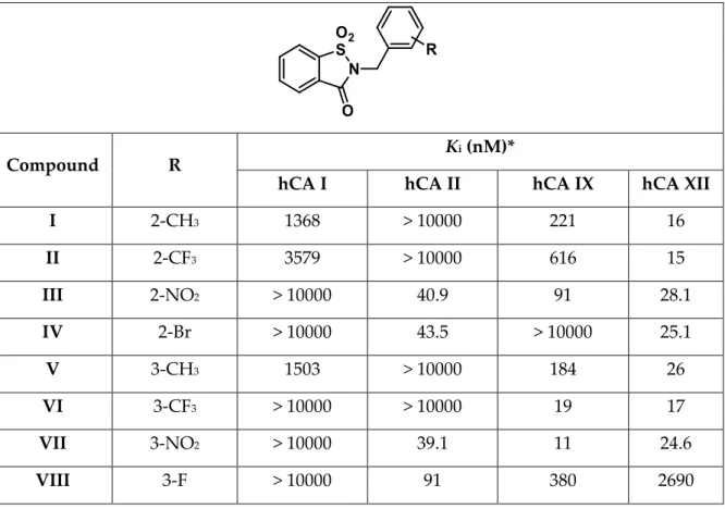
Table 2.1. Inhibitory activity of the saccharin parent drugs I-XXI and acetazolamide as a
reference drug, against selected hCA isoforms by a stopped-flow CO2 hydrase assay.
Compound R K
i (nM)*
hCA I hCA II hCA IX hCA XII
I 2-CH3 1368 > 10000 221 16 II 2-CF3 3579 > 10000 616 15 III 2-NO2 > 10000 40.9 91 28.1 IV 2-Br > 10000 43.5 > 10000 25.1 V 3-CH3 1503 > 10000 184 26 VI 3-CF3 > 10000 > 10000 19 17 VII 3-NO2 > 10000 39.1 11 24.6 VIII 3-F > 10000 91 380 2690
27 IX 3-Br > 10000 86.8 360 21.1 X 4-CH3 > 10000 > 10000 234 5.1 XI 4-CF3 1347 > 10000 51 4.7 XII 4-CN > 10000 47.6 240 150 XIII 4-NO2 324 > 10000 1169 29 XIV 4-F 2479 > 10000 539 41 XV 4-Cl 2361 > 10000 19 4.4 XVI 4-Br > 10000 > 10000 22 4.3 XVII 2,6-diF >10000 460 > 10000 1310 XVIII 3,4-diCl > 10000 > 10000 390 2760 XIX -CH=CH-CH=CH- > 10000 > 10000 > 10000 2540 XX H > 10000 > 10000 > 10000 1780 XXI 3-OCH3 1342 > 10000 1570 6.0 AAZ (acetazolamide) 250 12 25 6.0
*Mean from 3 different assays (errors were in the range of ±5–10% of the reported values).
Table 2.2. Inhibitory activity of the open saccharin-based derivatives 1-21 and acetazolamide
as a reference drug, against selected hCA isoforms by a stopped-flow CO2 hydrase assay.
Compound R K
i (nM)*
hCA I hCA II hCA IX hCA XII
1 2-CH3 >10000 >10000 218 4.6
2 2-CF3 >10000 >10000 200 54
28 4 2-Br >10000 >10000 113 323 5 3-CH3 >10000 >10000 223 4.3 6 3-CF3 >10000 >10000 238 4.4 7 3-NO2 >10000 >10000 176 382 8 3-F >10000 >10000 268 247 9 3-Br >10000 >10000 267 64 10 4-CH3 >10000 >10000 120 5.7 11 4-CF3 >10000 >10000 253 7.2 12 4-CN >10000 >10000 126 57 13 4-NO2 >10000 >10000 20 54 14 4-F >10000 >10000 26 63 15 4-Cl >10000 >10000 154 48 16 4-Br >10000 >10000 145 432 17 2,6-diF >10000 >10000 296 45 18 3,4-diCl >10000 >10000 298 40 19 -CH=CH-CH=CH- >10000 >10000 294 345 20 H >10000 >10000 224 64 21 3-OCH3 >10000 >10000 31 355 AAZ (acetazolamide) 250 12 25 6.0
*Mean from 3 different assays (errors were in the range of ±5–10% of the reported values).
2.4.2 Docking studies into the active site of hCA XII
The open saccharin analogs (compounds 1–21) were endowed with inhibition values in the nanomolar range against hCA XII isoform (Ki values: 4.3–432 nM, Table 1,
approximately 100-fold difference between lowest and highest Ki values). The
29 (Figure 2.2). Docking studies indicated that the hydroxymethyl group and one of the sulfonamide oxygen atoms could interact simultaneously with the Zn2+-ion, whereas
the other sulfonamide oxygen atom was water accessible. This oxygen might also form hydrogen bonds with the backbone of Thr199. The other polar substituents of the molecule were water accessible.
Figure 2.2 Docked pose of compound 6 in the active site of hCA XII. Hydrogen bonds and
interactions to the Zn2+-ion are depicted in red dashed lines. The Zn2+-ion is depicted as a
turquoise sphere. The three zinc-binding Histidines (H94, H96 and H119) are depicted in light grey for clarity.
2.5 Conclusions
The design, synthesis, characterization and in vitro pharmacological evaluation of several new secondary sulfonamides based on the open saccharin scaffold as selective inhibitors of human carbonic anhydrase, have been proposed. They were shown to be inactive against the two cytosolic off-target hCA I and II (Kis > 10 µM); conversely, all
these compounds inhibited hCA IX and XII in the low nanomolar range with Kis
ranging between 4.3 and 432 nM. The analysis of the Ki values showed as the H119 H94 H96 Q92 K67 H64 N62 W5 P202 T200 P201 T199 V121 L198
30 substituent on phenyl moiety that gives the best outcomes relative to inhibition of hCA XII isoform are methyl and trifluoromethyl groups. The results were also rationalized by means of docking studies into the active site of hCA XII. Since these two cancer-related hCA isoforms were recently validated as drug targets, these results provided the development of new anticancer candidates.
2.6 Experimental section General
Solvents and reagents were used as supplied without further purification. Where mixtures of solvents are specified, the stated ratios are volume:volume. Acetazolamide was purchased by Sigma-Aldrich (Italy) and used in the biological assays without further purification. All synthesized compounds have been fully characterized by analytical and spectral data. Column chromatography was carried out using Sigma-Aldrich® silica gel (high purity grade, pore size 60 Å, 200-425 mesh particle size).
Analytical thin-layer chromatography was carried out on Sigma-Aldrich® silica gel on
TLA aluminum foils with fluorescent indicator. Visualization was carried out under UV irradiation (254 nm). 1H-NMR spectra were recorded on a Bruker AV400 (1H: 400
MHz, 13C: 101 MHz). 19F-NMR spectra were recorded on a Bruker AVANCE 600
spectrometer (19F: 564.7 MHz). Chemical shifts are quoted in ppm, based on
appearance rather than interpretation, and are referenced to the residual non deuterated solvent peak. In the case of 19F, chemical shifts are referenced to an external
standard (CF3COOH, δ -76.55 ppm). Infra-red spectra were recorded on a Bruker
Tensor 27 FTIR spectrometer equipped with an attenuated total reflectance attachment with internal calibration. Absorption maxima (νmax) are reported in wavenumbers (cm -1). All melting points were measured on a Stuart® melting point apparatus SMP1 and
are uncorrected. Temperatures are reported in °C. Where given, systematic compound names are those generated by ChemBioDraw Ultra® 12.0 following IUPAC
31
Synthesis and characterization data of compounds I-XXI and 1-21
2-(2-Methylbenzyl)-1,2-benzothiazol-3(2H)-one 1,1-dioxide (I): anhydrous potassium carbonate (1.1 eq.) were added to a stirring solution of saccharin (1.0 eq.) in 10 mL of N,N-dimethylformamide. 2-Methylbenzyl bromide (1.1 eq.) was added and the reaction stirred at 80 °C for 48 h. The mixture was poured on ice and the resulting suspension was filtered. Purification via column chromatography on silica gel (ethyl acetate:n-hexane, 1:2) gave the title compound as a white solid (72% yield); mp 153-155 °C; IR νmax 3073 (ν Csp2-H), 1726 (ν C=O), 1335 (νas S=O), 1244 (ν C-N), 1181 (νs S=O),
754 (δ Csp2-H), 676 (δ Csp2-H) cm-1; 1H-NMR (400 MHz, CDCl3) δ 2.50 (3H, s, CH3), 4.98
(2H, s, CH2), 7.21-7.25 (m, 3H, Ar), 7.44 (d, J = 7.2 Hz, 1H, Ar), 7.83-7.91 (m, 2H, Ar),
7.95 (d, J = 6.8 Hz, 1H, Ar), 8.10 (d, J = 8.0 Hz, 1H, Ar); 13C-NMR (101 MHz, CDCl3) δ
19.3 (CH3), 40.6 (CH2), 121.0 (Ar), 125.3 (Ar), 126.3 (Ar), 127.3 (Ar), 128.3 (Ar), 128.7
(Ar), 130.5 (Ar), 132.1 (Ar), 134.4 (Ar), 134.9 (Ar), 136.3 (Ar), 137.9 (Ar), 159.0 (C=O). Anal. Calcd for C15H13NO3S: C, 62.70; H, 4.56; N, 4.87. Found: C, 62.52 ; H, 4.81; N, 5.06.
2-(2-Trifluoromethylbenzyl)-1,2-benzothiazol-3(2H)-one 1,1-dioxide (II): anhydrous
potassium carbonate (1.1 eq.) were added to a stirring solution of saccharin (1.0 eq.) in 10 mL of N,N-dimethylformamide. 2-Trifluoromethylbenzyl bromide (1.1 eq.) was added and the reaction stirred at 80 °C for 48 h. The mixture was poured on ice and the resulting suspension was filtered. Purification via column chromatography on silica gel (ethyl acetate:n-hexane, 1:2) gave the title compound as a white solid (60%
32 yield); mp 142-143 °C; IR νmax 3095 (ν Csp2-H), 1733 (ν C=O), 1336 (νas S=O), 1241 (ν
C-N), 1171 (νs S=O), 749 (δ Csp2-H), 676 (δ Csp2-H) cm-1; 1H-NMR (400 MHz, CDCl3) δ 5.20
(2H, s, CH2), 7.41-7.45 (t, 1H, Ar), 7.51-7.57 (m, 2H, Ar), 7.72 (d, J = 8.0 Hz, 1H, Ar),
7.87-7.95 (m, 3H, Ar), 8.13 (d, J = 7.6 Hz, 1H, Ar); 13C-NMR (101 MHz, CDCl3) δ 39.0 (CH2),
121.2 (Ar), 122.9 (Ar), 125.5 (Ar), 126.2 (Ar), 127.0 (Ar), 128.0 (Ar), 128.3 (Ar), 132.3 (Ar), 133.0 (Ar), 134.6 (Ar), 135.1 (Ar), 137.9 (Ar), 159.1 (C=O). 19F-NMR (564.7 MHz, CDCl3)
δ -57.34 (s, CF3). Anal. Calcd for C15H10F3NO3S: C, 52.79; H, 2.95; N, 4.10. Found: C,
52.55 ; H, 3.17; N, 3.91.
2-(2-nitrobenzyl)benzo[d]isothiazol-3(2H)-one 1,1-dioxide (III): anhydrous potassium carbonate (1.1 eq.) was added to a stirring solution of saccharin (1.0 eq.) in 10 mL of N,N-dimethylformamide at room temperature. 2-Nitrobenzyl chloride (1.1 eq.) was added and the reaction mixture was stirred at 80 °C for 24 h. The mixture was poured on ice and the resulting suspension was filtered. Purification by column chromatography on silica gel (ethyl acetate:n-hexane 1:2) gave title compound as a white solid (78% yield); mp 173-178 °C; IR νmax 3086 (ν Csp2-H), 1721 (ν C=O), 1526 (νas
N-O), 1333 (νas SO2), 1302 (νs N-O), 1259 (ν C-N), 1178 (νs SO2), 723 (δ Csp2-H), 670 (δ
Csp2-H) cm-1; 1H-NMR (400 MHz, DMSO-d6): δ 5.33 (s, 2H, CH2), 7.63-7.65 (m, 2H, 2 x
Ar), 7.75-7.76 (m, 1H, Ar), 8.04-8.18 (m, 4H, 4 x Ar), 8.37-8.38 (m, 1H, Ar); 13C-NMR
(101 MHz, DMSO-d6): δ 122.18 (Ar), 125.63 (Ar), 125.82 (Ar), 126.72 (Ar), 129.77 (Ar),
129.97 (Ar), 130.36 (Ar), 134.72 (Ar), 135.83 (Ar), 136.45 (Ar), 137.34 (Ar), 148.16 (Ar), 159.28 (C=O), (CH2 signal missing due to overlap with DMSO-d6).
33
2-(2-bromobenzyl)benzo[d]isothiazol-3(2H)-one 1,1-dioxide (IV): anhydrous
potassium carbonate (1.1 eq.) was added to a stirring solution of saccharin (1.0 eq.) in 10 mL of N,N-dimethylformamide at room temperature. 2-Bromobenzyl bromide (1.1 eq.) was added and the reaction mixture was stirred at 80 °C overnight. The mixture was poured on ice and the resulting suspension was filtered and washed with n-hexane and diethyl ether to give the title compound as a white solid (46% yield); mp 160-161 °C; IR νmax 3095 (ν Csp2-H), 1730 (ν C=O), 1334 (νas SO2), 1259 (ν C-N), 1173 (νs
SO2), 746 (δ Csp2-H), 673 (δ Csp2-H) cm-1; 1H-NMR (400 MHz, DMSO-d6): δ 4.98 (s, 2H,
CH2), 7.25-7.31 (m, 1H, Ar), 7.37-7.45 (m, 2H, Ar), 7.68 (d, J = 8.0 Hz, 1H, Ar), 8.01-8.11
(m, 2H, 2 x Ar), 8.16 (d, J = 7.6 Hz, 1H, Ar), 8.35 (d, J = 7.6 Hz, 1H, Ar); 13C-NMR (101
MHz, DMSO-d6): δ 42.48 (CH2), 122.13 (Ar), 122.70 (Ar), 125.79 (Ar), 126.69 (Ar), 128.52
(Ar), 129.72 (Ar), 130.41 (Ar), 133.15 (Ar), 134.02 (Ar), 135.81 (Ar), 136.44 (Ar), 137.35 (Ar), 159.08 (C=O).
2-(3-Methylbenzyl)-1,2-benzothiazol-3(2H)-one 1,1-dioxide (V): anhydrous potassium carbonate (1.1 eq.) were added to a stirring solution of saccharin (1.0 eq.) in 10 mL of N,N-dimethylformamide. 3-Methylbenzyl bromide (1.1 eq.) was added and the reaction stirred at 80 °C for 48 h. The mixture was poured on ice and the resulting suspension was filtered. Purification via column chromatography on silica gel (ethyl acetate:n-hexane, 1:2) gave the title compound as a white solid (72% yield); mp 94-96 °C; IR νmax 3065 (ν Csp2-H), 1729 (ν C=O), 1329 (νas S=O), 1260 (ν C-N), 1178 (νs S=O),
34 (2H, s, CH2), 7.14 (d, J = 7.6 Hz, 1H, Ar), 7.24-7.33 (m, 3H, Ar), 7.82-7.88 (m, 2H, Ar),
7.95 (d, J = 7.2 Hz, 1H, Ar), 8.08 (d, J = 8.0 Hz, 1H, Ar); 13C-NMR (101 MHz, CDCl3) δ
21.4 (CH3), 42.7 (CH2), 121.0 (Ar), 125.2 (Ar), 125.8 (Ar), 127.3 (Ar), 128.6 (Ar), 129.1
(Ar), 129.4 (Ar), 134.3 (Ar), 134.4 (Ar), 134.8 (Ar), 137.8 (Ar), 138.4 (Ar), 158.9 (C=O). Anal. Calcd for C15H13NO3S: C, 62.70; H, 4.56; N, 4.87. Found: C, 62.99; H, 4.28; N, 4.69.
8.2.4. 2-(3-Trifluoromethylbenzyl)-1,2-benzothiazol-3(2H)-one 1,1-dioxide (VI):
anhydrous potassium carbonate (1.1 eq.) were added to a stirring solution of saccharin (1.0 eq.) in 10 mL of N,N-dimethylformamide. 3-Trifluoromethylbenzyl bromide (1.1 eq.) was added and the reaction stirred at 80 °C for 48 h. The mixture was poured on ice and the resulting suspension was filtered.. Purification via column chromatography on silica gel (ethyl acetate:n-hexane, 1:2) gave the title compound as a white solid (79% yield); mp 128-130 °C; IR νmax 3092 (ν Csp2-H), 1720 (ν C=O), 1332 (νas S=O), 1263 (ν
C-N), 1175 (νs S=O), 752 (δ Csp2-H), 680 (δ Csp2-H) cm-1; 1H-NMR (400 MHz, CDCl3) δ 4.97
(2H, s, CH2), 7.49-7.52 (m, 1H, Ar), 7.60 (d, J = 8.0 Hz, 1H, Ar), 7.72 (d, J = 7.6 Hz, 1H,
Ar), 7.79 (s, 1H, Ar), 7.84-7.97 (m, 3H, Ar), 8.09 (d, J = 7.6 Hz, 1H, Ar); 13C-NMR (101
MHz, CDCl3) δ 42.1 (CH2), 121.1 (Ar), 125.3 (Ar), 125.4 (Ar), 125.6 (Ar), 127.1 (Ar), 129.3
(Ar), 131.0 (Ar), 132.1 (Ar), 134.5 (Ar), 135.0 (Ar), 135.5 (Ar), 137.7 (Ar), 158.9 (C=O).
19F-NMR (564.7 MHz, CDCl3) δ -60.08 (s, CF3). Anal. Calcd for C15H10F3NO3S: C, 52.79;
35
2-(3-nitrobenzyl)benzo[d]isothiazol-3(2H)-one 1,1-dioxide (VII): anhydrous potassium carbonate (1.1 eq.) was added to a stirring solution of saccharin (1.0 eq.) in 10 mL of N,N-dimethylformamide at room temperature. 3-Nitrobenzyl bromide (1.1 eq.) was added and the reaction mixture was stirred at 80 °C for 72 h. The mixture was poured on ice and extracted with dichloromethane. The organics were reunited, dried over sodium sulfate and concentrated in vacuo. Purification by column chromatography on silica gel (ethyl acetate:n-hexane 1:1) gave the title compound as a white solid (37 % yield); mp 179-181 °C; IR νmax 3091 (ν Csp2-H), 1734 (ν C=O), 1529 (νas
N-O), 1324 (νas SO2), 1294 (νs N-O), 1263 (ν C-N), 1180 (νs SO2), 751 (δ Csp2-H), 696 (δ
Csp2-H) cm-1; 1H-NMR (400 MHz, CD2Cl2): δ 4.83 (s, 2H, CH2), 7.39-7.43 (m, 1H, Ar),
7.68-7.82 (m, 4H, 4 x Ar), 7.91 (d, J = 7.2 Hz, 1H, Ar), 8.01 (d, J = 7.2 Hz, 1H, Ar), 8.18 (s, 1H, Ar); 13C-NMR (101 MHz, CD2Cl2): δ 41.58 (CH2), 121.13 (Ar), 123.16 (Ar), 123.40
(Ar), 125.30 (Ar), 127.03 (Ar), 129.76 (Ar), 134.59 (Ar), 134.69 (Ar), 135.27 (Ar), 136.94 (Ar), 137.55 (Ar), 148.10 (Ar), 158.94 (C=O).
2-(3-fluorobenzyl)benzo[d]isothiazol-3(2H)-one 1,1-dioxide (VIII): anhydrous
potassium carbonate (1.1 eq.) was added to a stirring solution of saccharin (1.0 eq.) in 10 mL of N,N-dimethylformamide at room temperature. 3-Fluorobenzyl chloride (1.1 eq.) was added and the reaction mixture was stirred at 80 °C for 48 h. The mixture was poured on ice and the resulting suspension was filtered and washed with petroleum ether to give compound 18a as a white solid (34% yield); mp 105-107 °C; IR νmax 3075
36 (δ Csp2-H) cm-1; 1H-NMR (400 MHz, DMSO-d6): δ 4.96 (s, 2H, CH2), 7.12-7.17 (m, 1H,
Ar), 7.26-7.28 (m, 2H, 2 x Ar), 7.39-7.44 (m, 1H, Ar), 7.94-8.18 (m, 3H, Ar), 8.35 (d, J = 7.2 Hz, 1H, Ar); 13C-NMR (101 MHz, DMSO-d6): δ 41.47 (CH2), 114.98 (Ar), 115.19 (Ar),
122.12 (Ar), 124.28 (Ar), 125.71 (Ar), 126.72 (Ar), 131.04 (Ar), 135.77 (Ar), 136.37 (Ar), 137.26 (Ar), 138.54 (Ar), 159.12 (C=O), 162.57 (d, J = 244.42 Hz, Ar).
2-(3-bromobenzyl)benzo[d]isothiazol-3(2H)-one 1,1-dioxide (IX): anhydrous potassium carbonate (1.1 eq.) was added to a stirring solution of saccharin (1.0 eq.) in 10 mL of N,N-dimethylformamide at room temperature. 3-Bromobenzyl bromide (1.1 eq.) was added and the reaction mixture was stirred at 80 °C for 72 h. The mixture was poured on ice and extracted with dichloromethane. The organics were reunited, dried over sodium sulfate, and evaporated in vacuo. Purification by column chromatography on silica gel (ethyl acetate:petroleum ether 1:2) gave the title compounds as a white solid (86% yield); mp 92-95 °C; IR νmax 1720 (ν C=O), 1330 (νas SO2), 1265 (ν C-N), 1180
(νs SO2), 705 (δ Csp2-H) cm-1; 1H-NMR (400 MHz, DMSO-d6): δ 4.95 (s, 2H, CH2),
7.31-7.35 (m, 1H, Ar), 7.43-7.45 (m, 1H, Ar), 7.49-7.51 (m, 1H, Ar), 7.65 (s, 1H, Ar), 7.98-8.12 (m, 3H, Ar), 8.34 (d, J = 7.2 Hz, 1H, Ar); 13C-NMR (101 MHz, DMSO-d6): δ 41.35 (CH2),
122.13 (Ar), 125.73 (Ar), 126.70 (Ar), 127.38 (Ar), 131.03 (Ar), 131.16 (Ar), 135.79 (Ar), 136.38 (Ar), 137.25 (Ar), 138.42 (Ar), 159.13 (C=O) (two aromatic carbon missing due to signals overlapping).
37
2-(4-Methylbenzyl)-1,2-benzothiazol-3(2H)-one 1,1-dioxide (X): anhydrous potassium carbonate (1.1 eq.) were added to a stirring solution of saccharin (1.0 eq.) in 10 mL of N,N-dimethylformamide. 4-Methylbenzyl bromide (1.1 eq.) was added and the reaction stirred at 80 °C for 48 h. The mixture was poured on ice and the resulting suspension was filtered. Purification via column chromatography on silica gel (ethyl acetate:n-hexane, 1:2) gave the title compound as a white solid (67% yield); mp 111-113 °C; IR νmax 2920 (ν Csp3-H), 1732 (ν C=O), 1329 (νas S=O), 1257 (ν C-N), 1174 (νs S=O),
747 (δ Csp2-H), 674 (δ Csp2-H) cm-1; 1H-NMR (400 MHz, CDCl3) δ 2.35 (3H, s, CH3), 4.89
(2H, s, CH2), 7.18 (d, J = 8.0 Hz, 2H, Ar), 7.42 (d, J = 8.0 Hz, 2H, Ar), 7.81-7.88 (m, 3H,
Ar), 8.06 (d, J = 8.0 Hz, 1H, Ar); 13C-NMR (101 MHz, CDCl3) δ 21.2 (CH3), 42.5 (CH2),
121.0 (Ar), 125.2 (Ar), 127.4 (Ar), 128.8 (Ar), 129.4 (Ar), 131.5 (Ar), 134.3 (Ar), 134.7 (Ar), 137.8 (Ar), 138.1 (Ar), 158.9 (C=O). Anal. Calcd for C15H13NO3S: C, 62.70; H, 4.56; N,
4.87. Found: C, 62.57; H, 4.34; N, 4.58.
2-(4-Trifluoromethylbenzyl)-1,2-benzothiazol-3(2H)-one 1,1-dioxide (XI): anhydrous potassium carbonate (1.1 eq.) were added to a stirring solution of saccharin (1.0 eq.) in 10 mL of N,N-dimethylformamide. 4-Trifluoromethylbenzyl bromide (1.1 eq.) was added and the reaction stirred at 80 °C for 48 h. The mixture was poured on ice and the resulting suspension was filtered. Purification via column chromatography on silica gel (ethyl acetate:n-hexane, 1:2) gave the title compound as a white solid (57% yield); mp 119-121 °C; IR νmax 2963 (ν Csp3-H), 1741 (ν C=O), 1319 (νas S=O), 1264 (ν
C-N), 1167 (νs S=O), 750 (δ Csp2-H), 679 (δ Csp2-H) cm-1; 1H-NMR (400 MHz, CDCl3) δ 4.97
(2H, s, CH2), 7.64 (bs, 4H, Ar), 7.84-7.98 (m, 3H, Ar), 8.08 (d, J = 8.0 Hz, 1H, Ar); 13
C-NMR (101 MHz, CDCl3) δ 42.0 (CH2), 121.2 (Ar), 125.4 (Ar), 125.6 (Ar), 125.7 (Ar), 127.1
38 (564.7 MHz, CDCl3) δ -60.12 (s, CF3). Anal. Calcd for C15H10F3NO3S: C, 52.79; H, 2.95;
N, 4.10. Found: C, 53.04; H, 2.78; N, 3.95.
4-((1,1-dioxido-3-oxobenzo[d]isothiazol-2(3H)-yl)methyl)benzonitrile (XII):
anhydrous potassium carbonate (1.1 eq.) was added to a stirring solution of saccharin (1.0 eq.) in 10 mL of N,N-dimethylformamide at room temperature. 4-(Bromomethyl)benzonitrile (1.1 eq.) was added and the reaction mixture was stirred at 80 °C for 72 h. The mixture was poured on ice and the resulting suspension was filtered. Purification by column chromatography on silica gel (ethyl acetate:petroleum ether 1:1) gave compound 10a as a white solid (71% yield); mp 178-180 °C; IR νmax 3096
(ν Csp2-H), 2227 (ν C≡N), 1723 (ν C=O), 1334 (νas SO2), 1254 (ν C-N), 1181 (νs SO2), 751
(δ Csp2-H), 673 (δ Csp2-H) cm-1; 1H-NMR (400 MHz, DMSO-d6): δ 5.05 (s, 2H, CH2), 7.62
(d, J = 7.2 Hz, 2H, 2 x Ar), 7.85 (d, J = 7.2 Hz, 2H, 2 x Ar), 8.01-8.16 (m, 3H, 3 x Ar), 8.37 (d, J = 7.6 Hz, 1H, Ar); 13C-NMR (101 MHz, DMSO-d6): δ 41.57 (CH2), 111.05 (CN), 119.08
(Ar), 122.18 (Ar), 125.77 (Ar), 126.69 (Ar), 129.03 (Ar), 132.94 (Ar), 135.85 (Ar), 136.45 (Ar), 137.26 (Ar), 141.37 (Ar), 159.15 (C=O).
2-(4-Nitrobenzyl)-1,2-benzothiazol-3(2H)-one 1,1-dioxide (XIII): anhydrous potassium carbonate (1.1 eq.) was added to a stirring solution of saccharin (1.0 eq.) in 10 mL of N,N-dimethylformamide. Then 4-nitrobenzyl bromide (1.1 eq.) was added and the reaction mixture was stirred at 80 °C for 24 h. The mixture was poured on ice
39 and the resulting suspension was filtered and washed with water and n-hexane. Purification by column chromatography on silica gel (ethyl acetate:petroleum ether, 1:3) gave the title compound as a yellow powder (91% yield); mp 178-180 °C; IR νmax
3092 (ν Csp2-H), 1727 (ν C=O), 1516 (νas N-O), 1338 (νas S=O), 1299 (νs N-O), 1248 (ν
C-N), 1197 (νs S=O), 755 (δ Csp2-H) cm-1; 1H-NMR (400 MHz, DMSO-d6) δ 5.10 (s, 2H, CH2),
7.69-7.71 (m, 2H, Ar), 8.03-8.36 (m, 6H, Ar); 13C-NMR (101 MHz, DMSO-d6) δ 41.3 (CH2),
122.2 (Ar), 124.1 (Ar), 125.7 (Ar), 126.7 (Ar), 129.3 (Ar), 135.8 (Ar), 136.4 (Ar), 137.2 (Ar), 143.3 (Ar), 147.5 (Ar), 159.1 (C=O). Anal. Calcd for C14H10N2O5S: C, 52.83; H, 3.17; N,
8.80. Found: C, 52.56; H, 3.41; N, 9.04.
2-(4-Fluorobenzyl)-1,2-benzothiazol-3(2H)-one 1,1-dioxide (XIV): anhydrous potassium carbonate (1.1 eq.) was added to a stirring solution of saccharin (1.0 eq.) in 10 mL of N,N-dimethylformamide. Then 4-fluorobenzyl bromide (1.5 eq.) was added and the reaction mixture was stirred at 80 °C for 72 h. The mixture was poured on ice and the resulting suspension was filtered and washed with water and petroleum ether. Purification by column chromatography on silica gel (ethyl acetate:n-hexane, 1:3) gave the title compound as a white powder (89% yield); mp 125-126 °C; IR νmax 1726 (ν
C=O), 1335 (νas S=O), 1295 (ν C-N), 1180 (νs S=O), 1158 (ν Csp2-F), 838 (δ Csp2-H) cm-1; 1H-NMR (400 MHz, DMSO-d6) δ 4.92 (s, 2H, CH2), 7.18 (t, J = 8.8 Hz, 2H, Ar), 7.47-7.50
(m, 2H, Ar), 7.97-8.11 (m, 3H, Ar), 8.31 (d, J = 7.6 Hz, 1H, Ar); 13C-NMR (101 MHz,
DMSO-d6) δ 41.4 (CH2), 115.6 (Ar), 115.9 (Ar), 122.1 (Ar), 125.6 (Ar), 126.7 (Ar), 130.6
(Ar), 130.7 (Ar), 131.8 (Ar), 135.7 (Ar), 136.3 (Ar), 137.3 (Ar), 159.1 (C=O), 162.2 (d, JC-F
= 244.9 Hz, Ar). 19F-NMR (564.7 MHz, CDCl3) δ -111.04 (tt, JF-H = 8.6 Hz (ortho), 5.2 Hz
(meta), CF). Anal. Calcd for C14H10FNO3S: C, 57.72; H, 3.46; N, 4.81. Found: C, 57.99; H,
40
2-(4-Chlorobenzyl)-1,2-benzothiazol-3(2H)-one 1,1-dioxide (XV): anhydrous potassium carbonate (1.1 eq.) was added to a stirring solution of saccharin (1.0 eq.) in 10 mL of N,N-dimethylformamide. Then 4-chlorobenzyl bromide (1.1 eq.) was added and the reaction mixture was stirred at 80 °C for 48 h. The mixture was poured on ice and the resulting suspension was filtered and washed with water, petroleum ether and diethyl ether. Purification by column chromatography on silica gel (ethyl acetate:petroleum ether, 1:3) gave the title compound as a white powder (89% yield); mp 153-155 °C; IR νmax 3092 (ν Csp2-H), 1726 (ν C=O), 1331 (νas S=O), 1307 (ν C-N), 1197
(νs S=O), 749 (δ Csp2-H) cm-1; 1H-NMR (400 MHz, DMSO-d6) δ 4.93 (s, 2H, CH2),
7.41-7.45 (m, 4H, Ar), 7.98-8.08 (m, 3H, Ar), 8.30-8.32 (m, 1H, Ar); 13C-NMR (101 MHz,
DMSO-d6) δ 41.4 (CH2), 122.1 (Ar), 125.6 (Ar), 126.7 (Ar), 128.9 (Ar), 130.3 (Ar), 133.0
(Ar), 134.6 (Ar), 135.6 (Ar), 136.3 (Ar), 137.2 (Ar), 159.1 (C=O). Anal. Calcd for C14H10ClNO3S: C, 54.64; H, 3.28; N, 4.55. Found: C, 54.90; H, 3.47; N, 4.27.
2-(4-Bromobenzyl)-1,2-benzothiazol-3(2H)-one 1,1-dioxide (XVI): anhydrous potassium carbonate (1.1 eq.) was added to a stirring solution of saccharin (1.0 eq.) in 10 mL of N,N-dimethylformamide. Then 4-bromobenzyl bromide (1.5 eq.) was added and the reaction mixture was stirred at 80 °C for 72 h. The mixture was poured on ice and the resulting suspension was filtered and washed with water and petroleum ether. Purification by column chromatography on silica gel (ethyl acetate:petroleum ether,
41 1:3) gave the title compound as a white powder (89% yield); mp 158-160 °C; IR νmax
1725 (ν C=O), 1332 (νas S=O), 1303 (ν C-N), 1180 (νs S=O or ν C-Br), 861 (δ Csp2-H) cm-1; 1H-NMR (400 MHz, CDCl3) δ 4.87 (s, 2H, CH2), 7.40 (d, J = 8.4 Hz, 2H, Ar), 7.49 (d, J =
8.4 Hz, 2H, Ar), 7.81-7.95 (m, 3H, Ar), 8.06 (d, J = 7.2 Hz, 1H, Ar); 13C-NMR (101 MHz,
CDCl3) δ 42.0 (CH2), 121.1 (Ar), 122.4 (Ar), 125.3 (Ar), 127.1 (Ar), 130.5 (Ar), 131.9 (Ar),
133.6 (Ar), 134.5 (Ar), 134.9 (Ar), 137.6 (Ar), 158.9 (C=O). Anal. Calcd for C14H10BrNO3S:
C, 47.74; H, 2.86; N, 3.98. Found: C, 47.92; H, 3.01; N, 4.15.
2-(2,6-difluorobenzyl)benzo[d]isothiazol-3(2H)-one 1,1-dioxide (XVII): anhydrous
potassium carbonate (1.1 eq.) was added to a stirring solution of saccharin (1.0 eq.) in 10 ml of N,N-dimethylformamide at room temperature. 2,6-Difluorbenzyl bromide (1.1 eq.) was added and the reaction mixture was stirred at 80 °C overnight. The mixture was poured on ice and the resulting suspension was filtered and washed with petroleum ether to give compound 20a as a white solid (81% yield); mp 173-175 °C; IR νmax 1733 (ν C=O), 1338 (νas SO2), 1295 (ν Csp2-F), 1252 (ν C-N), 1181 (νs SO2), 753 (δ Csp2
-H), 673 (δ Csp2-H) cm-1; 1H-NMR (400 MHz, DMSO-d6): δ 5.01 (s, 2H, CH2), 7.13-7.17 (m,
2H, 2 x Ar), 7.46-7.50 (m, 1H, Ar), 8.01-8.06 (m, 2H, 2 x Ar), 8.15 (d, J = 7.2 Hz, 1H, Ar), 8.28 (d, J = 7.2 Hz, 1H, Ar); 13C-NMR (101 MHz, DMSO-d6): δ 30.27 (CH2), 110.81 (Ar),
112.02 (Ar), 121.93 (Ar), 125.78 (Ar), 126.45 (Ar), 131.82 (Ar), 135.82 (Ar), 136.48 (Ar), 137.17 (Ar), 158.21 (C=O), 161.52 (d, J = 258.36 Hz, Ar).
42
2-(3,4-dichlorobenzyl)benzo[d]isothiazol-3(2H)-one 1,1-dioxide (XVIII): anhydrous
potassium carbonate (1.1 eq.) was added to a stirring solution of saccharin (1.0 eq.) in 10 mL of N,N-dimethylformamide at room temperature. 3,4-Dichlorobenzyl chloride (1.1 eq.) was added and the reaction mixture was stirred at 80 °C for 48 h. The mixture was poured on ice and the resulting suspension was filtered and washed with petroleum ether and diethyl ether to give the title compound as a white solid (46% yield); mp 135-137 °C; IR νmax 3079 (ν Csp2-H), 1726 (ν C=O), 1312 (νas SO2), 1263 (ν
C-N), 1182 (νs SO2), 749 (δ Csp2-H), 675 (δ Csp2-H) cm-1; 1H-NMR (400 MHz, DMSO-d6): δ
4.76 (s, 2H, CH2), 7.22 (d, J = 7.6 Hz, 1H, Ar), 7.43 (d, J = 8.0 Hz, 1H, Ar), 7.51 (s, 1H,
Ar), 7.81-7.93 (m, 3H, 3 x Ar), 8.15 (d, J = 7.2 Hz, 1H, Ar); 13C-NMR (101 MHz,
DMSO-d6): δ 40.83 (CH2), 122.14 (Ar), 125.74 (Ar), 126.74 (Ar), 128.66 (Ar), 130.39 (Ar), 130.98
(Ar), 131.17 (Ar), 131.54 (Ar), 135.79 (Ar), 136.38 (Ar), 136.89 (Ar), 137.22 (Ar), 159.12 (C=O).
2-(naphthalen-1-ylmethyl)benzo[d]isothiazol-3(2H)-one 1,1-dioxide (XIX):
anhydrous potassium carbonate (1.1 eq.) was added to a stirring solution of saccharin (1.0 eq.) in 10 mL of N,N-dimethylformamide at room temperature. 1-(Chloromethyl)naphthalene (1.1 eq.) was added and the reaction mixture was stirred at 80 °C for 72 h. The mixture was poured on ice and extracted with dichloromethane. The organics were reunited, dried over sodium sulfate and evaporated in vacuo. Purification by column chromatography on silica gel (ethyl acetate:petroleum ether 1:2) gave the title compound as a white solid (19% yield); mp 142-145 °C; IR νmax 3047
(ν Csp2-H), 1726 (ν C=O), 1336 (νas SO2), 1298 (ν C-N), 1171 (νs SO2), 748 (δ Csp2-H), 675
(δ Csp2-H) cm-1; 1H-NMR (400 MHz, DMSO-d6): δ 5.43 (s, 2H, CH2), 7.49-7.63 (m, 4H, 4
x Ar), 7.92-8.09 (m, 4H, 4 x Ar), 8.18-8.24 (m, 2H, Ar), 8.33-8.35 (m, 1H, Ar); 13C-NMR
43 126.65 (Ar), 127.12 (Ar), 129.07 (Ar), 129.16 (Ar), 130.48 (Ar), 130.94 (Ar), 133.71 (Ar), 135.80 (Ar), 136.45 (Ar), 137.46 (Ar), 159.27 (C=O), (CH2 signal missing due to overlap
with DMSO-d6).
2-(2-oxo-2-phenylethyl)benzo[d]isothiazol-3(2H)-one 1,1-dioxide (XX): anhydrous
potassium carbonate (1.1 eq.) was added to a stirring solution of saccharin (1.0 eq.) in 10 mL of N,N-dimethylformamide at room temperature. α-Bromoacetophenone (1.1 eq.) was added and the reaction mixture was stirred at 80 °C overnight. The mixture was poured on ice and the resulting suspension was filtered and washed with petroleum ether and diethyl ether to give the title compound as a yellow solid (89% yield); mp 192-194 °C; IR νmax 3070 (ν Csp2-H), 1735 (ν C=O), 1698 (ν C=O), 1332 (νas
SO2), 1296 (ν C-N), 1180 (νs SO2), 744 (δ Csp2-H), 673 (δ Csp2-H) cm-1; 1H-NMR (400 MHz,
DMSO-d6): δ 5.49 (s, 2H, CH2), 7.60 (t, J = 7.4 Hz, 2H, 2 x Ar), 7.74 (t, J = 7.2 Hz, 1H, Ar),
8.03-8.07 (m, 1H, Ar), 8.09-8.12 (m, 3H, 3 x Ar), 8.17 (d, J = 7.6 Hz, 1H, Ar), 8.37 (d, J = 7.6 Hz, 1H, Ar); 13C-NMR (101 MHz, DMSO-d6): δ 45.32 (CH2), 122.22 (Ar), 125.64 (Ar),
126.67 (Ar), 128.86 (Ar), 129.43 (Ar), 134.28 (Ar), 134.77 (Ar), 135.86 (Ar), 136.50 (Ar), 137.72 (Ar), 159.35 (C=O), 190.61 (C=O).
2-[2-(3-Methoxyphenyl)-2-oxoethyl]-1,2-benzothiazol-3(2H)-one 1,1-dioxide (XXI):
anhydrous potassium carbonate (1.1 eq.) was added to a stirring solution of saccharin (1.0 eq.) in 10 mL of N,N-dimethylformamide. 2-bromo-3’-methoxyacetophenone (1.1 eq.) was added and the reaction mixture was stirred at 80 °C for 72 h. The mixture was poured on ice and the resulting suspension was filtered and washed with water and
44 petroleum ether. Purification by column chromatography on silica gel (ethyl acetate:petroleum ether, 1:3) gave the title compound as a yellow powder (58% yield); mp 167-169 °C; mp 167-169 °C; IR νmax 1736 (ν C=O), 1694 (ν C=O), 1328 (νas S=O), 1258
(ν C-N), 1182 (νs S=O), 864 (δ Csp2-H) 752 (δo Csp2-H) cm-1; 1H-NMR (400 MHz, CDCl3) δ
3.86 (s, 3H, OCH3), 5.14 (s, 2H, CH2), 7.18-7.20 (m, 1H, Ar), 7.41-7.58 (m, 3H, Ar),
7.87-7.96 (m, 3H, Ar), 8.11 (s, 1H, Ar); 13C-NMR (101 MHz, CDCl3) δ 44.6 (CH2), 55.5 (OCH3),
112.4 (Ar), 120.7 (Ar), 120.9 (Ar), 121.2 (Ar), 125.5 (Ar), 127.3 (Ar), 129.9 (Ar), 134.5 (Ar), 134.9 (Ar), 135.3 (Ar), 137.9 (Ar), 159.1 (Ar), 160.0 (C=O), 188.7 (C=O). Anal. Calcd for C16H13NO5S: C, 58.00; H, 3.95; N, 4.23. Found: C, 58.22; H, 4.11; N, 4.02.
2-(Hydroxymethyl)-N-(2-methylbenzyl)benzenesulfonamide (1): (2-methylbenzyl)benzo[d]isothiazol-3(2H)-one 1,1-dioxide (1.0 eq.) was suspended in 20 mL of anhydrous methanol at room temperature. An excess of NaBH4 was added
portionwise and the reaction monitored by TLC. After 2 h the reaction was quenched with water and the aqueous phase was extracted with dichloromethane (3 x 30 mL). The organic layers were reunited, dried over sodium sulfate, and concentrated in vacuo to give a crude product which was purified by column chromatography (EtOAc/n-hexane, 1:4). The title compound was a white solid (52% yield); mp 64-66 °C; IR νmax
3459 (ν O-H), 3237 (ν N-H), 3065 (ν Csp2-H), 1319 (νas S=O), 1167 (νs S=O), 712 (δ Csp2
-H) cm-1; 1H-NMR (400 MHz, CDCl3) δ 2.25 (s, 3H, CH3), 2.77 (bs, 1H, OH, D2O exch.),
4.08 (s, 2H, CH2), 5.01 (s, 2H, CH2), 5.43 (bs, 1H, NH, D2O exch.), 7.09-7.19 (m, 4H, Ar),
7.47-7.61 (m, 3H, Ar), 8.01-8.04 (m, 1H, Ar); 13C-NMR (101 MHz, CDCl3) δ 18.7 (CH3),
45.5 (CH2), 64.2 (CH2), 126.2 (Ar), 128.2 (Ar), 128.5 (Ar), 128.9 (Ar), 129.6 (Ar), 129.9
45
2-(Hydroxymethyl)-N-(2-(trifluoromethyl)benzyl)benzenesulfonamide (2): (2-trifluoromethylbenzyl)benzo[d]isothiazol-3(2H)-one 1,1-dioxide (1.0 eq.) was suspended in 20 mL of anhydrous methanol at room temperature. An excess of NaBH4
was added portionwise and the reaction monitored by TLC. After 4 h the reaction was quenched with water and the aqueous phase was extracted with dichloromethane (3 x 30 mL). The organic layers were reunited, dried over sodium sulfate, and concentrated
in vacuo to give a crude product that was purified by column chromatography
(EtOAc/n-hexane, 1:3). The title compound was a white solid (47% yield); mp 66-68 °C; IR νmax 3446 (ν O-H), 3214 (ν N-H), 1310 (νas S=O), 1157 (νs S=O), 714 (δ Csp2-H) cm -1; 1H-NMR (400 MHz, CDCl3) δ 2.81 (t, 1H, J = 5.4 Hz, OH, D2O exch.), 4.17 (d, 2H, J =
6.0 Hz, CH2), 4.91 (s, 2H, CH2), 5.83 (m, 1H, NH, D2O exch.), 7.27 (t, 1H, J = 7.6 Hz, Ar),
7.35-7.41 (m, 3H, Ar), 7.46-7.50 (m, 3H, Ar), 7.89-7.91 (m, 1H, Ar); 13C-NMR (101 MHz,
CDCl3) δ 43.7 (CH2), 63.9 (CH2), 124.2 (C-F, 1JC-F =274.8 Hz, CF3), 125.9 (Ar), 126.0 (Ar),
127.9 (Ar), 128.6 (Ar), 129.7 (Ar), 130.6 (Ar), 131.6 (Ar), 132.3 (Ar), 133.3 (Ar), 135.0 (Ar), 138.0 (Ar), 138.1 (Ar); 19F-NMR (564.7 MHz, CDCl3) δ -56.89 (s, CF3).
2-(Hydroxymethyl)-N-(2-nitrobenzyl)benzenesulfonamide (3):
(2-nitrobenzyl)benzo[d]isothiazol-3(2H)-one 1,1-dioxide (1.0 eq.) was suspended in 20 mL of anhydrous methanol at room temperature. An excess of NaBH4 was added
portionwise and the reaction monitored by TLC. After 6 h the reaction was quenched with water and the aqueous phase was extracted with dichloromethane (3 x 30 mL). The organic layers were reunited, dried over sodium sulfate, and concentrated in vacuo
46 to give the title compound as a light brown solid (37% yield); mp 103-105 °C; IR νmax
3498 (ν O-H), 3238 (ν N-H), 2973 (ν Csp3-H), 1522 (ν N-O), 1327 (νas S=O), 1165 (νs S=O),
698 (δ Csp2-H) cm-1; 1H-NMR (400 MHz, DMSO-d6) δ 4.34 (d, 2H, J = 5.6 Hz, CH2), 4.88
(s, 2H, CH2), 5.45 (bs, 1H, OH, D2O exch.), 7.39 (t, 1H, J = 7.2 Hz, Ar), 7.50 (t, 1H, J = 7.6
Hz, Ar), 7.60-7.68 (m, 3H, 3 x Ar), 7.74-7.79 (m, 2H, 2 x Ar), 7.96 (d, 1H, J = 8.4 Hz, Ar), 8.29 (bs, 1H, NH, D2O exch.); 13C-NMR (101 MHz, DMSO-d6) δ 43.4 (CH2), 60.0 (CH2),
125.1 (Ar), 127.3 (Ar), 128.2 (Ar), 128.5 (Ar), 129.1 (Ar), 130.7 (Ar), 133.0 (Ar), 133.4 (Ar), 134.1 (Ar), 137.0 (Ar), 141.6 (Ar), 148.1 (Ar).
N-(2-Bromobenzyl)-2-(hydroxymethyl)benzenesulfonamide (4): (2-bromobenzyl)benzo[d]isothiazol-3(2H)-one 1,1-dioxide (1.0 eq.) was suspended in 20 mL of anhydrous methanol at room temperature. An excess of NaBH4 was added
portionwise and the reaction monitored by TLC. After 6 h the reaction was quenched with water and the aqueous phase was extracted with dichloromethane (3 x 30 mL). The organic layers were reunited, dried over sodium sulfate, and concentrated in vacuo to give a crude product which was purified by column chromatography (EtOAc/n-hexane, 2:1). The title compound was a white solid (0.05 g, 11% yield); mp 162-164 °C; IR νmax 3273 (ν O-H and ν N-H), 2920 (ν Csp3-H), 1402 (νas S=O), 1065 (νs S=O), 676 (δ
Csp2-H) cm-1; 1H-NMR (400 MHz, CDCl3) δ 3.45 (bs, 1H, OH, D2O exch.), 4.14 (d, 2H, J
= 6.4 Hz, CH2), 4.93 (s, 2H, CH2), 6.18 (m, 1H, NH, D2O exch.), 7.03 (t, 1H, J = 7.6 Hz,
Ar), 7.15 (t, 1H, J = 7.4 Hz, Ar), 7.28 (d, 1H, J = 6.8 Hz, Ar), 7.33-7.49 (m, 4H, Ar), 7.87 (d, 1H, J = 8.0 Hz, Ar); 13C-NMR (101 MHz, CDCl3) δ 47.4 (CH2), 63.5 (CH2), 123.4 (Ar),
127.6 (Ar), 128.3 (Ar), 129.4 (Ar), 129.6 (Ar), 130.3 (Ar), 131.3 (Ar), 132.7 (Ar), 133.1 (Ar), 135.6 (Ar), 137.8 (Ar), 138.4 (Ar).