Endocrinology
Adrenocortical carcinoma xenograft in zebrafish embryos as model to study in vivo
cytotoxicity of abiraterone acetate
--Manuscript
Draft--Manuscript Number: en.2019-00152R1Full Title: Adrenocortical carcinoma xenograft in zebrafish embryos as model to study in vivo cytotoxicity of abiraterone acetate
Article Type: Technical Resource Section/Category: Adrenal
Corresponding Author: Sandra SIGALA, M.D. Ph.D. University of Brescia Brescia, ITALY Other Authors: Alessandra Gianoncelli
Michela Guarienti Martina Fragni Michela Bertuzzi Elisa Rossini Andrea Abate Ram Basnet Daniela Zizioli Federica Bono Massimo Terzolo Maurizio Memo Alfredo Berruti Additional Information: Question Response
WELLCOME TRUST / RESEARCH COUNCILS UK:
In accordance with Wellcome Trust and Research Councils UK policies, the Endocrine Society will deposit to PubMed Central the final published article. For the full policy, see the Author Guidelines. Indicate if the paper you are submitting has received funding from any of the organizations listed below:
None of the above
DATA REPOSITORIES AND DATA REGISTRATION:
Not Applicable
I have read and agree to take appropriate action to comply with the following Data Repositories and Data Registration guidelines and confirm that I have included the appropriate registration numbers / information in the text of the manuscript being submitted.
CELL LINE AUTHENTICATION:
I have read and understood the Cell Line Authentication policy and describe my submission as follows:
My manuscript includes cell lines and meets the standards described in the Cell Line Authentication policy.
STEROID HORMONE MEASUREMENT:
I have read and understood the Steroid Hormone Measurement policy and describe my submission as follows:
My manuscript includes steroid hormone assays and meets the standards described in the Steroid Hormone Measurement policy.
SPECIAL REQUESTS:
Enter specific comments or requests to the editors here. Enter “none” if there aren’t any.
None
DATA AVAILABILITY:
The Endocrine Society requires that authors provide a statement about the availability of data generated or analyzed in the submitted manuscript. This statement will be included in the final version of accepted manuscripts. For more information, see the Author
All data generated or analyzed during this study are included in this published article or in the data repositories listed in References.
Guidelines.
Please select the statement below that describes the availability of the data generated or analyzed in your manuscript.
Funding Information: Associazione Italiana per la Ricerca sul Cancro
(IG17678)
Massimo Terzolo
Local grants from University of Brescia
(not applicable) Not applicable Requested Editor:
Author Comments: The authors have nothing to disclose
Reviewer 1: The authors present a technical resource describing their work employing zebrafish embryos
as xenograft model to study the in vivo cytotoxicity of abiraterone acetate. Overall, the paper appears
quite interesting and provides a good outlook on the use of relatively cheap in vivo models such as
zebrafish in the preclinical testing of drugs. The use of H295R cells can be regarded as a proof of principle
and one would be curious to see how primary tumor cells will behave in such a xerograph model.
Accordingly to the Reviewer suggestion, results obtained with a primary cell culture established from a
cortisol-secreting ACC patient, namely ACC29, have been added to the revised Ms. Results confirmed the
cytotoxic effect of AbiAC on cortisol-secreting ACC cells (revised Fig. 5). Exposure of ACC29 xenografted
embryos to 1 µM AbiAc significantly reduced the xenograft area: 56,987 ± 2,83 µM
2in solvent-treated
(control) embryos vs 39,776 ± 4,51 µM
2in ABiAC-treated embryos (* p <0.05). Furthermore, to strengthen
our results, as internal negative control, we xenografted the non secreting, AbiAc insensitive SW13 cell line
(
J Clin Endocrinol Metab. 2016;101(12):4594-602), that belong to adrenal gland, although its origin is
still under investigation (Mol Cell Endocrinol. 2012;58-65). Tumor area of SW13 xenograft in zebrafish
embryos was not modified after 1 µM AbiAc exposure up to 3 days (revised Fig. 6).
Due to the addition of these results, the Ms has been extensively revised throughout each part, included the
title that has been changed as: AdrenoCortical Carcinoma xenograft in zebrafish embryos as model to study
the in vivo cytotoxicity of abiraterone acetate.
The paper can benefit from some revisions.
1) It would be useful to have a figure or supplementary figure on the dose finding/ toxicity.
A figure showing the AbiAc dose toxicity in zebrafish embryos has been added as supplemental data
(https://figshare.com/s/0447ced4fc9703178db5).
2) It would be good to include schematic on the AbiAc treatment protocol in the paper. This would make
it easier to understand the treatment.
The scheme of AbiAc treatment has been added to the revised Ms (revised Fig. 1).
3) The authors have established abiraterone concentrations at 24 and 48 hpf. The experiments exploring
the effects in injected H295R cells have been conducted at a later time point (5 dpf). It would be useful to
get some information on abiraterone concentrations at 5 dpf and also if this affects interrenal cortisol
production due to blocking Cyp17a2.
Fig. 2 has been completed with Table 2, reporting the time-course of abiraterone concentrations up to 120
hpf (5 dpf).
The effect of 1 µM AbiAc on 5dpf embryo production of cortisol and progesterone has been measured and
results are reported in Table 3.
4) Line 46: Please introduce Δ4A in the text.
The correction has been made
5) Lines 96, 164: All androgen synthesis is blocked not only in the adrenal. Please amend the statement.
We corrected the wrong sentence at line 97. At line 164, only the hydrolysis from AbiAc to Abi was
mentioned.
6) Lines 165 and 166; Please specific which method had been used and stick with the abbreviation.
We corrected the method definition, eliminating high-performance and sticking to the acronym.
Furthermore, we add the full form of methods throughout the paragraph.
7) Line 239: States "were manually dechorionated at 24 hpf and divided", whereas the materials and
methods described 48 hours. What time points have been used?
We apologize for the mistake, the correct time point is 48hpf, as indicated now both in Methods and in the
Results section: “
. AB zebrafish embryos at 48 hpf were manually dechorionated, anesthetized withtricaine and…”
(lines 327-329 revised Ms).
8) Line 272: Please clarify the technical limits to readers not familiar working with zebrafish larvae.
This point has been clarified by adding the sentence: at this stage of development microinjection is not
recommended due to tissue fragility (lines 316-318 revised Ms)
9) Lines 320-322: Other groups have measured cortisol synthesis and precursors in zebrafish using
LC-MSMS. However, to assess an impact on endogenous cortisol production by abiraterone by far more
embryos/ larvae need to be used (50-100 plus). These experiments can be easily conducted and will be
very informative.
We thank the Reviewer for his/her criticism. As above indicated, the effect of 1 µM AbiAc on embryo
production of cortisol has been measured and results are reported (Table 3, lines 321-325 revised Ms): “AbiAc
at 1 µM concentration induced as well a modification of AB zebrafish embryo cortisol level. Results are
reported in Table 3. After 3 days exposure, the irreversible binding of AbiAc to CYP17A1 induced a significant
reduction of cortisol production in AbiAc-treated embryos. Accordingly, as expected, progesterone become
measurable in treated embryos, while it did not reach detection in solvent-treated embryos.”
Hormone determinations were performed as described in Methods and analyzed by LC-MS/MS as described
10) Figure 4: The figure legend does not guide through the figure in the order of presented data. The
labelling is hard to differentiate T3-AbiAc, T3+AbiAc; please make this a clearer.
Reviewer 2: In this study, the authors present results on a zebrafish model used to assess cytotoxicity of
abiraterone acetate (AbiAc), a CYP17A1 inhibitor, on growth of H295R ACC cell xenografts. After showing
that the drug is metabolized in zebrafish embryos, the authors show data suggesting that it has an
inhibitory effect on xenograft development.
Major concerns:
1)The authors should sequence the PCR product obtained to make sure that it has really been amplified
from the zebrafish 3beta-HSD transcript.
The gene expression evaluation was done in zebrafish embryos without xenograft to ensure that only
zebrafish mRNA could be amplified
2) The reported AbiAc effects on ACC cell growth might be due to a non-specific toxic effect of the drug. In
fact, as the authors report, a slightly higher concentration of the drug compared to the one used in
xenografts experiment had important effects on embryo viability and development. An essential control
needed is to measure the effect of AbiAc on the growth of xenografts from a human cell line not expressing
CYP17A1, in order to show that the effects of the drug on ACC cells are specific.
Accordingly to the Reviewer observation, to strenghten our results, as internal negative control, we
xenografted the non steroidogenic, CYP17A1 negative SW13 cell line (J Clin Endocrinol Metab.
2016;101(12):4594-602), that belong to adrenal gland, although its origin is still underi investigation (Mol Cell
Endocrinol. 2012;58-65). Tumor area of SW13 xenograft in zebrafish embryos was not modified after 1 µM
AbiAc exposure up to 3 days (revised Fig. 6).
Furthermore, results obtained with a primary cell culture established from a cortisol-secreting ACC patient,
namely ACC29, have been added to the revised Ms. Results confirmed the cytotoxic effect of AbiAC on
cortisol-secreting ACC cells (revised Fig. 5). Briefly, exposure of ACC29 xenografted embryos to 1 µM AbiAc
significanty reduced the xenograft area: 56,987 ± 2,83 µM
2in solvent-treated AB zebrafish embryos vs
39,776 ± 4,51 µM
2in ABiAC-treated AB zebrafish embryos (* p <0.05).
Due to this modification, the Ms has been extensively revised throughout each part, included the title that
has been changed in: AdrenoCortical Carcinoma xenograft in zebrafish embryos as model to study the in vivo
cytotoxicity of abiraterone acetate.
3) The manuscript is quite poor of data: the authors could take advantage of the zebrafish model by
showing the effect of the drug on xenograft histology (tumor structure, vascularization) and expression of
differentiation (steroidogenic enzymes) and proliferation markers (Ki67).
As the reviewer could see, the Ms has been extensively revised, adding new results, all of which strengthen
the aim of this work, that is to reproduce in zebrafish embryos the cytotoxic effect mediated by AbiAc,
reducing the ACC xenograft area in another in vivo animal model other than the athymic mice. Indeed, we
measured the effect of the CYP17A1 inhibition induced by AbiAc on cortisol and progesterone levels to
evaluate the AbiAc activity on zembrafish embryos.
We are taking into deep consideration the Reviewer indications, but we consider that they are outside the
main aim of this work, that was submitted to Endocrinology as “Technical Resource” article type.
Indeed, we are setting up the experiments (included those requested by the Reviewer), in order to study and
identify parameters that could become biomarkers of drug response to ACC in this model. Indeed, we
performed tumor vascularization experiments, but the NCI-H295R cells did not induce vascularization in this
model (see below).
Positive control: Embryos injected with FGF2-T-MAE (3F2T) cells.
T3-ABiAc:NCI-H295R cells at 3 days post injection.
T3+AbiAc: NCI-H295R cells treated with AbiAc at 3 days post injection.
Furthermore, we did the Ki67 immunofluorescence experiments, but we need to better set up the conditions
as we could count Ki67 positive cells in tumor xenograft in solvent-treated embryos, while, we can still count
Ki67 positive cells in AbiAc-treated embryos: they were significantly reduced from 58,22 ± 12,33 to 8,84 ±3,2
(n=10 solvent-treated and n=9 AbiAc-treated embryos). However, embryos presented a diffuse
immunofluorescent background that we have not been able to eliminate up to now. A hypothesis is that
AbiAc could modify the lipid homeostasis (Adv Exp Med Biol. 2018;1112:293-307), thus influencing the
unspecific secondary antibody binding. However, we are working out on these experiments.
Revised Manuscript - Changes Highlighted
Click here to access/download
Revised Manuscript - Changes Highlighted
Revised Ms -tc.docx
1
Adrenocortical carcinoma xenograft in zebrafish embryos as model to study the in
1
vivo cytotoxicity of abiraterone acetate
2
Alessandra Gianoncelli1#, Michela Guarienti1#, Martina Fragni1, Michela Bertuzzi1, Elisa Rossini1,
3
Andrea Abate1, , Ram M. Basnet1, Daniela Zizioli2, Federica Bono1, Massimo Terzolo3, Maurizio
4
Memo1, Alfredo Berruti4 and Sandra Sigala1
5
1 Section of Pharmacology, Department of Molecular and Translational Medicine, University of
6
Brescia, Brescia, Italy; 2 Section of Biotechnology, Department of Molecular and Translational
7
Medicine, University of Brescia, Brescia, Italy; 3 Department of Clinical and Biological Sciences,
8
University of Turin, Internal Medicine 1, San Luigi Gonzaga Hospital, Orbassano, Italy, 4 Oncology
9
Unit, Department of Medical and Surgical Specialties, Radiological Sciences, and Public Health,
10
University of Brescia and ASST Spedali Civili di Brescia, Brescia, Italy.
11
12
# These Authors equally contributed to the work
13
14
Short title: ACC cell xenograft in zebrafish embryos
15
16
Keywords: Adrenocortical carcinoma; ACC cell lines, ACC primary cells, abiraterone acetate;
17
zebrafish; xenograft model.
18
19
Corresponding Author:20
Sandra Sigala21
Section of Pharmacology22
Department of Molecular and Translational Sciences
23
V.le Europa 11, 25123 Brescia (Italy)
24
Phone: int + 0039 + 371766325
Fax: int + 0039 + 371752926
Email: [email protected]27
28
29
Funding: This work was supported by: AIRC project IG17678 (M.T.) and by local grants from
30
University of Brescia
31
32
Disclosure summary: The authors have nothing to disclose.
33
Revised Manuscript - Clean Click here to access/download;Revised Manuscript -Clean;Revised Ms - clean.docx
2
ABSTRACT
34
Purpose: Abiraterone acetate (AbiAc) inhibits tumor growth when administered to
35
immunodeficient mice engrafted with in vitro cell model of human adrenocortical carcinoma
36
(ACC). Here, we developed and validated a zebrafish model engrafted with cortisol-secreting
37
ACC cells to study the effects of AbiAc on tumor growth.
38
Methods: The experimental conditions for AbiAc absorption in AB zebrafish embryos as embryos
39
number, AbiAc concentrations and absorption time-curve by LC-MS/MS were set up. The AbiAc
40
effect on steroid production in AB zebrafish embryos was as well measured. ACC cells
(NCI-41
H295R cell line, the primary cells ACC29 and the negative control cells SW13) were treated with
42
the Dili fluorescent dye and about 240 cells/4 nl were injected in the subperidermal space of the
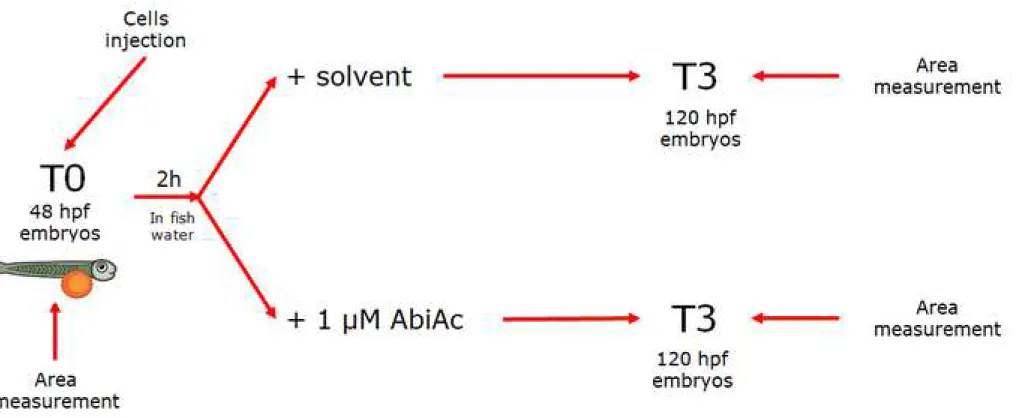
43
yolk sac of AB zebrafish embryos (n=80±10). Cell area was measured with Noldus
44
DanioScopeTM software.
45
Results: AbiAc absorption in AB zebrafish embryos was stage-dependent. Abiraterone (Abi)
46
concentration decreased while its main metabolite, namely Δ4A, increased. Accordingly, we
47
demonstrated that zebrafish expressed the enzyme 3β-hydroxysteroid dehydrogenase mRNA,
48
that converts Abi in Δ4A. Furthermore, ABiAc reduced zebrafish embryos cortisol production and
49
increased progesterone. Three days after cell injection (T3), the cortisol-secreting ACC cell area
50
in solvent-treated embryos was significantly higher compared to 1 µM AbiAC-treated embryos,
51
while no AbiAc effect was observed in SW13, that lacks the Abi target enzyme CYP17A1.
52
Conclusions: Zebrafish embryo xenografted with ACC tumor cells could be a useful, fast and reproducible
53
experimental model to preclinically test the activity of new drugs potentially active in human ACC
.
3
INTRODUCTION
55
AdrenoCortical Cancer (ACC) is a rare tumor with an estimated incidence between 0.7 and 2.0 per million
56
population per year (1). Surgery is the only potentially curative treatment modality. Systemic therapies have
57
a limited efficacy and the prognosis of locally advanced or metastatic ACC patients is often dismal. Mitotane
58
is the only drug approved to treat ACC both in adjuvant setting and metastatic disease (2, 3); the drug
59
pharmacokinetics and safety profile, however, limit its efficacy (4). Mitotane can be administered either
60
alone or in association with etoposide, doxorubicin and cisplatin (EDP-M) (5, 6). The overall 5-year survival
61
rate of metastatic ACC patients submitted to EDP-M is about 15%. In this scenario, the introduction of new
62
potentially effective drugs, or the demonstration of efficacy in ACC of already available drugs is of paramount
63
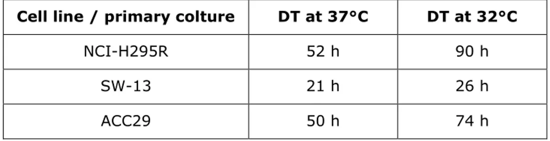
importance. Evaluating new targets and drugs using established cell lines is limited by the inexact correlation
64
between responsiveness observed in cell lines versus that elicited in the patient. Tumor cell xenografts in
65
athymic mice, generated from fresh tumor specimens, recapitulate the diversity of malignancies and have
66
been the most used in vivo model over the last 50 years (7). This model, however, is time-consuming and
67
expensive, and these are important limitations in the early phase of drug screening, when a large number of
68
drugs needs to be evaluated for their potential anti-tumor activity.
69
It is therefore mandatory, especially in the case of rare diseases like ACC, to identify and validate
70
reliable and faster experimental preclinical in vivo models that can offer a robust demonstration
71
of anti-tumor efficacy of new drugs.
72
The zebrafish (Danio rerio) animal model has been developed in the past years, and became a
73
widely used experimental vertebrate model in many fields of scientific research. In particular,
74
zebrafish embryos represent a valuable tool to study human diseases, including cancer (8, 9),
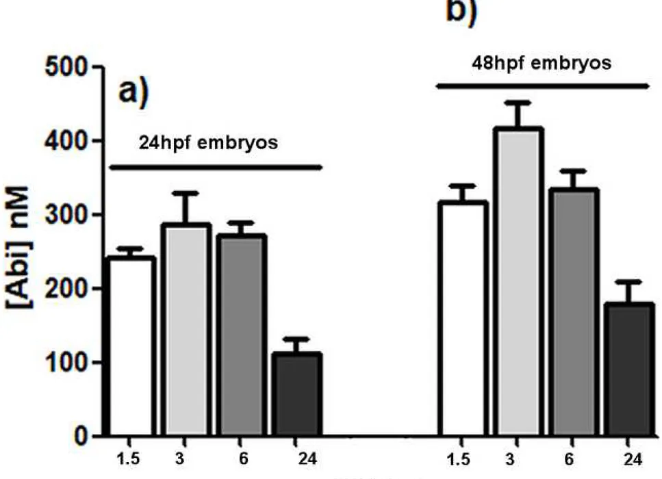
75
and to develop in vivo toxicological and pharmacological screenings (10-16).
76
Several zebrafish xenograft models of different tumor cells have been already validated for
pre-77
clinical anti-cancer drug screening (17-20). Indeed, very low amount of cancer cells and drugs
78
are needed to conduct the experiments; in addition only few days are necessary to obtain results
79
in zebrafish embryos/larvae, as opposed to the several weeks required in mice models. No
80
immunosuppressant treatment is necessary since lymphocytes mature after around 7 days post
81
fertilization (21) and the transparency of zebrafish embryos and larvae allow the easy
4
observation of engrafted tumor cells. Taken together, these observations allow to affirm that the
83
zebrafish xenograft models developed up to now are fast, simple and reproducible (17-20).
84
The aim of this study is to provide the first evidence that the zebrafish embryo model is a useful
85
tool to evaluate in vivo cytotoxicity of drugs potentially efficacious on ACC. To do so, we validated
86
in zebrafish embryo the results already obtained in immunodeficient mice xenografted with the
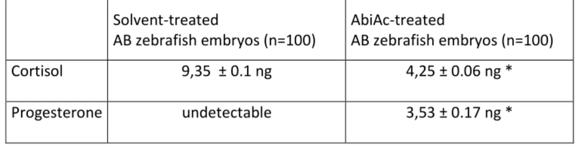
87
NCI-H295R cells treated with abiraterone acetate (AbiAc) (22). AbiAc is an irreversible inhibitor
88
of 17alpha-hydroxylase/17,20-lyase (CYP17A1), a key enzyme for steroid hormone synthesis
89
(23). Since AbiAc inhibits androgen synthesis throughout the body, it is effective in the
90
management metastatic prostate cancer (24, 25). Besides reducing androgen levels, the drug
91
rapidly impairs cortisol synthesis (26) and appears potentially effective in the management of
92
Cushing's syndrome often associated with ACC. Our group demonstrated in preclinical models
93
that AbiAc is not only able to inhibit cortisol secretion but also exerts a cytotoxic activity in the
94
ACC cell line and in ACC primary cell cultures (22), due to the drug induced increase of
95
progesterone levels (22, 27). AbiAc inhibited tumor growth when administered daily for 16 days
96
in NCI-H295R cells xenografted immunodeficient mice, thus confirming the in vitro findings (22).
97
In the present study, we were able to reproduce in the zebrafish embryos the in vivo cytotoxic
98
effect observed by AbiAc in the murine model and demonstrated that the zebrafish embryos
99
xenografted with ACC cells are a valuable in vivo preclinical model to screen drugs potentially
100
efficacious in ACC patients.
101
102
5
MATERIALS AND METHODS
103
Chemicals
104
AbiAc was purchased from Selleckchem (DBA, Segrate, MI, Italy) and resuspended in 100 %
105
ethanol (stock solution: 25 mM). When required, AbiAc was directly added to the fish water at
106
different concentration (0.5, 1, 2.5 µM), accordingly to the experimental conditions. Control
107
embryos were treated with solvent alone.
108
Reagent-grade methanol (MeOH) LC-MS CHROMASOLV® and formic acid (98%) were purchased
109
from Sigma Italia (Milan, Italy). Ultra-pure water was prepared using a Millipore Milli-Q
110
purification system (Millipore Corporation, Billerica, MA, USA). Abi standard, Abi deuterated
111
standard (SI) were purchased from Selleckchem; cortisol (1mg/ml in methanol), progesterone,
112
Δ4A standard was purchased from Sigma Aldrich (Sigma Italia, Milano, Italy). Abi was
113
resuspended in 100 % dimethylformamide; progesterone, Abi deuterated and Δ4A were
114
resuspended in 100 % dimethylsulfoxide. Drugs were subsequently diluted in methanol.
115
Cell culture and labeling
116
The human cell lines NCI-H295R and SW13 were obtained from the American Type Culture
117
Collection (ATCC) and cultured as suggested. The NCI-H295R cell line was established from a
118
secreting human ACC and represented the most widely used experimental cell model to study
119
ACC in vitro (28). The human SW-13 cell line has been established from a small cell carcinoma
120
in the adrenal cortex. These cells do not produce steroids and their exact histopathologic
121
characteristics are still under investigation (28). The human ACC primary cells, namely ACC29,
122
were derived from a female patient underwent surgery for ACC (29) and established as
123
previously described (22, 27). Cells were characterized as of adrenal origin, measuring the
124
Steroidogenic Factor 1 gene expression (30,31) by q-RT-PCR and measuring cortisol production,
125
as described in Fiorentini et al (22). The local Ethical Committee approved the project and written
126
informed consent was obtained from the patient. ACC culture conditions were as indicated for
127
NCI-H295R cells. Conditioned media from ACC29 were obtained as described in Fiorentini et al.
128
(22). The doubling-time was calculated according to ATCC indication with the following formula:
129
DT=T ln2/ln(Xe/Xb), where T is the incubation time in any units, Xb is the cell number at the
130
beginning of the incubation time, Xe is the cell number at the end of the incubation time. The
6
cell viability was evaluated by trypan blue exclusion test. Briefly, cell suspensions containing
132
0.25 % trypan blue were dropped in a haemocytometer chamber and the viable cells were
133
counted under a phase contrast microscope by two different operators. Cells (3x10⁶ cells) were
134
treated o/n with the vital red fluorescent dye CellTrackerTM CM-DiI (Thermo Fisher Scientific,
135
Milano, Italy, final concentration 0.66 ng/ml), then detached with trypsin/EDTA, washed in PBS,
136
resuspended in 50 μl of PBS and kept at 4 °C until use.
137
Measurement of cell viability
138
ACC29 cell viability was measured using the luminescence assay ATPliteTM (Perkin Elmer), that
139
measures the ATP production of viable cells. Cells were plated at the density of 5 x 10³ cells/well
140
in 96 wells plate and treated with increasing concentrations of AbiAc (1-200 nM); untreated and
141
AbiAc-treated cell viability was measured accordingly to the manufacturer instructions.
142
Experiments were conducted at least three times, each point run in triplicate.
143
Fish maintenance and eggs collection
144
All zebrafish embryos were handled according to national and international guidelines, following
145
protocols approved by the local Committee (OPBA protocol n. 211B5.24) and authorized by the
146
Ministry of Health (authorization n. 393/2017-PR).
147
Healthy adult wild-type zebrafish (AB strain) were used for egg production. Fish were maintained
148
under standard laboratory conditions as described (32), at 28 °C on a constant 14 h light/10 h
149
dark cycle. Fishes were fed thrice a day with a combination of granular dry food and fresh artemia
150
(Special Diet Services, SDS Diets). Nine months old male and female zebrafish were put in the
151
breeding tank overnight in a 1:2 ratio. Immediately after spawning, fertilized eggs were
152
harvested, washed and placed in 10 cm Petri dishes in fish water. The developing embryos were
153
incubated at 28 °C and maintained in 0.003 % (w/v) 1-phenyl-2-thiourea (PTU, Sigma-Aldrich)
154
to prevent pigmentation. Preliminary experiments were performed using different number of
155
embryos at different stages of development in order to obtain the best experimental conditions.
156
AbiAc absorption quantification by liquid chromatography–tandem mass
157
spectrometry (LC-MS/MS)
7
Due to the rapid hydrolysis of AbiAc in abiraterone (Abi) (33), AbiAc absorption from embryos
159
was evaluated by quantifying the concentration of Abi and its main metabolite Δ4A (34) by liquid
160
chromatography–tandem mass spectrometry (LC-MS/MS).
161
Ultra performance liquid chromatography (UPLC) was performed using a DionexTM UltiMateTM
162
3000 Thermo Fisher Scientific S.p.A (Milan, Italy) equipped with a LPG-3400SD quaternary
163
analytical pump, a WPS-3000SL analytical autosampler, a TCC-3000SD thermostated column
164
compartment. Chromatographic separation was performed using on XSELECT CSH C18 column
165
(150 mm × 2.1 mm ID, particle size 3.5 μm) (Waters, Milano, Italy). Mobile phase (A) was water
166
containing 0.1 % formic acid. Mobile phase (B) was methanol containing 0.1 % formic acid. An
167
isocratic mobile phase was used with 70 % of (B) and a runtime of 15 min. The UPLC flow rate
168
was 0.3 ml/min. The column temperature was 40 °C.
169
Collision-Induced Dissociation (CID)-MSn experiments were performed on an electrospray
170
ionization mass spectrometer (LCQ Fleet Ion Trap MSn, Thermo Fisher Scientific). The positive
171
ESI conditions were as follows. The source voltage was set at 4.5 kV and the sores current was
172
set at 100 µA. The capillary voltage was set at 7 kV and the capillary temperature was 350 °C.
173
The spray was stabilized with a nitrogen sheath gas (35 arb) and the auxiliary gas was set at 15
174
arb. The isolation width of precursor ions was 1 mass units. Ions were obtained in the range of
175
m/z 300–400. For all SRM analyses the scan time was equal to 100 ms, the collision energy (CE)
176
was fixed at 50 % and the isolation width of precursor ions was 2.5 mass units. Data were
177
treated with the Xcalibur software (Version 4.0, Thermo Fisher Scientific). The calibration curves
178
for the quantification of Abi and Δ4A were obtained both as described below.Twenty-five embryos
179
for each batch (up to (120 hpf) were put at 4 °C. 100 µl of Internal Standard (IS) (300 nM) was
180
added to each batch with 100 µl of Abi or Δ4A (dependent on the calibration curve) at different
181
dilutions in order to obtain the final concentrations, respectively, 5-1000 nM Abi and 12.5-500
182
nM Δ4A. Samples were broken up with a pestle, homogenized using a pipette, vortexed for one
183
minute, centrifuged at 15,000 rpm for 1 min at 4 °C, sonicated for 15 min, centrifuged at 15,000
184
× rpm for 10 min at 4 °C. The supernatant was transferred in a tube after filtration through a
185
0.2 μm PVDF filter and 5 μl were injected into the system LC-MS/MS. The SRM quantifier
186
transitions were 350-->156 for Abi and 348-->156 for Δ4A.
8
Linearity was determined by least-squares regression (data not shown).
188
Each batch of 25 embryos (treated as above indicated) was put at 4 °C. 100 µl of SI (300 nM)
189
was added to each batch with 100 µl of MeOH. Samples were homogenized using a pipette,
190
vortexed for one minute, centrifuged at 15,000 rpm for 1 minutes at 4 °C, sonicated for 15
191
minutes, centrifuged at 15,000 × rpm for 10 minutes at 4 °C. The supernatant was transferred
192
in a tube after filtration through a 0.2 μm PVDF filter.
193
Five µl of each samples were analyzed by Ultra-performance liquid chromatography (UPLC)
194
interfaced with the electrospray ionization (ESI) tandem mass spectrometer (MSn).
195
Cortisol and progesterone extraction and quantification by LC-MS/MS. Cortisol and
196
progesterone extraction was performed as indicated in Fiorentini et al. (22). For ACC29
197
conditioned media, samples were the reconstituted in 20 µl of methanol and a volume of 10 µl
198
was directly injected into the LC-MS/MS system. For AB zebrafish embryos, batch of 100
199
solvent- or AbiAc-treated embryos each were prepared. Briefly, 70 µl of MeOH was added to
200
each batch. Samples were homogenized using a pipette, vortexed for 1 min, centrifuged at
201
14,000 rpm for 1 min at 4 °C, sonicated for 15 min, centrifuged at 15,000 × rpm for 10 min at
202
4 °C. The supernatant was transferred in a tube after filtration through a 0.2 μm PVDF filter.
203
Ten µl of each samples were analyzed.
204
UPLC was performed using a DionexTM UltiMateTM 3000 Thermo Fisher Scientific S.p.A (Milan,
205
Italy) equipped with a LPG-3400SD quaternary analytical pump, a WPS-3000SL analytical
206
autosampler, a TCC-3000SD thermostated column compartment. Chromatographic separation
207
was performed with XSELECT CSH C18 column (150 mm × 2.1 mm ID, particle size 3.5 μm)
208
(Waters, Milano, Italy). Mobile phase (A) was water containing 0.1 % formic acid. Mobile phase
209
(B) was methanol containing 0.1 % formic acid. A gradient mobile phase was used. The gradient
210
program was as follows: 58% B for 2 minutes; then from 58 to 100% B in 6 min, then 100% B
211
for 2 min, from 100 to 58% B in 2 min and re-equilibration to 58% B for 8 min. All analyses
212
were performed at 30 °C. CID-MSn experiments were performed on an electrospray ionization
213
mass spectrometer (LCQ Fleet Ion Trap MSn, Thermo Fisher Scientific). The positive ESI
214
conditions were as follows. The source voltage was set at 3.8 kV and the source current was set
215
at 100 µA. The capillary voltage was set at 11 V and the capillary temperature was 300 °C. The
9
spray was stabilized with a nitrogen sheath gas (35 arb) and the auxiliary gas was set at 15 arb.
217
The isolation width of precursor ions was 1 mass units. Ions were obtained in the range of m/z
218
250–400. The calibration curves for the quantification of cortisol and progesterone were obtained
219
diluting both reference standard as follow: 500 ng/ml; 250 ng/ml; 100 ng/ml; 50 ng/ml. Ten
220
microliters of each standard dilution was analyzed by LC-MS/MS as previously described.
221
Linearity was determined by least-squares regression (data not shown).
222
Tumor xenograft
223
AB zebrafish embryos at 48 h post fertilization (hpf) were dechorionated, anesthetized with
224
0.042 mg/ml tricaine (ethyl 3-aminobenzoate methanesulfonate salt, Sigma-Aldrich) and
225
microinjected with the labeled tumor cells into the subperidermal space of the yolk sac (8, 18).
226
Microinjections were performed with the electronic microinjector FemtoJet coupled with the
227
InjectMan N12 manipulator (Eppendorf Italia, Milano, Italy). Approximately 240 cells in a volume
228
of 4 nl were injected into each embryo, which were then maintained in fish water plus PTU in a
229
32 °C incubator to allow tumor cell survival and growth. A picture of each injected embryo was
230
acquired under a Leica MZ16F fluorescence stereomicroscope two hours post treatment (T0).
231
AbiAc or solvent was directly added to the fish water. After three days (T3) pictures were taken
232
at above described. A scheme of the AbiAc protocol is shown in Fig. 1. The tumor area of T0 and
233
T3 AbiAc-treated and untreated groups was measured with Noldus DanioScopeTM software
234
(Noldus Information Technology) and analyzed by GraphPad Prism software 6.01 version.
235
In silico analysis
236
The human 3β-Hydroxysteroid dehydrogenase (3β-Hsd) protein information collected in the
237
UniProt database (35) were used to obtain the human 3β-HSD Ensemble gene entry (36).
238
Ensemble full length protein sequence of human 3β-Hsd protein was used to search the zebrafish
239
assembly on BLAST. The sequences of the zebrafish and human enzyme were aligned by using
240
ClustalOmega online software (37, 38).
241
RNA extraction and real-time PCR
242
Total RNA was extracted from batch of 30 embryos at five different stage of development (24,
243
48, 72, 96 and 120 hpf) using the RNAeasy kit (Qiagen Italia, Milano, Italy). RNA was quantified
244
by mySPEC microvolume spectrophotometer (VWR). One µg of each sample was transcribed into
10
cDNA using the M-MLV reverse transcriptase (Promega Italia, Milano, Italy). Relative gene
246
expression of 3β-HSD was evaluated by quantitative RT-PCR with the ViiA7 Real Time PCR
247
System (Applied Biosystems, Milano, Italy), using the iQ™SYBR Green Supermix method
(Bio-248
Rad, Segrate (Mi), Italy), according to manufacturer's instructions.
249
The zebrafish full length 3β-HSD transcript was employed to design zebrafish (ZF) specific
250
primers for PCR by using the Primer3web software version 4.1.0 (http://primer3.ut.ee/) (39).
251
The ZF-3β-HSD oligonucleotide sequences of were: F: CTTTCAACGCAGCGCTCTAC-3’, R:
5’-252
TCTTCCAGCAACAGTCGGAC-3’, while for the ZF β-actin (housekeeping gene) were: F
5’-253
AATCCCAAAGCCAACAGAGA-3’, R 5’-TCACACCATCACCAGAGTCC-3’.
254
Reactions were conducted under the following conditions: 1 cycle at 95 °C for 10 min, 40 cycles
255
at 95 °C for 15 s, 62 °C for 1 min. Differences in the threshold cycle Ct values between the
β-256
actin housekeeping gene and ZF-3β-HSD were then calculated as an indicator of the amount of
257
mRNA expressed. Analysis was performed in triplicate for each sample, using different groups
258
of embryos.
259
Statistical analysis
260
Statistical analyses were done using GraphPad Prism software 6.01 version. One-way ANOVA
261
followed by Dunnett’s test was performed to identify statistically significant differences among
262
different groups of data, considering a p value <0.05 as the threshold for a significant difference.
11
RESULTS
264
Cytotoxic and antisecretive effect of AbiAc in ACC29 primary cells. ACC29 primary cells
265
were exposed to increasing concentrations of AbiAc (1-200 nM) for 4 days and analyzed for cell
266
viability. AbiAc exerted a concentration-dependent reduction of cell viability, that reached the
267
plateau of 59,75 ± 2,3 % at 25 nM concentration and did not further increase. The calculated
268
IC50 was 6 nM (95%CI 2,9-11,3 nM) (40). Conditioned media of ACC29 were then analyzed for
269
cortisol and progesterone production. Results demonstrated that cortisol production of untreated
270
ACC29 cells was reduced from 2,24 ± 0,29 ng/ml/106 cells to 1,55 ± 0,29 ng/ml/106 cells in
271
100 nM AbiAc-treated cells (p<0.05 vs untreated cells). As expected, progesterone was
272
undetectable in untreated cells and raised up to 1,3 ng/ml/106 cells when exposed to AbiAc.
273
ACC cell viability and doubling time. The viability and the doubling time of ACC cells
274
maintained at 32 °C were investigated, in order to evaluate whether or not these cells can be a
275
suitable model for zebrafish embryo xenograft. Cell viability was evaluated by trypan blue
276
exclusion test at both 37 °C and at 32 °C and results demonstrated that at the lower
277
temperature, ACC cells are viable (not shown), although with a reduction of the cell proliferation
278
rate (Table 1).
279
Determination of AbiAc treatment concentration. To set up the method, AB strain zebrafish
280
embryos were treated with increasing concentrations of AbiAc, to determine the optimal drug
281
concentration to be used during in vivo experiments. Embryos maintained in fish water plus PTU
282
were manually dechorionated at 48hpf and divided into 5 groups (n=90±10 each). Three groups
283
were treated with 0.5, 1 and 2.5 μM AbiAc, respectively, directly added to the fish water; in the
284
fourth group the solvent alone (methanol) was added to the fish water plus PTU (control group),
285
while the last group was left untreated. Embryos were kept at 32 °C. After 3 days of treatment,
286
exposure of embryos the 2.5 μM AbiAc resulted in death or deformity (41). Embryos of the other
287
two treated groups, as well as embryos of the solvent-treated and control groups developed
288
normally and their phenotype was similar to that of untreated embryos (41). Based on these
289
findings, the concentration of 1 μM AbiAc was chosen for subsequent experiments.
290
Determination of AbiAc measurement. To evaluate the optimal embryo number to determine
291
absorbed drug concentration, 1 µM AbiAc-treated embryos at 24hpf were divided into groups of
12
10-15-20-25-30 embryos each and analyzed by LC-MS/MS. As indicated in Methods, AbiAc is
293
rapidly hydrolyzed to Abi (30), indeed AbiAc could not be detected in embryos, while Abi was
294
measurable. The LC-MS/MS analysis revealed that in samples obtained from groups of embryos
295
from 10 to 20, Abi was not detectable, while samples obtained from 25 embryos gave
296
measurable and reproducible results, as shown in Fig. 2a, where a time-curve of the absorbed
297
Abi concentration was performed. AB zebrafish embryos maintained in fish water plus PTU were
298
divided into 5 groups (n=120±10), each group was treated with 1 μM AbiAc, directly added to
299
the fish water, and embryos were collected at different time-points, up to 24 hrs, for the
300
quantification of Abi. In particular, experiments were conducted in both 24 hpf embryos and in
301
48 hpf embryos. The LC-MS/MS results reported in Fig. 2 showed that Abi absorption increased
302
with the stage of embryo development: interestingly, in 48hpf embryos, the Abi concentration
303
after 24hrs of incubation is about 179 ± 29.5 nM, that is very close to the highest Abi
304
concentration used in in vitro experiments with NCI-H295R cells, that was 200 nM (22). The
305
time course, at both development stages, displayed typical kinetic of time-dependent
306
concentration –curve observed in humans (42), suggesting the capability of embryos to
307
metabolize Abi. This hypothesis was supported by gene expression results demonstrating that
308
zebrafish expressed the mRNA encoding the enzyme 3β-HSD; indeed, by q-RT-PCR, we
309
demonstrated that the ΔCt was 4.8 ± 1.2 in 24 hpf embryos and 5.52 ± 0.75 in 48 hpf embryos.
310
The 3β-HSD gene was transcribed in its protein, as demonstrated by the capability of 48 hpf
311
embryos to metabolize Abi in its main metabolite Δ4A (Fig. 3a and Fig.3b). Interestingly, by in
312
silico analysis we observed a high level of similarity (63 %) and identity (46 %) with the human
313
counterpart (38). The experimental conditions were set up in both 24 hpf and 48 hpf embryos,
314
thus, based on results obtained, we chose to perform cell xenograft in 48 hpf embryos, due to
315
the Abi concentration time-course, to technical limits in injecting 24 hpf embryos (in particular,
316
at this stage of development microinjection is not recommended due to tissue fragility) and
317
according to the protocol of Nicoli et al. (8). Finally, the Abi metabolism time-course was
318
conducted at different hpf of embryos, up to 120 (Table 2) and results indicated that the
319
capability of AB zebrafish embryos to metabolize Abi increased with increasing hpf.
13
AbiAc at 1 µM concentration induced as well a modification of AB zebrafish embryo cortisol level.
321
Results are reported in Table 3. After 3 days exposure, the irreversible binding of AbiAc to
322
CYP17A1 induced a significant reduction of cortisol production in AbiAc-treated embryos.
323
Accordingly, as expected, progesterone become measurable in treated embryos, while it did not
324
reach detection in solvent-treated embryos.
325
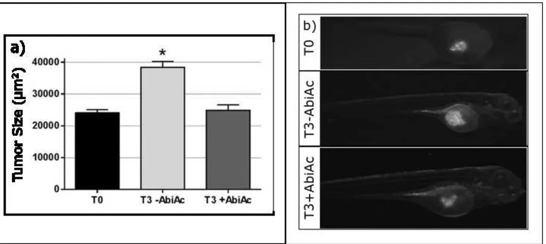
ACC cell xenograft in AB zebrafish embryos.
326
NCI-H295R cell line. AB zebrafish embryos at 48 hpf were manually dechorionated, anesthetized
327
with tricaine and microinjected with NCI-H295R labeled cells into the subepidermal space of the
328
yolk sac (n=80±10). A picture of each injected embryo was acquired under a fluorescence
329
stereomicroscope two hours post treatment (T0), then embryos were divided into two groups
330
(n=40±5): one maintained in fish water/PTU plus solvent (solvent-treated), while the other was
331
maintained in fish water/PTU plus 1μM AbiAc without medium change up to the 3 days treatment.
332
Embryos were then incubated at 32°C. Three days after treatment (T3), pictures were taken as
333
described and the injected cell area in solvent-treated and 1 μM AbiAc-treated embryos was
334
measured and analyzed as described in Methods. The injected cell area at T3 in solvent-treated
335
embryos was 38,390 ± 1,432 µM2 compared to 24,891 ± 1,302 µM2 in AbiAc-treated embryos
336
(p<0.01, Fig. 4a). The tumor area in solvent-treated embryos displayed a 1.63 ± 0.07 fold
337
increased at T3 compared to T0, while in AbiAc-treated embryos the tumor area was almost
338
unchanged, with a value of 1.01 ± 0.03 fold increase at T3 compared to T0. A representative
339
image is shown in Fig. 4b. Experiments were repeated changing zebrafish embryo medium every
340
day after xenograft, thus adding fresh AbiAc every day. Results demonstrated that the injected
341
cell areas measured was superimposable to that reported above (not shown).
342
ACC29 primary cell culture. The cytotoxic effect of 1 µM AbiAc was evaluated as well in a primary
343
culture obtained from a patient diagnosed with a cortisol-secreting ACC. Results are shown in
344
Fig. 5 and demonstrated that, although ACC29 cells seemed to be less sensitive to AbiAc both
345
in vitro (see above) and in vivo, at T3, the injected ACC29 cell area was significantly reduced
346
in 1 µM AbiAc-treated embryos compared to solvent-treated embryos. The area was indeed
347
56,987 ± 2,83 µM2 in solvent-treated embryos vs 39,776 ± 4,516 µM2 in ABiAC-treated
348
embryos (* p <0.05).
14
SW13 cell line. The non steroidogenic, AbiAc-insensitive (22, 28) cell line SW13 was used as the350
internal negative control. The tumor xenograft area of SW13 cells was not modified by exposure
351
to 1 µM AbiAc (Fig. 6), indeed the area of solvent-treated tumor was 52,842 ± 5,163 µM2 while
352
the area of AbiAc-treated tumor was 49,665 ± 3,705 µM2.
15
DISCUSSION
354
The zebrafish model, due to its distinctive characteristics, is nowadays progressively being
355
applied to design useful in vivo experimental studies in a number of human diseases (43),
356
including malignancies (reviewed in 44). In particular, zebrafish embryos provide a powerful tool
357
to develop functional cancer models that can be used for drug discovery and development and
358
drug toxicity. In a rare and severe disease such as ACC, the finding of reproducible and reliable
359
in vivo preclinical models is an open challenge, especially in light of the number of variables that
360
have to be taken into account when studying ACC xenograft in mouse model (45). The timing of
361
drug administration, the drug metabolism and the solvent in which the drug is dissolved (that
362
per se may affect the animal safety and the reproducibility of results) are among the most
363
relevant issues (43). The few available cell lines and their genomic instability, the immune
364
competence of the host and site of implantation are additional important drawbacks (45). Here,
365
we validated a NCI-H295R cell xenograft in AB zebrafish embryos as an in vivo model of ACC.
366
We were able indeed to confirm the in vivo cytotoxicity of AbiAc using an animal model which
367
offers several advantages over other models, like mice (17-21). It should be underlined that
368
AbiAc was directly added into the fish water and was significantly adsorbed by embryos, reaching
369
concentrations able to exert the cytotoxic effect, thus simplifying the treatment procedure.
370
Furthermore, the ACC xenograft in zebrafish was fast, as 3 days of AbiAc treatment were
371
sufficient to demonstrate a significant difference in the NCI-H295R cell proliferation rate. The
372
AbiAc effect was due to the direct binding on its target enzyme as the tumor area of xenografts
373
with non steroidogenic (28), CYP17A1 negative (22) SW13 cells was not modified after 3 days
374
exposure to 1 µM AbiAc, confirming the insensitivity of these cells to the cytotoxic effect of AbiAc
375
treatment, as observed in in vitro experiments (22). Interestingly, the effect of AbiAc lasted up
376
to 3 days without adding fresh drug, probably due to the irreversible binding of Abi to CYP17A1.
377
The inhibition of NCI-H295R cell area growth in AB zebrafish embryos was about 60% after 3
378
days, which is even higher than that we observed in immune incompetent mice, where it reached
379
the 34% inhibition about 60 days after the cell injection and 15 days after the end of 16 days
380
treatment (22). Furthermore, another finding that give support for the use of zebrafish embryos
381
as a useful model for in vivo animal studies on ACC is the expression in embryos of enzymes of
16
the steroidogenic pathways similar to what observed in more evolved animal models. Indeed,
383
they express the 3β-HSD enzyme that converted Abi in its active metabolite Δ4A, that itself
384
inhibits additional enzymes involved in steroidogenesis, including CYP17A1, 3β-HSD and the 5α
385
reductase SRD5A (34, 46). The combined effect of Abi and its main metabolite on CYP17A1
386
induced a decrease of cortisol and, as expected, an increase of progesterone levels. In the
387
personalized medicine era, the low number of cells needed and the lower length of the
388
experiments make the zebrafish model potentially candidate to prepare ACC patient-derived
389
xenografts in order to perform a real-time selection of the most appropriate cytotoxic drugs for
390
each patient. On these bases, we reproduced results obtained with the NCI-H295R cell line in a
391
patient-derived xenograft, obtained from primary cells established from a cortisol-secreting ACC,
392
thus giving support to the possibility to develop this in vivo method to screen available
393
therapeutic options for a cancer such as ACC, with a poor prognosis and a scarcity of therapeutic
394
options. We believe that the validation of this animal model can offer a useful tool to perform
395
a preclinical first-screening of a large number of drugs, and this is advantageous in a rare and
396
aggressive disease such as ACC, in which the treatment strategies are limited. We are aware
397
that this animal model cannot completely replace the others already in use, but we believe that
398
our findings could offer a suitable and fast model to perform an initial selection of potentially
399
effective drugs, also with regard to the identification of dose toxicity, and choose the most
400
promising compounds to be used in more advanced preclinical phases. Lastly, this procedure
401
could also reduce the number of animal models to finalize the research.
402
403
404
405
406
407
ACKNOWLEDGEMENT: We would like to thank the FIRM Foundation (Cremona, Italy) for their
408
support and generosity
409
410
17
REFERENCES
411
(1) Fassnacht M, Dekkers OM, Else T, Baudin E, Berruti A, de Krijger R, Haak HR, Mihai R,
412
Assie G, Terzolo M. European Society of Endocrinology Clinical Practice Guidelines on the
413
management of adrenocortical carcinoma in adults, in collaboration with the European
414
Network for the Study of Adrenal Tumors. Eur J Endocrinol. 2018;179(4):G1-G46. doi:
415
10.1530/EJE-18-0608.
416
(2) Berruti A, Grisanti S, Pulzer A, Claps M, Daffara F, Loli P, Mannelli M, Boscaro M, Arvat E,
417
Tiberio G, Hahner S, Zaggia B, Porpiglia F, Volante M, Fassnacht M, Terzolo M. Long-Term
418
Outcomes of Adjuvant Mitotane Therapy in Patients With Radically Resected
419
Adrenocortical Carcinoma. J Clin Endocrinol Metab. 2017;102(4):1358-1365. doi:
420
10.1210/jc.2016-2894
421
(3) Terzolo M, Daffara F, Ardito A, Zaggia B, Basile V, Ferrari L, Berruti A. Management of
422
adrenal cancer: a 2013 update. J Endocrinol Invest. 2014;37(3):207-17. doi:
423
10.1007/s40618-013-0049-2
424
(4) Paragliola RM, Torino F, Papi G, Locantore P, Pontecorvi A, Corsello SM. Role of Mitotane
425
in Adrenocortical Carcinoma - Review and State of the art. Eur Endocrinol.
426
2018;14(2):62-66. doi: 10.17925/EE.2018.14.2.62.
427
(5) Berruti A, Terzolo M, Sperone P, Pia A, Della Casa S, Gross DJ, Carnaghi C, Casali P,
428
Porpiglia F, Mantero F, Reimondo G, Angeli A, Dogliotti L. Etoposide, doxorubicin and
429
cisplatin plus mitotane in the treatment of advanced adrenocortical carcinoma: a large
430
prospective phase II trial. Endocr Relat Cancer. 2005;12(3):657-66. doi:
431
10.1677/erc.1.01025.
432
(6) Fassnacht M, Terzolo M, Allolio B, Baudin E, Haak H, Berruti A, Welin S, Schade-Brittinger
433
C, Lacroix A, Jarzab B, Sorbye H, Torpy DJ, Stepan V, Schteingart DE, Arlt W, Kroiss M,
434
Leboulleux S, Sperone P, Sundin A, Hermsen I, Hahner S, Willenberg HS, Tabarin A,
435
Quinkler M, de la Fouchardière C, Schlumberger M, Mantero F, Weismann D, Beuschlein
436
F, Gelderblom H, Wilmink H, Sender M, Edgerly M, Kenn W, Fojo T, Müller HH, Skogseid
437
B; FIRM-ACT Study Group. Combination chemotherapy in advanced adrenocortical
438
carcinoma. N Engl J Med. 2012;366(23):2189–97. doi: 10.1056/NEJMoa1200966.
18
(7) Kelland LR. Of mice and men: values and liabilities of the athymic nude mouse model in
440
anticancer drug development. Eur J Cancer. 2004;40(6):827-36. doi:
441
10.1016/j.ejca.2003.11.028.
442
(8) Nicoli S, Presta M. The zebrafish/tumor xenograft angiogenesis assay. Nature Protoc.
443
2007;2(11):2918-23. doi: 10.1038/nprot.2007.412.
444
(9) Mione MC and Trede NS. The zebrafish as a model for cancer. Dis Model Mech.
2010;3(9-445
10):517–523. doi: 10.1242/dmm.004747.
446
(10) Langheinrich U. Zebrafish: A new model on the pharmaceutical catwalk. Bioessays
447
2003;25:904-912. doi: 10.1002/bies.10326.
448
(11) Zon LI, Peterson RT. In vivo drug discovery in the zebrafish. Nat Rev Drug Discov.
449
2005;4:35-44. doi: 10.1038/nrd1606.
450
(12) Peterson RT, Macrae CA. Systematic approaches to toxicology in the zebrafish. Annu Rev
451
Pharmacol Toxicol. 2012;52:433-453. doi: 10.1146/annurev-pharmtox-010611-134751.
452
(13) Gianoncelli A, Bonini SA, Bertuzzi M, Guarienti M, Vezzoli S, Kumar R, Delbarba A, Mastinu
453
A, Sigala S, Spano P, Pani L, Pecorelli S, Memo M. An Integrated Approach for a Structural
454
and Functional Evaluation of Biosimilars: Implications for Erythropoietin. BioDrugs
455
2015;29(4):285-300. doi: 10.1007/s40259-015-0136-3.
456
(14) Guarienti M, Giacopuzzi E, Gianoncelli A, Sigala S, Spano P, Pecorelli S, Pani L, Memo M.
457
Computational and functional analysis of biopharmaceutical drugs in zebrafish:
458
Erythropoietin as a test model. Pharmacol Res. 2015;102:12-21. doi:
459
10.1016/j.phrs.2015.09.004.
460
(15) Basnet RM, Guarienti M, Memo M. Zebrafish Embryo as an In Vivo Model for Behavioral
461
and Pharmacological Characterization of Methylxanthine Drugs. Int J Mol Sci.
462
2017;18(3):E596. doi: 10.3390/ijms18030596.
463
(16) Gianoncelli A, Bertuzzi M, Guarienti M, Vezzoli S, Bonini SA, Mastinu A, Sigala S and Memo
464
M Parallelism of chemico-structural properties between filgrastim originator and three of
465
its biosimilar drugs.
J Chem.
In press466
(17) Marques IJ, Weiss FU, Vlecken DH, Nitsche C, Bakkers J, Lagendijk AK, Partecke LI,
467
Heidecke CD, Lerch MM, Bagowski CP. Metastatic behaviour of primary human tumours
19
in a zebrafish xenotransplantation model. BMC Cancer. 2009;9:128. doi:
10.1186/1471-469
2407-9-128.
470
(18) Jung DW, Oh ES, Park SH, Chang YT, Kim CH, Choi SY, Williams DR. A novel zebrafish
471
human tumor xenograft model validated for anti-cancer drug screening. Mol Biosyst.
472
2012;8(7):1930-9. doi: 10.1039/c2mb05501e.
473
(19) Tonon F, Zennaro C, Dapas B, Carraro M, Mariotti M, Grassi G. Rapid and cost-effective
474
xenograft hepatocellular carcinoma model in Zebrafish for drug testing. Int J Pharm.
475
2016;515(1-2):583-91. doi: 10.1016/j.ijpharm.2016.10.070.
476
(20) Bootorabi F, Manouchehri H, Changizi R, Barker H, Palazzo E, Saltari A, Parikka M, Pincelli
477
C, Aspatwar A. Zebrafish as a Model Organism for the Development of Drugs for Skin
478
Cancer. Int J Mol Sci. 2017;18(7). doi: 10.3390/ijms18071550.
479
(21) Chen AT, Zon LI. Zebrafish blood stem cells. J Cell Biochem. 2009;108(1):35-42. doi:
480
10.1002/jcb.22251.
481
(22) Fiorentini C, Fragni M, Perego P, Vezzoli S, Bonini SA, Tortoreto M, Galli D, Claps M,
482
Tiberio GA, Terzolo M, Missale C, Memo M, Procopio G, Zaffaroni N, Berruti A, Sigala S.
483
Antisecretive and Antitumor Activity of Abiraterone Acetate in Human Adrenocortical
484
Cancer: A Preclinical Study. J Clin Endocrinol Metab. 2016;101(12):4594-602. doi:
485
10.1210/jc.2016-2414
486
(23) Attard G, Reid AH, Auchus RJ, Hughes BA, Cassidy AM, Thompson E, Oommen NB, Folkerd
487
E, Dowsett M, Arlt W, de Bono JS. Clinical and biochemical consequences of CYP17A1
488
inhibition with abiraterone given with and without exogenous glucocorticoids in castrate
489
men with advanced prostate cancer. J Clin Endocrinol Metab. 2012;97(2):507–16. doi:
490
10.1210/jc.2011-2189
491
(24) Ryan CJ, Smith MR, Fizazi K, Saad F, Mulders PF, Sternberg CN, Miller K, Logothetis CJ,
492
Shore ND, Small EJ, Carles J, Flaig TW, Taplin ME, Higano CS, de Souza P, de Bono JS,
493
Griffin TW, De Porre P, Yu MK, Park YC, Li J, Kheoh T, Naini V, Molina A, Rathkopf DE.
494
Abiraterone acetate plus prednisone versus placebo plus prednisone in
chemotherapy-495
naive men with metastatic castration-resistant prostate cancer (COU-AA-302): final
20
overall survival analysis of a randomised, double-blind, placebo-controlled phase 3 study.
497
Lancet Oncol. 2015;16(2):152– 60. doi: 10.1016/S1470-2045(14)71205-7.
498
(25) Fizazi K, Scher HI, Molina A, Logothetis CJ, Chi KN, Jones RJ, Staffurth JN, North S,
499
Vogelzang NJ, Saad F, Mainwaring P, Harland S, Goodman OB Jr., Sternberg CN, Li JH,
500
Kheoh T, Haqq CM, de Bono JS. Abiraterone acetate for treatment of metastatic
501
castration-resistant prostate cancer: final overall survival analysis of the COU-AA-301
502
randomised, double-blind, placebo-controlled phase 3 study. Lancet Oncol.
503
2012;13(10):983–92. doi: 10.1016/S1470-2045(12)70379-0.
504
(26) Claps M, Lazzari B, Grisanti S, Ferrari V, Terzolo M, Sigala S, Vezzoli S, Memo M,
505
Castellano M, Berruti A. Management of severe cushing syndrome induced by
506
adrenocortical carcinoma with abiraterone acetate: a case report. AACE Clinical Case Rep.
507
2016;2(4):e337-e341
508
(27) Fragni M, Fiorentini C, Rossini E, Fisogni S, Vezzoli S, Bonini SA, Dalmiglio C, Grisanti S,
509
Tiberio GAM, Claps M, Cosentini D, Salvi V, Bosisio D, Terzolo M, Missale C, Facchetti F,
510
Memo M, Berruti A, Sigala S. In vitro antitumor activity of progesterone in human
511
adrenocortical carcinoma. Endocrine. 2018; Epub ahead of print. doi:
10.1007/s12020-512
018-1795-x
513
(28) Wang T, Rainey WE. Human adrenocortical carcinoma cell lines. Mol Cell Endocrinol.
514
2012;58-65. doi: 10.1016/j.mce.2011.08.041
515
(29) Gianoncelli A, Guarienti M, Fragni M, Bertuzzi M, Rossini E, Abate A, Basnet R M, Zizioli
516
D, Bono F, Terzolo M, Memo M, Berruti A, Sigala S. Supplemental data: Clinical and
517
immunohistochemical characteristics of ACC29 patient.
518
https://figshare.com/s/25f65cba83c6ca84f2f1.
519
(30) Sbiera S, Schmull S, Assie G, Voelker HU, Kraus L, Beyer M, Ragazzon B, Beuschlein F,
520
Willenberg HS, Hahner S, Saeger W, Bertherat J, Allolio B and Fassnacht M. High
521
diagnostic and prognostic value of steroidogenic factor-1 expression in adrenal tumors.
522
The Journal of clinical endocrinology and metabolism. 2010; 95:E161-171. doi:
523
10.1210/jc.2010-0653.