Parkinson’s disease and α-synuclein
Parkinson’s disease (PD) is the second most common neurodegenerative disorder after Alz-heimer’s disease, affecting almost 1% of the population over 50 years old. Clinical features of the disease include bradykinesia, resting tremor, muscle tone rigidity, and possible cogni-tive involvement [1, 2]. Pathologically, PD is characterized by selective degeneration of dopaminergic neurons in the substantia nigra pars compact (SNpc) and the presence of Lewy bodies (LBs), primarily composed of fibrillar α-synuclein (α-Syn) and ubiquitinated proteins, in the surviving neurons [3, 4].
Although more than 90% of PD cases occur spo-radically, the discovery of monogenic heredi-table forms of the disease has provided impor-tant clues as to the understanding of the mo-lecular mechanisms that lead to neuronal de-generation in this pathology [5]. Different genes have been associated with familial cases of PD, including α-Syn, LRRK2, parkin, DJ-1 and PINK1 [6]. However, the mechanism underlying
dis-ease onset and progression are still unclear. Evidences from genetic, pathological and bio-chemical studies supports critical involvement of α-Syn in the pathogenesis of the disease [7, 8].
α-Syn is a small natively unfolded protein widely expressed in neurons at the presynaptic level [9], with a conserved amphipathic N-terminus, an acidic C-terminus and a hydrophobic central region that is required for both oligomerization and fibrillization processes [10]. Although α-Syn biological function is still controversial, it has been suggested to play a role in synaptic vesi-cles biogenesis and modulation of synaptic transmission [9]. α-Syn (SNCA) gene mutations and single-nucleotide polymorphisms (SNPs) render α-Syn protein more prone to misfolding and accelerate formation of aggregates [5], which have been linked to autosomal-dominant forms of PD [11] and increased susceptibility to sporadic PD [12], respectively. While there is clear evidence that α-Syn plays an important role in the pathogenesis of PD, it is unclear what is/are the toxic form(s) of the protein
responsi-Review Article
Exosomes-associated neurodegeneration and progression
of Parkinson’s disease
Isabella Russo, Luigi Bubacco, Elisa Greggio
Department of Biology, University of Padova, Via Ugo Bassi 58/B, 35121 Padova, Italy
Received October 5 2012; Accepted November 12, 2012; Epub November 18, 2012; Published November 30, 2012 Abstract: Growing evidence indicates the role of exosomes in a variety of physiological pathways as conveyors of bio-logical materials from cell-to-cell. However the molecular mechanism(s) of secretion and their interaction with receiv-ing cells are yet unclear. Recently, it is emergreceiv-ing that exosomes are involved in pathological processes as potential carriers in the progression of neurodegenerative pathologies associated with misfolded proteins. In the current re-view we will discuss some recent findings on the key role of exosomes in the spreading of the aggregated products of α-synuclein from neuron-to-neuron and of inflammatory response propagation from immune cell-to-cell; we will high-light the implication of exosomes in the neurodegeneration and progression of the disease and the their potential interplay with genes related to Parkinson’s disease. Increasing our knowledge on the cell-to-cell transmissions might provide new insights into mechanism of disease onset and progression and identify novel strategies for diagnosis and therapeutic intervention in Parkinson and other neurodegenerative diseases.
ble for neuronal dysfunction and the mecha-nisms by which these purported toxic forms of α -Syn spreads from neuron to neuron triggering, in turn, neuronal degeneration [13].
Dopaminergic neuronal degeneration
The main pathological hallmark of PD is the progressive degeneration and death of dopa-minergic neurons in the SNpc [14], which leads to a severe dopamine depletion in the striatum, responsible for the motor symptoms observed in the disease [15]. In particular, the SNpc is subdivided into ventral and dorsal tiers, and neuronal death in PD mainly occurs in the ven-tral tier; in this regard, studies on post-mortem PD brains reported that the degeneration affect-ing the ventral tier account for 80-95% of neu-rons loss [16-18]. The pattern of cell loss and the selective vulnerability of dopamine (DA) neu-rons seen in PD do not occur in other neurode-generative diseases or in normal aging, suggest-ing that these neurons possess distinct features that render them more susceptible to degenera-tion [19]. Several mechanisms have been pro-posed to explain the observed vulnerability of dopaminergic neurons. First, it was suggested that the size and neurochemical phenotype of dopaminergic neurons which can make an ele-vated number of synaptic connections particu-larly prone to damage can account for the ob-served vulnerability [20]. Moreover, the large amounts of the dark-coloured pigment neuro-melanin and its ability to interact with transition metals, especially iron, and to mediate intracel-lular oxidative mechanisms has been suggested to sensitize this population of cells [21, 22]. In addition, an unbalanced DA homeostasis (consequent to impaired synthesis, storage, degradation and distribution in the synaptic vesicles) can lead to increased concentration of free cytosolic DA which favors ROS production and oxidative stress [19, 23] and, in its oxidized form, can itself covalently modify α-Syn and pro-mote the stabilization of toxic fibrils [24]. Fi-nally, these neurons exhibit enhanced sensitiv-ity to pro-inflammatory mediators compared to other neuronal populations, likely consequent to the high density of microglia cells in this region [25]. All these factors have been reported and suspected to contribute to the dopaminergic neurons vulnerability and degeneration.
Although it is generally accepted that the loss of midbrain dopaminergic neurons occurs and is
largely responsible for the major motor symp-toms, this is not the only region showing patho-logical hallmarks in PD patients. Lewy bodies and cell loss occur in other non-dopaminergic areas of the brain including brainstem cells nu-clei, olfactory bulb, limbic system and neocortex [26, 27] suggesting a mechanism involving propagation and progression of the pathology, similar to the one observed in prion diseases [28]. This model of a pathological propagation has recently gained particular attention with regard to neurodegenerative diseases linked to misfolded and aggregated proteins.
A few years ago, Braak and co-workers [26] pro-posed a pathological staging process for PD. In this system, based on the distribution of α-Syn pathology in post-mortem brains, early patho-logical events first occur in the autonomic nerv-ous system (stage 0), dorsal motor nucleus of the vagus and in the anterior olfactory nucleus (stage 1), then spread the locus coeruleus, SNpc and basal forebrain (stage 2). In the final stages, pathology extends to the neocortex, hip-pocampus and basal ganglia. Although the Braak staging does not fit all disorder associ-ated with LBs pathology and may be a simplified system, it supports the notion that α-Syn deposi-tions may spread to neighbor cells. There are several lines of evidence supporting cell-to-cell propagation of α-Syn. First, α-Syn exists as a soluble, natively unfolded monomer [29] and/or a folded alpha-helix tetramer [30, 31] and, in vitro, it can convert into soluble, oligomeric form and a beta-sheet, fibrillar form, the latter serv-ing as seed or template for the formation of larger aggregates [32]. Of interest, α-Syn dos-age is intimately linked to PD, being multiplica-tions of SNCA cause of hereditable, aggressive forms of the disease [33]. Increased cellular concentration of α-Syn can be the results of impaired protein homeostasis [34]. Accordingly, reduced protein clearance due to defects in the proteasome and autophagy machineries may lead to increased concentration of α-Syn and favor the formation of aggregates [35]. In sup-port of a cell-to-cell propagation α-Syn is the striking observation that neural grafts derived from midbrain healthy tissue transplanted into the striatum of individuals affected with PD showed LB pathology after 10-16 years in addi-tion to the expected Lewy pathology observed in the host tissue [36, 37].
α-Syn toxic forms can be secreted from cells in culture, enter other cells, and seed small intra-cellular aggregates in the receiving cells [38]. Although the precise mechanism is not yet clear, it is likely that α-Syn is released by the donor cell and uptaken by a recipient cell by energy dependent processes. In this regard, recent studies have reported the crucial role exerted by exosomes as material and structural templates transporters from cell-to-cell and as potential carriers in the dissemination of PD pathology [39, 40]. In the next paragraphs we discuss the recent findings on the key role of exosomes in the α-Syn spreading from neuron-to-neuron and in the inflammation propagation from immune cell-to-cell, highlighting their po-tential implication in the propagation and pro-gression of the PD.
Exosomes biogenesis and physiological functions
Exosomes are microvesicles whose biogenesis occurs within multivesicular bodies (MVBs) in the endosomal system. In details, proteins and signaling complexes that are sequestered to the limiting membrane of MVBs can be selectively incorporated into intraluminal vesicles (ILVs) by invagination of the MVB membrane. Here, MVB can either fuse with the lysosome membrane to degrade ILV and its content, or alternatively with the plasma membrane to release the ILV as exosomes into the extracellular space [40, 41]. Exosomes are small vesicles with a size ranging from 30 to 120 nm and appear with a charac-teristic round or cup-shaped morphology as ob-served by transmission or cryo-electron micros-copy [42]. These vesicles can be released from a variety of cells, both in vitro and in vivo, such as neuronal cells including neurons [43], micro-glia [44], astrocytes [45] and non-neuronal cells such as lymphocytes and monocytes [46, 47]. Recently, it has been shown that exosomes can transport messenger RNA, microRNA, protein and signaling complexes, all of which important in the modulation of gene and protein expres-sion in the receiving cells [40, 47]. They contain distinct proteins, some are common across all exosomes and are used as “marker” protein including Alix, tetraspanin (CD63, CD81) and heat shock protein (HSP70, 90) [47, 48], while others are specific and depend on the cell type from which they are secreted [49]. In this gard, it has been reported that exosomes
re-leased from antigen-presenting cells carry the major histocompatibility complex (MHC) [50] whilst those released from oligondendrocytes carry the myelin proteins [51].
Little is still known about the requirement and regulation of exosomes fusion with the plasma membrane of secreting and receiving cells. Dif-ferent studies have suggested that exosomes are secreted by a regulated process involving the tethering, docking and fusion of the vesicles at the plasma membrane probably through a specific combination of SNARE proteins, al-though this combination has not yet been identi-fied [41, 46]. It has been demonstrated that exosomes can be taken up by targeting cells; in this regard, different mechanisms have been proposed, including endocytosis, receptor-ligand binding, attachment or fusion with the plasma membrane to deliver their cytosolic content [49, 52, 53], however the mechanism of interaction and secretion has not yet been well established. Exosomes are biologically active entities and are important for a variety of physiological path-ways. In particular, the fusion of MVB with the lysosome results in the degradation of un-wanted cellular materials, and allows the cell to remove transmembrane proteins essential to down-regulate activated surface receptors, as growth factors or adhesion molecules [54]. Moreover, these vesicles have been shown to play a functional role in neuronal development and regeneration [55], and in the synaptic plas-ticity of the cortical and hippocampal neurons [56]. In addition, when released from immune cells, they exhibit immune-stimulatory proper-ties important to activate and propagate the inflammation in response to pathogenic stimuli or injury [57, 58]. Recently, in the context of pathological processes the role of exosomes has been the subject of intensive research, they have been implicated in the spread of toxic pro-teins within the brain and in the progression of different neurodegenerative diseases including PD [40].
Exosomes in the cell-to-cell communication and their role in the progression of PD
Cell-to-cell communication exerted by exosomes in the brain, in particular neuron-to-neuron and neuron-to-glia communication, has been in-tensely studied to investigate their role in the progression of the neurodegenerative disease
including PD. In this context, it was demon-strated that α-Syn deposits are released via exosomes and in this way are efficiently able to transfer and propagate α-Syn toxic forms to other cells [40, 59-61]. Different studies dem-onstrated that α-Syn transferred neuron-to-neuron is able to form aggregates in recipient cells [61, 62]. Moreover, Emmanouilidou and colleagues reported that secreted α-Syn via exosomes induces cell death of recipient neu-ronal cells, suggesting that α-Syn deposits spreading to the neighbors neurons causes propagation of the disease [60].
Of interest, immunologists started to focus on exosomes when they discovered that immune cells, B lynmphocytes and dendritic cells, were also able to secrete exosomes [57, 63]. Most notably, these exosomes when released by den-dritic cells (DCs) expressed the MHC II bound to antigenic peptide, and were able to present MHC-peptide complex to T cells promoting their activation [46, 57, 58, 64]. In addition, a recent study reported that exosomes released from infected macrophages expressed pathogen-associated molecular patterns (PAMPs) and can stimulate macrophages activation by pattern recognition receptor (PRP), and pro-inflammatory response both in vitro and in vivo [65]. This role of exosomes in antigen-specific immune response results in the spread of anti-gens or MHC-peptide complex between immune cells, causing the propagation of inflammatory response. Moreover, exosomes can contain pro-inflammatory mediators such as IL-1β and TNF-α, which are involved in the modulation and propagation of inflammatory response [46, 58]. Overall these evidences suggest that exosomes could play an active role in the immune re-sponse, thus contributing to the induction, am-plification and modulation of inflammatory re-sponse.
In this regard, several studies have reported that α-Syn deposits released from neurons can be endocytosed by astrocytes and microglia, the brain immune cells, probably in an attempt to clear toxic species of the protein [41, 66, 67]. However, excessive α-Syn uptake in these cells could produce glia inclusions, similar to the ones found in PD brains [68], and trigger inflam-matory response [67, 69]. Indeed, it has been reported that α-Syn released by injured neurons is able to induce and maintain inflammatory response through activation of microglia cells
[8, 70].
Inflammation is sustained by multiple interac-tions among cells, and in this context exosomes might act at different stages of the process, including the activation level, through neuron-to-glia communication, or during the propagation of the inflammatory response through glia-to-glia communication, as depicted in Figure 1. To date, it has been widely investigated and dis-cussed the pathogenic transmission of α-Syn-derived exosomes between neuron-to-neuron and neuron-to-glia cells, however studies on the role of exosomes in glia-to-glia communications and their potential impact in the progression of PD and other neurodegenerative diseases are missing. Although a well-regulated inflammatory process is essential for tissue repair, an exces-sive or protracted inflammatory response can result in a more severe and chronic neuroin-flammatory cycle that promotes the progression of the disease [71]. In fact, it is now established that inflammation is a key hallmark of PD patho-genesis and can contribute and exacerbate the nigral neurodegeneration observed in the dis-ease [72].
Notwithstanding it is established that exosomes are involved in the pathogenesis of neurodegen-erative diseases, we are still required to investi-gate the molecular pathways that regulate their assembly, secretion, interaction with receiving cells and the stimuli that trigger their release both in vitro and in vivo. Increasing our knowl-edge on the cell-to-cell transmission of α-Syn toxic forms and of inflammatory mediators be-tween brain cells, might provide new insights into mechanism of disease onset and progres-sion and identify novel strategies for diagnosis and therapeutic intervention in PD and other neurodegenerative diseases.
A role of PD related genes in exosome secre-tion?
As discussed earlier, exosomes derive from MVBs that can either fuse with the lysosome for degradation and with the plasma membrane for exosome release to the extracellular space. A number of genes linked to PD, either carrying causal mutations (LRRK2, ATP13A2 and VPS35) or more common variants acting as risk factors (heterozygous mutations in GBA), have been suggested to play a role in lysosomal-endosomal pathways. In particular, recent
stud-ies have provided strong evidence that LRRK2 directly impacts the secretory and endocytic machinery. Shin and colleagues [73] demon-strated that LRRK2 interacts with Rab5b, a regulator of endocytic vesicle trafficking. In addi-tion, LRRK2 has been observed to modulate synaptic vesicle trafficking via interaction with pre-synaptic proteins such as NSF, actin, syn-taxin and SV2A [74]. Xiong and colleagues over-expressed wild type LRRK2 in primary neuronal cultures and documented reduced rates of syn-aptic vesicle endocytosis and exocytosis in hip-pocampal neurons [75]. Interestingly, LRRK2 has been found to co-localize with MVBs [76]. Although a role for LRRK2 in exosome secretion has not been described yet, based on the strong links between LRRK2 function and endosome-lysosome related pathways, we can speculate
that LRRK2 activity is important in exosome secretion during fusion of MVBs with the plasma membrane. In particular, LRRK2 may function as scaffold to assemble components of the fu-sion machinery such as its partner NSF [74], which has been shown to participate in the fu-sion between MVBs with the plasma membrane to release exosomes into the extracellular space [77]. In addition, Alegre-Abarrategui and col-leagues [76] observed that R1441C LRRK2 pathological mutation induce the formation of skein-like abnormal MVBs. One possibility is that abnormally large MVBs due to pathological LRRK2 activity contain and release an in-creased number of exosomes, and, as conse-quence, more α-Syn toxic forms are present in the extracellular space leading thus to the spread of the disease.
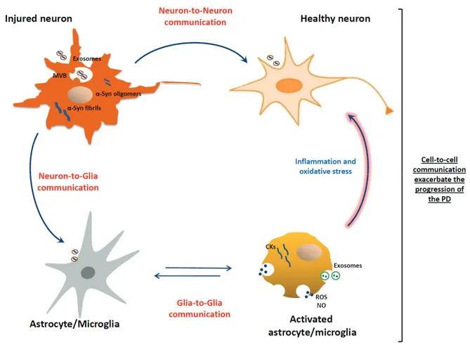
Figure 1. Exosomes in the cell-to-cell communications and their role in the progression of PD. Exosomes containing α-Syn released by injured neurons can be transmitted from neuron-to-neuron thus leading to α-α-Syn spreading, and from neuron-to-glia leading to activation of inflammatory response. In turn, exosomes released by activated glial cells, con-taining inflammatory mediators, can be transmitted from glia-to-glia leading to the propagation of inflammatory re-sponse. Consecutively, the propagation of inflammatory mediators and the exacerbated neuroinflammation could contribute to neuronal dysfunctions and to progression of the disease.
It has been reported that variation in tau gene (MAPT), which encoding for a protein involved in the pathogenesis of Alzheimer’s disease [78], can confer genetic risk for PD [79, 80]. In this regard, an interesting study reported that under pathological conditions tau protein can interact with α-Syn promoting the oligomerization and the toxicity of these proteins [81]. Taken to-gether these data suggest that tau can acceler-ate the exosomes-mediacceler-ated release containing α-Syn toxic forms from injured neurons.
Of interest, Vacuolar Sorting Protein 35 (VPS35), a protein that mutated is cause of late-onset autosomal dominant PD, is an essential component of the retromere complex, which performs the retrograde transport from the en-dosome to the Golgi apparatus. Sullivan et al. [82] observed that cells with defective retro-mere activity displayed increased exosomal se-cretion of amyloid precursor protein (APP). Al-though experimental models are not yet avail-able, we can predict that pathological mutations of VPS35 may lead to aberrant exosome secre-tion with consequent accumulasecre-tion of extracel-lular α-Syn and cell-to-cell propagation of the pathology. Establishing whether LRRK2, tau, and VPS35 operate within the exosome path-way might provide important clues on the patho-logical mechanisms of α-Syn spreading and pro-gression of the disease.
Acknowledgements
This work was supported by the PRIN (grant no. 2008-55YP79), the Rientro dei Cervelli Program (lncentivazione alla mobilita di studiosi stranieri e italiani residenti all’estero) from the Italian Ministry of Education, University and Research (EG), the Fondazione CARIPLO (grant no. 2011 0540) and the Micheal J Fox Foundation.
Address correspondence to: Dr. Isabella Russo or Dr. Elisa Greggio, Department of Biology, University of Padova, Via Ugo Bassi 58/B, 35121 Padova, ITALY. Tel: +390498276244; Fax: +390498276300; E-m a i l s : i s a b e l l a . r u s s o @ u n i p d . i t o r [email protected]
References
[1] Brundin P, Li JY, Holton JL, Lindvall O and Revesz T. Research in motion: the enigma of Parkinson's disease pathology spread. Nat Rev Neurosci 2008; 9: 741-745.
[2] Riess O, Kruger R and Schulz JB. Spectrum of phenotypes and genotypes in Parkinson's
dis-ease. J Neurol 2002; 249 Suppl 3: III/15-20. [3] Lees AJ, Hardy J and Revesz T. Parkinson's
disease. Lancet 2009; 373: 2055-2066. [4] Forno LS. Neuropathology of Parkinson's
dis-ease. J Neuropathol Exp Neurol 1996; 55: 259-272.
[5] Ali SF, Binienda ZK and Imam SZ. Molecular aspects of dopaminergic neurodegeneration: gene-environment interaction in parkin dysfunc-tion. Int J Environ Res Public Health 2011; 8: 4702-4713.
[6] Houlden H and Singleton AB. The genetics and neuropathology of Parkinson's disease. Acta Neuropathol 2012; 124: 325-338.
[7] Lee VM and Trojanowski JQ. Mechanisms of Parkinson's disease linked to pathological al-pha-synuclein: new targets for drug discovery. Neuron 2006; 52: 33-38.
[8] Lee HJ, Kim C and Lee SJ. Alpha-synuclein stimulation of astrocytes: Potential role for neuroinflammation and neuroprotection. Oxid Med Cell Longev 2010; 3: 283-287.
[9] Kim C and Lee SJ. Controlling the mass action of alpha-synuclein in Parkinson's disease. J Neurochem 2008; 107: 303-316.
[10] Giasson BI, Murray IV, Trojanowski JQ and Lee VM. A hydrophobic stretch of 12 amino acid residues in the middle of alpha-synuclein is essential for filament assembly. J Biol Chem 2001; 276: 2380-2386.
[11] Hardy J, Lewis P, Revesz T, Lees A and Paisan-Ruiz C. The genetics of Parkinson's syndromes: a critical review. Curr Opin Genet Dev 2009; 19: 254-265.
[12] Nalls MA, Plagnol V, Hernandez DG, Sharma M, Sheerin UM, Saad M, Simon-Sanchez J, Schulte C, Lesage S, Sveinbjornsdottir S, Stefansson K, Martinez M, Hardy J, Heutink P, Brice A, Gasser T, Singleton AB and Wood NW. Imputation of sequence variants for identification of genetic risks for Parkinson's disease: a meta-analysis of genome-wide association studies. Lancet 2011; 377: 641-649.
[13] Marques O and Outeiro TF. Alpha-synuclein: from secretion to dysfunction and death. Cell Death Dis 2012; 3: e350.
[14] Greggio E, Bisaglia M, Civiero L and Bubacco L. Leucine-rich repeat kinase 2 and alpha-synuclein: intersecting pathways in the patho-genesis of Parkinson's disease? Mol Neurode-gener 2011; 6: 6.
[15] Chinta SJ and Andersen JK. Dopaminergic neu-rons. Int J Biochem Cell Biol 2005; 37: 942-946.
[16] Damier P, Hirsch EC, Agid Y and Graybiel AM. The substantia nigra of the human brain. II. Patterns of loss of dopamine-containing neu-rons in Parkinson's disease. Brain 1999; 122: 1437-1448.
[17] Fearnley JM and Lees AJ. Ageing and Parkin-son's disease: substantia nigra regional selec-tivity. Brain 1991; 114: 2283-2301.
[18] Hirsch EC, Faucheux B, Damier P, Mouatt-Prigent A and Agid Y. Neuronal vulnerability in Parkinson's disease. J Neural Transm Suppl 1997; 50: 79-88.
[19] Double KL, Reyes S, Werry EL and Halliday GM. Selective cell death in neurodegeneration: why are some neurons spared in vulnerable re-gions? Prog Neurobiol 2010; 92: 316-329. [20] Hindle JV. Ageing, neurodegeneration and
Park-inson's disease. Age Ageing 2010; 39: 156-161.
[21] Korytowski W, Sarna T and Zar ba M. Antioxi-dant action of neuromelanin: the mechanism of inhibitory effect on lipid peroxidation. Arch Bio-chem Biophys 1995; 319: 142-148.
[22] Youdim MB, Ben-Shachar D and Riederer P. Is Parkinson's disease a progressive siderosis of substantia nigra resulting in iron and melanin induced neurodegeneration? Acta Neurol Scand Suppl 1989; 126: 47-54.
[23] Bisaglia M, Mammi S and Bubacco L. Kinetic and structural analysis of the early oxidation products of dopamine: analysis of the interac-tions with alpha-synuclein. J Biol Chem 2007; 282: 15597-15605.
[24] Conway KA, Rochet JC, Bieganski RM and Lans-bury PT Jr. Kinetic stabilization of the alpha-synuclein protofibril by a dopamine-alpha-synuclein adduct. Science 2001; 294: 1346-1349.
[25] Lawson LJ, Perry VH, Dri P and Gordon S. Het-erogeneity in the distribution and morphology of microglia in the normal adult mouse brain. Neuroscience 1990; 39: 151-170.
[26] Braak H, Ghebremedhin E, Rub U, Bratzke H and Del Tredici K. Stages in the development of Parkinson's disease-related pathology. Cell Tissue Res 2004; 318: 121-134.
[27] Braak H, Del Tredici K, Rub U, de Vos RA, Jansen Steur EN and Braak E. Staging of brain pathology related to sporadic Parkinson's dis-ease. Neurobiol Aging 2003; 24: 197-211. [28] Goedert M, Clavaguera F and Tolnay M. The
propagation of prion-like protein inclusions in neurodegenerative diseases. Trends Neurosci 2010; 33: 317-325.
[29] Fauvet B, Mbefo MK, Fares MB, Desobry C, Michael S, Ardah MT, Tsika E, Coune P, Prudent M, Lion N, Eliezer D, Moore DJ, Schneider B, Aebischer P, El-Agnaf OM, Masliah E and Lashuel HA. alpha-Synuclein in central nervous system and from erythrocytes, mammalian cells, and Escherichia coli exists predominantly as disordered monomer. J Biol Chem 2012; 287: 15345-15364.
[30] Bartels T, Choi JG and Selkoe DJ. alpha-Synuclein occurs physiologically as a helically folded tetramer that resists aggregation. Na-ture 2011; 477: 107-110.
[31] Wang W, Perovic I, Chittuluru J, Kaganovich A, Nguyen LT, Liao J, Auclair JR, Johnson D, Landeru A, Simorellis AK, Ju S, Cookson MR,
Asturias FJ, Agar JN, Webb BN, Kang C, Ringe D, Petsko GA, Pochapsky TC and Hoang QQ. A soluble alpha-synuclein construct forms a dy-namic tetramer. Proc Natl Acad Sci USA 2011; 108: 17797-17802.
[32] Volpicelli-Daley LA, Luk KC, Patel TP, Tanik SA, Riddle DM, Stieber A, Meaney DF, Trojanowski JQ and Lee VM. Exogenous alpha-synuclein fibrils induce Lewy body pathology leading to synaptic dysfunction and neuron death. Neuron 2011; 72: 57-71.
[33] Lesage S and Brice A. Parkinson's disease: from monogenic forms to genetic susceptibility factors. Hum Mol Genet 2009; 18: R48-59. [34] Spillantini MG, Crowther RA, Jakes R,
Hase-gawa M and Goedert M. alpha-Synuclein in filamentous inclusions of Lewy bodies from Parkinson's disease and dementia with lewy bodies. Proc Natl Acad Sci USA 1998; 95: 6469 -6473.
[35] Ebrahimi-Fakhari D, Cantuti-Castelvetri I, Fan Z, Rockenstein E, Masliah E, Hyman BT, McLean PJ and Unni VK. Distinct roles in vivo for the ubiquitin-proteasome system and the auto-phagy-lysosomal pathway in the degradation of alpha-synuclein. J Neurosci 2011; 31: 14508-14520.
[36] Li JY, Englund E, Holton JL, Soulet D, Hagell P, Lees AJ, Lashley T, Quinn NP, Rehncrona S, Bjorklund A, Widner H, Revesz T, Lindvall O and Brundin P. Lewy bodies in grafted neurons in subjects with Parkinson's disease suggest host-to-graft disease propagation. Nat Med 2008; 14: 501-503.
[37] Kordower JH, Chu Y, Hauser RA, Freeman TB and Olanow CW. Lewy body-like pathology in long-term embryonic nigral transplants in Park-inson's disease. Nat Med 2008; 14: 504-506. [38] Steiner JA, Angot E and Brundin P. A deadly
spread: cellular mechanisms of alpha-synuclein transfer. Cell Death Differ 2011; 18: 1425-1433.
[39] Aguzzi A and Rajendran L. The transcellular spread of cytosolic amyloids, prions, and prion-oids. Neuron 2009; 64: 783-790.
[40] Bellingham SA, Guo BB, Coleman BM and Hill AF. Exosomes: vehicles for the transfer of toxic proteins associated with neurodegenerative diseases? Front Physiol 2012; 3: 124.
[41] Schneider A and Simons M. Exosomes: vesicu-lar carriers for intercelluvesicu-lar communication in neurodegenerative disorders. Cell Tissue Res 2012.
[42] Thery C, Amigorena S, Raposo G and Clayton A. Isolation and characterization of exosomes from cell culture supernatants and biological fluids. Curr Protoc Cell Biol 2006; Chapter 3: Unit 3 22.
[43] Faure J, Lachenal G, Court M, Hirrlinger J, Chatellard-Causse C, Blot B, Grange J, Schoehn G, Goldberg Y, Boyer V, Kirchhoff F, Raposo G, Garin J and Sadoul R. Exosomes are released
by cultured cortical neurones. Mol Cell Neurosci 2006; 31: 642-648.
[44] Potolicchio I, Carven GJ, Xu X, Stipp C, Riese RJ, Stern LJ and Santambrogio L. Proteomic analy-sis of microglia-derived exosomes: metabolic role of the aminopeptidase CD13 in neuropep-tide catabolism. J Immunol 2005; 175: 2237-2243.
[45] Taylor AR, Robinson MB, Gifondorwa DJ, Tytell M and Milligan CE. Regulation of heat shock protein 70 release in astrocytes: role of signal-ing kinases. Dev Neurobiol 2007; 67: 1815-1829.
[46] Bobrie A, Colombo M, Raposo G and Thery C. Exosome secretion: molecular mechanisms and roles in immune responses. Traffic 2011; 12: 1659-1668.
[47] Simons M and Raposo G. Exosomes--vesicular carriers for intercellular communication. Curr Opin Cell Biol 2009; 21: 575-581.
[48] Lee TH, D'Asti E, Magnus N, Al-Nedawi K, Meehan B and Rak J. Microvesicles as media-tors of intercellular communication in cancer--the emerging science of cellular 'debris'. Semin Immunopathol 2011; 33: 455-467.
[49] Keller S, Sanderson MP, Stoeck A and Altevogt P. Exosomes: from biogenesis and secretion to biological function. Immunol Lett 2006; 107: 102-108.
[50] Thery C, Boussac M, Veron P, Ricciardi-Castagnoli P, Raposo G, Garin J and Amigorena S. Proteomic analysis of dendritic cell-derived exosomes: a secreted subcellular compartment distinct from apoptotic vesicles. J Immunol 2001; 166: 7309-7318.
[51] Kramer-Albers EM, Bretz N, Tenzer S, Winter-stein C, Mobius W, Berger H, Nave KA, Schild H and Trotter J. Oligodendrocytes secrete exosomes containing major myelin and stress-protective proteins: Trophic support for axons? Proteomics Clin Appl 2007; 1: 1446-1461. [52] Thery C, Zitvogel L and Amigorena S. Exosomes:
composition, biogenesis and function. Nat Rev Immunol 2002; 2: 569-579.
[53] van Niel G, Porto-Carreiro I, Simoes S and Ra-poso G. Exosomes: a common pathway for a specialized function. J Biochem 2006; 140: 13-21.
[54] Dello Sbarba P and Rovida E. Transmodulation of cell surface regulatory molecules via ectodo-main shedding. Biol Chem 2002; 383: 69-83. [55] Marzesco AM, Janich P, Wilsch-Brauninger M,
Dubreuil V, Langenfeld K, Corbeil D and Huttner WB. Release of extracellular membrane parti-cles carrying the stem cell marker prominin-1 (CD133) from neural progenitors and other epithelial cells. J Cell Sci 2005; 118: 2849-2858.
[56] Lachenal G, Pernet-Gallay K, Chivet M, Hem-ming FJ, Belly A, Bodon G, Blot B, Haase G, Goldberg Y and Sadoul R. Release of exosomes from differentiated neurons and its regulation
by synaptic glutamatergic activity. Mol Cell Neu-rosci 2011; 46: 409-418.
[57] Raposo G, Nijman HW, Stoorvogel W, Lie-jendekker R, Harding CV, Melief CJ and Geuze HJ. B lymphocytes secrete antigen-presenting vesicles. J Exp Med 1996; 183: 1161-1172. [58] Thery C, Ostrowski M and Segura E. Membrane
vesicles as conveyors of immune responses. Nat Rev Immunol 2009; 9: 581-593.
[59] Alvarez-Erviti L, Seow Y, Schapira AH, Gardiner C, Sargent IL, Wood MJ and Cooper JM. Ly-sosomal dysfunction increases exosome-mediated alpha-synuclein release and trans-mission. Neurobiol Dis 2011; 42: 360-367. [60] Emmanouilidou E, Melachroinou K, Roumeliotis
T, Garbis SD, Ntzouni M, Margaritis LH, Stefanis L and Vekrellis K. Cell-produced alpha-synuclein is secreted in a calcium-dependent manner by exosomes and impacts neuronal survival. J Neurosci 2010; 30: 6838-6851. [61] Desplats P, Lee HJ, Bae EJ, Patrick C,
Rocken-stein E, Crews L, Spencer B, Masliah E and Lee SJ. Inclusion formation and neuronal cell death through neuron-to-neuron transmission of al-pha-synuclein. Proc Natl Acad Sci USA 2009; 106: 13010-13015.
[62] Hansen C, Angot E, Bergstrom AL, Steiner JA, Pieri L, Paul G, Outeiro TF, Melki R, Kallunki P, Fog K, Li JY and Brundin P. alpha-Synuclein propagates from mouse brain to grafted dopa-minergic neurons and seeds aggregation in cultured human cells. J Clin Invest 2011; 121: 715-725.
[63] Zitvogel L, Regnault A, Lozier A, Wolfers J, Fla-ment C, Tenza D, Ricciardi-Castagnoli P, Ra-poso G and Amigorena S. Eradication of estab-lished murine tumors using a novel cell-free vaccine: dendritic cell-derived exosomes. Nat Med 1998; 4: 594-600.
[64] Chaput N, Flament C, Viaud S, Taieb J, Roux S, Spatz A, Andre F, LePecq JB, Boussac M, Garin J, Amigorena S, Thery C and Zitvogel L. Den-dritic cell derived-exosomes: biology and clini-cal implementations. J Leukoc Biol 2006; 80: 471-478.
[65] Bhatnagar S, Shinagawa K, Castellino FJ and Schorey JS. Exosomes released from macro-phages infected with intracellular pathogens stimulate a proinflammatory response in vitro and in vivo. Blood 2007; 110: 3234-3244. [66] Lee HJ, Suk JE, Bae EJ, Lee JH, Paik SR and Lee
SJ. Assembly-dependent endocytosis and clear-ance of extracellular alpha-synuclein. Int J Bio-chem Cell Biol 2008; 40: 1835-1849.
[67] Lee HJ, Suk JE, Patrick C, Bae EJ, Cho JH, Rho S, Hwang D, Masliah E and Lee SJ. Direct trans-fer of alpha-synuclein from neuron to astroglia causes inflammatory responses in synuclei-nopathies. J Biol Chem 2010; 285: 9262-9272.
[68] Halliday GM and Stevens CH. Glia: initiators and progressors of pathology in Parkinson's
disease. Mov Disord 2011; 26: 6-17.
[69] Vekrellis K, Xilouri M, Emmanouilidou E, Ride-out HJ and Stefanis L. Pathological roles of alpha-synuclein in neurological disorders. Lan-cet Neurol 2011; 10: 1015-1025.
[70] Alvarez-Erviti L, Couch Y, Richardson J, Cooper JM and Wood MJ. Alpha-synuclein release by neurons activates the inflammatory response in a microglial cell line. Neurosci Res 2011; 69: 337-342.
[71] Gao HM and Hong JS. Why neurodegenerative diseases are progressive: uncontrolled inflam-mation drives disease progression. Trends Im-munol 2008; 29: 357-365.
[72] Panaro MA and Cianciulli A. Current opinions and perspectives on the role of immune system in the pathogenesis of Parkinson's disease. Curr Pharm Des 2012; 18: 200-208.
[73] Shin N, Jeong H, Kwon J, Heo HY, Kwon JJ, Yun HJ, Kim CH, Han BS, Tong Y, Shen J, Hatano T, Hattori N, Kim KS, Chang S and Seol W. LRRK2 regulates synaptic vesicle endocytosis. Exp Cell Res 2008; 314: 2055-2065.
[74] Piccoli G, Condliffe SB, Bauer M, Giesert F, Boldt K, De Astis S, Meixner A, Sarioglu H, Vogt-Weisenhorn DM, Wurst W, Gloeckner CJ, Matte-oli M, Sala C and Ueffing M. LRRK2 controls synaptic vesicle storage and mobilization within the recycling pool. J Neurosci 2011; 31: 2225-2237.
[75] Xiong Y, Coombes CE, Kilaru A, Li X, Gitler AD, Bowers WJ, Dawson VL, Dawson TM and Moore DJ. GTPase activity plays a key role in the pathobiology of LRRK2. PLoS Genet 2010; 6: e1000902.
[76] Alegre-Abarrategui J, Christian H, Lufino MM, Mutihac R, Venda LL, Ansorge O and Wade-Martins R. LRRK2 regulates autophagic activity and localizes to specific membrane microdo-mains in a novel human genomic reporter cellu-lar model. Hum Mol Genet 2009; 18: 4022-4034.
[77] Fader CM, Sanchez DG, Mestre MB and Co-lombo MI. TI-VAMP/VAMP7 and VAMP3/ cellubrevin: two v-SNARE proteins involved in specific steps of the autophagy/multivesicular body pathways. Biochim Biophys Acta 2009; 1793: 1901-1916.
[78] Clavaguera F, Bolmont T, Crowther RA, Abramowski D, Frank S, Probst A, Fraser G, Stalder AK, Beibel M, Staufenbiel M, Jucker M, Goedert M and Tolnay M. Transmission and spreading of tauopathy in transgenic mouse brain. Nat Cell Biol 2009; 11: 909-913.
[79] Edwards TL, Scott WK, Almonte C, Burt A, Pow-ell EH, Beecham GW, Wang L, Zuchner S, Koni-dari I, Wang G, Singer C, Nahab F, Scott B, Sta-jich JM, Pericak-Vance M, Haines J, Vance JM and Martin ER. Genome-wide association study confirms SNPs in SNCA and the MAPT region as common risk factors for Parkinson disease. Ann Hum Genet 2010; 74: 97-109.
[80] Satake W, Nakabayashi Y, Mizuta I, Hirota Y, Ito C, Kubo M, Kawaguchi T, Tsunoda T, Watanabe M, Takeda A, Tomiyama H, Nakashima K, Hase-gawa K, Obata F, Yoshikawa T, Kawakami H, Sakoda S, Yamamoto M, Hattori N, Murata M, Nakamura Y and Toda T. Genome-wide associa-tion study identifies common variants at four loci as genetic risk factors for Parkinson's dis-ease. Nat Genet 2009; 41: 1303-1307. [81] Giasson BI, Forman MS, Higuchi M, Golbe LI,
Graves CL, Kotzbauer PT, Trojanowski JQ and Lee VM. Initiation and synergistic fibrillization of tau and alpha-synuclein. Science 2003; 300: 636-640.
[82] Sullivan CP, Jay AG, Stack EC, Pakaluk M, Wad-linger E, Fine RE, Wells JM and Morin PJ. Retro-mer disruption promotes amyloidogenic APP processing. Neurobiol Dis 2011; 43: 338-345.