Molecular Oncology
University of Pisa
Establishment and Characterization of
Four New Human Pancreatic Cancer Cell
Lines:
Proposal for an
in vitro
Research Platform
Supervisor:
Prof. Generoso Bevilacqua
Tutors:
Dr. Andrea Cavazzana
Student:
Barbara Chifenti
Dr.ssa. M.Teresa Locci
A mia sorella Cecilia,
a Lucia, Denise, a tutti i miei mici-amici e a me stessa
INTRODUCTION………...1
The normal Pancreas………...1
Pancreatic Cancer………...6
Classification of Pancreatic Cancer………..6
Epidemiology………7
Incidence and mortality……….7
Risk Factors………8
PDCA Morphological Characteristic and Progression Model……….10
Pancreatic desmoplastic reaction………10
The “Adenoma-Carcinoma Sequence” ………… 14
PDCA Molecular Genetic………..19
Oncogenes in PDAC………..20
KRAS………..20
Tumor Suppressor Genes in PDAC……….28
CDKN2A……….28
TP53………35
SMAD4………...44
Origin of Pancreatic Cancer……….52
Experimental in vivo and in vitro models 58
In vivo models………..59
Mouse model: xenografts………59
Mouse model: genetically engineered mice (GEM) ………60
MATERIALS AND METHODS………...……..….65
Establishment of pancreatic cancer cell lines ...65
Serial passage and storage in vitro ……….65
RT-PCR Analysis………66
Mutational Analysis of TP53, KRAS, CDKN2A, SMAD4 and BRAF genes……….66
DNA-fingerprinting analysis………...67
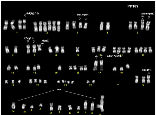
Kariotype analysis………...68
Cytoskeleton filaments………...69
Western Blot analysis………...69
Assay of Ras and Ras effectors………....70
Ras-GTP Assay………..…71
In vitro Migration assay………..….71
Bromodeoxyuridine (BrdU) assay……….…..71
Proliferation assays………..72
RESULTS………...……….………...….73
Primary tumor samples………...73
In vitro Morphology...73
Growth Kinetics and Doubling Time …….…….……75
DNA fingerprinting profile…………...….75
and BRAF analysis. ……….…82
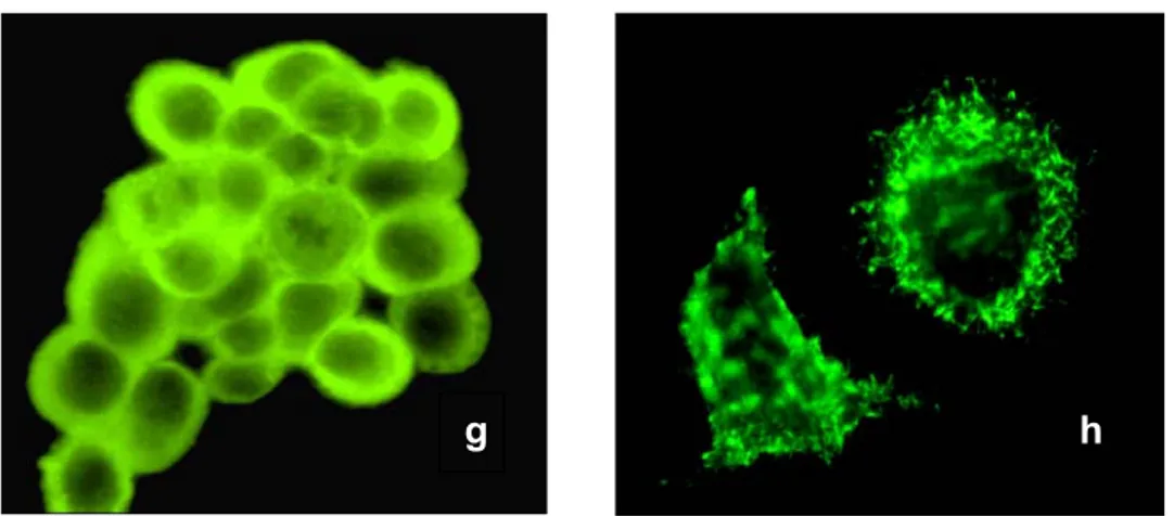
Cytoskeleton filaments pattern ………...83
Western Blot analysis………...85
Ras/MAPK-inase and AKT Pathway Analysis...86
Migration assay………....…..87
DISCUSSION……….…..88
INTRODUCTION
The normal Pancreas
The name Pancreas derives from the Greek roots ‘pan’ meaning ‘all’ and ‘creas’ meaning ‘flesh’. The pancreas, gland of endodermal origin, is the key regulator of protein and carbohydrate digestion and glucose homeostasis (Hezel AF et al, 2006). The pancreas is located deep in the abdomen, with the liver, intestine, and other organs surrounding it. The pancreas is divided into three parts: head, body, and tail. The head is the thicker part of the organ and borders the duodenum, the common bile duct and the right renal artery and vein (Fig.1). The middle section, the body, and the thinnest part, the tail, of the organ are elongated in shape and extend to the left anterior to the aorta, and posterior to the stomach.
Functionally, the pancreas is a composite exocrine-endocrine gland: the prevalent exocrine portion forms up to the 84 percent of the pancreas volume, blood vessel and duct cells represent about 4 percent, and the endocrine component is only 2 percent. The remaining (about 10 percent) is formed by extracellular matrix.
The Exocrine Pancreas is a lobulated gland composed of a branching network of acinar and duct cells that produce and deliver digestive zymogens into the gastrointestinal tract (Slack JMW et al, 1995). The secretory acinar cells are grouped into functional units termed acini dislocated along the duct network. The acinar cells are pyramidal in shape with a broad base and a narrow luminal face (Fig.2). In the apical cytoplasm, they contain zymogen granules that contain a number of digestive enzymes in their inactive form, including trypsinogen, chymotrypsinogen, ribonuclease, and amylase. In response to cues from the stomach and the duodenum acinar cells secrete zymogens into the ductal lumen. The duct system in the pancreas begins within the acini themselves. (Grapin-Botton A , 2005). A narrow intercalated (intralobular) duct is deeply inserted into each secretory mass, the initial part of the duct wall being lined by low cuboidal centroacinar cells thought to secrete non-enzymatic components of the pancreatic juice. More distally these cells are replaced by taller cuboidal, and eventually columnar cells. The larger ducts are interlobular in position, and are surrounded by loose connective tissue which contains some nonstriated muscle fibres, and autonomic nerve fibres (Gray’s Anatomy, Longman 35th edition, Edhinburg, 1975). The interlobular ducts drain directly into the duct of
Wirsung, the main pancreatic duct. It runs the length of the pancreas parallel to its long axis and joins the common bile duct before entering the duodenum at the papilla of Vater.
The acini and smaller ducts are invested with a delicate, loose connective tissue, which becomes more extensive around the larger ducts.
Fig.2 Acinar Unit
The Endocrine Pancreas regulates metabolism and glucose homeostasis through the secretion of hormones into the bloodstream. The endocrine cells are mainly grouped into the Langerhans islets (about 2% of the pancreas), which are compact spheroidal clusters embedded in to exocrine tissue (Fig.3). There are four major cell types that have been definitively identified in the islet. The β (or B) cells are the most numerous of the islet cell types. They synthesize, store, and secrete insulin, and also an insulin antagonist called amylin.
The
α
(or A) cells constitute 15 to 20 percent of the islet cells and they secrete glucagon. The δ (or D or A1) cells are mainlydistributed at the islet periphery and secrete somatostatin. The PP (or F) cells secrete pancreatic polypeptide, and PP-rich islet are found particularly in the posterior part of the head of the pancreas. All islet cell types also express a number of products characteristic of neuroendocrine cells, such as neuron-specific enolase, tetanus toxin receptor, and the homeodomain-LIM protein islet-1.
Fig.3 Endocrine Pancreas
In addition to the glandular components, the pancreas has a rich blood supply, an extensive lymphatic drainage, and a rich sympathetic and parasympathetic nerve supply.
The main distinctive markers of the normal pancreatic cells are the Cytoskeleton Intermediate Filament (IF) proteins (Bouwens L, 1998; Schussler MH et al, 1992). Keratins (CK), the major IF proteins of epithelial cells, comprise a family of nearly 20 different polypeptides and are classified into two subfamilies according to the Moll’s catalogue (Moll R at al,
1982): type I CK (keratins 9-20) are acidic and have a low molecular weight; type II CK (keratins 1-8) are neutral to basic. Keratin filaments contain at least one member from the type I subfamily and one member from the type II subfamily (Chu PG et al, 2002), and the different pairs of keratins are expressed in a tissue-specific manner. This finding means that a given specific epithelial cell can be characterized by the specific pattern of its keratin components.
Keratin immunohistochemistry is a very useful tool for identifying different cell types in the pancreas. The normal pancreas is composed of simple, non-stratified epithelia and so it expresses the typical combination of keratin 8 and 18, and keratin 7 and 19. The acinar cells express CK 8 and 18, while CK 7 and 19 are not express in this type of cells. Centroacinar cells and ductal cells share a similar CK pattern: CK 7, 8, 18, and 19 are in fact consistently and homogeneous expressed. Keratin staining is more intense in exocrine than in endocrine cells, with the highest levels of staining in the larger ducts. In the endocrine pancreas, islet cells constantly express only CK8 and CK18. The main distinctive cytokeratin proteins of pancreatic cells are listed in Table N.1
Table N.1: Expression of CKs in Normal Pancreas
Cell type CK 8,18 CK 7,19
Ductal + +
Centroacinar + +
Acinar + -
Pancreatic Cancer
Classification of Pancreatic Cancer
The Pancreas is a particularly important organ from the point of view of human medicine because it suffers from two important disease: diabetes mellitus and pancreatic cancer.
‘Pancreatic cancer’ is a broad term to describe as many as 20 different types of tumor that can occur in the pancreas. Pancreatic cancer is categorised as being either endocrine or non-endocrine (exocrine). The phenotypic classification of pancreatic neoplasms is based on their cellular lineage, thus tumor with a ductal, acinar, and endocrine phenotype can be distinguished.
Endocrine tumors represent only about one percent of all pancreatic cancers. They affect the hormone-producing cells of the pancreas and can have dramatic effects on the amounts of hormones produced. These type of tumors regard, for example, Insulinomas, which can cause excessive insulin production resulting in low blood sugar, and Glucagonomas, which may cause excessive glucagons production, resulting in a distinctive skin rash.
Exocrine tumors make up the majority of pancreatic cancer (more than 90 percent). Most pancreatic neoplasms show a ductal line of differentiation and therefore classified as ductal adenocarcinomas. The fine structure of PDAC cells resembles that of the pancreatic duct cells. The luminal surface
of the cells shows microvilli; the apical cytoplasm contains mucin granules, and the basal part of the cells contains a round nucleus with a small distinct nucleolus. The morphologic features of this tumor include infiltrating duct-like and tubular structures embedded in a high desmoplastic stroma. The tumor cells, like the ductal cells of the normal pancreas, produce mucins. Other duct cells markers that are typically found in PDAC are the cytokeratin 7, 8, 18, and 19, CA 19.9, and CEA. Pancreatic Ductal Adenocarcinoma (PDAC) is the most common form of pancreatic cancer comprising 85 to 90 percent of all pancreatic tumours. Less common tumors with a ductal phenotype are the variants of ductal adenocarcinoma, intraductal papillary mucinous neoplasm (including colloid carcinoma), mucinous cystic neoplasm, medullary carcinoma, and other rare tumors (Kloppel G et al, 2004).
Epidemiology
Incidence and mortality
PDAC is characteristically a tumour of elderly individuals: approximately 80 per cent of cases occurs in patients between the ages 60 to 80; patients below the age of 40 are rar (Lüttges J, et al 2004).
PDAC ranks eight in a worldwide ranking of cancer deaths, (Lowenfels AB et al, 2006) while with respect to incidence, it ranks 13th being then considered a relatively rare tumor. PDAC is one of the most lethal human cancers and continues to be a major unsolved health problem at the start of the 21st century
(Li D et al, 2004). The poor 5-year survival rate (5%) and the median survival of less than 6 months are largely the result of PDAC’s silent nature and of a late diagnosis (Sakorafas et al, 2000). The vast majority of patients do not exhibit specific symptoms until the disease is advanced. Specific symptoms usually only develop after invasion or obstruction of nearby structures. As most pancreatic cancers arise in the head of the pancreas, obstruction of the biliary tree resulting in jaundice is the hallmark presentation (Lillemoe KD et al, 2000) .
Moreover close to 100% of patients with pancreatic cancer develop metastases at the time of diagnosis, as a consequence, only 20% of pancreatic cancer patients are eligible for radical surgical resection, which currently remains the only potentially curative therapy.
Conventional therapeutic cancer strategies such as chemotherapy and radiotherapy have failed to significantly improve the prognosis of advanced pancreatic cancer, due to its unusual resistance to chemotherapy and radiation therapy. These information indicate that current interventions to prevent, diagnose, and cure the disease are still far from being satisfactory.
Risk factors
Large studies and country specific differences in cancer frequency, have showed that environmental factors are the most important cause of most type of cancer, including pancreatic cancer.
Data from early epidemiologic studies of pancreatic cancer (Wynder EL et al, 1973) showed that carcinogens derived from
tobacco, occupational or other environmental sources might enter the bile and then cause pancreatic cancer by reflux through the pancreatic duct. This hypothesis has never been disproved and could explain the excess of pancreatic cancer known to occur in the head of the pancreas. Current data confirm only three types of risk factors linked with pancreatic cancer: cigarette smoking, chronic pancreatitis and diabetes (Li D et al, 2004).
About 30 percent of cases of pancreatic cancer are attributable to smoking (de Braud F et al, 2004). Cigarette smokers develop this disease 2-3 times more than non-smokers, while exposure to environmental smoking (passive smoking) cause a non-significant increase in the risk of pancreatic cancer. The harmful effect of smoking is mainly related to mutagens present in the metabolites of tobacco smoke (Lowenfels AB et al, 2006). Recent findings from large-scale population-based case-control studies support the hypothesis that individuals who have deficient detoxifying and/or DNA repair systems are at increase risk of pancreatic cancer.
In Western countries the most common cause of chronic pancreatitis is heavy alcohol consumption. Cohort studies have confirmed a link between pancreatitis and pancreatic cancer. Another cause of chronic pancreatitis is hereditary pancreatitis. This is a rare inherited autosomal disease with an onset in childhood or early adulthood. Patients with hereditary pancreatitis experience a 53-fold increased incidence of pancreatic cancer (Bardeesy N et al, 2002). The link between chronic pancreatitis and pancreatic cancer has not fully
investigated at the molecular level. Increased cell turnover, defective DNA repair, loss of p16 expression along with K-ras gene mutations, have been found in nearly all pancreatic cancer and also in chronic pancreatitis samples.
Diabetes is a disorder found in 5-10 % of the general population. A recent study found a modest relationship between longstanding diabetes of >5 years and pancreatic cancer, while the higher risk was found for less than 5 year diabetic patients. These studies are biased since diabetes can be an early manifestation of pancreatic cancer.
PDCA Morphological Characteristic and
Progression Model
Pancreatic desmoplastic reaction
The importance of stromal interactions with epithelial cells is well established in embryonic development and tumorigenesis. The concept of a link between stromal and adjacent epithelial cells was introduced more that 20 years ago (Cunha GR, et al, 1980). The interaction is mediated by soluble paracrine signals and extracellular matrix (ECM) components secreted from developing mesenchimal cells that induce adjacent epithelial cells to proliferate rapidly (Bissel MJ at al, 2001). Therefore tissue differentiation appears to be a coordinate process involving both epithelial and mesenchimal cells . Differentiated stromal cells generally express lower quantities of growth factors, and differentiated epithelia express cytokines for the
maintenance of stromal differentiation, suggesting that a new balance of mesenchimal-epithelial crosstalk is reached during maturation. (Bhowmick NA et al. 2004). This fine tuning of epithelial/stromal interactions is deeply disturbed during tumorigenesis so that tumor microenvironment may influence tumor characteristics such as tumor differentiation, growth rate and drug sensitivity. On the other hand, tumor cells can regulate the development of the tumor stroma by aberrant expression of growth factors or induction of growth factor receptors in stroma compartment (Micke P and Ostman A, 2004).
In many human tumors the stroma microenvironment is both morphologically and biologically different from the corresponding normal stroma. This “reactive stroma” is characterized by the presence of fibroblasts with an “activated” phenotype embedded in a modified ECM with an increased microvessel density and presence of inflammatory cells. These modified fibroblasts, often termed myofibroblasts, reactive stroma or cancer-associated fibroblasts (CAFs), are considered to play a central role in the complex process of tumor-stroma interaction and consequently tumorigenesis (Desmouliere A et al, 2004).
Extreme representations of the desmoplastic reaction is particularly evident in certain tumor types, such as scirrhous breast cancer, biliary and pancreatic carcinomas. A pathologic characteristic of pancreatic cancer is, in fact, a pronounced desmoplastic (stromal) reaction around tumour tissue (Luttges J et al, Med Klin 2004). Neoplasms like pancreatic carcinoma are in fact typically composed of infiltrating ductlike structures
surrounded by a dense fibrous or desmoplastic stroma. Rather unique to ductal adenocarcinoma is the consistently low ratio of the infiltrating adenocarcinoma cells relative to the abundant desmoplastic stromal environment, with pancreatic cancer cells represent on average 25% of the cells in the tumor mass (Apte MV et al, Pancreas 2004). Pancreatic desmoplastic reaction is composed of connective tissue (predominantly collagen type I and other glycoproteins) and new blood vessels and is associated with proliferation of fibroblastic cells. The reactive stroma itself contains a variety of cells including proliferating fibroblasts, small endothelial-lined vessels, inflammatory cells and residual parenchymal components of the invaded organ. It is now clear that host stromal reaction is far from being a passive actor in the neoplastic process and may strongly influence the behaviour of neoplastic cells (Iacobuzio-Donahue CA, Ryu B, Hruban RH, Kern SE, 2002). Cytokines, growth factors and angiogenic factors released by these cells may positively influence tumor growth, metastatic capabilities and contribute to drug resistance (Kopfstein L, Christofori G, 2006).
Fig.4 Crosstalk between tumour cells and their activated stroma
Despite the importance of tumor-stroma interaction in cancer progression, the underlying molecular mechanisms have not been yet well characterized. Several molecules have been identified to participate in tumor-stroma interaction including Hepatocyte growth factor (HGF), Interleukin-8 (IL-8) SPARC/osteonectin, Transforming Growth Factor Beta, several Matrix Metalloproteinases (MMPs) and reactive oxygen species (COX) (Müerköster S, et al 2004). Moreover, different gene expression profiles have been generated by experiments done using co-cultures of pancreatic cancer cells and stromal fibroblasts (Fig.4). These experiments confirmed the existence of and active cross-talk between cancer and normal cells (Sato N, Maehara N and GogginsM, 2004). The possibility to identify the molecular pathways involved in this cross-talk may allow us to provide new targets susceptible of therapeutic intervention
(Corsini C et al, 2003). In fact, stromal therapy is emerging as
Irradiated fibroblasts can cause activation of c-Met oncogene in pancreatic cancer cells enhancing its invasiveness while the administration of a specific antagonist of c-Met may block this effect (OhuchidaK, et al, 2004). A phase-I dose-escalation trial recently published (Scott AM, et al , 2003) utilizing humanised monoclonal antibodies (sibrotuzumab) against the fibroblast activating protein (FAP) showed promising results in controlling colorectal cancer.
The “Adenoma-Carcinoma Sequence”
Invasive PDAC is an almost uniformly fatal disease also because of lack of effective screening test for early pancreatic cancer. A better understanding of the pathogenesis of pancreatic cancer and more effective screening techniques are required to improve current survival rates. To this purpose it is important to know the characteristics of pre-cancerous lesions of PDC because they provide a unique opportunity to understand the early events in the development of an infiltrating carcinoma, and a better understanding of these events, in turn, may lead to the development of novel diagnostic techniques to detect this cancers in early stages. (Brat DJ et al, 1998).
The first evidence that there may be a distinctive precursor lesion to infiltrating adenocarcinoma of the pancreas came from morphological studies. In 1976 Cubilla and Fitzgerald identified histologically distinct proliferative non-invasive lesions in the pancreatic ducts and ductules adjacent to infiltrating adenocarcinoma of the pancreas. They called these duct lesion “hyperplasia” and showed that they were more commonly found
in tissues adjacent to cancer than in fully healthy organ. Thus, morphological observation provided the first critical step in the recognition that intraductal proliferations in the pancreas could represent the morphologic precursors to invasive ductal cancer. These precursor lesions were later termed pancreatic intraepithelial neoplasia (PanIN),( Hruban RH et al, 2004; Hruban RH et al, 2000) by the National Cancer Institute-sponsored Pancreatic Think Tank, and they were graded from stage I to stage III (PanIN-1A to PanIN-3) (Fig.5). PanIN-1A are characterized by the transition from cuboidal to tall columnar cells with still basally located non-atypical nuclei. PanIN-1B is cytologically indistinguishable from PanIN-1A, but show a distinct papillary or pseudo-stratified architecture. PanIN-2 lesions manifest architecturally as flat or papillary lesions but they show a higher degree of nuclear atypia. Finally the PanIN-3 lesions (also known as “carcinoma in situ”) bear a cytologic resemblance to infiltrating carcinoma despite the fact they are still contained within the ductal basal lamina. The cells are characterized by profound nuclear atypia, prominent nucleoli and mitotic activity (Baumgard M et al, 2005; Takaori K et al, 2004; Hruban RH et al, 2000).
Fig.5 Histologic Progression Model for Pancreatic Cancer.
These morphological observations were, however, static, and there was not universally agreement upon whether these pancreatic duct lesions could represented the true intraductal phase of an invasive cancer (Luttges J et al, 1999). Molecular analyses have provided strong evidences that pancreatic intraductal lesions are indeed the precursors of infiltrating adenocarcinomas.
Most genetic abnormalities commonly identified in infiltrating ductal adenocarcinoma, such as k-ras, p16, p53, and DPC4, have also been found in PanIN duct lesions. Moreover molecular studies have helped to define the time axis of the progression of the PanIN lesions to invasive PDAC showing that the genetic mutations that take place in the precursors lesions appear to occur in a temporal sequence rather than a random fashion (Vimalachandran D et al, 2004), so that relative frequencies of a given molecular abnormality increases with the increasing grade of the PanINs with an overall cumulative effect. These findings shape a progression model similar to that described for colorectal cancer. K-ras mutations are the first molecular abnormality detectable in PanIN – Carcinoma
Normal PanIN-1A PanIN-1B PanIN-2 PanIN-3 Infiltrating
sequence being also found in the adjacent normal-appearing pancreatic ducts (Luttges J et al, 1999). K-ras mutation rate increases up to virtually all PanIN-3 lesions and infiltrating adenocarcinomas, much to be considered a sort of “signature” of pancreatic cancer. Telomerase activity is significantly higher in invasive lesions compare to normal adjacent structures, nevertheless it is peculiar that this high activity is preceded by a shortage of telomere that have been documented in all types of pre-invasive lesions. (Heek NT et al, 2002). Considering the fundamental role exerted by telomeres in normal chromosomal separation this finding may predispose these non-invasive ductal lesions to accumulate progressive chromosomal abnormalities and to develop toward the stage of invasive carcinoma.
The second step include p16, p53, DPC4, and BRCA2 inactivation. Loss of p16 INK4A is found to be the next hit, occurring slightly later than K-ras (Fig.6). Allelic loss has been detected in PanIN-2, and inactivation has been found up to 100% in invasive cancer (Moskaluk CA et al, 1997). The p53 gene inactivation is a later event, usually occurring in PanIN-3 lesion and in near 75% of infiltrating PDAC. Inactivation of DPC4 and BRCA2 genes appear to be a late event in genetic progression of pancreatic cancer, occurring in histologically high- grade lesions and infiltrating PDAC. The validity of this histologic-genetic progression model is also been confirmed by a study of the proliferative rate of the epithelial cells in the successive grades of PanINs (Klein WM et al, 2002). The Ki-67 expression was used as a proliferative marker and correlated with the PanIN lesions histology. Data from this study showed
that the Ki-67 labelling rate parallel an increasing grade of dysplasia, leading to a better understanding of the pancreatic progression model.
Fig.6 Histologic-Genetic Progression Model for Pancreatic Cancer.
Normal PanIN-1A PanIN-1B PanIN-2 PanIN-3 Infiltrating
carcinoma
K-ras Shortened
telomeres p16 p53; DPC4
BRCA2
PDCA Molecular Genetic
A careful molecular analysis of infiltrating PDAC revealed that this carcinoma, like other cancers, showed a characteristic pattern of genetic lesions comprising activation of oncogene and inactivation of tumor suppressor genes (Fig.7). PDAC it is characterized by a well-established molecular fingerprint comprising activation of K-ras oncogene (90%) of cases and inactivation of tumor suppressor genes like p16, p53, and DPC4 (90%, 60%, and of 50% cases respectively).
Fig.7 Major pathways in the development of pancreatic adenocarcinoma.
Oncogenes in PDAC
Oncogenes are defined as dominant genes that can induce or maintain cellular transformation. They are often related to normal cellular genes, called proto-oncogenes, which have critical functions in cellular growth or differentiation. Oncogenes may be activated in various ways, by point mutations, amplification and chromosomal translocation. Most oncogenes code for proteins that are thought to be major players in the control of cell growth.
KRAS
The mammalian ras gene family consist of three members: H-ras, K-H-ras, and N-H-ras, were originally identified in the mild-1960s as the transforming elements of the Harvey and Kirsten strains of rat sarcoma viruses. H-ras and K-Ras oncoproteins were identified as viral (v-Ras) oncoproteins of Harvey and Kirsten murine sarcoma viruses, and were found to be capable of cellular transformation. The N-Ras oncoprotein was identified in a neuroblastoma cell line (Macaluso M et al, 2002). H-ras, K-Ras, and N-Ras genes are located in different chromosomes, 11p15.5, 12p12.1, and 1p13, respectively. RAS-family proteins are low-molecular-weight guaninenucleotide-binding proteins and the three ras genes encode four highly related p21kDa GTPases of 188 (K-RasB) or 189 (K-RasA, H-Ras, and N-Ras) amino acids in length. The A and B forms of K-Ras are generated by an alternative splicing of the fourth exon of this gene, and the abundance of the K-RasB transcripts is higher in comparison of that of K-RasA transcripts. Ras is ubiquitously expressed although mRNA analysis suggests different tissue expression
levels. H-Ras is highly expressed in the skin and in skeletal muscles, K-Ras is mostly expressed in the colon and the thymus, and N-Ras in male germinal tissue and the thymus. Ras proteins are essential components of receptor-mediated signal transduction pathways that control cell proliferation and differentiation (Bar-Sagi D, 2001). Ras is synthesized as a biologically inactive cytosolic propeptide (Pro-Ras) and is localized to the inner surface of the plasma membranes only after it has undergone a series of closely linked posttranslational modifications at the C-terminus, thereby increasing its hydrophobicity and facilitating its association with the plasma membrane where they can interact with their upstream activators and downstream targets. The association of Ras proteins with the plasma membrane involves a series of posttranslational modifications in their C termini. These modifications increase the hydrophobicity of the C terminal region of the proteins allowing its inner in plasma membrane (Hancock JF, 2003; Prior IA et al, J 2001 ) . All the Ras proteins contain in fact a terminal CAAX box in the 186-189 position. In this box, the “C” represents cysteine, “A” represents an aliphatic amino acid (leucine, serine, or valine) and “X” is metionine, serine, leucine, or glutamine. The first step in this process is the farnesylation of the cysteine in the CAAX box at the C-terminal end. Subsequently, the AAX amino acids are cleaved off by Ras-converting enzyme I, and then the farnesylated cysteine is carboxymethylated by isoprenylcysteine carboxyl methyltransferase. Upon palmitoylation (HRas, N-Ras, and KRas-4A) or through the presence of a polybasic domain (KRas-4B), the protein is anchored in the cell membrane (Chiu ViK et al, 2002) (Fig.8).
Fig.8 Posttranslational modification of Ras proteins (from Hancock JF,2003).
Ras proteins function is regulated by cycling between inactive GDP-bound state and active GTP-bound forms, and this ability makes them ideal signal transducers (Joneson T, 1997). In quiescent cells, RAS exists predominantly in the GDP-bound state. Ras proteins are activated transiently in response to a diverse array of extracellular signals such as growth factors, cytokines, hormones and neurotransmitters that stimulate cell surface receptors, and integrins. The best-studied activation mechanism involves the assembly of complexes of activated, autophosphorylated growth-factor receptor tyrosine kinases with the GEF SOS through the adaptor protein GRB2, and possibly SHC, resulting in the recruitment of SOS to the plasma membrane, where RAS is located (Downward J , 2003).
Fig.9 Signalling upstream of RAS (from Downward J, 2002).
In the GDP-bound form they are inactive, but upon replacement of bound GDP with fresh GTP they undergo an activating conformational change (Fig.9). The intrinsic GDP/GTP exchange activity of Ras is very low and hence increase in GDP/GTP nucleotide exchange involves interaction between Ras and the regulatory protein guanine exchange factors (GEFs) (Campbell SL et al, 1998). Once activated, Ras is able to interact with and activate effector proteins, which in turn stimulates signalling cascades that induce a variety of cellular responses designed to prolong cell survival and promote cell proliferation. Ras-GTP exist for only a short time because it is subsequently inactivated by a class of proteins known GTP-ase-activating proteins (GAPs), which enhance the Ras intrinsic low GTPase activity (Singh A et al, 2005).
GTP-bound RAS is able to bind and activate several families of effector proteins, resulting in stimulation of their catalytic activity. The main effectors are the RAF–MEK– ERK/MAPK, Phosphatidylinositol 3-kinase (PI3K) pathways, RAL, and Phospholipase Cε (PLCε) (Shields JM et al, 2000).
The first mammalian effector of RAS to be characterized is the protein serine/threonine kinase RAF. GTP-bound RAS binds to, and activates the three closely related RAF proteins, c-RAF1, BRAF, and ARAF. This interaction cause RAF to be relocated to the plasma membrane, which is crucial for its activation. Downstream of this, activated RAF, phosphorylates and actives activates the dual-specificity mitogen- tyrosine/threonine kinase MEK (MEK1 and MEK2). Active MEK then phosphorylates ERK/MAPK, which translocates to the nucleus and activates a variety of transcription factors. As a result of stimulating these transcriptional regulators, protein such as D-type cyclins are expressed. So RAF activation can promote cell-cycle progression, at least in conjunction with other signals (Fig.10).
RAS can interact directly binds and activates the catalytic subunit of Phosphatidylinositol 3-kinase (PI3K), a lipid kinase that generates phosphatidylinositol 3,4,5- trisphosphate (PtdIns(3,4,5)P3) second messengers, which bind to a large number of proteins through the plecktrin homology and other domains. In this way PI3K controls the activity of a large number of downstream enzymes such as phosphoinositide-dependent kinase-1 (PDK1) and AKT/protein kinase B (AKT/PKB) kinases . AKT/PKB has a strong anti-apoptotic function by phosphorylating various targets and is an important part of the survival signal that is generated by RAS. In addiction, PI3K activation leads to stimulation of RAC, a RHO family protein that is involved in the regulation not only of the actin cytoskeleton, but also of transcription-factor pathway-for example nuclear factor kB (NF-kB).
RALGDS proteins are guanine nucleotide exchange factors (GEFs) for RAL, a RAS-related protein. Through these proteins, RAS is able to stimulate RAL, resulting in control of downstream effectors such as the inhibition of Forkhead transcription factors of the FoxO family. These have been implicated in promoting cell-cycle arrest through KIP1 (p27), and apoptosis.
Phospholipase Cε (PLCε) is another RAS effector that as been reported recently. It catalyses the hydrolysis of phosphatidylinositol-4,5-bisphosphate to diacylglycerol and inositol trisphosphate, resulting in protein kinase C (PKC) activation and calcium mobilization from intracellular stores.
Fig.10 Signalling downstream of RAS (from Downward J, 2002). Activating point mutations of ras genes is the most commonly event detected in human tumors, occurring in approximately 20% of all cancers. With regard to the three ras genes, mutation of K-RAS is the most commonly found (about 85% of total), whereas N-RAS mutations are encountered less often (about 15%) and H-RAS mutations rarely (less than 1%) (Downward J, 2003). KRAS mutations are one of the most
common mutational events in the carcinogenesis of human malignancies. It has been estimated that as many as 75-90% of pancreatic cancer, 50% of colorectal carcinoma, and 25% of non-lymphocytic leukemias have ras gene mutations. having mutations in human cancer samples. RAS mutations are somatic rather than in the germ-line, and consist of single base-pair substitutions which lead to the change of one amino acid in the protein. The hotspots mutations are located at codon 12 , codon 13, and, with a low frequency, at codon 61. All these mutations involve amino acid substitution near the bound nucleotide making RAS protein insensitive to GAPs. This results in a RAS mutant protein persistently activated in the absence of extracellular signalling . Mutations in codon 12 or in codon 13 reduces the intrinsic GTPase activity of the ras gene product, but mutation in codon 61 results in ras protein with no catalytic activity at all. The reason for the specificity of these mutations to codon 12 is not entirely clear and may be due to etiologic agents binding preferentially to this codon and to poor repair of adducts formed at this codon compared with DNA damage occurring at other codons. However, a closer look at the mutation spectrum of K-ras suggest that the explanation may be more complex (Howe JR et al, 1997).
The highest incidence of KRAS mutations has been found in PDAC, with a range of K-ras mutations from 60% to 100% of cases. Activating KRAS mutations are the first genetic changes that are detected in the pancreatic cancer progression series, occurring occasionally in histologically normal pancreas and in about 30% of lesion that show the earliest stages of histological disturbance (Bardeesy N et al, 2002). KRAS mutations increase
in frequency with disease progression, and are found in nearly all 100% PDAC. The KRAS mutations in PDAC are almost exclusively at K-ras codons 12 and 13 and , to a lesser extent, at codon 59, 61, and 63 (Deramaudt T et al, 2005).
K-RAS point mutations have been also described in the majority of pancreatic ductal adenocarcinoma cancer cell lines that are commonly used in laboratory-based investigation and xenografts models in nude mice.
KRAS mutations are an initiating step in PDAC pathogenesis, and detailed pathological studies have demonstrated that KRAS mutations are one of the early genetic events seen in human PanIN progression. KRAS plays an essential role in maintenance of advanced PDAC, as suggested by dominant-negative mutant study .
The high frequency of KRAS mutations has led to speculation regarding its application as a diagnostic tumor marker, as well as a prognostic indicator. Since now, however only two studies found that KRAS mutations offer prognostic information post resection. In these studies patients with KRAS mutation exhibited a shorter survival time and recurrence after surgery than those with the wild-type gene (Castells A et al, 1999; Niedergethmann M et al, 2002). However, the subtype or number of KRAS mutations may offer greater information regarding survival. In the largest number of pancreatic cancer specimens so far studied, GaT,cGT and GcT mutations demonstrated shorter median survival times when compared with other mutations (Kawesha A et al, 2000).
There have been attempts to inhibit KRAS in PDAC, mainly through inhibition of post-translational modifications. Despite
promise in vitro and in PDAC xenografts, farnesyltransferase inhibitors (FTIs) have not been clinically effective. Among the explanations for the clinical failure of FTIs, compensatory geranyltransferase activity preserving RAS function has been suggested.
Tumor Suppressor Genes in PDAC
The tumor suppressor genes contribute to tumorigenesis through their loss of function. When only one copy of tumor suppressor gene harbours an inactivating mutation, the remaining wild-type copy may be adequate to maintain the tumor suppressor function. Therefore, mutations in tumor suppressor genes generally involve both copies of the gene and the second inactivating mutation is most often the loss of a chromosome region containing the gene, also called allelic loss or loss of heterozygosity (LOH). Proteins encoded by tumor suppression genes have important role in regulating cellular differentiation and proliferation.
CDKN2A
The INK4a/Arf locus, situated on chromosome 9p21, is among the most frequent sites of genetic loss in human cancer. Deletion of this locus is seen with high frequency in a variety of malignancies, including melanoma, glioblastoma, and pancreatic adenocarcinoma (Sharpless NE, 2005). The INK4a/Arf locus
encodes two tumor suppressors P16 ink4a and p14ARF, whose expression enhances the growth-suppressive functions of the retinoblastoma protein (Rb) and the P53 transcription factor, respectively. The genetic organization of the INK4a/Arf locus is unusual because the two protein products are encoded in part by common nucleotide sequences that are read in alternative frames (Fig.11). This open reading frame has its own promoter and first exon (exon 1β for p14ARF and exon 1α for P16 ink4a), but splices into the common second and third exons. Consequently, the two proteins are encoded in alternative reading frames, and therefore are not isoforms and have no amino acid homology. While the encoding of open reading frames in overlapping stretches of the genome is common in viruses and bacterial, such genomic structure is practically unique in the mammalian genome.
The P16Ink4a and p14ARF tumor suppressor proteins function in distinct anti-cancer pathways: P16 ink4a regulates Rb and p14ARF regulates p53.
p16INK4a is an inhibitor of the cyclin-dependent kinases, CDK4 and CDK6, which promote proliferation. The binding of the p16INK4a protein to CDK4 and CDK6 induces an allosteric change that abrogates the binding of these kinases to D-type cyclins, inhibiting CDK4/6-mediated phosphorylation of retinoblastoma (Rb) family members. Thus, expression of p16INK4a maintains Rb-family proteins in a hypophosphorylated state, which promotes binding E2F to effect a G1 cell-cycle arrest (Kim WJ et al, 2006) (Fig.12).
Fig.12 The P16Ink4a function in cell cycle arrest (from Chin L, 1998).
p14ARF stabilizes p53 by inhibiting its Mdm2-dependent proteolysis. ARF can interfere with all of the known functions of Mdm2, including its ability to block p53 transcription; to ubiquinate p53; and to enforce p53 transport into the cytoplasm, where it is degraded in proteasomes (Sherr CJ, 2000) (Fig.13).
Fig.13 The P14ARF function in cell cycle arrest (from Chin L, 1998).
The critical property that distinguish p16INK4a seems to be its capacity to be transcriptionally upregulated in response to senescence stress. These evidences were motivated by the seminal observation of Sherr and colleagues (Zindy et al., 1997), that the expression of p16INK4a increases markedly with aging in many tissues of rodents and humans. Moreover, as aging is characterized in part by a reduced ability of reservoirs of self-renewing tissue stem cells to regenerate lost or damaged cells, this observation has suggested the possibility that an age-induced increase in p16INK4a expression contributes to the decline of replicative potential of certain self-renewing compartments with aging.
p16INK4a age-dependent antiproliferative effects has been documented in true stem cells (HSCs and NSCs) as well as unipotent progenitors (pancreatic β cells). However, β cells from old p16INK4a knockout mice demonstrated less regenerative capacity than those of young wild-type mice, meaning that p16INK4a loss did not completely abrogate the effects of aging. Thus, p16INK4a-independent aging may occur in the
compartments of the organ studied (bone marrow, brain, or endocrine pancreas). Recent studies point out a role for p16INK4a in preventing centrosome dysfunction and genomic instability in primary cells. Loss of p16INK4a generates supernumerary centrosomes in human diploid epithelial cells with the consequence production of aneuploid daughter cells as a result of unequal segregation of the genomic material during mitosis (McDermott KM et al, 2006; Jacobs JJ and de Lange T, 2005). p14ARF was initially found to be a mediator of p53-dependent apoptosis in response to the cell cycle dysregulation associated with Rb deficiency, suggesting ARF linked oncogenic stress to p53. Several additional form of oncogenic stress including expression of v-abl, c-myc, or loss of RB have been demonstrated to induce ARF and thereby stabilize p53. Of interest, this response to oncogenic expression depends on cellular context, as RAS potently induces ARF in murine, but not human, cells. In murine embryo fibroblasts, the expression of p19ARF correlates with the onset of senescence, in contrast p14ARF does not appear to play a major role in the replicative senescence of normal human cells, a phenotype mediated by p16INK4a and/or p53.
As cancers frequently harbour homozygous deletions of the INK4a/ARF/ locus, several studies have been performed to highlight the relative importance of the two INK4a/ARF/ locus encoded proteins in tumors.
In murine cancers, in vivo experiments indicate that p19ARF, the mouse homolog of p14ARF, play an important protective role in oncogenic transformation and tumorigenesis (Kamijo et al, 1997).
In human cancers, epidemiological and experimental data (Voorhoeve PM and Agami R, 2003) implicate p16INK4a as the more important tumor suppressor gene product in the human INK4A locus. Specific somatic loss of p16INK4a, through point mutation or small deletion, has been reported in thousands of human cancers (Forbes et al., 2006). p16INK4a was one of the first genes noted to be silenced epigenetically in human cancers, and silencing through promoter methylation is well described at high frequency in numerous types of human cancers (Esteller et al, 2001). Somatic ARF-specific mutations and promoter methylation have been reported in studies of colon cancer (Burri et al, 2001; Esteller et al, 2001), but such specific targeting events appear less common in human cancer than those affecting only p16INK4a (Kim WJ et al, 2006).
The role of p16INK4a in tumorigenesis is important particularly in melanoma and pancreatic carcinoma. The findings of germline p16INK4a mutations in some melanoma prone kindred and the loss or somatic mutation in many melanoma cell lines, strongly suggest that p16INK4a is a melanoma tumor suppressor gene (Kamb A et al, 1994; Hussussian CJ et al, 1994).
The genetic inactivation of the p16INK4a gene in almost all pancreatic cancer means that a critical regulator of the cell cycle is lost in pancreatic cancer. p16INK4a alterations are considered to be an early event in pancreatic carcinogenesis, detected even in PanIN-1A, but they represent a more typical finding in duct lesion with significant atypia.
The function connections among Rb, p16, cyclin D1, and Cdk4 imply that abrogation of the Rb/p16 pathway through alteration of any of these four proteins should result in similar effects upon
tumor progression. Analysis of a variety of human cancers has revealed that only one of the four members of the Rb/p16 pathway is inactivated in a given tumor. In pancreatic carcinoma, the Rb/p16 pathway appears to be almost exclusively inactivated by alteration of the p16 protein and, only rarely, the Rb protein (Schutte M et al, 1997). p16INK4a is inactivated in almost all PDAC: in 40% of pancreatic cancer by homozygous deletion, in another 40% by an intragenic mutation in one allele of the gene coupled with loss of the second allele, and in 15% by silencing associated with hypermetylation of the gene’s promoter (Caldas C et al, 1994; Attri J et al, 2005; Maitra et al, 2006). Although p16INK4a alterations have been described in the majority of pancreatic ductal adenocarcinoma cancer cell lines, inactivation of this tumor suppressor gene occurs more frequently in tumor-derived cell lines than in primary pancreatic ductal carcinoma. A possible explanation may lay in the marked desmoplastic reaction characteristic of PDAC: contaminating normal cells may in fact led to an underestimated frequency of p16INK4a alteration in tumor samples (Huang L et al, 1996).
Recent studies indicate that p16INK4a alterations may be associated with pancreatic cancer prognosis. For the most part, studies examining loss of p16INK4a expression have found an association with decreased survival, increased tumor size and increased risk of metastasis. In the study of Ohtsubo et al, (Ohtsubo K et al, 2003) was investigated p16INK4a expression in 60 pancreatic carcinoma cases by immunohistochemistry. Pancreatic tumors were found to be larger and the survival period significantly shorter for patients with p16INK4a mutations, suggesting that p16INK4a alterations may participate in the
aggressiveness of pancreatic carcinoma. Similarly, two other studies (Hu YX et al, 1997; Gerdes B et al, 2002) reported that the survival period was shorter and metastasis was more likely in those patients that do not showed p16INK4a expression.
TP53
The p53 gene was the first tumor suppressor gene to be discovered in 1979. It is located on the short arm of chromosome 17 (17p13) and encode a 393 amino acid polypeptide of 53 kDa (p53) . p53 is a nuclear homotetrameric transcription factor that regulates the expression of genes controlling DNA repair, cell cycle progression, cellular senescence, induction of apoptosis, and other functions involved in cellular responses to stress or damage (Fig.14). For its ability to integrate many signals that control life and death, TP53 is known as the “Guardian of the Genome” (Levine AJ et al, 1997).
Fig.14 TP53 Network (from Hofseth LJ, 2004).
The p53 protein can be divided structurally an functionally in several domains (Fig.15).
The amino-terminus part (1-44) contains the transactivation domain, which is responsible for activating downstream target genes.
A proline-rich domain (58-101) mediates p53 response to DNA damage through apoptosis.
The DNA-binding domain (102-292) is a core domain which consists of a variety of structural motifs. It is the target of 90% of p53 mutations found in human cancers as a single mutation within this domain can cause a major conformational change. The oligomerization domain (325-356) consists of a β-strand, which interacts with another p53 monomer to form a dimer, followed by an α-helix which mediates the dimerization of two p53 dimers to form a tetramer.
In the C-terminus are localized three putative nuclear localization signals (NLS) have been identified in, through sequence similarity and mutagenesis. The most N-terminal NLS (NLSI), which consists of 3 consecutive Lysine residues to a basic core, is the most active and conserved domain. Two putative nuclear export signals (NES) have been identified.
The leucine-rich C-terminal NES, found within the oligomerization domain, is highly conserved and it has been suggested that oligomerization can result in masking of the NES, resulting in p53 nuclear retention (Arrowsmith Ch , 1999).
Normally, in a cell, the p53 protein is kept at low concentration by its relatively short half-time (about 20 min) attributable to the action of the Mdm2 oncoprotein. Mdm2 display a very peculiar relationship with p53. On one hand, the Mdm2 protein binds to p53 and acts as its major cellular antagonist. Mdm2 has two activities which likely mediate its effect on p53 stability. The first is Mdm2’s E3 ubiquitin ligase activity, which results in direct ubiquitination of p53. Once ubiquitinated, p53 is targeted for proteosoma-mediated degradation. The second manner in which Mdm2 may regulate p53 stability is through a potential role in regulating nuclear export of p53, thereby permitting degradation to occur (Fisher DE, 2001). Importantly, Mdm2 also serves as a major transcriptional target gene for p53, thus p53 directly activates expression of it’s own negative regulator, producing a potent negative feedback regulatory loop (Levine AJ et al, 1997) (Fig.16). As long as the loop is fully closed, the balance between p53 and Mdm2 seems to in favour of the latter, resulting in continuous repressing of p53 activity and its maintenance in a biological inert state (Ore M et al 2002).
The p53, normally “off”, is activated only when cells are stressed or damaged. Such cells poses a threat to the organism: they are more likely than undamaged cells to contain mutations and exhibit abnormal cell-cycle control, and present a greater risk of becoming cancerous. The p53 protein shuts down the multiplication of stressed cells, inhibiting progress the cell-cycle. In many cases it even causes the programmed death (apoptosis) of cells in an attempt to contain the damage and protect the organism (Vogelstein B et al, 2000). In response to stresses the activation of p53 as a transcription factor involve firstly its stabilization. Upon cellular stresses, p53 protein is in fact accumulated in the cells as a consequence of a lowering of the rate at which p53 is degraded. The most known stress able to active p53 is DNA damage (Fig.17). DNA break, such as that produced by γ-irradiation, is sensed by ‘checkproteins’ that retard progress through the cell cycle until the damage is mended. The main kinase in question is called AMT (ataxia telangiectasia mutated). Phosphorylation of p53 by ATM at amino-terminal sites near the Mdm2-binding region block p53 interaction with Mdm2, leading to stabilization of p53. Moreover Mdm2 is a rapid, DNA damage/ATM-dependent target for phosphorylation, thus becoming impaired in its ability to promote p53 degradation (Anderson CW and Appella E , 2002) .
Another pathway is triggered by aberrant growth signals, such as those resulting from oncogenes activation. Oncogenes such as RAS or c-MYC when activated or overexpressed, stabilize p53, and dependent on the cell type, induce senescence (RAS) or apoptosis (c-MYC) through signalling pathways that activates
ARF, the product of the alternative reading frame of the cell-cycle regulatory gene INK4a/CDKN2a. Specifically, AFR binds to Mdm2 sequestering Mdm2 into the nucleolus away from p53. Thus, the oncogenic pathway neutralizes Mdm2 function and physical removal of Mdm2 rather than by modifying the substrate, p53 (Moll Um et al, 2003).
Tumor hypoxia occurring in most solid tumors, induce p53 stabilization as a result of the down regulation of Mdm2 with concomitant phosphorylation of p53.
In addition to DNA damage, hypoxia is able to activate p53 protein.
Fig.17. Regulatory processes in the p53 network (from Moll UM,
2003).
P53 accumulation alone is not sufficient to fully activate p53-dependent transcription. Studies by Hupp at al indicated that p53 is synthesized in a latent from that is incompetent for sequence-specifying DNA binding because the C-terminus of p53 normally fold back and negatively regulate the sequence-specific
DNA-binding domain of the protein. As a consequence of cellular stresses, acetylation or phosphorylation of residues near the C-terminus of p53 cause conformational changes in the protein, leading to the binding of p53 with the DNA.
The increased concentration and the subsequent activation of p53 allows the protein to carry out its major function: to bind to particular DNA sequences and activate the expression (transcription) of adjacent genes. The downstream events mediated by p53 take place by two major pathways: cell cycle arrest and apoptosis. One of the first effects of p53 expression, in nearly all mammalian cell type, is a block in the cell-division cycle. The p53 protein directly stimulates the expression of p21WAF1/CIP1, an inhibitor of expression of cyclin-dependent kinase (CDKs). Through its negative effects on various CDKs, p21WAF1/CIP1 inhibits both the G1-to-S and G2-to mitosis transitions. In epithelial cells p53 also stimulates the expression of 14-3-3σ, which sequesters cyclin B1-CDK1 complexes outside the nucleus and thereby helps to maintain a G2 block (Fig.18). Some cells in which p53 is activated undergo programmed cell death. There are several potential mediators of p53-induced apoptosis. The Bax protein is an apoptosis-inducing member of the Bcl-2 protein family. Transcription of the Bax gene in some human cells is directly activated by p53-binding sites in the regulatory region of the gene. More recently, the NOXA and P53AIP1 genes have been discovered to be directly activated by p53. Like Bax, NOXA and P53AIP1proteins are located in mitochondria. When overexpressed, these proteins induce apoptosis (Vogelstein B et al, 2000).
Fig.18. p53 pathways.
Analysis of many tumors has shown that TP53 is mutated in about half of all cancers, resulting in loss of apoptotic function. From the data available it would seem that 95% of mutations occur in the central region of TP53, which is responsible for sequence-specific DNA binding (amino acids 100-300). Tumor-associated mutations in TP53 are predominant point mutations (93,%) that result in single amino acid substitutions (Redston MS et al, 1994). Furthermore, certain TP53 codons show an unexpectedly high mutation frequency, with 28% of the mutations affecting only six residues -175, 245, 248, 249, 273 and 282- of p53. The result of the mutational inactivation of TP53 by single-amino acid substitutions is that many tumor cells retain the ability to express the mutant p53 protein (Fig.19). These proteins are often more stable than wilde-type p53, and are present at high levels in the tumor cells (Vousden KH et al, cer 2002). As a
result, the accumulation of p53 protein reaches concentrations detectable by immunohistochemistry.
Fig.19. Loss of p53 activity.
One explanation for the selection of such mutations is that the mutant p53 proteins can act as dominant-negative inhibitors of the wild-type p53, which function as a tetramer. P53 protein contain oligomerization domains and form homotetramers, and this association is likely to be necessary for efficient transcriptional activity. Point mutations in the DNA-binding domain not only inactivate the mutant protein but also, as a consequence of oligomerization between mutant and wild-type protein, can lead to the inactivation of wilde-type p53 expressed from an unaffected allele (Lohrum M et al 2000). The observation that many tumors that harbour TP53 point mutations also show loss of heterozygosity – effectively eliminating the wild-type allele- indicates that the efficiency of dominant-negative inhibition might not be complete.
The TP53 is inactivated in 55-75% of PDAC, almost always by an intragenic mutation in one allele coupled with loss
of the second allele (Maitra et al, 2006). P53 mutation appears in later-stage PanINs that have acquired significant feature of dysplasia. In these more advanced PanINs, the selective pressure to eliminate TP53 may stem in part from a collective accumulation of genetic damage, from telomere erosion and ROS, for example, resulting in the activation of p53-dependent DNA damage checkpoint responses (Hezel AF et al, 2006). Thus, TP53 loss probably facilities the rampant genetic instability that characterize this malignancy and provide a basis for its resistance to therapeutic modalities (Chen M et al, 2000; Galmarini CM et al, 2002). Given that human PDAC is characterized by profound aneuploidy and complex chromosomal rearrangements, as well as great intratumoral genomic heterogeneity, a clear understanding of how p53 participates in genomic stability would provide insight into PDAC pathogenesis. A recent study (Onizuka S et al, 2004) suggests that evasion of apoptosis may play an important role in PDAC. In particular the authors report that p53- positive immunohistochemical staining, combined with Bcl-2-negative staining in pancreatic cancer was related to both histologic and clinical staging, and that the expression of wild-type p53 in Panc-1 cells induced apoptosis . Recent research has revealed that apoptosis and angiogenesis are closely interrelated. Wild-type p53 was found to inhibit angiogenesis in fibroblast through regulation of the synthesise of thrombpspondin-1 a potent antiangiogenic protein . Thus, these observations suggest that inactivation of p53 in PDAC may be associated with apoptosis and also angiogenesis. However, p53 mutations have surprisingly been associated with increasing chemosensitivity. It has been postulated that p53 inactivation,
through mutation, may render tumor cells more sensitive to certain anti.cancer drugs, such as cisplatin, due to loss of ability to repair drug-induced DNA damage. In light of the presente evidences, p53 mutations alone are unlikely offer useful prognostic information in patient with pancreatic cancer.
SMAD4
SMAD4, also known as DPC4 ( deleted in pancreatic cancer) was originally isolated from the human chromosome 18q21 (Hahn SA et al, 1996). SMAD4 is a member of the SMAD family and regulates transduction of the TGFβ super family. TGFβ signalling pathway involves membrane receptors and SMAD transcriptional factors. TGFβ is a member of a family of dimeric polypeptide growth factors and virtually every cell in the body, including epithelial, endothelial, haematopoietic, neural, and connective-tissue cells, produce TGFβ. There are three isoforms of TGFβ: TGFβ1, TGFβ2, and TGFβ3. Each form is encoded by a distinct gene and its expressed in a tissue-specific and a developmentally regulated fashion, with TGFβ1 the most abundant and universally expressed among the three isoform. TGFβ is secreted in the extracellular matrix as a latent protein, and the mechanism of its activation, which is required for biological function, occurs through poorly understood mechanisms (Blobe GC et al, 2000). One activated TGFβ regulates cellular processes by binding to three high affinity cell surface receptors known as type I (TβRI), II (TβRII), and III (TβRIII). TβRIII are the most abundant type and they function as non-signal receptors as they function only by binding TGFβ. The
TβRI and TβRII type are instead serine/threonine kinase signalling receptor, being able to transduce the TGFβ signal inside the cell (Fig.20).
In brief, the TGFβ ligand either binds to TβRIII, which present TGFβ to TβRII, or binds to TβRII directly. Once bound to TGFβ, TβRII recruits, binds, and transphosphorylates TβRI, thereby stimulating its protein kinase activity. The activated TβRI specifically recognizes and phosphorylates a family of transcription factors, the SMADs. SMAD2 and SMAD3 are receptor-activeted proteins, because they are phosphorylated by TβRI. Phosphorylated SMAD2 or SMAD3 then can associate with SMAD4- a share partner of all receptor-activated SMADs that is not phosphorylated in response to TGFβ but is essential for the formation of the SMAD transcriptional complex. The resulting SMAD complex then moves to the nucleus, where interacts in a cell-specific manner with transcription factors to regulate the transcription of a multitude of TGFβ-responsive genes (Elliott RL and Blobe GC, 2005). SMAD6 and SMAD7 are instead inhibitory SMADs that block the phosphorylation of SMAD2 or SMAD3, thus inhibiting TGFβ signalling.
Although TβRs and SMADs are the core of TGFβ signalling pathway, SMAD-independent signalling has also been reported. These alternative pathway comprise mitogen-activated protein kinase (MAPK) signalling pathway, Rho guanosine triphosphatases, PI3Kinase/Akt; precise molecular mechanisms by which the TGFβ signalling pathway signals to these pathway have not been established (Elliott RL and Blobe GC, 2005).
FIG.20. Mechanism of Signalling Transduction Mediated by TGFβ (from Blobe GC, 2000).
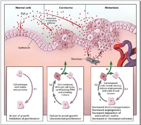
In normal cells TGFβ pathway control many fundamental aspect of cell behaviour, and TGFβ acts as a tumor suppressor by inhibiting cellular ptoliferation or by promoting cellular differentiation or apoptosis.
In human cancer TGFβ pathway has a complex role, being involved in the hallmark events that occur in virtually all cancers. Among these hallmarks are the ability of tumor cells to proliferate in the absence of exogenous growth factors, invade e metastasize, recruit a blood supply through angiogenesis, and evade apoptosis (Fig.21).
TGFβ is an important regulator of cellular proliferation. In normal cells it potently inhibits epithelial, endothelial, and
hematopoietic cell proliferation, preventing the cell progression through the cell cycle first by induction of cell cycle regulators such as p15INK4B and p21CIP1, and second by directly suppressing c-MYC expression. In cancer cells, mutations in the TGFβ pathway have been described that confer resistant to growth inhibition by TGFβ, thus allowing uncontrolled proliferation of the cells.
TGFβ normally stimulates the production of the extrcellular matrix by increasing the production of the extracellular proteins, decreasing the production of enzymes that degrade the extrcellular matrix. However, during tumorigenesis, TGFβ frequently stimulates the proteolytic activity of cancer cell, leading to a decreased cell adhesiveness and increased cell invasive capability.
TGFβ is a cytokine that regulates angiogenesis, and can function either as a proangiogenic or antiangiogenic factor in vitro. Recent evidences support a TGFβ proangiogenic role in vivo: mice with deletion of TGFβI or TGFβII receptors result to have a decreased and defective vasculogenesis. Stimulation of angiogenesis may be another mechanism by which TGFβ stimulates the growth of late-stage tumors.
The mechanism by which TGFβ induces and regulates apoptosis are cell and context specific, but in most cases the TGFβ signalling is proapoptotic and frequently activated by the SMADs-dependent pathway. TGFβ-induced apoptosis may occur through both p53-dependent and p53-independent mechanisms, and involves caspase activation, upregulation of proapoptotic factors( i.e., Bax), and /or downregulation of antiapoptotic factors (i.e., Bcl-2 and Bcl-xL). Resistant to
TGFβ-induced apoptosis is an essential component particularly for cancer arising from tissue in which TGFβ is a prominent regulator of apoptosis, such as prostate cancer.
FIG. 21. Role of TGFβ in Cancer (from Blobe GC, 2000).
The role of the TGFβ pathway in human tumor progression is complex, TGFβ exerting both growth-inhibitory and growth promoting effects depending on the cell type and cell context. The precise mechanism for the dichotomous function of TGFβ in human cancer remains elusive. In one proposed model TGFβ function as a tumor suppressor on precancerous epithelial cells. During tumor progression the epithelial-derived cancer cells become resistant to the tumor suppressor effect of TGFβ, then increase the production of TGFβ which now acts as tumor promoter predominantly through effect on the stroma compartment (Fig.22).
Another scenario proposes that TGFβ acts only on tumor cells: TGFβ switch from tumor suppressor to tumor promoter when epithelial-derived cancer cells undergo an EMT. Tumor cells that have lost cytostatic response but retained TGFβ signalling component can undergo EMT in response to TGFβ, becoming more invasive (Siegel PM and Massague J, 2003).
FIG. 22. Role of TGFβ in Cancer as a Tumor Suppressor and Tumor Promoter (from Elliott RL 2005).
Given the antiproliferative and apoptotic functions of TGFβ in normal tissue , it is not surprising that TGFβ pathway is disrupted by mutation in human cancers.
Mutations in TβRII are frequently observed in colon cancers that posses microsatellite instability (MSI), but are also found in 15% of microsatellite stable colon cancers. These mutations arise within a region that encodes the TβRII extracellular domain, and result in a truncated and inactivated form of the
receptor. These kind of mutations can also be found in MSI gastric tumors and gliomas, but are infrequent in MSI cancer of the endometrium, liver and breast (Siegel PM and Massague J, 2003). Mutation targeting TβRI have been observed in ovarian, breast, and pancreatic cancer (Goggins M et al, Cancer Research 1998).
Mutation resulting in loss of function or decreased production of the TGFβ signalling molecules SMAD2 and SMAD4 have been also identified in human cancers.
Although infrequent, SMAD2 mutations are found in human colorectal and lung cancers.
SMAD4 was first identified as a tumor-suppressor gene (originally named deleted in pancreatic carcinoma locus 4, DPC4). SMAD4 mutations have been identified in near 10% of all colon cancers and 30% of metastatic colon cancers, with lower frequencies in other cancer types.
The importance of TGFβ signalling in PDAC is illustrated by the fact that SMAD4 genetic inactivations appear to be specific for pancreatic cancer, being rarely observed in other tumor types. 90% of pancreatic tumors show loss of heterozygosity (LOH) at the SMAD4 locus, with 55% of PDAC having either homozygous deletion (35%) or mutational inactivation of the second allele (20%). All SMAD4 mutants are affected in their nuclear localization through impaired heterodimerization with other SMAD proteins, resulting unable to mediate gene transcription. Loss of SMAD4 expression occurs biologically late in the pancreatic neoplastic progression, at the stage of histologically recognizable carcinoma (Wilentz RE et al, 2000). The exact relationship between SMAD4 and
prognosis in PDAC has not yet been elucidate. Despite one report, which found that loss of SMAD4 expression resulted in significantly shorter survival following resection, other studies have failed to corroborate this. One possible explanation for this discrepant result is that immunohistochemical analysis may not differentiate between wild-type and mutate SMAD4 proteins (Garcea G et al, 2005).
The dichotomous function of TGFβ, TGFβ exerting both growth-inhibitory and growth promoting effects, has been elegantly confirmed in PDAC by Bardeesy and colleagues (Bardeesy N et al, 2006). Using genetically engineered mice, the authors determined the role of SMAD4 deficiency on the initiation/progression of PDAC in combination with activated KRAS(G12D) allele. Results from this study provide genetic confirmation that SMAD4 is a PDAC tumor suppressor, functioning to block the progression of KRAS(G12D)-initiated neoplasms, whereas in a subset of advanced tumors, intact SMAD4 facilitates epithelial-to mesenchimal transition (EMT) and TGFβ-dependent growth.
In vitro experiment of restoration of SMAD4 in SMAD4 deleted pancreatic cancer cells showed the abolition of tumorigenesis in immunodeficient mice due to suppression of angiogenesis. This evidence suggest that loss of SMAD4 in PDAC may have a primary role in modulating the interaction between epithelial cells and stromal cells rather than in growth control of the tumor cells themselves (Schwarte-Waldhoff I et al, 2000).