Calcif Tissue Int (1993) 52:125-129
Calcified Tissue
International
9 1993 Springer-Verlag New York Inc.
Inhibitory Effect of Salmon Calcitonin on Bone Resorption:
Morphological Study of the Tibial Growth Plate in Rats
Ugo E. Pazzaglia, 1 Giovanni Zatti, 1 Amalia Di Nucci, 2 and Anna Coci 3
lClinica Ortopedica, II Facolt~t di Medicina e Chirurgia and 2Istituto di Farmacologia II dell'Universita' di Pavia, and 3Dip. Patologia Umana ed Ereditaria, Sez. Anatomia Patologica, Universit~t Di Pavia, Pavia, Italy
Received June 13, 1992, and in revised form July 23, 1992
Summary. Salmon calcitonin (sCT) at doses of 100 and 50 UI given subcutaneously to growing rats produced in vivo evidence of osteoclastic activity inhibition. Histological as- sessment was carried out by measuring the perichondrial ring of Lacroix height, and a dose-correlated effect was found. These aspects were coupled with an increase in the osteoclast number and suggested that in studies with bone resorption inhibitors, morphological evaluation based on os- teoclasts count is not reliable. The changes of the metaphy- sis suggested also that sCT affects the activity of hyper- trophic chondrocytes of the growth plate. Plasma calcium levels did not differ significantly between treated rats and controls; an increased phosphatemia was observed in sCT- treated animals.
Key words: Bone resorption - Osteoclasts - Calcitonin - Growth plate.
The capacity of calcitonin to inhibit bone resorption has been demonstrated with organ culture techniques [1-4] and in experiments with isolated osteoclasts cultured on calcified substrates [5-7]; on the other hand, there are few histological evidences of such an effect in experimental animals [8], al- though morphological changes of osteoclasts have been ob- served with electron microscopy [9, 10]. The reported sig- nificant decrease in n u m b e r of osteoclasts following admin- istration of calcitonin [1, 11, 12] is not necessarily related to the level of resorption activity; on the contrary, other potent inhibitors of bone resorption, such as diphosphonates, in- crease the n u m b e r of osteoclasts, but they are not active [13]. The tibial growth plate cartilage and the related peri- chondrial ring of Lacroix [14, 15] are characterized by fast modeling, therefore, they are a suitable model for the study of early changes of osteoclasts morphology and activity. No effects of calcitonin were observed employing histological techniques in rats, which received current therapeutic doses [8]. In this study, the experiment was carried out with high doses of salmon calcitonin to investigate the effectiveness of the hormone in inhibiting bone resorption in the fast model- ing metaphysis of growing rats. The conical shape of the metaphysis is the result of high rate modeling of the peri- chondrial ring and metaphyseal primary trabeculae, and thus an inhibitory effect on bone resorption should soon become apparent as a modification of the morphology of the metaph- ysis [16].
Offprint requests to: U. E. Pazzaglia
Table 1. Mean plasmatic levels of calcium and phosphates in rats treated with sCT (100 and 50 U daily) for 21 days and controls
Plasma calcium Plasma phosphate
Group (mg/100 ml -+ SD) (mg/100 ml +- SD)
A (CT 100 U) n = 10 10.61 -+ 0.33 9.65 -+ 0.64 a B (CT 50 U) n = 10 10.09 -+ 0.76 10.18 +- 0.858 C (Controls) n = 10 10.34 +- 0.45 8.70 -+ 0.49 Mean of group A and B were compared with C using Student's t test a p < 0.002; b p < 0.001
Materials and Methods
Thirty Sprague-Dawley rats (Stefano Morini, S. Polo d'Enza, Reg- gio Emilia, Italy) weighing about 150 g were used, randomly distrib- uted in three groups of 10 animals each. The rats in group A received 100 U of sCT daily (Sandoz S.p.A., Milano, Italy) subcutaneously and those in group B received 50 U of sCT daily: the 10 control rats of group C were treated daily with the same volume of the vehicle alone for 21 days.
Rats were housed three to a cage with free access to food and water. They were killed with an ether overdose after 21 days. The interval between the sacrifice and the last administration of sCT was 8 hours. About 30 cc of blood was drawn from the heart with a hep- arinized syringe; the samples were centrifuged and the plasma was stored in a freezer at -30~ until analyzed.
Plasma levels of calcium and phosphates were assessed with the method of o-cresolphthalein-complexon in alkaline solution [17], plasma phosphate with the direct phosphomolybdate reaction with- out deproteinization [18].
The right and left tibia of each animal were carefully dissected from soft tissues without damaging periosteum and perichondrium; the left tibia was fixed in neutral formalin (10%). Eight specimens were decalcified with EDTA, cut in the middle, coronal plane of the metaphysis and embedded in paraffin. Sections were stained with hematoxylin-eosin. The remaining two specimens were embedded undecalcified in Technovit resin (Kulzer and Co., Gmgh, Wehrheim, FRG), and sections of the middle coronal plane of the metaphysis were prepared with a cutting-grinding machine (Exact Apparatebau, Norderstadt, FRG). Sections of 25-1~m thickness were stained by the Von-Kossa method (counterstained with neutral red). The number of osteoclasts was determined in a median coronal sec- tion of the proximal tibia of each rat, including the whole epiphysis, metaphysis, and proximal diaphysis. Only multinuclear cells in con- tact with a resorption lacunae on the bone surface were identified as osteoclasts. The height of the perichondrial ring was measured un- der the microscope with a graduated reticulum.
Right tibias were freed of periosteum and all soft tissues, stored for 7 days in an ethanol-methanol solution (50:50) at 27~ to extract lipids and water, and then dried in an oven at 100~ for 48 hours. They were then weighed and the lengths were measured with a caliber.
126
Table 2. Mean dry weight and length of tibia in rats treated with sCT (100 and 50 U daily) and controls for 21 days
Tibias weight Tibias length
Group (g -+ SD) (mm -+ SD)
A (CT 100 U) n = 10 0.361 -+ 0.41 34.6 -+ 0.3 B (CT 50 U) n = 10 0.345 -+ 0.035 34.8 -+ 0.7 C (Controls) n = 10 0.340 +- 0.033 35.2 -+ 1.2 Mean of group A and B were compared with C using Student's t test
U. E. Pazzaglia et al.: Effect of sCT on Bone Resorption Observations
Treatment was well tolerated by the rats: no animals died during the experiment, and the behavior and weight gain of the animals in groups A and B were normal. Eight hours after the last administra- tion of sCT there were no significant differences in plasma calcium levels between treated animals and controls; mean plasma phos- phate was significantly higher in both treated groups compared with controls (Table 1).
The difference in tibial mean dry weight and length between rats
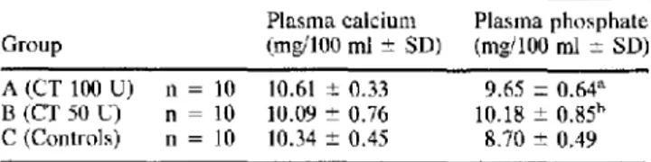
Fig. 1. Left: proximal tibial metaphysis of control rats. Right: proximal tibial metaphysis of rats treated with sCT 100 U daily for 21 days. Thickening of the outer part of the growth plate is evident as well as the cylindrical shape of one side of the metaphysis, due to failure of remodeling. Original magnification x 3.3,
hematoxylin-eosin.
Fig. 2. Failure of calcification of intercolumnar septa of the thickened growth plate cartilage. The metaphyseal trabeculae are thicker and denser than normal, due to inhibition of remodeling, x40, Von KossaJneutral red.
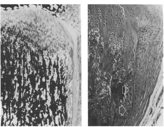
Fig. 3. Normal aspect of the perichondrial bone bark. It does not extend below the line of primary metaphyseal trabeculae and is resorbed at its distal end by osteoclasts (arrows). x 100, hematoxylin-eosin.
U. E. Pazzaglia et al.: Effect of sCT on Bone Resorption 127
Fig. 4. The effect of sCT on the height of
perichondrial bone bark (left: control, right: CT 100 U daily for 21 days), x30, hematoxylin-eosin.
treated with sCT and controls was not significant (Table 2). The morphology of the growth plate cartilage and metaphysis in groups A and B was different from that in controls in quantity but not in quality. The growth plate cartilage showed an increased thickness of its outer part (Fig. 1). This thickening was not symmetrical and in most bones it involved only the medial side of the tibia. Thickening was due to persistence of hypertrophic chondrocytes because of failure of vascular invasion from the metaphysis. The proximal tibial metaphysis on the affected side showed an inversion of the concav- ity of its profile (failure of conization). Primary metaphyseal trabec- ulae were denser and thicker in this area (Fig. 2), as is usually observed when inhibition of resorption is coupled with a normal rate of osteoblastic apposition [25]. In the thickened growth plate failure of calcification of the inlercolumnar septa was present (Fig. 2); cal- cification and vascular invasion were normal in the other sectors of the plate. No pathological osteoid border was present on trabeculae or cortical bone.
The perichondrial ring [14, 15, 19-21], which normally does not extend below the line of the first primary metaphyseal trabeculae (Fig. 3) and has the appearance of a very thin lamina without osteo- cytes, was higher in the sCT-treated animals than in controls be- cause it lined the thickened growth plate and the unremodeled me- taphysis. Also, the thickness of the ring was increased and osteo- cytes were present in the structure.
Because the proximal and distal ends of the perichondrial ring were easily identified on histological sections, its height could be measured (Fig. 4) and a significant difference was found when groups A and B were compared with controls (Table 3).
In the lower part of the metaphysis, a row of osteoclasts lined the bone below the periosteum. The number of osteoclasts and the ratio nuclei/osteoclast were significantly higher than in controls (Table 4). The nuclei had a dense chromatin and appeared to have an abnor- mally high number of picnotic nuclei (Fig. 5).
Discussion
N o e f f e c t s o f c a l c i t o n i n o n t h e c h e m i s t r y a n d h i s t o l o g y o f t h e r a t s ' b o n e s w e r e o b s e r v e d in a p r e v i o u s r e p o r t [8], b u t t h e d o s e u s e d w a s s m a l l (200 m U / d a y ) . I n t h e p r e s e n t e x p e r i -
Table 3. Mean height of the perichondrial ring in rats treated with sCT (100 and 50 U daily) for 21 days and controls
Group Height (ram -+ SD)
A n = 15 2.70 + 1.05 a
B n = 12 2.21 --- 0 . 8 4 b
C n = 14 1.35 -+ 0.32
Mean of groups A and B were compared with C using Student's t test
a p < 0.001; bp = 0.001
Table 4. Mean number of osteoclasts/mm 2 and mean ratio nuclei/ osteoclast in rats treated with sCT (100 and 50 U daily) for 21 days and controls
Number of
Group osteoclasts/mm 2 Nuclei/Osteoclast
A n = 10 1.14 +- 0.56 a 5.24 -+ 0.37 a B n = 10 0.98 - 0.31 a 4.98 -+ 0.39 b
C n = 10 0.41 - 0.17 4.25 -+ 0.67
Mean of groups A and B were compared with C using Student's t test a p <0.001; bp = 0.008 m e n t , 50 a n d 100 I U / d a y w e r e u s e d , as c u r r e n t l y g i v e n in clinical t h e r a p y . C o n s i d e r i n g t h e b o d y w e i g h t o f r a t s , t h e s e d o s e s are h i g h , a l t h o u g h c o m p a r i s o n o f t h e d o s e / b o d y w e i g h t r a t i o in d i f f e r e n t s p e c i e s is q u e s t i o n a b l e . I f c a l c i t o n i n is r a p - idly c l e a r e d f r o m t h e c i r c u l a t i o n in t h e r a t , h i g h d o s e s a r e n e c e s s a r y to i n d u c e t h e i n h i b i t o r y r e s p o n s e o n b o n e t u r n - o v e r . H o w e v e r , t h e a i m o f this s t u d y w a s n o t to i n v e s t i g a t e t h e e f f e c t u a l d o s e s o f c a l c i t o n i n in t h e r a t , b u t to c o n f i r m h i s t o l o g i c a l l y in vivo t h e e f f e c t s o b s e r v e d in in vitro s t u d i e s .
128 U.E. Pazzaglia et at.: Effect of sCT on Bone Resorption
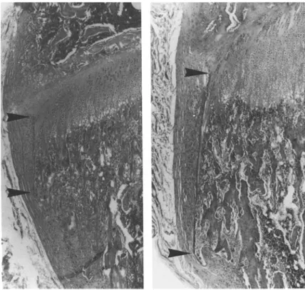
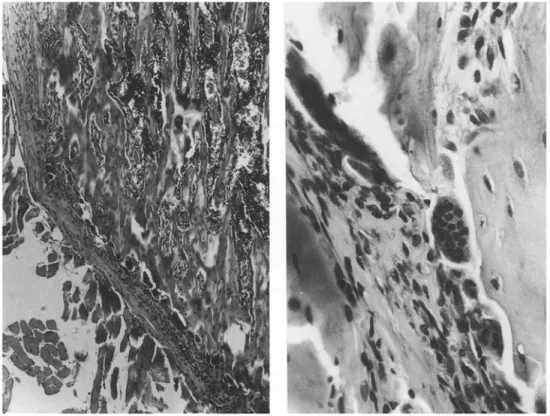
Fig. 5. Proximal tibial metaphysis of rats treated with sCT 100 U daily for 21 days. A row of osteoclasts is evident on the outer aspect of the metaphysis; they appear abnormally large and with an abnor-
really high number of picnotic nuclei, x 100 and x 400, hematoxilin- eosin.
Most changes of the growth plate and metaphyseal mor- phology observed by us can be explained in terms of inhibi- tion of bone resorption and vascular invasion of cartilage [16]; the inhibition due to sCT involved only the outer part of the epiphyseal plate and this area was characterized by three basic phenomena: (1) inhibition of osteoclastic resorption; (2) reduced mineralization of intercolumnar septa; and (3) failure of vascular invasion.
It is difficult to explain the different response of the same anatomical structure to sCT, as well as the association of inhibition of mineralization and failure of bone resorption and vascular invasion of cartilage. The conical shape of the proximal tibial metaphysis results from a high remodeling rate, and under normal conditions, many more osteoclasts are observed on the outer border of the metaphysis than anywhere else in the bone. As osteoclasts are the major target cells of calcitonin, the inhibitory effects of the hor- mone obviously becomes evident earlier at sites where os- teoclastic activity is higher. This could account for a differ- ent response to the hormone of different parts of the growth plate cartilage and metaphysis.
Attachment of osteoclasts to bone surface is a necessary
condition for the activity of these cells both in vitro and in
vivo; there are evidences that calcitonin is capable of inhib-
iting both mobility of osteoclasts in vitro and their attach-
ment to the bone surface [6, 22]; therefore, a quantitative
evaluation of in vivo bone resorption inhibition based on
morphology is questionable. The osteoclast count has been used [11, 23], but it is not a reliable index, as the number of cells does not necessarily correlate with their degree of ac- tivity. In this study, the number ofosteoclasts was observed to increase in the presence of an arrest of bone modeling.
Explanation of these observations can only be specula- tive: either the formation of osteoclasts is enhanced, or the
life-span of inactive osteoclasts is lengthened and the cells accumulate at the resorption sites, or the large polykarios are easier identified in the histological slides, and the ob- served differences represent a technical artifact. On the con- trary measurement of the perichondrial ring of Lacroix proved to be a simple and reliable method for assessing os- teoclastic resorption inhibition and allowed comparison be- tween groups. The inhibitory effect was proportional to the dose of the hormone (Table 3). On the other hand, there was no inhibitory effect on osteoblastic activity, as demonstrated by the thickness of perichondrial ring and primary metaph- yseal trabeculae.
The sequence of phenomena that characterizes growth plate cartilage maturation suggests a correlation between cartilage mineralization and cartilage invasion by vessels. The nature of this relationship remains obscure. However, recent observations have cast doubts on the notion that chondrocytes in the lowermost hypertrophic zone are degen- erating [24] and have suggested that the hypertrophic chon- drocyte is a highly differentiated cell, which synthetizes and secretes into extracellular matrix a set of macromolecules, such as type • collagen, chondrocalcin, and S-100 protein, which may be directly involved in the processes of matrix mineralization and capillary invasion [25-27]. The effects on mineralization of intercolumnar septa and vascular invasion could indicate that calcitonin has an influence on the activity of hypertrophic chondrocytes.
In another study [28] in which sCT 10 IU was given for 3 and 7 days to rats weighing 20-30 g (approximately the same dose as given to group A animals in our experiment), a wid- ening of the metatarsal growth plate cartilage was observed. Although these authors explain their findings in terms of stimulation of chondrocyte maturation, their observations confirm our results.
U. E. Pazzaglia et al.: Effect of sCT on Bone Resorption
Calcitonin has been reported to have a hypocalcemic ef- fect whose magnitude and duration depends on the dose [29]; 4 hours after administration of high doses of sCT (0.4 and 2 IU/100 g), h y p o c a l c e m i a and s e c o n d a r y h y p e r p a r a t h y - roidism have been observed [14]; the consequent increase of PTH and 1,25-dihydroxycolecalciferol secretion are known to enhance the formation of osteoclasts, while this effect is prevented by parathyroidectomy [30]. The increased number of osteoclasts and nuclei/osteoclast in the present experi- ment probably reflects transient changes of plasmatic cal- cium levels. However, as blood sampling was performed 8 hours from the last sCT administration, the animals have possibly compensated for the low calcium levels in plasma, whereas phosphate remained higher.
In conclusion, treatment with very high doses of sCT was well tolerated by rats and produced histological evidences of osteoclastic activity inhibition; these aspects were coupled with an increase of the osteoclast n u m b e r and suggested that morphological evaluation based on osteoclast count is not reliable.
Acknowledgments:
The authors would like to thank Mr. G. Viani and Mr. F. Rossi for their technical assistance.References
1. Friedman B, Raisz LG (1965) Thyrocalcitonin: inhibitor of bone resorption in tissue culture. Science 1465-1467
2. Reynolds J, Dingle JT (1970) A sensitive in vitro method for studying the induction and inhibition of bone resorption. Calcif Tissue Res 4:339-349
3. Stewart PJ, Stern PH (1987) Vertebral bone resorption in vitro: effects of parathyroid hormone, calcitonin, 1,25-dihydroxy vi- tamin D3, epidermal growth factor, prostaglandin E2 and estro- gen. Calcif Tissue Int 40:21-26
4. Yamamoto I, Kitamura W, Aoki J, Shigeno C, Hiro H, Ajo- numa K, Torizuka K, Fujii N, Otaka A, Yajima H (1986) Hu- man calcitonin gene-related peptide possesses weak inhibitor potency of bone resorption in vitro. Calcif Tissue Int 38:339-341 5. Chambers TJ, Revell PA, Fuller K, Athanasou NA (1984) Re- sorption of bone by isolated rabbit osteoclasts. J Cell Sci 66: 383-388
6. Chambers TJ, McSheehy PMJ, Thomson BM, Fuller K (1985) The effect of calcium regulating hormones and prostaglandins on bone resorption by osteoclasts disaggregated from neonatal rabbit bone. Endocrinology 116:234-239
7. Chambers TJ, Fuller K, McSheehy PMJ, Pringle JAS (1985) The effects of calcium regulating hormones on bone resorption by isolated human osteoclastoma. J Pathol 145:297-307
8. Russell RGG, Kislin AM, Casey PA, Fleisch H (1973) Effect of diphosphonates and calcitonin on the chemistry and quantita- tive histology of rat bones. Calcif Tissue Res 11:179-195 9. Norimatsu H, Van der Wiel CJ, Talmage RV (1979) Electron
microscopic study of the effects of calcitonin on bone ceils and their extracellular milieu. Clin Orthop 139:250-258
10. Norimatsu H, Yamamoto T, Ozawa H, Talmage RV (1982) Change in calcium phosphate on bone surfaces and in lining cells after the administration of parathyroid hormone and calci- tonin. Clin Orthop 164:271-278
129 11. Baron R, Vignery A (1981) Behavior of osteoclasts during rapid change in their number induced by high doses of parathyroid hormone or calcitonin in intact rat. Metab Bone Dis Rel Res 2:339-346
12. Foster GV, Doyle FH, Bordier P, Matrajat H (1966) Effect of thyrocalcitonin on bone. Lancet II: 1428-1431
13. Miller SC, Jee WSS, Kimmel DB, Woodbury L (1977) Ethane- 1-hydroxy-l,l-diphosphonate (EHDP). Effects on incorpora- tion and accumulation of osteoclast nuclei. Calcif Tissue Res 22:243-252
14. Lacroix P (1951) The organization of bones. JA Churchill Ltd, London
15. Ranvier LA (1989) Traite technique d'histologie (ed 2) Sary, Paris
16. Schenk R, Merz WA, Muhlbauer R, Russel RGG, Fleisch H (1973) Effect of ethane-hydroxy- 1,1-diphosphonate (EHDP) and dichloro-methylene-disphosphonate (CI2MDP) on the calcifica- tion and resorption of cartilage and bone in the tibial epiphysis and metaphysis of rats. Calcif Tissue Res 11:196-214 17. Baginski ES, Marie SS, Clark WL, Zak B (1973) Direct micro-
dermination of serum calcium. Clin Chir Acta 46:49
18. Weismann N, Pileggi VJ (1974) Clinical chemistry, principles and techniques (2nd ed) In: Henry RJ, Harper & Tow, Ha- gerstown, pp 723-754
19. Pratt CWM (1959) The significance of the "perichondrial zone" in a developing long bone of the rat. J Anat 93:110-122 20. Shapiro F, Holtrop ME, Glimcher MJ (1977) Organization and
cellular biology of perichondral ossification groove of Ranvier. A morphological study in rabbits. J Bone Joint Surg 59A:703- 723
21. Speer DP (1982) Collagenous architecture of the growth plate and perichondrial ossification groove. J Bone Joint Surg 64A: 399-407
22. Feldman RS, Krieger NS, Tashjian AH Jr (1980) Effect of para- thyroid hormone and calcitonin on osteoclasts formation in vitro. Endocrinology 107:1137-1145
23. Hedlund T, Hulth A, Johnell O (1983) Early effects of parathor- mone and calcitonin on the number of osteoclasts and on serum calcium in rats. Acta Orthop Scand 54:802-804
24. Hunziker EB, Schenk RK, Cruz-Orive LM (1978) Quantitation of chondrocytes performance in growth plate cartilage during longitudinal bone growth. J Bone Joint Surg 69A: 162-1873 25. Boden SD, Kaplan FS, Fallon MD, Ruddy R, Belik J, Anday E,
Zakai E, Ellis J (1987) Metatropic dwarfism. Uncoupling of en- dochondrial and perichondral growth. J Bone Joint Surg 69A: 174-184
26. Cowell HR, Hunziker EB, Rosemberg L (1987) The role of hy- pertropic chondrocytes in endochondral ossification and in the development of secondary centers of ossification. J Bone Joint Surg 69A: 159-161
27. Floyd WE, Taleske DJ, Schiller AL, Trahan C, Mankin HJ (1987) Vascular events associated with the appearance of the secondary center of ossification in the murine distal femoral epiphysis. J Bone Joint Surg 69A:185-190
28. Burch WM, Corda G (1985) Calcitonin stimulates maturation of mammalian growth plate cartilage. Endocrinology 116:1724-
1728
29. Azria M (1989) The calcitonins. Physiology and pharmacology. Karger AG, Basel
30. Glajchen N, Thomas S, Jowell P, Epstein S, Ismail F, Fallon M (1990) The effect of high-dose salmon calcitonin on bone me- tabolism in the normal rat. Calcif Tissue Int 46:28-32