Journal of the American Heart Association
ORIGINAL RESEARCH
Papillary Muscles Abnormalities in Athletes
With Otherwise Unexplained T-Wave
Inversion in the ECG Lateral Leads
Manuel De Lazzari , MD, PhD*; Alessandro Zorzi , MD, PhD*; Natascia Bettella, MD; Alberto Cipriani , MD; Kalliopi Pilichou , BSc, PhD; Marco Cason, BSc; Teresina Vessella, MD; Patrizio Sarto, MD; Maria Rita Gualea, MD; Francesca Chianura, MD; Lucia Tardini , MD; Giuseppe Ricci , MD; Ilaria Mazzanti, MD; Fabrizio Ricci , MD, PhD; Raffaella Motta, MD; Martina Perazzolo Marra , MD, PhD; Domenico Corrado , MD, PhD
BACKGROUND: Papillary muscles (PMs) abnormalities may be associated with ECG repolarization abnormalities. We aimed to
evaluate the relation between lateral T-wave inversion (TWI) and PMs characteristics in a cohort of athletes with no clinically demonstrable cardiac disease.
METHODS AND RESULTS: We included 53 athletes (median age, 20 years; 87% men) with lateral TWI and no evidence of heart
disease on clinical and cardiac magnetic resonance evaluation. A group of healthy athletes with normal ECG served as con-trols. We evaluated the PMs dimensions, such as diameters, area, volume, mass, and ratio between PMs and left ventricular mass, and the prevalence of PMs apical displacement. Compared with controls, athletes with TWI showed PMs hypertrophy with significantly increased PMs diameters, area, volume, and mass. The ratio between PMs and left ventricular mass was 4.4% in athletes with TWI and 3.0% in controls (P<0.001). A PMs/left ventricular mass ratio >3.5% showed 85% sensitivity and 76% specificity for differentiating between athletes with TWI and controls. Apical displacement of PMs was found in 25 (47%) athletes with TWI versus 9 (17%) controls (P=0.001). At multivariable analysis, PMs/left ventricular mass ratio and apical displacement remained independent predictors of TWI. Clinical outcome of the athletes with TWI and PMs abnormalities was uneventful despite continuation of their sports activity.
CONCLUSIONS: PMs hypertrophy and apical displacement may underlie otherwise unexplained lateral TWI in the athlete. Lateral
TWI associated with PMs abnormalities appears as a distinct anatomo-clinical condition characterized by a favorable outcome.
Key Words: cardiac magnetic resonance ■ electrocardiography ■ imaging ■ preparticipation screening ■ sports cardiology
T
he presence of T-wave inversion (TWI) in the athlete’s ECG is a warning sign of a potential cardiovascular disease at risk of sudden car-diac death (SCD) during sports.1–3 TWI in lateral leads, with or without involvement of inferior leads, is the repolarization pattern most frequently associ-ated with structural heart diseases such as ischemic heart disease, cardiomyopathy, myocarditis, aortic valve disease, left ventricular (LV) noncompaction,and idiopathic LV fibrosis.4 Accordingly, in the athlete with lateral TWI, any forms of heart disease need to be accurately excluded by a comprehensive clinical and imaging workup, including contrast-enhanced cardiac magnetic resonance (CMR).1–3,5 However, 1% to 3% of athletes have lateral TWI that remains unex-plained even after an accurate clinical evaluation.6–8 Whether these unexplained repolarization changes reflect the presence of a concealed or unrecognized
Correspondence to: Domenico Corrado, MD, PhD, Department of Cardio-Thoraco-Vascular Sciences and Public Health, University of Padua, Via Giustiniani, 2, 35128 Padua, Italy. E-mail: [email protected]
*Dr De Lazzari and Dr Zorzi contributed equally to this work as co-first authors. For Sources of Funding and Disclosures, see page 10.
© 2021 The Authors. Published on behalf of the American Heart Association, Inc., by Wiley. This is an open access article under the terms of the Creative Commons Attribution-NonCommercial License, which permits use, distribution and reproduction in any medium, provided the original work is properly cited and is not used for commercial purposes.
JAHA is available at: www.ahajournals.org/journal/jaha
heart muscle abnormality remains to be determined. Papillary muscles (PMs) abnormalities in isolation, that is, not associated with cardiomyopathy—par-ticularly hypertrophic cardiomyopathy (HCM)—are considered a normal anatomic variant without clinical relevance.9 Past clinico-echocardiographic studies reported the association between TWI with isolated hypertrophy and/or the apical displacement of LV PMs.10,11 In more recent years, CMR has become the most accurate technique for imaging study of struc-tural heart disease in the athlete.12 Due to its high resolution, multiplanar capabilities, and soft-tissue contrast, CMR is the most valuable imaging modality in the evaluation of PMs, providing detailed morpho-logic and functional information.9 However, system-atic studies correlating lateral TWI with the anatomy and structure of PMs are lacking. Hence, the present ECG-CMR correlation study was designed to eval-uate the relation between unexplained lateral TWI and morpho-functional PMs abnormalities, either hy-pertrophy or displacement, in a cohort of apparently healthy athletes with no clinically demonstrable car-diac disease.
METHODS
The data that support the findings of this study are available from the corresponding author upon reason-able request. The study included a consecutive group of athletes with TWI in lateral leads (V4–V6), with or without involvement of inferior leads (L2-aVF), who were identified on preparticipation screening during the time interval January 2014 to December 2019 in 9 Italian sports medicine centers and referred to the University of Padova for CMR study. The study was approved by the local institutional review board and because of its retrospective nature no consent was required.
All athletes with lateral TWI underwent a routine cardiovascular evaluation including medical history, physical examination, and 12-lead ECG. In addition, all underwent CMR imaging study for deeper investi-gation of a possible underlying heart muscle disease, with particular reference to HCM including the “apical” variant. Athletes older than 40 years with risk factors for coronary artery disease also performed either a coronary computed tomography or a stress imaging test to exclude an ischemic heart disease. Athletes with lateral TWI that remained unexplained after the clinical and imaging study comprised the study population. Athletes with abnormal CMR findings, such as patho-logical increase in wall thickness or with evidence of late gadolinium enhancement (LGE)/myocardial fibrosis other than the isolated “junctional” spotty LGE (that is normal in the athlete5) were excluded from the study. Healthy athletes, matched for age class (5-year inter-vals), sex, and type of sport, who had a normal CMR performed for evaluation of cardiac arrhythmias or for investigational purposes served as controls.
Molecular genetic testing was performed in a sub-set of athletes with lateral TWI. Indications to genetic testing were provided by each participating center according to local practice and based on family his-tory of SCD in 2 athletes, borderline LV wall thickness (12–13 mm) in 4, and persistence of lateral TWI after detraining in 3.
Standard ECG
A standard 12-lead ECG was recorded at the time of CMR scan. The tracing was interpreted by 2 ob-servers who were unaware of clinical data. Lateral T-wave inversion was defined as negative T waves ≥0.1 mV in depth in ≥2 contiguous lateral leads (V4– V6), with or without involvement of inferior leads (L2-aVF).1
Cardiac Magnetic Resonance
Acquisition Protocols
All images were performed on a 1.5 T scanner (Magnetom Avanto Siemens AG, Germany) using a protocol
CLINICAL PERSPECTIVE
What Is New?
• Athletes with lateral (or inferolateral) T-wave in-version and normal cardiac magnetic resonance demonstrated a higher prevalence of papillary muscles abnormalities such as hypertrophy (in-crease papillary muscles/left ventricular mass ratio) and apical displacement compared with control athletes with normal ECG.
What Are the Clinical Implications?
• Isolated papillary muscles abnormalities suchas hypertrophy and apical displacement may underlie lateral (or inferolateral) T-wave inversion in a sizeable proportion of athletes with other-wise structurally normal heart and appear asso-ciated with a favorable medium-term outcome despite continuation of sports activity.
Nonstandard Abbreviations and Acronyms
HCM hypertrophic cardiomyopathy
LGE late gadolinium enhancement
PMs papillary muscles
SCD sudden cardiac death
TWI T-wave inversion
including post contrast sequences. Biventricular mor-pho-functional assessment was performed by a set of steady-state free precession sequence cine loops in se-quential short-axis views and long-axis views as previ-ously reported.13 After 10 minutes since administration of gadolinium-based contrast agent (gadobenate dime-glumine, Multihance or gadobutrol, Gadovist, typically 0.2 mmol/kg of body weight), 2-dimensional segmented fast low-angle short inversion recovery sequences were acquired in the same views of the cine images, cover-ing the entire ventricles (repetition time, 5.4–8.3 ms; echo time, 1.3–3.9 ms; average in-plane spatial resolu-tion, 1.4–1.5×2.2–2.4 mm; 6-mm slice thickness; 2-mm gap; and flip angle, 20°–25°). Inversion times were ad-justed to null normal myocardium using a Look-Locker sequence, and images were repeated in 2 separate phase-encoding directions to exclude artefacts.
Functional and LGE Analysis
Global ventricular volumes, systolic function, and maximum basal and apical wall thickness were cal-culated from the short-axis cine images, excluding PMs from the myocardium, using a computer-aided analysis package (CMR42; Circle Cardiovascular Imaging Inc, Calgary, Canada). The presence and re-gional distribution of LGE were visually assessed in-dependently by 2 experienced observers (European Association of Cardiovascular Imaging third-degree certification) who were blinded to patient clinical data. Ambiguous cases were reviewed by a third expert.
PMs Morphologic Analysis
Assessment of PMs was performed according to a previously reported protocol.10,11,14 In brief, horizontal and vertical diameters and area of both PMs were measured on end-diastolic midventricular short axis cine images (Figure 1A and 1B); while PMs volume and mass were calculated by manually drawing the PMs profile on each short-axis cine slice that contained PMs (Figure 1C) using a computer-aided analysis package (CMR42; Circle Cardiovascular Imaging Inc, Calgary, Canada). PMs volumes and mass were used in addition to linear dimensions to overcome possible errors attributable to measurements in slices of differ-ent thickness and from PMs of differdiffer-ent geometry. In order to index the PMs volume for the different degree of LV hypertrophy of the athlete’s heart, we also cal-culated the ratio between PMs and LV mass for each athlete. Apical displacement of PMs was identified when their implant was in the apical one-third of the left ventricle using multiplanar views (4- and 3-cham-ber long-axis images and their cross section in short-axis view [Figure 1D]). Measures were performed by 2 independent experts (M.D.L. and N.B.).
Follow-Up
All study athletes were followed up yearly according to the Italian guidelines for management of athletes with apparently unexplained TWI, by routine cardiovascular evaluation including medical history, physical examina-tion, 12-lead ECG, and echocardiography.15 Six study athletes underwent control ECG and CMR study after 3 to 6 months of detraining as a therapeutic measure after traumatic injury (N=4) or for evaluation of potential reversibility of “borderline” LV maximal wall thickness (N=3).
Statistical Analysis
Data are expressed as median with 25th to 75th per-centiles because normality could not be assumed for any variable. Categorical differences among groups were evaluated by the χ2 test or the Fisher exact test, as appropriate. Differences among continuous vari-ables were compared using the Mann-Whitney U test. The best cutoff value to differentiate between athletes with and without TWI were calculated with the receiver operating characteristic curve. The following covariates were entered into a binary logistic regression analysis for predictors of the presence of TWI: PMs/LV mass ratio, PMs displacement, age, and sex. A 2-tailed prob-ability value of 0.05 was considered statistically sig-nificant. The intraobserver (M.D.L.) and interobserver (M.D.L. and N.B.) reliability analysis for quantitative measures of PMs characteristics were assessed with the Pearson correlation and the intraclass correlation coefficient. Intraobserver agreement for the presence of apical displacement was assessed with Cohen’s κ coefficient for qualitative parameters. All analyses were performed using SPSS 23 (SPSS Inc, Chicago, IL).
RESULTS
Clinical and Imaging Characteristics
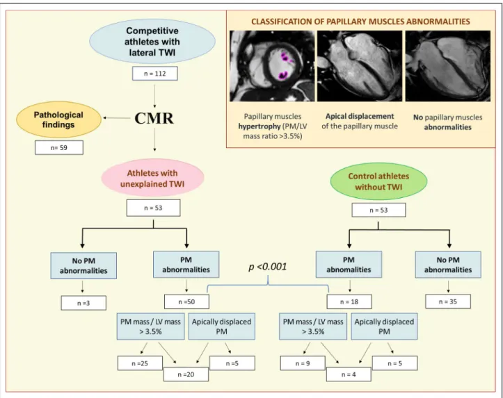
Of 112 athletes referred for investigation of lateral or inferolateral TWI, clinical evaluation including CMR identified an underlying heart disease in 59 (52%). Thirty-nine athletes (35%) had overt HCM, 11 (10%) iso-lated nonischemic LV scar, 3 (3%) mitral valve prolapse, 2 (2%) dilated cardiomyopathy, 2 (2%) myocardial non-compaction and 2 (2%) ischemic heart disease. These 59 athletes diagnosed with a heart disease were ex-cluded from the study.
The remaining 53 athletes (32%), with TWI and no evidence of heart disease on clinical and CMR evaluation comprised the study population. Athletes with TWI had a median age of 20 years (16–43), and 46 (87%) were males. Eighteen subjects practiced endurance sports, 2 power sports, and 33 mixed sports. Family history of SCD was ascertained in 3
athletes. Of 9 athletes undergoing molecular genetic testing, none showed pathogenic or likely pathogenic mutations of genes responsible for inherited cardio-myopathy (including HCM) and channelopathies.
In 21 athletes (40%), TWI was confined to the lateral leads and in 32 (60%) was recorded in both lateral and inferior leads.
CMR findings are summarized in Table 1. Volume and ejection fraction of both LV (median LV indexed end-diastolic volume=85 mL/m2 and LV ejection frac-tion=65%) and right ventricle (median right ventricu-lar indexed end-diastolic volume=89 mL/m2 and right ventricular ejection fraction=60%) were within normal values. The median LV indexed mass was also normal (70 g/m2).
Dimensions of PMs
Comparison of morpho-functional PMs features in ath-letes with lateral TWI and control athath-letes is summarized
in Table 2. Diameter, area, and volume of the antero-lateral papillary muscle were greater than those of the posteromedial.
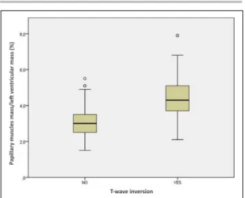
Compared with controls, athletes with lateral TWI showed significantly greater diameters, areas, vol-umes, and mass of both PMs. The ratio between the PMs mass and the LV mass was 4.4% among athletes with TWI versus 3.0% among control athletes (P<0.001) (Figure 2). Intraobserver and interobserver variability in the calculation of quantitative PMs characteristics are shown on Table 3.
Apical Displacement of PMs
The prevalence of apical displacement of PMs was significantly higher among athletes with TWI than con-trols (47% versus 17%; P<0.001) (Figure 3). Ten (19%) athletes with TWI showed isolated displacement of the anterolateral papillary muscle, 3 (6%) an isolated displacement of the posterolateral papillary muscle,
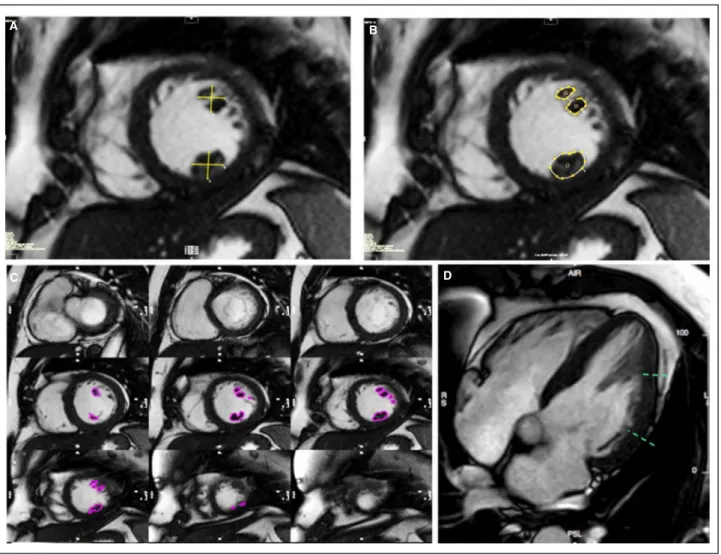
Figure 1. Papillary muscles hypertrophy quantitative analysis.
A, Linear quantification of orthogonal diameters of anterolateral and posteromedial papillary muscles. B, Papillary muscles area quantification in the same slice. C, Papillary muscles volume analysis in the entire stack of short-axis views. D, Apical displacement assessment in long axis view.
A B
C D
and 12 (23%) the displacement of both PMs. In control athletes, an isolated apical displacement of the ante-rolateral papillary muscle was found in 2 cases (4%), isolated apical displacement of posteromedial papil-lary muscle in 2 (4%) and apical displacement of both PMs in 5 (9%) (Figure 3). The interobserver agreement for PM apical displacement was 101 of 106 (95%, P<0.001; k=0.89).
Correlation of Results
The relationship between apical displacement and dimensions of the PMs in athletes with lateral TWI is reported in Table 3. Normally implanted and apically displaced PMs did not differ significantly with regard to diameter, area, volume, mass, and PMs/LV mass ratio.
According to the receiver operating characteristic curve (area under the curve, 0.84; P<0.001), a ratio be-tween the PMs/LV mass ratio >3.5% showed a 85% sensitivity and a 76% specificity for the presence of lateral TWI (Figure 4).
Five of 8 (63%) athletes with lateral TWI, who showed a PMs/LV mass ratio <3.5%, had an apical
displacement of the anterolateral papillary muscle. Three (6%) athletes with lateral TWI versus 35 (66%) control athletes showed neither a PMs/LV mass ratio <3.5% nor apical displacement (P<0.001) (Figure 5).
At multivariate analysis both the PMs/LV mass ratio (odds ratio [OR], 3.8 per 1% increase; 95% CI 2.1–6.7; P<0.001) and the apical displacement of the PMs (OR, 3.2; 95%, CI 1.1–9.6; P=0.03) were independent pre-dictors of lateral TWI.
Detraining
Six athletes with lateral TWI underwent control ECG and CMR study after 3 to 6 months of detraining. All 3 athletes with persistence of lateral TWI on control ECG had an apical displacement of the PMs with un-changed hypertrophy. In 2 of 3 athletes (1 with api-cal displacement of the anterolateral papillary muscle), showing ECG normalization after detraining, there was a significant decrease of the PMs/LV mass ratio; 1 ath-lete had ECG normalization with persistence of PMs hypertrophy.
Follow-Up
All athletes with TWI were allowed to continue the prac-tice of competitive sports because the abnormalities
Table 1. Clinical and Cardiac Magnetic Resonance Characteristics of Athletes With and Without TWI
Characteristics TWI (N=53) No TWI (N=53) P Value Male sex 46 (87) 46 (87) … Age, y 20 (16–43) 24 (20–40) 0.34 Sport Endurance sports 18 (34) 18 (34) … Power sports 2 (4) 2 (4) … Mixed sports 33 (62) 33 (62) … ECG features LV hypertrophy (Sokolow-Lyon Index ≥40 mm) 32 (60) 24 (45) 0.173
Distribution of T-wave inversion
Lateral only (V4–V6) 21 (40) … … Inferolateral (V4–
V6+DII and aVF)
32 (60) … … CMR features LV EDVi, mL/m2 85 (80–103) 96 (85–100) 0.11 LV EF (%) 65 (61–69) 62 (60–66) 0.12 LV mass indexed, g/m2 70 (63–84) 74 (63–86) 0.38 LV mass, g 130 (112–151) 139 (116–158) 0.51 Basal septal maximum
wall thickness, mm
9 (8–11) 9 (8–10) 0.06
Apical maximum wall thickness, mm
8 (6–8) 6 (5–6) <0.001
RV EDVi, mL/m2 89 (78–100) 85 (75–101) 0.31 RV EF (%) 60 (55–66) 59 (56–65) 0.74 Values are expressed as number/total (percentage) of subjects or median (25th–75th percentiles). EDVi indicates end-diastolic indexed volumes; EF, ejection fraction; LV, left ventricular; RV, right ventricular; and TWI, T-wave inversion.
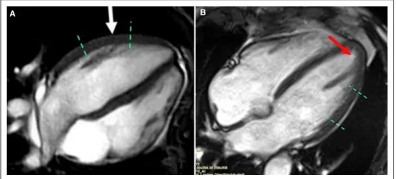
Table 2. PMs Quantitative Analysis Relationship Between Athletes With and Without TWI
TWI (N=53) No TWI (N=53) P Value Anterolateral papillary muscle
Orthogonal diameter 1, mm 12.4 (10.3–13.4) 10 (9–11.6) 0.001 Orthogonal diameter 2, mm 9.8 (8.6–11) 8.3 (7–9.1) <0.001 Area, cm2 1.1 (0.9–1.4) 0.8 (0.7–1) <0.001 Volume, cm3 3.2 (2.5–3.8) 2.1 (1.7–2.7) <0.001 Posteromedial papillary muscle
Orthogonal diameter 1, mm 10 (8–12.2) 9.2 (8–10.4) 0.09 Orthogonal diameter 2, mm 8.1 (7.4–10) 7.6 (6.7–8.9) 0.024 Area, cm2 0.9 (0.7–1.1) 0.7 (0.6–0.9) <0.001 Volume, cm3 2.5 (1.9–3) 1.6 (1.3–2.1) <0.001 PMs mass, g 5.6 (4.8–7.2) 3.9 (3.4–4.9) <0.001 PMs mass indexed, g/m2 3.1 (2.6–3.7) 2.3 (1.8–2.7) <0.001 PMs mass/LV mass, % 4.4 (3.8–5.2) 3 (2.5–3.6) <0.001 Apical displacement 25 (47%) 9 (17%) 0.001 Anterolateral papillary muscle 22 (42%) 7 (13%) 0.001 Posteromedial papillary muscle 15 (28%) 7 (13%) 0.055
Values are expressed as number/total (percentage) of subjects or median (25th–75th percentiles). LV indicates left ventricular; PMs, papillary muscles; and TWI, T-wave inversion.
of PMs in isolation were not considered a cause of dis-qualification by the Italian guidelines for sports eligibil-ity.15 During a mean follow-up of 3.6 ± 1.2 years, the outcome of the entire study population (with and with-out TWI) was uneventful. During the follow-up period, no athlete showed evidence of delayed progression toward any definite cardiac disease, with particular reference to HCM, at yearly control echocardiogram.
DISCUSSION
This case-control observational study was designed to evaluate the relationships between lateral TWI and morpho-functional abnormalities of PMs in a cohort of athletes who underwent CMR for exclusion of heart disease. The use of CMR imaging allowed us to obtain detailed and accurate measurements of diameters, area, volume, and mass of PMs in the study athletes.
The main study findings were the following: (1) In more than half of the athletes, the lateral TWI was
explained by CMR demonstration of an underlying heart disease; (2) the remaining athletes with unexplained lateral TWI showed both a higher PMs/LV mass ratio and a higher prevalence of apical displacement of the PMs compared with controls; (3) persistence of lateral TWI after detraining was always associated with lack of PMs hypertrophy regression and/or apical displace-ment of the PMs, while normalization of the ECG was associated with reduction of the PMs/LV mass ratio in 2 of 3 cases; (4) over follow-up, the clinical outcome of all athletes with lateral TWI and PMs abnormalities, who continued to participate in their sports activity, was uneventful, and there was no late occurrence of a cardiomyopathy phenotype.
Pathological Substrates of Lateral TWI in
the Athlete
Previous studies consistently showed that lateral TWI in athletes is associated with a high prevalence of cardiac pathologies that may predispose to SCD.1–3,5 Pelliccia
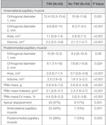
Figure 2. Papillary muscles area and volume quantification in athletes with and without T-wave inversion.
Papillary muscles area and volume quantification in an athlete with TWI in inferolateral leads (A) and in a control athlete with a normal ECG (B). AL indicates anterolateral; and PM, posteromedial.
A
B
et al6 reported that 24 of 123 athletes with TWI had ei-ther cardiomyopathy or oei-ther heart disease diagnosed by echocardiography. In addition, 5 (6%) athletes with TWI and initial normal echocardiography, subsequently developed an overt cardiomyopathy phenotype (3 HCM, 1 arrhythmogenic right ventricular cardiomyopa-thy, 1 dilated cardiomyopathy) over a 9-year follow-up. Schnell et al7 performed a comprehensive investigation including CMR of 155 athletes with TWI; 69 (44%) were diagnosed with structural heart disease, predominantly HCM (n=55). Of note, 88% of the athletes diagnosed with a cardiomyopathy had lateral TWI. The prevalence of disease in the present study was higher than previ-ously reported (59 of 113 athletes; 52%), although HCM
was the most common identified disease (35% of ath-letes) in agreement with previous studies. The use of contrast-enhanced CMR may have accounted for our high prevalence of diagnoses of structural heart dis-eases, which included 11 cases (10%) of isolated noni-schemic LV scar.
PMs Hypertrophy
A previous study by Kobashi et al10 reported that patients with echocardiographic evidence of PMs hypertrophy may show TWI in the absence of other demonstrable heart disease, including HCM. Isolated PMs hypertrophy was subsequently observed on CMR scan in anecdotal young subjects and healthy athletes with TWI of unknown etiology.16–18 Our study results confirm and extend previous observations by providing a systematic and accurate CMR analysis of morpho-functional features of PMs in a cohort of athletes with otherwise unexplained lateral TWI. Our study results demonstrated a significant association between lateral TWI and increased diameters, area, volume, mass, and PMs/LV mass ratio; this latter index was used to normalize the PMs mass with the LV mass, which may be increased in athletes. The mean PMs volume and PMs/LV mass ratio among athletes with TWI were greater than those of control athletes with a normal ECG. According to the receiver operat-ing characteristic curve analysis, a PMs/LV mass ratio >3.5% provided a sensitivity of 85% (ie, 8 of 53 ath-letes with lateral TWI had PMs/LV mass ratio ≤3.5%) and a specificity of 76% (ie, 13 of 53 controls without lateral TWI had PMs/LV mass ratio >3.5%).
PMs Displacement
In our study, lateral TWI was also significantly as-sociated with apical displacement of the PMs,
Table 3. Intraobserver and Interobserver Variability for PMs Quantitative Analysis in Athletes With TWI
Interobserver Variability
Intraobserver Variability
ρ ICC ρ ICC Anterolateral papillary muscle
Orthogonal diameter 1 0.97 0.98 0.98 0.98 Orthogonal diameter 2 0.98 0.98 0.97 0.98 Area 0.96 0.98 0.96 0.97 Volume 0.96 0.94 0.95 0.93 Posteromedial papillary muscle
Orthogonal diameter 1 0.98 0.98 0.98 0.98 Orthogonal diameter 2 0.94 0.96 0.95 0.97 Area 0.92 0.94 0.93 0.94 Volume 0.89 0.91 0.90 0.92 PMs mass indexed 0.89 0.91 0.90 0.92 PMs mass/LV mass 0.88 0.90 0.91 0.91
ICC indicates intraclass correlation coefficient; LV, left ventricular; PMs, papillary muscles; and TWI, T-wave inversion.
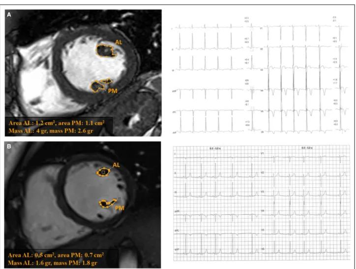
Figure 3. Papillary muscles implantation in athletes with and without T-wave inversion.
Normal papillary muscles implantation on the mid left ventricular wall in a control athlete with normal ECG (A). Papillary muscles apical displacement in an athlete with TWI in the inferolateral leads (B).
A B
particularly the anterolateral. In sarcomeric gene-related HCM, the apical displacement of PMs is a recognized accessory abnormality that can contrib-ute to the dynamic LV outflow tract obstruction.19 Lee et al11 demonstrated that an apically displaced (nonhypertrophic) anterolateral papillary muscle may be associated with an hypertrophy of LV apicolateral segments (but no apicoseptal), which may mimic an apical HCM, also regarding the ECG presentation with giant TWI. This finding indicated that an abnor-mal implant of the anterolateral papillary muscle may cause repolarization abnormality.
In our cohort of athletes with lateral TWI, the apical displacement of the anterolateral papillary muscle was isolated and occurred in the absence of increased thick-ness of apical LV segments and without LV outflow tract gradient, leading to a clear exclusion of an apical HCM. It is noteworthy that PMs hypertrophy was unrelated to the PMs apical displacement, and multivariable analysis showed that both PMs abnormalities were independent predictors of lateral TWI, with an OR of 3.4 and 3.2, re-spectively. Of note, 5 of 8 athletes (63%) with PMs/LV mass ratio <3.5% showed an apical displacement of the PMs.
Clinical Implications
In the study of Kobashi et al,10 one-third of patients with isolated PMs hypertrophy had a family history of HCM, suggesting that this patient subset had a “forme frus-tre” of HCM (without increase of LV thickness). In our study, athletes with lateral TWI associated with PMs abnormalities had a negative family history for HCM and did not show any clinical and imaging features of
HCM, such as symptoms, arrhythmias, LV hypertro-phy, and LGE of LV myocardium.
Pelliccia et al6 reported that failure to detect struc-tural abnormalities on imaging in athletes with TWI does not conclusively exclude an underlying structural heart disease. Indeed, TWI may represent the initial phenotypic expression of a cardiomyopathy (including HCM) that may precede by years the development of structural changes detectable on cardiac imaging. In our study, athletes underwent a repeat imaging study during follow-up with no evidence of subsequent de-velopment of LV hypertrophy consistent with late phenotypic expression of HCM. Finally, the clinical out-come of all athletes with lateral TWI and PMs abnor-malities, who continued to participate in their sports activity, was uneventful.
Nine athletes (17%) underwent molecular screen-ing of a large panel of genes associated with familial cardiomyopathies and channelopathies, which failed to identify any pathogenic or likely pathogenetic muta-tions, including those responsible for HCM. These find-ings are in agreement with those of a previous study showing the negligible added value of molecular geno-typing for identification of an underlying heart disease in athletes with lateral TWI, in the absence of family history and lack of phenotypic expression of cardiomy-opathy, especially HCM.8
All these findings indicate that lateral TWI in asso-ciation with PMs abnormalities, hypertrophy, and/or displacement may occur as a phenotype distinct from that of HCM and may be associated with a favorable outcome in the athlete.
Study Limitations
The study is limited by the retrospective nature, the relatively small sample size, and the short follow-up duration. Although athletes underwent yearly echo-cardiography that did not show progression toward any definite cardiomyopathy, it should be recognized that the exam may be insufficiently sensitive to cap-ture early/minor phenotypic expression because of the limitations of the technique in accurately meas-uring the LV wall thickness, mostly in regions such as the apex and anterior wall. These study limita-tions do not allow us to draw definite conclusions about the clinical meaning and outcome of isolated PMs abnormalities in athletes. However, the princi-pal aim of our study was the correlation, previously unaddressed, between unexplained lateral TWI and isolated PMs abnormalities in the athlete, while the preliminary and short-term follow-up data should be regarded as a parallel but secondary study objective. Molecular genetic testing was performed in a minor-ity of athletes with TWI and PMs abnormalities, pre-cluding any definite conclusion about the etiology of
Figure 4. Box and whisker graph showing the ratio
between papillary muscles mass and left ventricular mass in athletes with and without T-wave inversion in the lateral or inferolateral leads.
PMs abnormalities. Likewise, findings of control ECG and CMR after detraining are inconclusive because the study was limited to few athletes, although they indicate the potential for a normalization of repolari-zation abnormalities in the presence of PMs hyper-trophy reversion.
CONCLUSIONS
The results of our study showed that isolated hyper-trophy and/or displacement of LV PMs may underlie lateral TWI in a sizeable proportion of athletes with oth-erwise structurally normal heart. Overall, 94% of our athletes with unexplained lateral TWI showed a PMs/ LV mass ratio ≥3.5%, an apical displacement of the PMs, or both. According to our study findings, the as-sociation between lateral TWI and PMs abnormalities represents a distinct anatomo-clinical condition char-acterized by a favorable medium-term outcome even in individuals engaged in competitive sports activity.
These findings have important implications for evalu-ation and management of athletes with repolarizevalu-ation abnormalities without clinically demonstrable heart disease.
Future studies on a larger population of athletes with this condition over a long-term follow-up are war-ranted to better define background, clinical meaning, and outcome. Further research should focus on sys-tematic molecular genetic analysis and control ECG and CMR study after detraining.
ARTICLE INFORMATION
Received September 30, 2020; accepted December 11, 2020.
Affiliations
From the Department of Cardiac, Thoracic and Vascular Sciences and Public Health, University of Padova, Italy (M.D.L., A.Z., N.B., A.C., K.P., M.C., M.P.M., D.C.); Center for Sports Medicine, ULSS2 Marca Trevigiana, Treviso, Italy (T.V., P.S.); Interdipartimental Center for Biology and Sports Medicine, University of Pavia, Italy (M.R.G.); San Raffaele Resnati/IRCCS Ospedale San Raffaele, Milano, Italy (F.C.); UOC Cardiologia, Ospedale Ramazzini, Carpi
Figure 5. Summary of main study findings.
CMR indicates cardiac magnetic resonance; LV, left ventricular; PM, papillary muscles; and TWI, T-wave inversion.
(MO), Italy (L.T.); San Tommaso Sports Medicine Center, Ascoli Piceno, Italy (G.R.); Azienda Sanitaria Regionale Marche, Ancona, Italy (I.M.); Department of Neuroscience, Imaging and Clinical Sciences, University of Chieti-Pescara, Chieti Scalo, Italy (F.R.); and Department of Medicine, Radiology Unit, University of Padova, Padova, Italy (R.M.).
Acknowledgments
The authors thank Drs Marino Tonelli, Pierluigi Russo, and Carlo Stefenelli for referring athletes with T-wave inversion.
Sources of Funding
None.
Disclosures
None.
REFERENCES
1. Sharma S, Drezner JA, Baggish A, Papadakis M, Wilson MG, Prutkin JM, La Gerche A, Ackerman MJ, Borjesson M, Salerno JC, et al. International recommendations for electrocardiographic interpretation in athletes. Eur
Heart J. 2018;39:1466–1480. DOI: 10.1093/eurhe artj/ehw631.
2. D’Ascenzi F, Anselmi F, Adami PE, Pelliccia A. Interpretation of T-wave inversion in physiological and pathological conditions: current state and future perspectives. Clin Cardiol. 2020;43:827–833. DOI: 10.1002/ clc.23365.
3. Zorzi A, Vio R, Bettella N, Corrado D. Criteria for interpretation of the athlete’s ECG: a clinical appraisal. Pacing Clin Electrophysiol. 2020;43:882–890.
4. Hanna EB, Glancy DL. ST-segment depression and T-wave inversion: classification, differential diagnosis, and caveats. Cleve Clin J Med. 2011;78:404–414. DOI: 10.3949/ccjm.78a.10077.
5. Zorzi A, Perazzolo Marra M, Rigato I, De Lazzari M, Susana A, Niero A, Pilichou K, Migliore F, Rizzo S, Giorgi B, et al. Nonischemic left ventricular scar as a substrate of life-threatening ventricular arrhyth-mias and sudden cardiac death in competitive athletes. Circ Arrhythm
Electrophysiol. 2016;9:e004229. DOI: 10.1161/CIRCEP.116.004229.
6. Pelliccia A, Di Paolo FM, Quattrini FM, Basso C, Culasso F, Popoli G, De Luca R, Spataro A, Biffi A, Thiene G, et al. Outcomes in athletes with marked ECG repolarization abnormalities. N Engl J Med. 2008;358:152– 161. DOI: 10.1056/NEJMo a060781.
7. Schnell F, Riding N, O’Hanlon R, Axel Lentz P, Donal E, Kervio G, Matelot D, Leurent G, Doutreleau S, Chevalier L, et al. Recognition and significance of pathological T-wave inversions in athletes. Circulation. 2015;8:e003454.
8. Sheikh N, Papadakis M, Wilson M, Malhotra A, Adamuz C, Homfray T, Monserrat L, Behr ER, Sharma S. Diagnostic yield of genetic testing in young athletes with T-wave inversion. Circulation. 2018;138:1184–1194. DOI: 10.1161/CIRCU LATIO NAHA.118.034208.
9. Rajiah P, Fulton NL, Bolen M. Magnetic resonance imaging of the papillary muscles of the left ventricle: normal anatomy, variants, and abnormalities. Insights Imaging. 2019;10:83. DOI: 10.1186/s1324 4-019-0761-3.
10. Kobashi A, Suwa M, Ito T, Otake Y, Hirota Y, Kawamura K. Solitary pap-illary muscle hypertrophy as a possible form of hypertrophic cardiomy-opathy. Jpn Circ J. 1998;62:811–816. DOI: 10.1253/jcj.62.811. 11. Lee SP, Park K, Kim HK, Kim YJ, Sohn DW. Apically displaced
pap-illary muscles mimicking apical hypertrophic cardiomyopathy. Eur
Heart J Cardiovasc Imaging. 2013;14:128–134. DOI: 10.1093/ehjci/
jes113.
12. Gati S, Sharma S, Pennell D. The role of cardiovascular magnetic res-onance imaging in the assessment of highly trained athletes. JACC
Cardiovasc Imaging. 2018;11:247–259.
13. De Lazzari M, Zorzi A, Cipriani A, Susana A, Mastella G, Rizzo A, Rigato I, Bauce B, Giorgi B, Lacognata C, et al. Relationship between electro-cardiographic findings and cardiac magnetic resonance phenotypes in arrhythmogenic cardiomyopathy. J Am Heart Assoc. 2018;7:e009855. DOI: 10.1161/JAHA.118.009855.
14. Harrigan CJ, Appelbaum E, Maron BJ, Buros JL, Gibson CM, Lesser JR, Udelson JE, Manning WJ, Maron MS. Significance of papillary mus-cle abnormalities identified by cardiovascular magnetic resonance in hypertrophic cardiomyopathy. Am J Cardiol. 2008;101:668–673. DOI: 10.1016/j.amjca rd.2007.10.032.
15. Protocolli Cardiologici Per il Giudizio Dell’idoneità Allo Sport Agonistico
(COCIS). Rome: Casa Editrice Scientifica Internazionale; 2017.
16. Correia AS, Pinho T, Madureira AJ, Araujo V, MacIel MJ. Isolated pap-illary muscle hypertrophy: a variant of hypertrophic cardiomyopathy? Do not miss a hypertrophic cardiomyopathy. Eur Heart J Cardiovasc
Imaging. 2013;14:827–828.
17. Ferreira C, Delgado C, Vázquez M, Trinidad C, Vilar M. Isolated papillary muscle hypertrophy: a gap in our knowledge of hypertrophic cardio-myopathy? Rev Port Cardiol. 2014;33:379.e1–379.e5. DOI: 10.1016/j. repc.2014.01.015.
18. Işilak Z, Uz Ö, Uzun M, Cebeci BS. An unusual cause of electrocar-diographic abnormality: solitary papillary muscle hypertrophy. Turk
Kardiyol Dern Ars. 2012;40:473.
19. Kwon DH, Setser RM, Thamilarasan M, Popovic ZV, Smedira NG, Schoenhagen P, Garcia MJ, Lever HM, Desai MY. Abnormal papillary muscle morphology is independently associated with increased left ventricular outflow tract obstruction in hypertrophic cardiomyopathy.
Heart. 2008;94:1295–1301. DOI: 10.1136/hrt.2007.118018.