Crucial pathophysiological role of CXCR2 in experimental
ulcerative colitis in mice
Pasquale Buanne,* Emma Di Carlo,
†,‡Lorenzo Caputi,* Laura Brandolini,* Marco Mosca,*
Franca Cattani,* Luigi Pellegrini,* Leda Biordi,
§Gino Coletti,
¶Carlo Sorrentino,
†,‡Guido Fedele,
㛳Francesco Colotta,*
,#Gabriella Melillo,* and Riccardo Bertini*
,1*Department of Preclinical Pharmacology, Dompe´ pha.r.ma s.p.a., L’Aquila, Italy;
†Department of Oncology and
Neurosciences, Surgical Pathology Section, "G. d'Annunzio" University, Chieti, Italy;
‡Ce. S.I. Aging Research
Center, “G. d'Annunzio” University Foundation, Chieti, Italy;
§Experimental Medicine Department, University of
L’Aquila, L’Aquila, Italy;
¶Operative Unit of Pathological Anatomy, S. Salvatore Regional Hospital, L’Aquila, Italy;
㛳Biometry Unit, Dompe´ pha.r.ma s.p.a., Milan, Italy; and
#Nerviano Medical Sciences, Milan, Italy
Abstract:
Polymorphonuclear leukocyte
infiltra-tion and activainfiltra-tion into colonic mucosa are
be-lieved to play a pivotal role in mediating tissue
damage in human ulcerative colitis (UC). Ligands
of human CXC chemokine receptor 1 and 2
(CXCR1/R2) are chemoattractants of PMN, and
high levels were found in the mucosa of UC
pa-tients. To investigate the pathophysiological role
played by CXCR2 in experimental UC, we induced
chronic experimental colitis in WT and CXCR2
ⴚ/ⴚmice by two consecutive cycles of 4% dextran
sulfate sodium administration in drinking water. In
wild-type (WT) mice, the chronic relapsing of
DSS-induced colitis was characterized by clinical signs
and histopathological findings that closely resemble
human disease. CXCR2
ⴚ/ⴚmice failed to show
PMN infiltration into the mucosa and, consistently
with a key role of PMN in mediating tissue damage
in UC, showed limited signs of mucosal damage and
reduced clinical symptoms. Our data demonstrate
that CXCR2 plays a key pathophysiological role in
experimental UC, suggesting that CXCR2
activa-tion may represent a relevant pharmacological
tar-get for the design of novel pharmacological
treat-ments in human UC. J. Leukoc. Biol. 82:
1239 –1246; 2007.
Key Words:
rodent
䡠neutrophils
䡠inflammation
INTRODUCTION
Ulcerative colitis is a chronic inflammatory bowel disease
(IBD), clinically characterized by frequent diarrheic attacks
and anal bleeding. Histological hallmark of UC are the
inva-sion of crypt epithelium and lamina propria by PMN,
disrup-tion of the epithelial lining, and, consequently, mucosal
ulcer-ation and crypt abscess formulcer-ation in the bowel wall [1, 2]. PMN
infiltration is believed to play a pivotal role in mediating tissue
damage in UC. In animal studies an association between
colonic neutrophilia and progression of acute colitis was
served [3, 4]. In humans, PMN accumulation has been
ob-served in rectal biopsies of patients with active UC relative to
healthy controls [5, 6].
Interleukin-8 (IL-8/CXCL8), a member of CXC chemokines,
is a chemoattractant of PMN. Two high-affinity human CXCL8
receptors are known, CXC chemokine receptor 1 (CXCR1) and
CXC chemokine receptor 2 (CXCR2). Although a mouse
or-tholog of CXCL8 has not been identified, the two corresponding
receptors have been identified in the mouse [7, 8]. By
recruit-ing and activatrecruit-ing PMN, CXCL8 has been implicated in a wide
range of disease states characterized by PMN infiltration in
different organs [9], including UC [10]. CXCL8 is expressed in
the colonic mucosa of patients affected by UC [11, 12] and IBD
[13, 14] and a correlation between CXCL8 levels, PMN number
in mucosal tissue, and the severity of UC has also been
described [15, 16].
To investigate the pathophysiological role played by CXCR2
and its specific ligands in experimental UC, we induced
chronic relapsing DSS-mediated colitis in WT and CXCR2
⫺/⫺mice. Among animal models of UC, the chronic relapsing
DSS-induced colitis resembles human UC under several
fea-tures, including clinical symptoms and histopathological
changes [17]. CXCR2
⫺/⫺mice were reported to bear and
develop normally, but their PMN were unable to migrate in
response to its specific ligands CXCL1 (GRO
␣/⌲C) and
CXCL2 (GRO
/⌴⌱P-2) when compared with their WT
litter-mates [18]. The results reported hereafter point out that the
functional lacking of CXCR2 produced an amelioration of
experimental UC, as shown by a significant improvement of
clinical conditions paralleled by a reduction of the
histopatho-logical hallmarks of this disease model. These data are in
keeping with a key role of CXCR2 in mediating PMN
recruit-ment and tissue damage in experirecruit-mental UC, suggesting that
CXCR2 activation may represent a relevant pharmacological
target for the design of novel pharmacological treatments in
human UC.
1Correspondence: Department of Preclinical Pharmacology, Dompe´ pha.r.ma s.p.a., Via Campo di Pile 67100 L’Aquila, Italy. E-mail: [email protected]
Received February 19, 2007; revised March 28, 2007; accepted June 21, 2007.
MATERIALS AND METHODS
Animals
Male, 6-wk-old BALB/c mice weighing 19-23 g were obtained from Charles River Laboratories (Calco, Lecco, Italy). Male CXCR2⫺/⫺mice on BALB/c background (strain C129S2 (B6)-IL8RB) were derived from founders provided by Jackson Laboratories (Bar Harbor, ME, USA) and genotyped by polymerase chain reaction (PCR) [19]. The animals were housed in cages up to five mice each and acclimated for 1 wk under conditions of controlled temperature (20°C⫾2), humidity (55%⫾10), and lighting (7:00 AM–7:00 PM). Sterilized diet No. 48 pellets (Laboratorio Dottori Piccioni, Gessate, Milan, Italy) and water were supplied ad libitum during acclimatization and experimental ses-sions.
All the procedures were performed in the animal operating rooms, according to ethical guidelines for the conduct of animal research (Authorization Italian Ministry of Health No. 50/2001-B; Italian Legislative Decree 116/92, Gazzetta Ufficiale della Repubblica Italiana No. 40, February 18, 1992; EEC Council Directive 86/609, OJL 358, 1, December 12, 1987; National Institutes of Health Guide for the Care and Use of Laboratory Animals, NIH Pub. No. 85-23, 1985).
Chronic DSS colitis model
Chronic colitis was induced by 4% DSS (36 –50 kDa, ICN Biochemicals, Milan, Italy) in drinking water in two consecutive cycles, as previously reported [20, 21]. The first DSS cycle was from day 0 to day 6. This cycle was followed by 14 days of simple ultrafiltered water (1st washout phase), from day 6 to day 20. The second DSS cycle (from day 20 to day 24) induced the relapse of the disease and was followed by three days of ultrafiltered water (2nd washout phase), from day 24 to day 27. Control group received water during all the experimental sessions. During the setup of the model, colitis was induced in Balb/c mice obtained from Charles River Laboratories and WT mice (CXCR2⫹/⫹) derived from founders by Jackson Laboratories. No differences were seen in terms of clinical and histopathological response between the two inbred Balb/c strains. In all of the experiments reported hereafter, WT mice derived from founders by Jackson Laboratories were used.
Clinical evaluation of DSS-induced colitis
Disease progression was determined at crucial time points by assessment of disease activity index (DAI), which ranged from 0 to 7. DAI score was calculated accordingly to previous reports [17, 22], adding two separate scores: Stool score (0⫽Normal; 1⫽⬎30% pellets with smooth consistency or ⬍30% pellets with diarrheic consistency; 2⫽30–70% pellets with diarrheic consis-tency; 3⫽⬎70% pellets with diarrheic consistency) and Emo score (0⫽ negative; 1⫽occult blood positive; 2⫽small blood drops on the pellet; 3⫽ gross anal bleeding; 4⫽blood drops at the bottom of the cage). Occult blood in feces was determined by Hemo-fec (Roche Diagnostics, Mannheim, Germany).
Histopathological, ultrastructural,
immunohistochemical, and immunofluorescent
analysis
Colon from the ileocecal valve to the anus was removed, washed in ice-cold saline, fixed in 10% neutral buffered formalin, embedded in paraffin and stained with hematoxylin and eosin. Histopathological score was assigned by two blinded pathologists. According to grading scales of histological assess-ment in human UC [23, 24], histopathological score was calculated as sum of single scores (ranged from 0 to 15), taking into account crypt distortion, loss of muco-secretive capability, percentage of severe ulcers, amount of inflammatory infiltrate in the severe ulcers expressed as granulocytes/severe ulcer thickness ratio (G/U ratio), and extension of tissue damage. The amount of inflammatory infiltrate in severe ulcers (expressed as G/U ratio) was singularly considered and defined as inflammatory score ranging from 0 to 5.
For electron microscopy, specimens were fixed in cacodylate buffered 2.5% glutaraldehyde, postfixed in osmium tetroxide and embedded in Epon 812. Ultrathin sections were stained with uranyl acetate-lead citrate.
For immunohistochemistry, large intestines were embedded in optimum cutting temperature compound (Miles), snap-frozen in liquid nitrogen and stored at – 80°C. Then, frozen samples were sectioned, air dried overnight,
fixed with acetone, and immunostained with anti-CXCL1 (Peprotech, Rocky Hill, NJ), CD11c (Chemicon International, Temecula, CA, USA), anti-CD11b (Mac-1; Sera-Lab), anti-cytokeratins (CK) 8/18 (Progen Biotechnick, Heidelberg, Germany), anti-CD8 (Ly/T2; Sera-Lab), and anti-CD4 (LT34; Sera-Lab), anti-GR-1 (American Type Culture Collection) Abs. After washing, sections were overlaid with biotinylated goat anti-rat, anti-hamster, anti-rabbit and horse anti-goat Ig (Vector Laboratories, Burlingame, CA, USA). Unbound Ig was removed by washing, and slides were incubated with ABC (avidin/biotin complex)/alkaline phosphatase (DakoCytomation, Glostrup, Denmark).
For double immunofluorescent staining and confocal analysis, acetone-fixed frozen sections were incubated with the first primary antibodies and then with biotinylated secondary antibodies. After washing, sections were incubated with the second primary antibodies and, after washing, with biotinylated secondary antibodies and then with Alexa Fluor 594 conjugated StreptAvidin. Cross-reactions between the first secondary antibodies and Alexa Fluor 594 was prevented by saturation of all of its binding sites with Alexa Fluor 488. Slides were mounted with Vectashield medium (Vector Laboratories) and examined with a LSM 510 Meta laser scanning confocal microscope (Zeiss, Jena, Germany).
Myeloperoxidase activity
Three centimeters of colon from anus were ice-homogenized (Ultra–Turrax, Ika-Werk) in a tube containing 1 ml/100 mg of tissue of 50 mM PBS, pH 6.0 and 0.5% HTAB (Sigma-Aldrich, St. Louis, MO, USA). Then samples were ice-sonicated (Ultrasonic processor XL) and ultracentrifuged (Sorvall RC5C) at 40,000 g at 4°C [25]. The supernatants were assayed for MPO activity [26]. Protein assay was performed by bicinchoninic acid method (Pierce Biotech-nology, Rockford, IL, USA).
CXCL1 and CXCL2 production
Three centimeters of colon from the anus were removed, ice-homogenized with protease inhibitors (5g/ml leupeptin, pepstatin, chimostatin, aprotinin and 1 mM PMSF; Sigma-Aldrich) and centrifuged at 20,800 g at 4°C (Centrifuge 5417 R, Eppendorf). Measurement of CXCL1 and CXCL2 was performed in the supernatants by using mouse CXCL1 and CXCL2 ELISA kits (R&D Systems, Minneapolis, MN, USA), according to manufacturer’s instructions.
Statistical analysis
Data are expressed as means⫾SEM. All the analyses were conducted by using SAS 9,1 TS Level 1M3 in Windows XP professional environment.
DAI score was analyzed by Cochran-Mantel-Haenszel test (CMH) [27] and mixed linear models of analysis of variance (ANOVA) for repeated measures on data transformed by ranks [28]. Histopathological and inflammatory scores were processed by applying both Exact2-test or CMH and mixed linear models of ANOVA for a model completely random on data transformed by ranks. ANOVA followed by Dunnett’s multiple comparison test was performed for CXCL1 and CXCL2 production data.
RESULTS
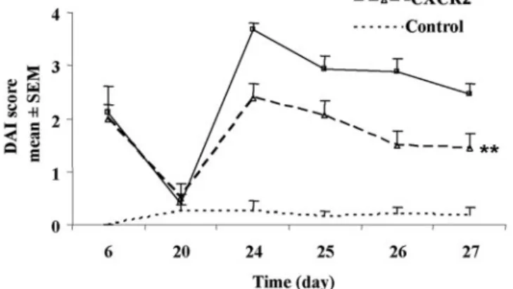
Lacking of CXCR2 reduces clinical signs in
DSS-induced chronic colitis
The chronic relapsing DSS-induced colitis was induced by the
administration of two cycles of DSS, with a washout period in
between. In this model, as described previously [17, 22], the
first 6-day cycle of DSS induced in WT mice an acute
inflam-mation with a spontaneous gradual resolution when DSS
ad-ministration was stopped.
To induce relapsing colitis, a second cycle of DSS
admin-istration was started at day 20 until day 24, as described
previously [20]. DAI score increased in a time-dependent
manner, reaching the maximum value at day 24 and persisting
until day 27.
To assess the role of CXCR2 in the chronic relapsing
dis-ease, DSS colitis was induced in CXCR2
⫺/⫺mice. As shown in
Fig. 1
, the first cycle of DSS administration induced in
CXCR2
⫺/⫺mice a clinical outcome similar to WT mice
(2.00
⫾ 0.51 and 2.11 ⫾ 0.50 in CXCR2
⫺/⫺and WT mice at
day 6, respectively). DSS-second cycle caused a
time-depen-dent increase in DSS-treated WT mice, reaching the maximum
value at day 24. A significant reduction (P
⬍0.001) of clinical
signs of colitis (from 30% up to 48% of inhibition at day 24 and
day 27, respectively) was observed in CXCR2
⫺/⫺mice in
comparison with WT. At day 27, the time of sacrifice, the DAI
score of CXCR2
⫺/⫺group was 42% lower than that of WT
group (1.44
⫾ 0.22 and 2.47 ⫾ 0.19 in CXCR2
⫺/⫺and WT
mice, respectively) (Fig. 1). The clinical score improvement
observed in CXCR2
⫺/⫺mice was evident both as stool
con-sistency and as blood in feces.
Reduction of colonic mucosa damage in
CXCR2
⫺/⫺mice
To gain insights into the cellular mechanisms underlying the
reduced severity of experimental colitis in CXCR2
⫺/⫺mice,
we carried out a histopathological analysis of gut lesions
in-duced in both CXCR2
⫺/⫺and WT mice. The histopathological
analysis included both nonspecific gut lesions (e.g., crypt
dis-tortion) and inflammatory-specific lesions (e.g., severe ulcer
formation).
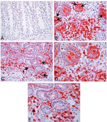
According to previous reports [17, 22], the first cycle of DSS
administration induced a pathological status with a low
in-volvement of inflammatory cells and reduced tissue damage.
Only few PMN, lymphocytes, and plasma cells, incoming from
submucosa, were detected between crypts and into crypt
epi-thelium. Initial erosions were also observed but without PMN
stratifications (data not shown).
During the second DSS cycle, clinical symptoms (see above)
were related by progressive histopathological changes,
charac-terized by a dramatic infiltration of PMN from the lamina
propria into the colonic mucosa causing severe ulcers (Fig.
2C–E)
and microabscesses formation (Fig. 2F). No
histopatho-logical change was reported in control group (Fig. 2A, B). As
shown in Fig. 3, massive PMN recruitment into colonic
seg-ment was further confirmed by increased tissue MPO, a
well-known specific marker of PMN (5.81
⫾ 0.11 and 171.25 ⫾
12.16 mU/mg protein in water and DSS-treated WT mice at day
27, respectively). Large tracts of mucosa lost the epithelium
Fig. 1. Clinical evaluation of DSS-induced colitis in wild-type (WT) and CXC chemokine receptor 2 (CXCR2⫺/⫺) mice. The clinical progression of the colitis was determined by disease activity index (DAI), calculated by adding two separate scores (ranging from 0 to 7): Stool score (0⫽normal; 1⫽⬎30% pellets with smooth consistency or⬍30% pellets with diarrheic consistency; 2⫽30– 70% pellets with diarrheic consistency; 3⫽⬎70% pellets with diarrheic consistency) and Emo score (0⫽negative; 1⫽occult blood positive; 2⫽small blood drops on the pellet; 3⫽gross anal bleeding; 4⫽blood drops at the bottom of the cage). Colitis was induced in WT and CXCR2⫺/⫺mice by giving 4% DSS in drinking water in two cycles. Control group received water. Data are reported as means ⫾ SEM (24 –35 animals for each time point from one experiment of three). **, P⬍ 0.01 vs. DSS-treated WT group by ANOVA.
Fig. 2. Histopathological analysis of DSS-induced colitis in WT and CXCR2⫺/⫺mice. Colon from the ileocecal valve to the anus was removed and stained with hematoxylin and eosin. (A) and (B) control mice. At day 27, DSS induced massive infiltration of PMN into the crypt epithelial layer with necrosis and ulcerative lesions of colonic mucosa (C), (D), large tracts of mucosa lost crypt epithelium appeared to be replaced by granulation tissue (E) associated with a dramatic infiltration of PMN, causing microabscesses forma-tion (F) in WT mice. The overall aspect of colonic mucosa was less severe in CXCR2⫺/⫺mice. A significant decrease of PMN aggregates was associated with a reduction of severe ulcers formation (G), (H). The inflammatory cells (indicated by arrow) were mainly confined into microvascular space, confined around the crypts (I). Muco-secretive ability preserved in CXCR2⫺/⫺mice (G), (H), (L). Original magnification,⫻100 (A), (C), (D),(G); ⫻200 (B), (H); ⫻400 (E), (I), (L);⫻1000 (F).
and was replaced by granulation tissue (Fig. 2E). The overall
histopathological and inflammatory scores peaked at day 27
(Fig. 4A, B).
When CXCR2
⫺/⫺mice were examined at day 27,
DSS-treated CXCR2
⫺/⫺mice showed PMN accumulation in the
lumen of blood vessels and only limited extravasation and
infiltration into the tissue. The few infiltrating PMN remained
in close proximity of microvasculature, confined around the
crypts and rarely penetrated the basal membrane, (Fig. 2I; 5C,
E; and 6C–D) whereas in DSS-treated WT mice, PMN
infil-trated the crypt epithelial layer, reaching tight contact with
epithelial crypts thus causing necrotic and ulcerative lesions in
colonic mucosa (Fig. 5B, D and Fig. 6A, B). In addition to
neutrophils, also eosinophils infiltrate largely the mucosa of
DSS-treated WT mice (Fig. 6A, B). These data are in keeping
with previous descriptions of DSS-induced chronic colitis in
the mouse [17, 29]. As shown in Fig. 3, reduction of PMN
infiltration in CXCR2
⫺/⫺mice was also confirmed by a
signif-icant (P
⬍ 0.01) reduction of mucosal MPO activity (171.25 ⫾
12.16 and 76.61
⫾ 8.42 mU/mg of protein in WT and
CXCR2
⫺/⫺mice, respectively). Areas of the mucosa retaining
muco-secretive properties (maintenance of goblet cells) were,
unlike WT counterparts (Fig. 2C–E), evident in CXCR2
⫺/⫺mice (Fig. 2G–H and 2L). However, the presence of mitotic
figures and “pseudo-stratification” of the crypt epithelium were
observed in CXCR2
⫺/⫺mice, providing evidence of the insult
and the following regenerative process (Fig. 2L). Moreover, in
CXCR2
⫺/⫺mice, a significant decrease in the number of
severe ulcers was observed (Fig. 2G). The overall
histopatho-logical and inflammatory scores were significantly reduced in
CXCR2
⫺/⫺mice at day 27 (53% and 56% of reduction,
respectively) (Fig. 4A, B). At day 20, starting time of second
cycle of DSS, no significant differences between WT and
Fig. 3. MPO activity in the colon of DSS-treated WT and CXCR2⫺/⫺mice.WT and CXCR2⫺/⫺mice were killed at day 27 after DSS administration, and colonic mucosa were processed for determination of MPO activity. Control group received normal water. (**, P⬍0.01 vs. DSS-treated WT group by ANOVA). Data are expressed as mean⫾SEM(14 animals for each experi-mental group from one experiment of three).
Fig. 4. Histopathological and inflammatory scores in DSS-treated WT and CXCR2⫺/⫺ mice. Animals were killed during the chronic phase of DSS-induced colitis, and histopathological analysis was performed in the tract of the colon mainly involved by disease. (A) Histopathological score in CXCR2⫺/⫺ and WT mice (**, P ⬍ 0.01 vs. DSS-treated WT group by ANOVA). (B) Inflammatory score in CXCR2⫺/⫺and WT mice (**, P⬍0.01 vs. DSS-treated WT group by ANOVA). Data are expressed as means⫾SEM(14 –25 animals for each time point from one experiment of three).
Fig. 5. Immunohistochemical analysis of colon section from WT and CXCR2⫺/⫺ mice. Immunohistochemistry on intestinal sections from mice exposed to normal water or two cycles of DSS and killed at day 27. (A) Control (water-treated) WT animals. (B) DSS-treated WT mice. PMN infiltrated (ar-rows) and destroyed the crypt epithelial layer. (C) DSS-treated CXCR2⫺/⫺ mice. PMN were mostly confined among the crypts (arrows). (D) DSS-treated WT mice. Magnification of details indicated by arrows in (B). (E) DSS-treated CXCR2⫺/⫺mice. Magnification of details indicated by arrows in (C). Original magnification,⫻400.
CXCR2
⫺/⫺mice were observed. However, in both
experimen-tal groups, there was an increase over baseline of
histopatho-logical and inflammatory scores in comparison with control
group (Fig. 4A, B), indicating that a few histopathological signs
of the insult still remained, even though they were not
clini-cally detectable.
Thus, whereas WT mice develop a chronic colitis
charac-terized by massive PMN infiltration and mucosal erosion,
CXCR2
⫺/⫺mice show no PMN infiltration into the mucosa and
limited signs of mucosal damage, confirming the key role of
PMN in mediating tissue injury in colitis.
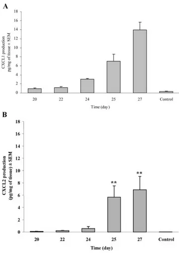
Time-dependent increase of production of
CXCR2 ligands in DSS-treated WT
To further evaluate the different clinical and histopathological
course of colitis in WT vs. CXCR2
⫺/⫺mice, we examined the
expression of CXCR2 ligands into colonic mucosa after DSS
administration in both WT and CXCR2
⫺/⫺mice. As shown in
Fig. 7A
, CXCL1 expression was almost absent in
water-treated WT mice. During the chronic colitis, CXCL1 was
consistently expressed in WT mice, peaking at day 27
(13.96
⫾1.73 pg/mg of tissue). CXCL1 mucosal production was
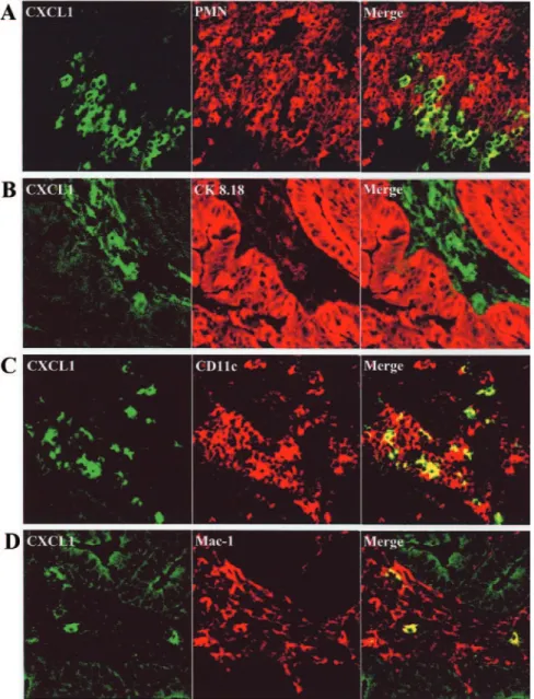
further confirmed by confocal microscopy (Fig. 8). CXCL1 was
found to be expressed in the stroma of the lamina propria,
especially close to the areas of PMN recruitment (Fig. 8A),
mainly by (CD11c
⫹) dendritic cells (Fig. 8C) and (CD11b
⫹)
macrophages to a lesser extent (Fig. 8D), whereas epithelial
cells of the intestinal crypts were mostly negative (Fig. 8B).
Also CXCL2 was consistently expressed in WT mice, with
CXCL2 levels peaking again at day 27 (0.03
⫾0.00 and
6.89
⫾2.16 pg/mg of tissue in water-treated and DSS-treated
animals, respectively (Fig. 7B).
When CXCR2
⫺/⫺mice exposed to DSS were examined, we
found that the CXCR2 ligand CXCL1 was expressed in the gut
mucosa at levels similar to WT mice (e.g., 13.96
⫾1.73 and
15.91
⫾ 1.13 pg/mg of tissue at day 27 in WT and CXCR2
⫺/⫺Fig. 6.Ultrastructural features of colonic lamina pro-pria from DSS-induced chronic colitis in WT and CXCR2⫺/⫺ mice. Colon sections from WT and CXCR2⫺/⫺mice killed at day 27 after DSS adminis-tration. (A, B) WT mice. In the colonic lamina propria, both eosinophils (E) and neutrophils (N) largely extrav-asated. Endothelial cells are indicated by arrow; vessel lumen indicated by asterisk. Eosinophils and neutro-phils spread to the stroma, reaching tight contact with epithelial crypts (indicated by arrows). (C, D) CXCR2⫺/⫺ mice. PMN extravasated in the colonic lamina propria but did not migrate, remaining in close proximity to microvessel walls (indicated by arrows). PMN stopped and filled microvessels. Bars in A and B⫽ 100 m. Bars in C and D ⫽ 50 m.
Fig. 7. Expression of CXCL1 and CXCL2 in the colon of DSS-treated WT mice. WT mice were killed after different times of DSS administration, and colonic mucosa was processed for CXCL1 and CXCL2 detection. Control group received normal water. (A) Colonic production of CXCL1 (**, P⬍0.01 vs. control group by ANOVA). (B) Colonic production of CXCL2 (**, P⬍0.01 vs. control group by ANOVA). Data are expressed as means⫾SEM(14 animals for each time point).
mice, respectively). An analogous result was observed for
CXCL2 colonic expression (e.g., 5.24
⫾4.42 and 2.58⫾1.25
pg/mg of tissue at day 27 in WT and CXCR2
⫺/⫺mice,
respec-tively).
DISCUSSION
The infiltration of PMN into the colonic mucosa is believed to
play a key role in mediating tissue damage and clinical symptoms
in both human and experimental UC. We demonstrate here that
PMN infiltration in the model of chronic colitis is mainly mediated
by CXCR2. CXCR2
⫺/⫺mice have limited, if any, PMN
infiltra-tion into the colonic mucosa in the relapsing phase of the disease.
Accordingly with a key role played by PMN in mediating tissue
damage in colitis, histopathologically detected mucosal damage,
including mucosal ulcerations, and clinical signs were reduced in
mice lacking a functional CXCR2.
Ligands of CXCR2 were expressed at high levels in
exper-imental colitis of both WT and CXCR2
⫺/⫺mice, and their
production was strongly related to the increase of PMN
recruit-ment into colonic mucosa of WT mice.
To assess the role of CXCR2, we used a model of
DSS-induced colitis that closely resembles human UC under several
features [30, 31]. In this model, repeated DSS administration
induced epithelial atrophy, extensive loss of mucus production,
widespread hyperplasia in large areas of the colonic mucosa,
complete loss of crypt epithelium in large tract of mucosa,
crypt abscesses, severe ulcers and, most notably, a massive
infiltration of PMN into colonic mucosa. Also, from the clinical
point of view, morphological changes observed after chronic
administration of DSS were paralleled by clinical symptoms
induced by repeated DSS administration that closely resemble
severe human UC, including diarrhea and gross rectal bleeding
[30]. Moreover, selected drugs currently used in the therapy of
UC are also efficacious in this model of repeated DSS
admin-istration, including cyclosporin A [32] and infliximab [21, 33],
a monoclonal antibody anti-TNF-
␣ [34–36], further suggesting
that the DSS model we used closely resembles the human
condition.
Fig. 8. Confocal analysis of CXCL1 expression in colonic mucosa of DSS-treated WT mice. Colon sec-tions from WT mice exposed to two cycles of DSS and killed at day 27. (A) Massive influx of PMN (red stained) close to the mucosal areas rich in CXCL1 expression (green stained) without aspects of colocal-ization, as shown by the merge image. (B) Expression of CXCL1 (green stained) was mainly among the intestinal epithelial (red stained) crypts. (C) Most of dendritic cells (CD11c, red stained) infiltrating the lamina pro-pria produce CXCL1 (green stained), as revealed by the yellow color in the merge image. (D) Macrophages (CD11b, red stained) infiltrating the lamina propria cooperate to CXCL1 production (green stained), as re-vealed by the yellow color in the merge image. Original magnification,⫻400.
In the DSS-induced colitis, both high levels of CXCR2
ligands and a massive recruitment of PMN were evident in WT
mice during the chronic phase of disease.
The main histopathological changes and clinical symptoms
in the DSS-induced experimental colitis were strongly reduced
in CXCR2
⫺/⫺mice. Lack of functional CXCR2 prevented
PMN infiltration in the mucosa, suggesting that PMN
recruit-ment into the gut mucosa is mainly attributable to CXCR2
activation. Inhibition of PMN recruitment was associated with
a strong reduction in all histopathological changes associated
with experimental colitis, including necrotic and ulcerative
lesions in the colonic mucosa. Large areas of mucosa with
intact muco-secretive properties were also observed in
CXCR2
⫺/⫺mice, whereas in WT mice, there was a strong
reduction of mucus production, and large tracts of mucosa lost
all crypt epithelium, leaving in its place granulation tissue.
These data are further in keeping with the concept that PMN
recruitment and activation in UC are key events in inducing
mucosal damage and clinical symptoms. This evidence was
apparently in contrast to a previous report [37], in which the
abrogation of PMN has a deleterious effect on DSS-induced
colitis. However, these results are referred to a single cycle of
DSS administration, an experimental condition that does not
resemble UC condition. Indeed, a single cycle of DSS
admin-istration induces a pathological state, with a low involvement of
inflammatory cells and reduced tissue damage, mainly caused
by the toxic effect of DSS on colonic epithelial cells [17, 22].
In addition, the efficacy of treatment with a monoclonal
anti-body anti-TNF-
␣ and corticosteroids clinically proved in
UC was not experimentally observed after a single cycle of
DSS [38].
Whereas intraepithelial infiltration of PMN seems to be
mediated mainly by CXCR2, we found that lack of CXCR2 has
no effect on intravascular accumulation of leukocytes. These
findings suggest that intravascular leukocyte recruitment might
be under the control of other leukocyte-activating factors, such
as TNF-
␣ [20] and products of the complement system [39]. On
the other hand, the impairment of PMN to cross endothelial
surface and subsequently infiltration into the crypt epithelial
barrier strongly suggest that PMN recruitment into the mucosa
could be mainly dependent upon CXCR2 activity on
endothe-lial and epitheendothe-lial cells. In keeping with this hypothesis,
sev-eral reports support the crucial role played by CXCR1 and
CXCR2 activity on endothelial and epithelial cell layer in
chemokine-mediated PMN arrest and subsequent
transmigra-tion into inflamed tissue [40 – 43]. Indeed, PMN of CXCR2
⫺/⫺mice were unable to cross the epithelial barrier and
accumu-late in the tissue in an experimental model of infection of the
urinary tract [42], and immunoneutralization of CXCL1 and
CXCL2 did not reduce endotoxin-induced leukocyte
intravas-cular accumulation, whereas PMN transmigration and
ex-travascular tissue accumulation was abolished [40]. Similarly,
CXCR1 and CXCR2 overexpression was detected in infected
human uroepithelial cell lines, and neutralization of CXCR1
strongly inhibited CXCL8-dependent PMN migration across
the epithelial cell layer [42]. Finally, the crucial role of CXCR2
for transendothelial and transepithelial migration of PMN, but
not for their accumulation in the vasculature, was also reported
in a murine model of acute lung injury in which PMN of
CXCR2
–/–mice did not migrate into the lung interstitium
following endotoxin aerosolization, remaining into the
pulmo-nary vasculature without inducing lung damage [44].
Although PMN infiltration is believed to be a key event in
DSS-induced colitis, information as to the soluble factors
in-volved in the regulation of PMN recruitment in this model are
scanty. Here, we demonstrate that CXCR2 ligands (CXCL1 and
CXCL2) are highly expressed in the stroma of the lamina
propria, close to the areas of PMN recruitment. Production of
these PMN attractants was mainly attributable to mononuclear
and dendritic cells in the colonic mucosa. High levels of both
CXCR1/R2 expression and CXCR1/2 ligands have also been
found in the mucosa of UC patients [45].
Taken together, these findings demonstrate that CXCR2
plays a key, nonredundant role in mediating PMN infiltration
into the mucosa in an animal model of colitis that closely
resembles human UC. Prevention of PMN infiltration was
as-sociated with a strong reduction in both histopathological signs
of the disease and clinical symptoms. These data may provide
the rationale for testing CXCL8 receptors inhibitors as a new
therapeutic approach to UC in the clinic.
REFERENCES
1. Ajuebor, M. N., Swain, M. G. (2002) Role of chemokines and chemokine receptors in the gastrointestinal tract. Immunology 105, 137–143. 2. Podolsky, D. K. (2002) Inflammatory bowel disease. N. Engl. J. Med.
347,417– 429.
3. Palmen, M. J., Dijkstra, C. D., van der Ende, M. B., Pena, A. S., van Rees, E. P. (1995) Anti-CD11b/CD18 antibodies reduce inflammation in acute colitis in rats. Clin. Exp. Immunol. 101, 351–356.
4. Natsui, M., Kawasaki, K., Takizawa, H., Hayashi, S. I., Matsuda, Y., Sugimura, K., Seki, K., Narisawa, R., Sendo, F., Asakura, H. (1997) Selective depletion of neutrophils by a monoclonal antibody, RP-3, sup-presses dextran sulphate sodium-induced colitis in rats. J. Gastroenterol.
Hepatol. 12, 801– 808.
5. Mazzucchelli, L., Hauser, C., Zgraggen, K., Wagner, H., Hess, M., Lais-sue, J. A., Mueller, C. (1994) Expression of interleukin-8 gene in matory bowel disease is related to the histological grade of active inflam-mation. Am. J. Pathol. 144, 997–1007.
6. Uguccioni, M., Gionchetti, P., Robbiani, D. F., Rizzello, F., Peruzzo, S., Campieri, M., Baggiolini, M. (1999) Increased expression of IP-10, IL-8, MCP-1, and MCP-3 in ulcerative colitis. Am. J. Pathol. 155, 331–336. 7. Fan, X., Patera, A. C., Pong-Kennedy, A., Deno, G., Gonsiorek, W., Mandra, J. D., Vassileva, G., Zeng, A., Jackson, C., et al. (2006) Murine CXCR1 is a functional receptor for GCP-2/CXCL6 and IL-8/ CXCL8.
J. Biol. Chem., In press.
8. Lee, J., Cacalano, G., Camerato, T., Toy, K., Moore, M. W., Wood, W. I. (1995) Chemokine binding and activities mediated by the mouse IL-8 receptor. J. Immunol. 155, 2158 –2164.
9. Gerard, C., Rollins, B. J. (2001) Chemokine and disease. Nat. Immunol. 2,108 –115.
10. Keshavarzian, A., Fusunyan, R. D., Jacyno, M., Winship, D., MacDermott, R. P., Sanderson, I. R. (1999) Increased interleukin-8 (IL-8) in rectal dialysate from patients with ulcerative colitis: evidence for a biological role for IL-8 in inflammation of the colon. Am. J. Gastroenterol. 94, 704 –712.
11. Banks, C., Bateman, A., Payne, R., Johnson, P., Sheron, N. (2003) Chemokine expression in IBD. Mucosal chemokine expression is unselec-tively increased in both ulcerative colitis and Crohn’s disease. J. Pathol. 199,28 –35.
12. Katsuta, T., Lim, C., Shimoda, K., Shibuta, K., Mitra, P., Banner, B. F., Mori, M., Bernard, G. F. (2000) Interleukin-8 and SDF1-alpha mRNA expression in colonic biopsies from patients with inflammatory bowel disease. Am. J. Gastroenterol. 95, 3157–3164.
13. McCormack, G., Moriarty, D., O’Donoghue, D. P., McCormick, P. A., Sheahan, K., Baird, A. W. (2001) Tissue cytokine and chemokine expres-sion in inflammatory bowel disease. Inflamm. Res. 50, 491– 495.
14. Daig, R., Andus, T., Aschenbrenner, E., Falk, W., Scho¨lmerich, J., Gross, V. (1996) Increased interleukin-8 expression in the colon mucosa of patients with inflammatory bowel disease. Gut 38, 216 –222.
15. Izzo, R. S., Witkon, K., Chen, A. I., Hadjiyane, C., Weinstein, M. I., Pellecchia, C. (1992) Interleukin-8 and neutrophils markers in colonic mucosa from patients with ulcerative colitis. Am. J. Gastroenterol. 87, 1447–1452.
16. Mitsuyama, K., Toyonaga, A., Sasaki, E., Watanabe, K., Tateishi, H., Nishiyama, T., Saiki, T., Ikeda, H., Tsuruta, O., Tanikawa, K. (1994) IL-8 as an important chemoattractant for neutrophils in ulcerative colitis and Crohn’s disease. Clin. Exp. Immunol. 96, 432– 436.
17. Cooper, H. S., Murphy, S. N., Shah, R. S., Sedergran, D. J. (1993) Clinicopathologic study of dextran sulfate sodium experimental murine colitis. Lab. Invest. 69, 238 –249.
18. Cacalano, G., Lee, J., Kikly, K., Ryan, A. M., Pitts-Meek, S., Hultgren, B., Wood, W. I., Moore, M. W. (1994) Neutrophil and B cell expansion in mice that lack the murine IL-8 receptor homolog. Science 265, 682– 684. 19. Del Rio, L., Bennouna, S., Salinas, J., Denkers, E. Y. (2001) CXCR2 deficiency confers impaired neutrophil recruitment and increased suscep-tibility during Toxoplasma gondii infection. J. Immunol. 167, 6503– 6509.
20. Myers, K. J., Murthy, S., Flanigan, A., Witchell, D. R., Butler, M., Murray, S., Siwkowski, A., Goodfellow, D., Madsen, K., Baker, B. (2003) Antisense oligonucleotide blockade of tumor necrosis factor-alpha in two murine models of colitis. J. Pharmacol. Exp. Ther. 304, 411– 424.
21. Murthy, S., Flanigan, A., Coppola, D., Buelow, R. (2002) RDP58, a locally active TNF-␣ inhibitor, is effective in the dextran sulphate mouse model of chronic colitis. Inflamm. Res. 51, 522–531.
22. Okayasu, I., Hatakeyama, S., Yamada, M., Ohkusa, T., Inagaki, Y., Nakaya, R. (1990) A novel method in the induction of reliable experi-mental acute and chronic ulcerative colitis in mice. Gastroenterology 98, 694 –702.
23. Geboes, K., Riddell, R., O¨ st, A., Jensfelt, B., Persson, T., Lo¨fberg, R. (2000) A reproducible grading scale for histological assessment of inflam-mation in ulcerative colitis. Gut 47, 404 – 409.
24. Seldenrijk, C. A., Morson, B. C., Meuwissen, S. G., Schipper, N. W., Lindeman, J., Meijer, C. J. (1991) Histopathological evaluation of colonic mucosal biopsy specimens in chronic inflammatory bowel disease: diag-nostic implications. Gut 32, 1514 –1520.
25. Krawisz, J. E., Sharon, P., Stenson, W. F. (1984) Quantitative assay for acute intestinal inflammation based on myeloperoxidase activity. Assess-ment of inflammation in rat and hamster models. Gastroenterology 87, 1344 –1350.
26. Klebanoff, S. J., Waltersdorph, A. M., Rosen, H. (1984) Antimicrobial activity of myeloperoxidase. In: Methods in Enzymology, Academic Press, New York. NY pp. 399 – 403.
27. Stokes, M. E., Davis, C. S., Koch, G. G. (1995) Categorical Data Analysis
Using the SAS System. SAS Institute Inc., Cary. NC, 449 pp.
28. Wolfinger, R. D. (1997) An example of using mixed models and PROC MIXED for longitudinal data. J. Biopharm. Stat. 7, 481–500.
29. Forbes, E., Murase, T., Yang, M., Matthaei, K. I., Lee, J. J., Lee, N. A., Foster, P. S., Hogan, S. P. (2004) Immunopathogenesis of experimental ulcerative colitis is mediated by eosinophil peroxidase. J. Immunol. 172, 5664 –5675.
30. Talbot, I. C., Price, A. B. (1987) Biopsy Pathology in Colorectal Disease. Cambridge University Press, Cambridge, UK.
31. Whitehead, R., Johansen, A., Rubio, C. A. (1995) Gastrointestinal and
Oesophageal Pathology. Churchill Livingstone. New York, NY.
32. Murthy, S. N., Cooper, H. S., Shim, H., Shah, R. S., Ibrahim, S. A., Sedergran, D. J. (1993) Treatment of dextran sulfate sodium-induced murine colitis by intracolonic cyclosporin. Dig. Dis. Sci. 38, 1722–1734. 33. Murthy, S., Cooper, H. S., Yoshitake, H., Meyer, C., Meyer, C. J., Murthy, N. S. (1999) Combination therapy of pentoxifilline and TNFalpha mono-clonal antibody in dextran sulphate-induced mouse colitis. Aliment.
Phar-macol. Ther. 13, 251–260.
34. Baert, F. J., D’Haens, G. R., Peeters, M., Hiele, M. I., Schaible, T. F., Shealy, D., Geboes, K., Rutgeerts, P. J. (1999) Tumor necrosis factor alpha antibody (infliximab) therapy profoundly down-regulates the inflammation in Crohn's ileocolitis. Gastroenterology 116, 22–28.
35. D’haens, G.R., Van Deventer, S., Van Hogezand, R., Chalmers, D., Kothe, C., Baert, F., Braakman, T., Schaible, T., Geboes, K., Rutgeerts, P. (1999) Endoscopic and histological healing with infliximab anti-tumor necrosis factor antibodies in Crohn's disease: A European multicenter trial.
Gas-troenterology 116, 1029-1034.
36. Sandborn, W. J. (2005) New concepts in anti-tumor necrosis factor therapy for inflammatory bowel disease. Rev. Gastroenterol. Disord. 5, 10 –18. 37. Hans, W., Scho¨lmerich, J., Gross, V., Falk, W. (2000) Interleukin-12
induced interferon-␥ increases inflammation in acute dextran sulfate sodium induced colitis in mice. Eur. Cytokine Netw. 11, 67–74. 38. Kojouharoff, G., Hans, W., Obermeier, F., Mannel, D. N., Andus, T.,
Scho¨lmerich, J., Gross, V., Falk, W. (1997) Neutralization of tumour necrosis factor (TNF) but not of IL-1 reduces inflammation in chronic dextran sulphate sodium-induced colitis in mice. Clin. Exp. Immunol. 107,353–358.
39. Woodruff, T. M., Arumugam, T. V., Shiels, I. A., Reid, R. C., Fairlie, D. P., Taylor, S. M. (2003) A potent human C5a receptor antagonist protects against disease pathology in a rat model of inflammatory bowel disease. J. Immunol. 171, 5514 –5520.
40. Li, X., Klintman, D., Liu, Q., Sato, T., Jeppsson, B., Thorlacius, H. (2004) Critical role of CXC chemokines in endotoxemic liver injury in mice.
J. Leukoc. Biol. 75, 443– 452.
41. Zhang, X. W., Liu, Q., Wang, Y., Thorlacius, H. (2001) CXC chemokines, MIP-2 and KC, induce P-selectin-dependent neutrophil rolling and ex-travascular migration in vivo. Br. J. Pharmacol. 133, 413– 421. 42. Godaly, G., Hang, L., Frende´us, B., Svanborg, C. (2000) Transepithelial
neutrophil migration is CXCR1 dependent in vitro and is defective in IL-8 receptor knockout mice. J. Immunol. 165, 5287–5294.
43. Smith, M. L., Olson, T. S., Ley, K. (2004) CXCR2 and E-selectin-induced neutrophil arrest during inflammation in vivo. J. Exp. Med. 200, 935– 939.
44. Reutershan, J., Morris, M. A., Burcin, T. L., Smith, D. F., Chang, D., Saprito, M. S., Ley, K. (2006) Critical role of endothelial CXCR2 in LPS-induced neutrophil migration into the lung. J. Clin. Invest. 116, 695–702.
45. Williams, E. J., Haque, S., Banks, C., Johnson, P., Sarsfield, P., Sheron, N. (2000) Distribution of the interleukin-8 receptors, CXCR1 and CXCR2, in inflamed gut tissue. J. Pathol. 192, 533–539.