1060
—
3
12
Intestinal Perforation Due to an Ingested Foreign
Body
AEGiuseppe Cicero
BSimona Caloggero
EMarco Cavallaro
ELuciano Frosina
BCarmela Visalli
FVelio Ascenti
DAlfredo Blandino
AESilvio Mazziotti
Corresponding Author: Giuseppe Cicero, e-mail: [email protected]
Conflict of interest: None declared
Patient: Female, 73
Final Diagnosis: Ileal perforation due to the ingestion of a foreign body
Symptoms: Abdominal discomfort • nausea • vomiting
Medication: —
Clinical Procedure: CT-scan
Specialty: Radiology
Objective: Unusual clinical course
Background: Diagnosis and management of accidental or intentional ingestion of foreign bodies is a common problem at in emergency departments. This condition is generally observed in patients with limited consciousness or atten-tion, such as children, elders, or psychiatric patients. Here, we report a case of intestinal perforation caused by ingestion of a foreign body that occurred during the performance of a contrast-enhanced CT scan.
Case Report: A 73-year-old diabetic woman was admitted to the emergency room of our hospital with postprandial abdom-inal discomfort, nausea, and vomiting. Under the suspicion of bowel ischemia, the patient underwent a con-trast-enhanced CT scan. A thickened ileal loop with an endoluminal bone-density foreign body was detected. The following contrast-enhanced acquisitions additionally showed air bubbles adjacent to the loop, as the sign of an intestinal perforation that occurred between the basal and the contrast-enhanced acquisitions.
Conclusions: Caution should be always exercised in patients with suspected gastrointestinal perforation, especially if caused by ingested foreign bodies. A high degree of suspicion and a CT scan may prevent delays in the diagnosis and clinical management of these patients.
MeSH Keywords: Eating • Foreign Bodies • Intestinal Perforation • Tomography Scanners, X-Ray Computed
Full-text PDF: https://www.amjcaserep.com/abstract/index/idArt/915290 Authors’ Contribution: Study Design A Data Collection B Statistical Analysis C Data Interpretation D Manuscript Preparation E Literature Search F Funds Collection G
Section of Radiological Sciences, Department of Biomedical Sciences and Morphological and Functional Imaging, University of Messina, Policlinico “G. Martino”, Messina, Italy
Background
Intentional or unintentional ingestion of a foreign body (FB) is a relatively common clinical problem, especially in children and the elderly.
The FBs ingested, particularly if small and blunt, are usually ex-pelled from the gastrointestinal tract without any complaint. However, they may occasionally lead to serious complications, including intestinal perforation (1% of cases), which requires a prompt diagnosis [1].
Although plain abdominal radiography can demonstrate the presence of radiopaque FBs and pneumoperitoneum, multide-tector computed tomography (MDCT) is the most reliable im-aging modality for accurate localization of FBs and detection of a small amount of free air within the abdominal cavity [2]. To the best of our knowledge, we report the first case of an ongoing bowel perforation caused by an FB with a real-time leakage of intestinal gas occurring during the performance of a contrast-enhanced CT scan.
Case Report
A 73-year-old diabetic woman was admitted to the Emergency Department of our hospital with a clinical picture of abdomi-nal discomfort, nausea, and vomiting, which began during the late postprandial phase (3–4 hours after eating). At physical examination, mild distension of the abdomen with mild cen-tral tenderness were found. Blood tests showed a mild rela-tive neutrophilia and a mild rise of transaminases and lac-tate dehydrogenase. Since the physical examination and the post-prandial onset of symptoms were suspicious for intesti-nal ischemia, we decided to directly perform an intravenous contrast-enhanced CT scan of the abdomen.
Immediately after the acquisition of the unenhanced phase, the patient experienced a sudden worsening of the pain, with tachy-cardia, tachypnea, and hypotension. The examination was sus-pended and prompt assistance was provided by the nursing staff, but no medications or resuscitation were necessary because her clinical condition quickly improved. As soon as she was stable (approximately 4–5 minutes later), the CT scan was completed with the injection of intravenous contrast medium. Oral con-trast medium was not necessary and thus was not administered. During evaluation of the unenhanced phase, a thickened il-eal loop with a parietal pneumatosis and a stranding of the
A
B
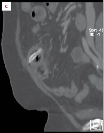
Figure 1. Unenhanced CT scan performed on axial-oblique (A) and sagittal (B) planes showing the FB (arrow), the parietal pneumatosis (arrowheads) of the thickened bowel loop, and the stranding of the surrounding fat tissue (asterisk). No free air was detectable.
surrounding mesenteric fat tissue were noted. On the endo-luminal side of the thickened loop, an oblong-shaped bone-density element was also detected (Figure 1).
However, no air-fluid levels within the intestinal loops nor free air in the abdominal cavity were detected.
Backwards, the evaluation of the contrast-enhanced acquisi-tions showed the presence of some gas bubbles strictly adja-cent to the thickened ileal loop (Figure 2).
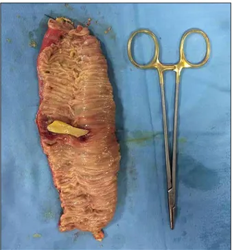
The patient underwent laparotomy with resection of the ne-crotic loop (approximately 13 cm) and removal of the foreign body consistent with a goat ossicle, unknowingly swallowed by the patient during her last meal (Figure 3). A 3-mm perfo-ration of the ileum was found, surrounded by a small amount of small-bowel content.
The operation was followed by 6-day hospitalization for re-covery. Afterwards, the patient was discharged from the hos-pital after a few days without early or delayed complica-tions. Therefore, no follow-up examinations were needed or performed.
Discussion
Ingestion of FBs is a frequent cause of admission to the Emergency Department.
FBs may vary in shape and composition (e.g., coins, batteries, fish and animal bones, pins, and needles) and their ingestion is
A
C
B
Figure 2. Axial-oblique (A) and sagittal (B) contrast-enhanced CT scan with soft-tissue windowing demonstrating a small amount of free air (arrow) adjacent to the thickened loop, as the sign of intestinal perforation. Detail of sagittal contrast-enhanced CT scan with bone windowing (C), better demonstrating the structure of the goat ossicle.
more frequent in people with limited consciousness (e.g., chil-dren or old people, psychiatric patients, alcohol abusers) [1,3,4]. Generally, most FBs ingested pass harmlessly through the en-tire digestive tube, but some of them cause discomfort and lead to complications such as intestinal perforation [5]. This latter condition can occur anywhere within the gut, but the tracts with a physiologic acute angulation or shrinkage of the lumen (e.g., ileocecal valve, colic flexures) are the most af-fected. Nevertheless, some pathological conditions, (e.g., ste-nosis, thickening of the bowel walls in patients with Crohn’s disease) can further increase the risk [6].
Radiological imaging plays a pivotal role in achieving the cor-rect diagnosis since the symptoms are often non-specific and a clinical history of FBs ingestion is seldom available, especially considering the patients generally affected [7].
Usually, plain abdominal radiography is the initial imaging exam-ination performed in the diagnostic workup for acute abdomi-nal pain with a suspicion of intestiabdomi-nal perforation [8]. However, its sensitivity in detecting abdominal free air is not very high (50–70%, or even lower at early stages with small amounts of gas) and the site of the perforation is rarely recognizable [9]. Nevertheless, the detection of the FBs can be very challeng-ing because of their dimensions (often very small) and density
Figure 3. Surgical specimen of the ileal loop resected with the goat ossicle.
(radiolucent or insufficiently radiopaque) or due to the over-lap of bones or intestinal gas [10].
On the other hand, CT scan is the imaging modality of choice for the evaluation of acute abdomen conditions, with a high sen-sitivity in recognizing the site of an eventual perforation [11]. The most important CT signs of perforation are thickening of the perforated intestinal wall and stranding of the surround-ing mesenteric fat tissue, caused by the infiltration of fluid and inflammatory cells, where air bubbles also can be found. The presence of intestinal ischemia, demonstrated in our case by intramural pneumatosis, is very rare in cases of ingested foreign bodies, but has been described in literature and is re-lated to traumatic mucosal damage [12].
Even the detection of a FB within the bowel lumen is not al-ways easy, depending on its size, density, and orientation. In this sense, the post-processing reconstructions and the win-dow setting adjustments can significantly improve the confi-dence of the radiologist.
The uniqueness of our case is the ongoing intestinal perfo-ration appreciable during the different acquisitions of a con-trast-enhanced CT scan. In fact, no evidence of localized or dif-fuse pneumoperitoneum was detectable on the basal phase, showing that the serosa was still containing the progression of the perforation.
This “sealing” process ceased right after the ending of the pre-contrast phase, with consequent abdominal pain consistent with the perforation and CT-proven by the air bubbles found near the thickened bowel wall.
Conclusions
The ingestion of FBs can be unconscious and therefore should always be included in the differential diagnosis of acute ab-dominal pain, particularly in children and the elderly. We de-scribed the first case in which the onset of intestinal perfora-tion was appreciable during the performance of a multiphase CT scan. This case report demonstrates not only the impor-tance of multidetector CT scan and the usefulness of multi-planar reconstructions, but also the high degree of attention necessary in the management of these particular clinical con-ditions for both clinical physicians and emergency radiologists.
Conflict of interest
References:
1. Coulier B, Tancredi MH, Ramboux A: Spiral CT and multidetector-row CT diagnosis of perforation of the small intestine caused by ingested foreign bodies. Eur Radiol, 2004; 14(10): 1918–25
2. Singh JP, Steward MJ, Booth TC et al: Evolution of imaging for abdominal perforation. Ann R Coll Surg Engl, 2010; 92(3): 182–88
3. Goh BK, Tan YM, Lin SE et al: CT in the preoperative diagnosis of fish bone perforation of the gastrointestinal tract. Am J Roentgenol, 2006; 187(3): 710–14
4. Gayer G, Petrovitch I, Jeffrey RB: Foreign objects encountered in the ab-dominal cavity at CT. Radiographics, 2011; 31(2): 409–28
5. Hunter TB, Taljanovic MS: Foreign bodies. Radiographics, 2003; 23(3): 731–57 6. Venkatesh SH, Venkatanarasimha Karaddi NK: CT findings of accidental
fish bone ingestion and its complications. Diagn Interv Radiol, 2016; 22(2): 156–60
7. Nicolodi GC, Trippia CR, Caboclo MF et al: Intestinal perforation by an in-gested foreign body. Radiol Bras, 2016; 49(5): 295–99
8. Kothari K, Friedman B, Grimaldi GM, Hines JJ: Nontraumatic large bowel perforation: Spectrum of etiologies and CT findings. Abdom Radiol, 2017; 42(11): 2597–608
9. Lo Re G, Mantia FL, Picone D et al: Small bowel perforations: What the ra-diologist needs to know. Semin Ultrasound CT MR, 2016; 37(1): 23–30 10. Kuzmich S, Burke CJ, Harvey CJ et al: Perforation of gastrointestinal tract
by poorly conspicuous ingested foreign bodies: Radiological diagnosis. Br J Radiol, 2015; 88(1050): 20150086
11. Hainaux B, Agneessens E, Bertinotti R et al: Accuracy of MDCT in predicting site of gastrointestinal tract perforation. Am J Roentgenol, 2006; 187(5): 1179–83
12. Kuroda, Zakimi M, Hirata K, Kikuchi K: Pneumatosis intestinalis caused by an ingested denture. Intern Med, 2017; 56(4): 471–72