Assessing QT interval in patients with
autoimmune chronic in
flammatory
diseases: perils and pitfalls
Pietro Enea Lazzerini, Pier Leopoldo Capecchi, Franco Laghi-Pasini
To cite: Lazzerini PE, Capecchi PL, Laghi-Pasini F. Assessing QT interval in patients with autoimmune chronic inflammatory diseases: perils and pitfalls. Lupus Science & Medicine 2016;3:e000189. doi:10.1136/lupus-2016-000189 PLC and FL-P contributed equally. Accepted 3 November 2016 Department of Medical Sciences, Surgery and Neurosciences, University of Siena, Siena, Italy
Correspondence to Dr Pietro Enea Lazzerini; [email protected]
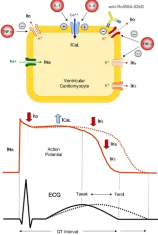
The QT interval on the ECG reflects the dur-ation of action potential (AP) in ventricle cardiomyocytes, in turn representing the sum of ventricular depolarisation and repo-larisation.1 AP is caused by transmembrane flow of ions, including inward depolarising currents mainly through sodium and calcium channels, that is, the sodium current (INa) and the L(long-lasting)-type calcium current (ICaL), and outward repolarising currents mainly through potassium channels, particu-larly the transient outward current (Ito), the rapid and the slow component of the delayed rectifier potassium current (rapid component, IKr, and slow component, IKs) and the inward rectifier potassium current (IK1) (figure 1).1
The long QT syndrome (LQTS) is a multi-factorial disorder characterised by a pro-longed heart rate-corrected QT interval (QTc), which predisposes to life-threatening ventricular arrhythmias, particularly torsades de pointes (TdP), that can degenerate into ventricular fibrillation and cause sudden cardiac death.2 3Although there is no thresh-old of QTc prolongation at which TdP is certain to occur, the risk of TdP gradually increases as the QTc prolongs over 440 ms, with an approximately 5%–7% exponential increase in risk for each 10 ms prolongation of QTc. In particular, studies indicate that a QTc>500 ms is associated with a twofold to threefold higher risk for TdP.3
The LQTS is traditionally classified as con-genital or acquired,2 even though it has becoming clear how in many cases the clin-ical phenotype is the result of a complex interaction of multiple aetiological factors operating concomitantly in the single patient.3 Congenital LQTS is caused by gen-etically determined abnormalities affecting directly or indirectly the function of specific ionic channels involved in ventricular AP, that is, potassium (loss of function), sodium or calcium channels (gain of function).2
To date, over 1000 mutations in 15
LQTS-susceptibility genes have been identi-fied. Acquired LQTS is much more prevalent than the congenital form, in most cases representing an adverse effect of drugs or the result of electrolyte disturbances interfer-ing with cardiomyocyte electrophysiology.2In particular, molecular basis of drug-induced LQTS almost exclusively involves the reduc-tion of IKr through hERG-potassium channel blockade.2 3 Other currently recognised causes of acquired LQTS include structural heart diseases, bradycardia, endocrine disor-ders, liver diseases, nervous system injuries, HIV infection, starvation, hypothermia and toxins.2–4
Recently, a number of basic and clinical studies strongly suggest that inflammation and immunity represent further important determinants of acquired LQTS.4
Accumulating evidence indicates that inflammatory activation profoundly impacts the electrophysiological properties of cardio-myocytes via multiple effects, ultimately resulting in a prolongation of AP duration (APD), and thereby QTc. In this scenario, the key mediators seem to be the in flamma-tory cytokines ( particularly tumour necrosis factor-α, interleukin (IL)-6, IL-1β) which may affect myocardium either directly, by modu-lating the expression and function of speci-fic ion channels critically involved in AP (figure 1) or indirectly, by increasing central nervous system sympathetic drive on the heart.5Among systemic autoimmune diseases
(ADs), the largest evidence has been
reported for rheumatoid arthritis (RA) and connective tissue diseases (CTDs). Recent studies demonstrated that QTc is frequently prolonged in RA, associates with disease severity and inflammatory markers and inde-pendently predicts all-cause mortality.4–7 Notably, in these patients QTc duration cor-related with circulating levels of in flamma-tory cytokines,8 and anti-IL-6 therapy with
tocilizumab resulted in a rapid and significant QTc shortening.9 Moreover, marked QTc prolongation/TdP occurrence has been reported in chronic inflammatory arthiritis patients with elevated C reactive protein (CRP) and IL-6 levels.10 Several studies performed in patients with different CTDs reported a high overall prevalence of QTc prolongation (up to ∼30%),4 with circulating IL-1β levels independently predicting the presence of a prolonged QTc.11 Specifically, patients with SLE display a longer mean QTc than controls, a 7%–15% incidence of QTc prolongation (marked QTc prolongation, ie,
>500 ms, in ∼3%), which is significantly associated with disease activity and overall inflammatory burden.4 12
Besides inflammatory activation, specific autoantibody-mediated mechanisms may also contribute to QTc pro-longation occurring in patients with ADs.4In particular, it has been demonstrated that anti-Ro/SSA antibodies (specifically the anti-Ro/SSA-52kD subtype) are respon-sible for a novel form of acquired LQTS of autoimmune origin, by directly cross-reacting with the extracellular loop of the hERG-potassium channel pore-forming region, as a result of molecular mimicry mechanisms.13 14 Such an interaction leads to IKr inhibition, resulting in APD and QTc prolongation (figure 1). Accordingly, several clinical studies demonstrated that patients with anti-Ro/SSA-positive CTD (and their newborns) fre-quently display QTc prolongation, which correlated with circulating autoantibody levels and complex ventricular arrhythmias incidence.4 14 Moreover, a high prevalence hERG-binding and IKr blocking Ro/SSA-52kD anti-bodies was also found in a cohort of unselected patients with TdP, in most cases without any history of AD, thus raising the distinct possibility that these autoantibodies may represent a clinically silent novel risk factor for QTc prolongation and TdP in the general population as well.15 Despite the fact that most of the data strongly suggest that anti-Ro/SSA-mediated electrophysiological effects have a significant clinical impact, some studies reported partially conflicting results (either slight differ-ences but very close to statistical significance, or no associ-ation between anti-Ro/SSA and QTc prolongassoci-ation), both in adults and in children.14 Notably, even among studies showing a significant association, markedly different per-centages of QTc prolongation in patients with anti-Ro/ SSA-positive CTD were observed (from∼10% to 60%).14 Although this QTc variability can be attributed to differ-ences among CTD cohorts in terms of autoantibody con-centration and specificity (high levels of anti-Ro/ SSA-52kD are particularly frequent in Sjögren syndrome, much less in SLE and systemic sclerosis),16 it may also be in part explained by electrophysiological considerations. In fact, based on the evidence that (i) besides the hERG-channel, anti-Ro/SSA are able to bind to Ca2+ -channels (L-type and T-type) and inhibit related cur-rents17 and (ii) Ca2+-channels and K+-channels have opposing effects on APD (figure 1), it is conceivable that a concomitant inhibitory effect of anti-Ro/SSA on Ca2+ -channels can partially counteract IKr inhibition-dependent APD prolongation in vivo, thus reducing the extent of QTc prolongation observed. On the basis of this hypothesis, recently confirmed by mathematical simula-tion data,18 intrinsic (inherited or acquired) differences in K+-channel and Ca2+-channel expression (ion channel reserves) on patients’ cardiomyocytes may also contribute to the QTc variability observed.19 20
In a recent publication in Lupus Science and Medicine, Geraldino-Parrilla et al21 analysed ECG repolarisation parameters in a cross-sectional study involving 189
patients with autoimmune chronic inflammatory
Figure 1 QT interval, inflammation and autoimmunity. From the cell to the surface ECG. (i) Inflammatory cytokines prolong APD and QTc by directly modulating the expression and function of potassium or calcium channels in cardiomyocyte, resulting in changes of the related currents: TNF-α inhibits IKr, IKs and IK1 currents, while IL-1 and IL-6 enhance the L-type Ca2+current. (ii) Anti-Ro/SSA-52kD directly cross-react with the extracellular loop of the hERG-potassium channel pore-forming region, leading to IKr inhibition with APD and QTc prolongation. APD, action potential duration; QTc, heart rate-corrected QT interval; TNF-α, tumour necrosis factor α; IL-1, interleukin-1; IL-6, interleukin-6; Na+, sodium; Ca2+, calcium; K+, potassium; INa, sodium current; Ito, transient outward current; ICaL, L(long-lasting)-type calcium current; IKr, rapid component of the delayed rectifier potassium current; IKs, slow component of the delayed rectifier potassium current; IK1, inward rectifier potassium current; anti-Ro/SSA-52kD, anti-Ro/SSA-52kD antibodies. Modiified from: Lazzerini et al.4
diseases, 50 affected with SLE and 139 with RA. The authors reported that non-specific ST-T abnormalities were significantly more common (∼fivefold), and mean QTc significantly longer (∼25 ms) in SLE when com-pared with patients with RA. Since RA is associated with a similarly increased cardiovascular risk, thus represent-ing a good control group for patients with SLE, the authors concluded that the different prevalence of repo-larisation alterations observed between the two popula-tions is a robust finding. The risk of presenting non-specific ST-T abnormalities increased with older age in patients with SLE, while was associated with male sex in patients with RA. As regards QTc duration, statins use was inversely associated with this parameter in patients with SLE, while in the RA group female sex, CRP levels and disease activity measured by disease activity score (DAS)-28 were associated with an increased QTc length.
Although the authors try to further corroborate their conclusions by adjusting data for confounders, neverthe-less it should be stressed that the two groups analysed are too different, thus rising concerns about the actual significance of the results, particularly QT interval find-ings. In fact, while an increased prevalence of non-specific ST-T abnormalities was found in patients with SLE despite the presence of a ‘favourable’ matching ( patients with SLE were younger, predominantly females and with shorter disease duration than patients with RA) thus supporting the validity of these findings, this con-sideration is not true for the QTc, whose assessment may have been also biased by some additional procedural issues. In this view, the paper of Geraldino-Parrilla et al21 gives us the opportunity to alert about perils potentially sneaking in this kind of investigation. In particular, we here discuss a number of crucial methodological aspects that require careful attention in order to avoid pitfalls when a study assessing the QT interval in patients with autoimmune chronic inflammatory diseases is performed.
First, the degree of inflammatory activation as reflected by circulating markers and DAS needs accurate consideration.
Given the above discussed effects of inflammatory cyto-kines in modulating QTc duration, particularly in RA,4 5 it is possible that the shorter QTc duration observed in patients with RA versus patients with SLE may be at least in part the result of the fact that this RA cohort did not have a sufficiently high disease activity, thus underesti-mating a key QT-prolonging mechanism operating in the disease (accordingly, in the present RA cohort CRP levels and DAS28-CRP score were associated with an increased QTc length). In fact, although the authors stated that their patients with RA had moderate-to-severe activity (median DAS28-CRP: 3.6, using for this definition the presence of a DAS28-CRP>3.2), current American College of Rheumatology (ACR)-recommended cut-offs (remission <2.6; low/minimal disease activity 2.6–3.2; moderate disease activity 3.2–5.1; high/severe disease activity >5.1)22 indicate that a DAS28-CRP value of 3.6
corresponds at most to a moderate disease activity. Even, for other authors DAS28-CRP values ranging from 2.3 to 3.8 corresponded to low disease activity.23Accordingly, in this RA cohort, median CRP levels were 2.1 mg/L (ie, 0.21 mg/dL, a value below the cut-off of 1 mg/dL recently selected by ACR/European League Against Rheumatism to define remission).24 Indeed, also IL-6 levels in this RA cohort were rather low (median 3.6 pg/ mL), approximately three times lower when compared with those found in a large French cohort of high-disease activity patients with RA (median DAS28-erythrocyte sedimentation rate (ESR): 5.1; median IL-6 levels ∼10 pg/mL).25 Thus, in the RA cohort selected in this study systemic inflammation could be not high enough to produce the expected RA-associated QT prolongation. Indeed, CRP levels in patients with RA were significantly lower, approximately two times than patients with SLE. Moreover, in a previous study demon-strating high prevalence of QTc prolongation in patients with RA, severe disease activity was present in most patients (65%; mean DAS28-ESR 5.5; mean DAS28 CRP 4.9), and mean CRP levels approximately seven times higher than in the present RA cohort (1.5 mg/dL, ie, 15 mg/L).9
Second, the specific autoantibody profile of patients with AD under study has to be carefully characterised, particularly the presence, subtype and titre of anti-Ro/ SSA antibodies.
In the present study, the authors found no association between anti-Ro/SSA positivity and QTc length in patients with SLE, thereby concluding that these auto-antibodies do not contribute to the pathogenesis of the QTc prolongation observed. However, they neither ana-lysed anti-Ro/SSA subtypes nor their circulating levels, both representing critical factors for the clinical appear-ance of anti-Ro/SSA-associated QTc prolongation. In fact, it has been demonstrated that the anti-Ro/ SSA-52kD subtype only can inhibit the IKr current,13 15 and that high anti-Ro/SSA-52kD levels are required for developing QTc prolongation in positive patients, at least 10 times higher than the upper normal limit.26 Thus, it is possible that in this SLE cohort prevalence and/or circulating levels of the anti-Ro/SSA-52kD subtype were too low to produce a clinically evident QTc prolongation. Notably, a very recent paper27 identified Tpeak-Tend (Tp-e) interval as a better ECG predictor of anti-Ro/SSA-52kD-associated ventricular repolarisation abnormalities in positive patients. Indeed, IKr current is activated after the peak of T wave (figure 1). Thus, Tp-e may be more sensitive than QTc to detect the electro-physiological consequences of anti-Ro/SSA-52kD-asso-ciated hERG-channel inhibition in the clinical setting.
Third, the potential impact of demography on QTc findings represents another important factor that cannot be disregarded in this type of study. Indeed, it is well recognised that gender and ethnic characteristic markedly impact on the epidemiology of ADs in general, of SLE and RA in particular.28
However, the two groups of patients studied by Geraldino-Parrilla et al21 are very different, maybe too much, in terms of sex and ethnicity, and this could have significantly biased the results. In particular, in the general population it is well-established that female sex is associated with longer QTc duration (accordingly QTc prolongation cut-offs are different in males vs females) and higher TdP risk, via complex effects of sexual hor-mones on cardiac ion currents.3 29 Thus, the longer QTc duration observed in patients with SLE versus patients with RA may be influenced by the higher per-centage of females in the SLE group (92% vs 61%; p<0.0001). Accordingly, in the RA cohort QTc duration correlated with female sex (in the SLE cohort, the large preponderance of females probably did not allow to evaluate the weight of the gender on QTc). Moreover, also ethnicity is very different between the two groups. Indeed, RA cohort mainly consisted of whites (87%), while patients with SLE were in the large majority (96%) Hispanic or blacks (74% and 22%, respectively). Many large studies involving both general population patients and patients with cardiac diseases found that in blacks and Hispanics the QTc interval is longer than in whites.30–32 As a result, higher percentages of females and Hispanic/blacks may have also contributed to the longer QTc observed in the SLE cohort when compared with patients with RA.
In conclusion, cardiovascular involvement is currently recognised as a main cause of morbidity and mortality in autoimmune chronic inflammatory diseases. In this context, growing recent evidence indicates inflammation and autoimmunity as novel cardiovascular risk factors functionally impacting ventricular repolarisation, par-ticularly promoting QTc prolongation and associated life-threatening arrhythmias. If on one hand these con-siderations fully justify the increasing attention that this ECG parameter is receiving in these patients, on the other hand it is essential that researchers are alerted on the specific perils of this type of study in order to avoid methodological pitfalls when assessing QT interval in a population with many peculiarities such as the patients with AD.
Competing interests None.
Provenance and peer review Commissioned; internally peer reviewed.
Data sharing statement No additional data are available.
Open Access This is an Open Access article distributed in accordance with the Creative Commons Attribution Non Commercial (CC BY-NC 4.0) license, which permits others to distribute, remix, adapt, build upon this work non-commercially, and license their derivative works on different terms, provided the original work is properly cited and the use is non-commercial. See: http:// creativecommons.org/licenses/by-nc/4.0/
REFERENCES
1. Grant AO. Cardiac ion channels.Circ Arrhythm Electrophysiol
2009;2:185–94.
2. Viskin S. Long QT syndromes and torsade de pointes.Lancet
1999;354:1625–33.
3. Drew BJ, Ackerman MJ, Funk M, et al. Prevention of torsade de pointes in hospital settings: a scientific statement from the American Heart Association and the American College of Cardiology Foundation.Circulation2010;121:1047–60.
4. Lazzerini PE, Capecchi PL, Laghi-Pasini F. Long QT syndrome: an emerging role for inflammation and immunity.Front Cardiovasc Med
2015;2:26.
5. Lazzerini PE, Capecchi PL, Laghi-Pasini F. Systemic inflammation and arrhythmic risk: lessons from rheumatoid arthritis.Eur Heart J
2016. Published Online First.
6. Chauhan K, Ackerman MJ, Crowson CS, et al. Population-based study of QT interval prolongation in patients with rheumatoid arthritis. Clin Exp Rheumatol 2015;33:84–9.
7. Panoulas VF, Toms TE, Douglas KM, et al. Prolonged QTc interval predicts all-cause mortality in patients with rheumatoid arthritis: an association driven by high inflammatory burden.Rheumatology (Oxford)2014;53:131–7.
8. Adlan AM, Panoulas VF, Smith JP, et al. Association between corrected QT interval and inflammatory cytokines in rheumatoid arthritis.J Rheumatol2015;42:421–8.
9. Lazzerini PE, Acampa M, Capecchi PL, et al. Antiarrhythmic potential of anticytokine therapy in rheumatoid arthritis: tocilizumab reduces corrected QT interval by controlling systemic inflammation.
Arthritis Care Res2015;67:332–9.
10. Lazzerini PE, Capecchi PL, Bertolozzi I, et al. Marked QTc prolongation and torsades de pointes in patients with chronic inflammatory arthritis.Front Cardiovasc Med2016;3:31. 11. Pisoni CN, Reina S, Arakaki D, et al. Elevated IL-1β levels in
anti-Ro/SSA connective tissue diseases patients with prolonged corrected QTc interval. Clin Exp Rheumatol 2015;33:715–20. 12. Sham S, Madheshwaran M, Tamilselvam T, et al. Correlation
of QT interval with disease activity in newly detected SLE patients at baseline and during flare.Indian J Rheumatol
2015;10:121–4.
13. Yue Y, Castrichini M, Srivastava U, et al. Pathogenesis of the novel autoimmune-associated long-QT syndrome.Circulation
2015;132:230–40.
14. Boutjdir M, Lazzerini PE, Capecchi PL, et al. Potassium channel block and novel autoimmune-associated long QT syndrome.Card Electrophysiol Clin2016;8:373–84.
15. Lazzerini PE, Yue Y, Srivastava U, et al. Arrhythmogenicity of anti-Ro/SSA antibodies in patients with torsades de pointes.Circ Arrhythm Electrophysiol2016;9:e003419.
16. Dugar M, Cox S, Limaye V, et al. Diagnostic utility of anti-Ro52 detection in systemic autoimmunity.Postgrad Med J2010;86: 79–82.
17. Karnabi E, Boutjdir M. Role of calcium channels in congenital heart block.Scand J Immunol2010;72:226–34.
18. Fabris F, Yue Y, Qu Y, et al. Induction of autoimmune response to the extracellular loop of the HERG channel pore induces QTc prolongation in guinea-pigs.J Physiol2016;594:6175–87. 19. Lazzerini PE, Capecchi PL, Laghi-Pasini F. The“invulnerability” of
the adult conduction system to anti-Ro/SSA antibodies? A matter of calcium channel expression on the cardiomyocyte.J Cardiovasc Electrophysiol2011;22:E88.
20. Lazzerini PE, Capecchi PL, Boutjdir M, et al. Comment on“Absence of an association between anti-Ro antibodies and prolonged QTc interval in systemic sclerosis: a multicenter study of 689 patients”.
Semin Arthritis Rheum2015;44:e16–17. 21. Geraldino-Parrilla L, Gartshteyn Y, Piña P, et al.
Electrocardiographic non-specific ST-T and QTc abnormalities in patients with systemic lupus erythematosus compared with rheumatoid arthritis. Lupus Sci Med 2016.
22. Anderson J, Caplan L, Yazdany J, et al. Rheumatoid arthritis disease activity measures: American College of Rheumatology recommendations for use in clinical practice.Arthritis Care Res
2012;64:640–7.
23. Castrejón I, Ortiz AM, Toledano E, et al. Estimated cutoff points for the 28-joint disease activity score based on C-reactive protein in a longitudinal register of early arthritis.J Rheumatol2010;37: 1439–43.
24. Felson DT, Smolen JS, Wells G, et al. American College of Rheumatology; European League Against Rheumatism. American College of Rheumatology/European League Against Rheumatism provisional definition of remission in rheumatoid arthritis for clinical trials.Arthritis Rheum2011;63:573–86.
25. Baillet A, Gossec L, Paternotte S, et al. Evaluation of serum interleukin-6 level as a surrogate marker of synovial inflammation and as a factor of structural progression in early rheumatoid arthritis: results from a French national multicenter cohort.Arthritis Care Res
26. Lazzerini PE, Capecchi PL, Acampa M, et al. Anti-Ro/SSA-associated corrected QT interval prolongation in adults: the role of antibody level and specificity.Arthritis Care Res2011;63: 1463–70.
27. Tufan AN, Sag S, Oksuz MF, et al. Prolonged Tpeak-Tend interval in anti-Ro52 antibody-positive connective tissue diseases.Rheumatol Int2016. Published Online First.
28. Salem JE, Alexandre J, Bachelot A, et al. Influence of steroid hormones on ventricular repolarization.Pharmacol Ther2016. 29. Amur S, Parekh A, Mummaneni P. Sex differences and genomics in
autoimmune diseases.J Autoimmun2012;38:J254–65.
30. Ramirez AH, Schildcrout JS, Blakemore DL, et al. Modulators of normal electrocardiographic intervals identified in a large electronic medical record.Heart Rhythm2011;8:271–7.
31. Williams ES, Thomas KL, Broderick S, et al. Race and gender variation in the QT interval and its association with mortality in patients with coronary artery disease: results from the Duke Databank for Cardiovascular Disease (DDCD).Am Heart J
2012;164:434–41.
32. Hebert K, Quevedo HC, Tamariz L, et al. Prevalence of conduction abnormalities in a systolic heart failure population by race, ethnicity, and gender.Ann Noninvasive Electrocardiol2012;17:113–22.
perils and pitfalls
autoimmune chronic inflammatory diseases:
Assessing QT interval in patients with
Pietro Enea Lazzerini, Pier Leopoldo Capecchi and Franco Laghi-Pasini
doi: 10.1136/lupus-2016-000189
2016 3: Lupus Sci Med
http://lupus.bmj.com/content/3/1/e000189
Updated information and services can be found at:
These include:
References
#BIBL http://lupus.bmj.com/content/3/1/e000189
This article cites 28 articles, 8 of which you can access for free at:
Open Access
http://creativecommons.org/licenses/by-nc/4.0/
non-commercial. See:
provided the original work is properly cited and the use is
non-commercially, and license their derivative works on different terms, permits others to distribute, remix, adapt, build upon this work
Commons Attribution Non Commercial (CC BY-NC 4.0) license, which This is an Open Access article distributed in accordance with the Creative
service
Email alerting
box at the top right corner of the online article.
Receive free email alerts when new articles cite this article. Sign up in the
Notes
http://group.bmj.com/group/rights-licensing/permissions
To request permissions go to:
http://journals.bmj.com/cgi/reprintform
To order reprints go to:
http://group.bmj.com/subscribe/