1
DOTTORATO INTERNAZINALE DI RICERCA IN
NEUROBIOLOGIA
XXVI° CICLO
UNIVERSITA’ DEGLI STUDI DI CATANIA
SEDI CONSORZIATE: UNIVERSITA’ DI CATANIA, ROMA E PAVIA SEDE AMMINISTRATIVA: UNIVERSITA’ DI CATANIA
Dott.ssa Maria Concetta Scuto
_______________
BIOGENESI MITOCONDRIALE, MISFOLDING PROTEICO E RISPOSTA CELLULARE ALLO STRESS NELL’AGING E NEI DISORDINI NEURODEGENERATIVI: APPROCCIO
PROTEOMICO
MITOCHONDRIAL BIOGENESIS, PROTEIN MISFOLDING AND CELLULAR STRESS RESPONSE IN AGING AND IN NEURODEGENERATIVE DISORDERS: PROTEOMIC
APPROACH
_______________
TESI DI DOTTORATO
_______________
Tutor
Chiar.mo Prof. Vittorio Calabrese
Coordinatore
Chiar.mo. Prof. Roberto Avola
2
INDEX
INTRODUCTION
3
1. REDOX BALANCE AND ANTIOXIDANT DEFENSE SYSTEMS
-Oxidative stress 5
-Reactive oxygen speciaes (ROS) 7
-Ros Toxicity 9
- Mechanism of antioxidant defence 11
- Defense mechanism “Heat Shock Response” 16
- Antioxidant and Neuroblastoma cells 24
-Oxidative stress and type II Diabetes 25
- Diabetic nephropathy: effects of carnosine and cellular stress response in
podocyte cells 27
- Vitagenes and UCP proteins in Alzheimer’s disease 28
- Oxidative stress and cellular stress response in glaucoma: implications with
Alzheimer’s disease 30
2. AIM OF THE RESEARCH 33
3. MATERIAL AND METHODS 38
4. RESULTS 51
5. DISCUSSION 64
6. FIGURES 82
3
INTRODUCTION
Life is the interplay between structure and energy, yet the role of energy deficiency in human disease has been poorly explored by modern medicine (1).
Numerous experimental evidence supporting the idea that the reduction of the expression cell, the activity of antioxidant proteins and the consequent increase in oxidative stress are the fundamental cause of the aging process and neurodegenerative disorders (2).
Several studies suggest that the accumulation of oxidative molecules damaged is one of the factors that could cause senescence. The predominant molecular cause of aging is the accumulation of protein mutated.
Moreover, several conditions including protein, lipid or glucose oxidation disrupt redox homeostasis and lead to accumulation of unfolded or misfolded proteins in the aging brain. Alzheimer’s and Parkinson’s diseases or Friedreich ataxia are neurological diseases sharing, as a common denominator, production of abnormal proteins, mitochondrial dysfunction and oxidative stress, which contribute to the pathogenesis of these so called ‘‘protein conformational diseases’’(3).
The central nervous system has evolved the conserved mechanism of unfolded protein response to cope with the accumulation of misfolded proteins (3).
Harman in 1972 first proposed that mitochondria may have a central role in the process of aging. According to this theory, free radicals generated through mitochondrial metabolism can act as causative factor of abnormal function and cell death (4).
The mitochondrial genome plays a key role in aging and neurodegenerative disorders. Mitochondrial dysfunction is indeed characteristic of various disorders, and damage lead to altered mitochondrial respiratory chain activity as found in Parkinson's disease, in Alzheimer's disease and in Huntington's disease.
These dysfunctions in the activity of respiratory complex, possibly associated with alterations of the oxidant/antioxidant balance, are thought to underlie defects in energy metabolism and induce cellular degeneration (5).
The mitochondrial DNA mutations, both inherited and acquired, cause a malfunction of the electron transport chain (ETC), which leads to the decrease of ATP production, increased formation of toxic free radicals and altered homeostasis of Ca2+. These determine the toxic consequences of ETC dysfunction that promote mitochondrial
4
damage, including oxidation of mitochondrial DNA, proteins, lipids and altered mitochondrial permeability, an event associated with the degeneration and cell death (6).
Mitochondria are the main significant source of oxidants, and in vitro studies have indicated that approximately 1–2% of electron flow through the ETC results in the univalent generation of superoxide (4).
Moreover, various toxins in the environment can injure mitochondrial enzymes, leading to increased generation of free radical that over the life span would eventually play a major role in aging.
During the last few years, cellular oxidant/antioxidant balance has become the subject of intense study, especially by those interested in brain aging and in neurodegenerative mechanisms (7).
As one of the main intracellular redox systems involved in neuroprotection, , the
vitagene system is emerging as a neurohormetic potential target for novel
cytoprotective interventions.
Vitagenes encode for cytoprotective heat shock proteins (Hsp) Hsp70 and heme oxygenase-1, as well as thioredoxin reductase and sirtuins.
Nutritional studies show that ageing in animals can be significantly influenced by dietary restriction. Thus, the impact of dietary factors on health and longevity is an increasingly appreciated area of research.
Reducing energy intake by controlled caloric restriction or intermittent fasting increases lifespan and protects various tissues against disease.
Genetics has revealed that ageing may be controlled by changes in intracellular NAD/NADH ratio regulating sirtuin, a group of proteins linked to aging, metabolism and stress tolerance in several organisms (3).
Consistently, the neuroprotective roles of dietary antioxidants including curcumin, acetyl-L-carnitine and carnosine have been demonstrated through the activation of these redox-sensitive intracellular pathways.
Although the notion that stress proteins are neuroprotective is broadly accepted, still much work needs to be done in order to associate neuroprotection with specific pattern of stress responses (3).
5
-1-
REDOX BALANCE AND ANTIOXIDANT
DEFENSE SYSTEMS
Oxidative stress
The overproduction of free radicals, molecular species that are continuously produced in small quantities in our bodies, is an important cause of cellular aging and plays a role in the pathogenesis of a wide variety of pathological states (8).
Under normal conditions, cells have efficient antioxidant defense systems. When the rate of generation of free radicals exceeds the capacity of antioxidant defenses, a state of "oxidative stress" is established which is the “primum movens" for the consequences that can lead to irreversible cell damage (9).
Numerous experimental data shows the involvement of oxidative stress in the mechanism of aging and neurodegeneration (10).
Oxidative stress is therefore characterized by an imbalance of the redox state of oxidants/antioxidants that may lead to altered cellular function and oxidative damage of fundamental biological macromolecules like protein (protein carbonyls, nitration of tyrosine), lipids (products of lipid peroxidation) and nucleic acids (11,12).
Oxidative stress is induced by both exogenous and endogenous sources (13). The first include drugs and toxic chemicals that change the balance of oxidants / antioxidants on behalf of oxidation; the latter includes overproduction of reactive oxygen intermediates by the mitochondrial electron transport chain. One of the main causes of oxidative stress is therefore the excessive release of reactive oxygen species (ROS).
6
Modification of the normal balance between ROS and antioxidants (from Nutrition and oxidative stress.
7
Reactive oxygen species (ROS)
ROS are defined as molecular entities that react with cellular components, causing harmful effects on their functions. ROS include both free radicals (containing highly reactive unpaired electrons) such as superoxide anion (O2-•), nitric oxide (NO•) and hydroxyl radical (OH•) and other molecular species, such as hydrogen peroxide (H2O2)
and peroxynitrite (ONOO-).
The majority of cellular ROS are generated during the incomplete metabolic reduction of oxygen to water (see figure below).
Reactive oxygen species (ROS)
Free radicals are highly reactive molecules, have a very short half-life and can have a provenance endogenous and exogenous.
Reactive oxygen intermediates are produced continuously for a process of "leakage or short circuiting" of electrons from the normal locations of biological origin, which are rapresented mainly by the mitochondrial respiration, the tissue content of xanthine oxidase and, to a lesser extent, by the metabolism of arachidonic acid and by the processes of auto-oxidation of catecholamines or heme proteins (14).
8
The oxygen molecule is a biological paradox that on the one hand is a molecule essential for aerobic life, the other a biological hazard due to its high toxicity. In fact, the oxygen that is taken from the external environment is required in the mitochondrial respiration for the production of energy in the form of ATP according to a complex process that takes the name of "oxidative phosphorylation"; oxygen acting as a final acceptor of electrons removed from the molecules and combining with the protons subtracted under the same, allows the complete oxidation to water and carbon dioxide molecules of various nature (glycidol, fatty acids, amino acids, etc.), with release of all the energy contained in them (14).
The activation of molecular oxygen can usually be done in two ways: one by using electrons and energy (8). The free radicals which can be formed during the sequential reduction mono electronics O2 are the superoxide anion (O2ˉ), hydrogen peroxide
(HOOH) hydroxyl radical (OH˙).
During activation energy, it takes 22 kcal of energy because you have electronic transitions in molecular orbital of oxygen leading to the formation of singlet oxygen (O2) which is not a radical, as there is an unpaired electron, but has a strong oxidizing
ability, and, for degradation, generates superoxide anion (O2ˉ).
Ionizing radiation, the photosensitizing substances, heat, death of a cell, are all phenomena entropic thermodynamically favoring the release of the amount of energy and, therefore, sufficient because these electronic transitions take place (14).
Even the excessive production of NO, a molecule physiologically important for the regulation of vascular tone and processes immunomodulators, can generate radical forms if associated with a concomitant overproduction of superoxide anion (15). The endothelium appears to continuously produce small amounts of superoxide can react with nitric oxide (both are free radicals) to form nitrate ions, a product not radical. For this reason, variations in the production of nitric oxide and superoxide by the endothelium may represent a mechanism for the regulation of vascular tone. The peroxynitrite anion, degrading, form a hydroxyl radical (15,16).
9
If two radicals react with each other, cancel each other, and when a radical species reacts with a molecule not radical, it produces a new free radical and triggers a chain of reactions until it will form a stable compound. It then passes from the stage of "generation" to that of "propagation" of the free radical (14).
The formation of reactive oxygen species is therefore an eventuality cannot be eliminated in the cellular environment.
ROS Toxicity
Certain clinical situations or the intensification of external factors such as environmental pollution, smoking, a diet rich in fats, alcohol abuse, solar radiation, the use of certain medications, physical and mental stress, are all conditions generally associated with the overproduction of free radicals.
When the generation of free radicals exceeds the capacity of detoxifying antioxidant defenses, it establishes a condition of "oxidative stress". This represents a risk to the structural and functional integrity of important molecules such as DNA, proteins and lipids (13).
Free radicals and in particular the OH˙, which is the most reactive free radical, can react with various molecules, dramatically changing both the chemical state and profoundly altering the function. The proteins can be oxidized at the level of sulfhydryl groups through a process that involves the deactivation of channel proteins, receptor or important enzyme activities (16,17); for example, enzymes such as phosphofructokinase, complex I and complex IV of mitochondrial respiratory chain are inactivated with severe deterioration of the cell's ability to supply energy. The calcium pump is inactivated with a consequent tendency to maintain high levels of calcium citosolubile.
Nucleic acids are sensitive to free radical attack at both of the bases of the pentose resulting in rupture of the propellers with the formation of modified bases such as 8-hydroxy-guanine and alteration of the genetic code (14).
10
The best known harmful effect of ROS is lipid peroxidation a chain reaction that leads to the formation of lipid peroxides and hydroperoxides from the oxidation of a methylene bridge at the level of a polyunsaturated fatty acid of membrane lipids such as arachidonic acid and linolenic acid. The lipohydroperoxides tend to move from the hydrophobic membrane to the surface leading to a disorganization of the structure of the membrane itself.
Consequently it causes irreversible damage to the morpfofuctionality of intracellular and cellular membranes or lipoproteins (18).
The continued against oxidative damage important molecules like DNA, proteins and lipids, elicited by activated oxygen species and of NO, is considered, in light of current experimental and clinical evidence, the most important cause of physiopathogenetics and biochemical changes observed during aging of the CNS including neurodegenerative disorders (15).
Several lines of evidence suggest that accumulation of oxidative molecular damage is a causal factor in senescence.
Among the correlative evidence supporting the involvement of oxidative stress are the following: (a) oxidative molecular damage to DNA and proteins increases exponentially with age, and concomitantly, the rates of mitochondrial O2¯· and H2O2
generation as well as the susceptibility of tissues to experimentally induced oxide. Among the correlative evidence supporting the involvement of oxidative stress are the following: (b) experimental regimens that extend the lifespan, such as caloric restriction in mammals and reduction of metabolic rate in insects, decrease the accumulation rates of oxidative damage; (c) mitochondria make two rather contradictory contributions to cell survival. The classically recognized function is the synthesis of ATP for energizing endergonic reactions, the other is generation of reactive oxygen species which may compromise the long-term survival of cells and constitute a major underlying cause of the aging process. Indeed, these two rather conflicting functions are part of the same process, namely mitochondrial respiration. The alteration in the redox and mitochondrial dysfunction that follows, is involved in so many disease states neuropathogenesis including neurodegenerative disorders such as multiple sclerosis (MS), Parkinson's disease (PD) the Alzheimer's disease, (AD) and the aging (19).
CNS has a large potential oxidative capacity (20) due to the high level of tissue oxygen consumption. However, the ability of the brain to withstand oxidative stress is limited
11
because of: (a) a high content of easily oxidizable substrates, such as polyunsaturated fatty acids and catecholamines; (b) relatively low levels of antioxidants such as glutathione and vitamin E and antioxidant enzymes (such as glutathione peroxidase, catalase and superoxide dismutase); (c) the endogenous generation of reactive oxygen free radicals via several specific reactions; (d) the elevated content of iron in specific areas of the human brain, such as globus pallidus and substantia nigra (SN), while cerebrospinal fluid has very little iron-binding capacity owing to its low content of transferrin; (e) CNS contains non-replicating neuronal cells which, once damaged, may be permanently dysfunctional or committed to programmed cell death (apoptosis). Numerous experimental evidence lead to the conclusion that the dysfunction at the level of cellular energy metabolism is an important factor in the neurotoxicity mediated by NO and that the cellular content of thiols is crucial in determining the sensitivity of cells to oxidative stress and nitrosative (21).
Mechanisms of Antioxidant Defence
In the cell, in the normal conditions, there is a steady-state balance between pro-oxidants and antipro-oxidants which is necessary to ensure optimal efficiency of antioxidant defenses, that, during the normal cellular metabolism, can adequately cope with all the free radicals generated (6).
Furthermore, recent studies show that a minimum amount of free radicals, which until now have been considered only due to aging, is the necessary condition for the functionality of the cells, because it leads to a greater efficiency of defense systems and increased cell survival (21).
In the cell, as regards oxidative insult, at the level of the cytoplasm, in mitochondria and also in the extracellular fluid, there are efficient antioxidant defense mechanisms of enzymatic and non-enzymatic nature. In the first case it is cytoprotective enzymes (superoxide dismutase, catalase, glutathione peroxidase), said "scavengers", because they function as "scavengers" in respect of free radicals, tending to remove them as formats or to prevent their formation. In the second case it is of antioxidants can react with free radicals, abducting or neutralizing them, and therefore capable of blocking the series of reactions of lipid peroxidation, and then to prevent the detrimental action
12
is that the propagation of free radicals; between these molecules are able to bind the O2
singlet (β-carotenes, retinoids); xanthine oxidase inhibitors (allopurinol); molecules of low molecular weight, is water-soluble such as ascorbic acid (vitamin C) and glutathione, which operate in blood plasma and in the cytosol, both fat-soluble such as α-tocopherol (vitamin E), bilirubin, uric acid and estrogen that instead exert their action in the heart hydrophobic cell membranes or of plasma lipoproteins (14) .
In recent years, since oxidative stress has been felt at the base of some if not all aspects of neurodegeneration, numerous experimental studies were conducted in order to reduce the effects of oxidative stress through the use of scavengers of free radicals. Among these are some nutritional compounds.
There are two classes of antioxidants, endogenous and exogenous. Among the former are the tripeptide glutathione (GSH), various vitamins, and products of reactions catalyzed by enzymes that are subjected to upregulation in response to oxidative stress, such as bilirubin dall'emeossigenasi, and products ARE (antioxidant responsive element) (22). Among the latter, the nutritional antioxidants, there are several classes of molecules: those that increase the endogenous levels of GSH or otherwise in functionality reactive-SH; vitamins, phenolic and polyphenolic compounds (23,24). The tripeptide glutathione (γ-glutamyl-cysteinyl-glycine) is an endogenous antioxidant of great importance. Glutathione (GSH) is required for the maintenance of the thiol redox state of the cell, for the protection from oxidative damage, for the detoxification of electrophilic and reactive metals endogenous and exogenous, for the storage and transport of cysteine, as well as for the synthesis of proteins and DNA, for the regulation of the cell cycle, and for cell differentiation (25). The glutathione and glutathione-dependent enzymes play a key role in protecting the cell from the effects of reactive oxygen species. The key element of the functional cysteine glutathione is the part that provides the thiol reactive group. Glutathione is the main defense against reactive oxygen species (ROS), which are reduced by glutathione in the presence of glutathione peroxidase.
As a consequence of the GSH is oxidized to GSSG, which, in turn, is rapidly reduced back to GSH by glutathione reductase at the expense of NADPH. The thiol-disulfide redox cycle contributes also to maintain the reduced protein thiols and enzymatic. In the absence of a process of reduction of protein disulfides, vulnerable cysteine residues essential enzymes may remain oxidized, with consequent alteration of the catalytic activity.
13
Glutathione contributes, also, for the storage and transfer of cysteine also. The cysteine to cystine autoossida quickly producing toxic oxygen radicals. To escape the toxicity of cystine, most of the protein is collected in the non-cysteine glutathione. Moreover, exerting a protective action by reactive oxygen species, glutathione is an excellent scavenger of the products of lipid peroxidation, such as HNE and acrolein, which bind proteins inhibiting its activity. Glutathione also reacts with the carbon atoms, saturated, unsaturated and aromatic. This detoxification involves the nucleophilic attack on an electrophilic carbon atom by GSH.
This reaction may start spontaneously, but very often is catalyzed by glutathione S-transferase. Glutathione also forms metal complexes through non-enzymatic reactions. The GSH intervenes to storage, mobilization and release of metal ions between ligands, in the transport of metals across cell membranes, serves as a source of cysteine to bind the metals, and by reductant for redox reactions involving metals (25). The sulfhydryl group of the cysteine portion of GSH has a high affinity for metal ions such as mercury, silver, cadmium, arsenic, lead, gold, zinc and copper, forming a complex thermodynamically stable which can be eliminated by the body. Glutathione reacts with free radical molecules. Thus, the possibility of increasing glutathione levels could prove useful against oxidative stress.
Vitamin E, a phenolic compound, acts as an antioxidant by removing the free radicals
by the atom-H phenolic. The reactions of vitamin E, vitamin C, and glutathione may be connected with different recycling processes, contributing, thus, increasing the efficiency of these parts against oxidative stress.
Polyphenols are natural substances ubiquitously present in fruits and vegetables, as
well as, beverages obtained from plants such as tea, red wine and olive oil. Flavonoids compose the largest group of polyphenols. Their skeletal structure consists of an aromatic ring condensed to a heterocyclic ring, attached to a second aromatic ring. Flavonoids are mainly divided into:
anthocyanins, glycosylated derivative of anthocyanidin, present in colorful flowers and fruits, and anthoxantins, colorless compounds further divided in several categories including flavones, flavans, flavonols, flavanols, and isoflavones. The remarkable antioxidant activity of these compounds is conferred by the numerous phenolic hydroxyl groups on the aromatic ring.
The rapid donation of a hydrogen atom to lipid peroxyl radical results in the formation of the polyphenol phenoxyl radical (PP•) according to the reaction ROO• +
14
PPH→ROOH → PP• that can be stabilized by further donation of another hydrogen or by reacting with another radical. In addition, flavonoids present efficient iron chelating activity, for which the 3-OH is important (26). The physiological effects of flavonoids are particularly significant in those pathologies where
the oxidative stress hypothesis is accepted and supported by experimental data, such as AD. In vitro, flavonoids are capable of scavenging superoxide anions (27) and hydroxyl radicals (28). Once ingested, these compounds are capable of elevating the redox and antioxidant level (29). In red blood cells, polyphenols enhance cell resistance to oxidative insult (30), as well as inhibit LDL oxidation in plasma (31). The importance of these molecules in protecting cells from oxidative stress goes beyond the simple radical oxygen species (ROS) scavenging properties. In a recent study on neuronal Cells (32), three different mechanisms of protection have been identified: Flavonoids can prevent cell death after glutamate injury by scavenging radicals, maintaining the correct glutathione levels and inhibiting Ca2+ influx, which represents the last step in the cell death cascade. These properties, together with anti-inflammatory properties attributed to some polyphenols (33), renders this class of compounds suitable for application where oxidative stress, together with inflammation and antioxidant defense depletion take place, such as AD.
Spices and herbs often contain phenolic substances with potent antioxidative and
chemopreventive properties (34). Spices form an important class of food adjuncts and are used to enhance the sensory quality of food.
Recent studies show that some of the biochemical effects of spices are due to their active principles. Turmeric (Curcuma longa Linn, family: Zingiberacee) has been used as a coloring agent and food additive in Indian culinary preparations from time immemorial (35).
The active antioxidant principle in Curcuma longa has been identified as curcumin (diferuloyl methane). It is generally assumed that the phenol moiety is responsible for antioxidant properties of any plant phenolic compound. Consequently, the free radical chemistry of curcumin (an omethoxyphenol derivative) has focused on its phenol ring (36). The possible involvement of the _-diketone moiety in the antioxidant action of curcumin has been considered and, as recently shown (37), the H-atom donation from the β-diketone moiety to a lipid alkyl or a lipid peroxyl radical has been reported as the potentially more important mechanism underlying its antioxidant action.
15
Similar to the well-known synergism between lipid soluble antioxidant action (38) might position itself at the border of the cell membrane adjacent to the acqueous milieu, in short “pops out” of the membrane to be repaired by water soluble antioxidant, such as catechins or ascorbate. Curcumin, as a powerful lipid soluble antioxidant, positions itself within the cell membrane, where it intercepts lipid radicals and becomes a phenoxyl radical. Being more polar than curcumin, phenoxyl radical travels to the membrane surface. Owing to the high reduction potential of phenoxyl curcumin radical (0.8 V, at physiological pH 7), this allows the curcumin intermediate to be easily repaired by electron donors with favorable oxidation potential, such as epigallocatechin gallate (0.43 V), catechin (0.55 V), or vitamin C (0.28 V). Such electron and associated proton transfer reaction will maintain optimal concentrations of curcumin at expense of water soluble antioxidants, in spite of its fast turnover and low physiological uptake. Moreover, curcumin free radicals can also react with each other or with other free radicals forming, either stable products such as curcumin dimers, vanilin and ferulic acid (39) or, through a peroxyl linkage at the 3’ position of the curcumin phenolic ring, coupling products which generate, via intramolecular Diels-Alder reaction, not radical stable compounds (40). Curcumin contains two electrophilic α,β-unsaturated carbonyl groups, which can react with nucleophiles such as glutathione (41). By virtue of its Michael reaction acceptor function and its electrophilic characteristics, curcumin and several other polyphenolic compounds have been recently demonstrated to induce the activities of Phase I and Phase II detox system (42,43), e.g., inhibition of COX-1 and COX-2 enzymes (44) and stimulation of glutathione-S-transferase (45). In addition to its ability to scavenge carcinogenic free radicals (46), curcumin also interferes with cell growth through inhibition of protein kinases. Although the exact mechanisms by which curcumin promotes these effects remains to be elucidated, the antioxidant properties of this yellow pigment appear to be an essential component underlying its pleiotropic biological activities. Of particular interest is the ability of curcumin to inhibit lipid peroxidation and effectively to intercept and neutralize ROS (superoxide, peroxyl, hydroxyl radicals) (47) and NO-based free radicals (nitric oxide and peroxynitrite) (48). In this regard, curcumin has been demonstrated to be several times more potent than vitamin E (49).
It should be noted cheanche though most of the cytoprotective enzymes is localized in the intracellular antioxidant agents are located within both intra-and extracellular (6).
16
Mechanism by which an antioxidant neutralizes a free radical, (from www.gmvirtual.com/acaibasics.html)
Defense mechanism “Heat Shock Response”
Oxidative stress has been shown that alters the expression of antioxidant enzymes in mammals (50) and also stimulates the expression of several transcription factors that bind to DNA including AP-1, fos, jun, myc, erg-1, NFkB and the SAPK (51).
Moreover, it is well known that brain cells are continually challenged by conditions which may cause acute or chronic stress.
In order to adapt to different environmental conditions and to the different types of insults eukaryotic cells have developed a system with different responses, which reveal and control various forms of stress (52). One of these answers, known as "heat shock response", has attracted considerable interest as a fundamental mechanism for cell survival under different conditions.
In mammalian cells, the induction of the "heat shock response" requires the activation and translocation into the nucleus of one or more dell'heat shock transcription factors that control the expression of a series of specific genes, denominated vitagenes (14), which encode for cytoprotective HSPs.
Recent studies have demonstrated that the heat shock response contributes to establishing a condition cytoprotective in a great variety of pathological conditions, such as ischemia and reperfusion injury, inflammation, cancer, as well as metabolic and neurodegenerative disorders. The "stress response" determines the gene expression
17
in response to environmental changes such as high temperature, radiation, exposure to excitotoxins and other stressful conditions that lead to protein denaturation. Thus, the induction of Hsps is not only an index of physiological stress, but is used by cells to repair processes but is used by cells to repair processes after the insults to prevent the damage resulting to the accumulation of these proteins denatured (53).
The HSPs can be either inducible and constitutive. The constitutive form performs basic physiological functions. However, some of these are up-regulated by stress. The inducible form prevents the denaturing of proteins and assembly of abnormal polypeptides during exposure to conditions stressogene. Denatured proteins induce stress proteins. The family of HSPs of 70-kDa is one of the most studied. This family includes the form constitutively expressed Hsc-70 and the inducible form of Hsp-70 also called Hsp-72.
Another important family of HSPs is represented by the Hsp-32 or heme oxygenase (HO). There are three isoforms of heme oxygenase: HO-1, or inducible isoform; HO-2, or constitutive isoform; and the recently discovered HO-3.
Heme oxygenase-1 exerts protective role, by degrading the intracellular levels of prooxidant heme and by producing biliverdin, the precursor of bilirubin, this latter being an endogenous molecule with potent antioxidant and antinitrosative features and also produces carbon monoxide, a molecule involved in regulating vessel active pathway of NO (54).
Increasing evidence suggests that the HO-1 gene is redox regulated and contains in its promoter region the antioxidant responsive element (ARE), similarly to other antioxidants that bind specific transcription factors Nrf2 as sensitive to the alteration or NFkB redox balance; in fact, nitrosative stress and depletion of GSH up-regulate the protein. It was seen that the cells overexpressing the Hsps are resistant to several types of oxidizing agents and to the heat shock; the Hsps play a protective role against oxidative damage to DNA (55).
Previous studies suggest a correlation between the mechanisms of oxidative stress and the induction of HSPs as: (i) inhibition of antioxidant defense increases susceptibility to killing by heat shock (Mitchell and Russo, 1983); (ii) hsp confers resistance to oxidative stress (56) (iii) induction of HSP70 protein is inhibited by antioxidant compounds, such as flavonoids, furthermore,a role for NO in inducing hsp70 proteins HSP70 induction (16).
18
The thioredoxin system, originally identified in Escherichia coli, in 1964, as a
hydrogen donor for ribonucleotide reductase required for DNA synthesis, plays a key role in cell function by limiting oxidative stress directly via antioxidant effects and indirectly by protein–protein interactions (57).
The thioredoxin system, comprising primarily by the enzyme thioredoxin reductase (TrxR) and its related protein thioredoxin (Trx), has aroused a keen interest by different researchers, and in the light of new experimental evidence, is a leading multifunctional systems redox ubiquitous deputy to the redox regulation of the cell (58).
Thioredoxin (Trx) is one of the members of a family of proteins redox-active evolutionarily conserved feature of a catalytic center disulfidrilic / dithiol within the sequence of the active site (-Cys-Gly-Pro-Cys-), which is subjected to reversible reactions of oxidation at the level of cysteine residues following the reduction of flight disulfidrilic of a wide variety of target proteins oxidized (59). The thioredoxin (Trx) has evolved similar to a protein chaperone by ensuring the maintenance of the structure dithiol / disulfidrilic protein biological function. Indeed, several studies show that the scientific Trx binds to specific proteins, modulating its structural conformation.
Several experimental evidences show that in mammals the redox regulation of many cellular processes is regulated by the cooperation between the system of the thioredoxin and the glutathione (60). In fact, both systems are involved in a variety of redox-dependent pathway as the supply of reducing equivalents for ribonucleotide reductase (an enzyme involved in the first phase of the synthesis of deoxyribonucleotides for DNA repair), and for methionine sulfoxide reductase (an enzyme involved in antioxidant defense) and end-in the regulation of cellular redox balance (58).
The TRX system is considered one of the key systems Members of the redox homeostasis of the cellular microenvironment in synergistic cooperation with the redox system of GSH. Taken together, both systems, constitute a powerful antioxidant mechanism deputy to redox control of gene expression, signal transduction, cell proliferation, the cellular protection against oxidative stress, anti-apoptotic function, of the effects mediated by factors of growth and by cytokines, as well as the adjustment of the redox state of the extracellular environment (61).
19
The Trx gene promoter contains a number of elements of the stress response, several transcription factors that bind sites like: SP1, AP-1, NF-kB and antioxidant response elements (ARE) (3). A difference of reduced glutathione (GSH), which is largely responsible for the low power and redox of the global content of thiol groups free of organelles within the cell and because of its high intracellular concentration (1-10 mM), the system of thioredoxin may play a critical role in the regulation of protein thiol redox target, mainly involved in signal transduction and in the regulation of gene expression (62).
In addition, the thioredoxin (Trx), which behaves essentially as a soluble protein after disintegration of the cells, exists in an isoform predominant cytoplasmic (Trx-1) and a mitochondrial (Trx-2) (63). Molecular studies show that both the cytoplasmic isoform of the mitochondrial Trx protect against oxidative stress and both are essential for the viability of mammalian cells (64).
Given the huge amount of functions carried out by the redox Trx, it is plausible to say that it is a critical molecule essential for cell viability. The overexpression of the system of Trx / TrxR is generally associated with the activation of cellular mechanisms of tolerance to stress and, in general, to a resistance to oxidative damage and / or nitrosative mediated by a wide variety of stress agents, including compounds such as doxorubicin and etoposide (65,67). The Trx plays a cytoprotective role against different forms of stress in a variety of biological systems.
It has been characterized as a fundamentally stress-inducible protein with a typical cytosolic intracellular localization (63). Many chemical and physical stimuli, such as UV irradiation and hydrogen peroxide (H2O2), induce the expression and secretion of Trx as redox-sensitive molecule with cytokine-like activity and chemokine-like which prevents cell damage by oxidative stress. Furthermore treatment of cells in culture with phorbol esters, hydrogen peroxide (H2O2), hypoxia, chemotherapeutic agents such as cisplatin and hemin, causing the translocation of TRX from the cytoplasm to the nucleus where it regulates the activation and the redox activity of DNA binding of transcription factors (Jun, Fos, p53, CREB, PEBP2/CBF, Myb) critics who are involved in fundamental processes such as gene expression, cell growth and apoptosis. The Trx-1, the most extensively studied isoform, is primarily a cytosolic soluble protein without a specific localization signal (63). Several studies indicate that the Trx is expressed constitutively associated with protein sulfhydryl on the surface of the plasma membrane of different cell types (63).
20
Thioredoxin plasma levels in normal individuals vary between 20 and 30 ng/ml (68,69) and increase in certain human diseases including HIV infection and cancer (3). Elevated Trx levels may contribute to increased cancer cell proliferation and resistance to chemotherapy by several mechanisms as the stimulation of DNA synthesis and the activation of redox-modulated transcription factors.
Recent work suggests that Trx-1 is involved in nerve growth factor (NGF) signaling pathways. NGF, a neurotrophic factor regulating development, maintenance and function of the CNS, has been shown to activate Trx-1 expression via cyclic AMP (cAMP)-response elements (CREs) present in the Trx-1 gene promoter, and also to induce nuclear translocation of Trx1 (3). Recent data suggest that, beyond its ability to regulate the function of proteins through thiol-disulfide exchange reactions, Trx and its substrates may also have beneficial effects during oxidative stress by upregulating HO-1(70), with important cytoprotective pleiotropic effects deriving from heme degradation and bilirubin formation (71). Besides the role as a source of reducing equivalents, Trx per se acts as antioxidant or ROS scavenger. In fact, Trx eliminates singlet oxygen, hydroxyl radical and hydrogen peroxide. Finally, the NO-dependent expression of Trx has been shown to be involved in the neuroprotection against oxidative stress mediated by estrogens (3).
The sirtuins are a group of proteins linked to aging, metabolism and stress tolerance in several organisms. In mammalian cells seven sirtuins have been identified. SIRT1, 2, 3, 6 and possibly 5 are NAD-dependent deacetylases, SIRT4 and 6 are ADP-ribosyltransferases, and the activity of SIRT7 has not been defined (72).
The sirtuin family of histone deacetylases (HDACs) was named after their homology to the Saccharomyces cerevisiae gene silent information regulator 2 (Sir2). In the yeast, Sir2 has been shown to mediate the effects of caloric restriction on the extension of life span, with high levels of Sir2 activity promoting longevity (73). Like their yeast homologs, the mammalian sirtuins (SIRT1-7) are class III HDACs and require NAD+ as a cofactor to deacetylate substrates ranging from histones to transcriptional regulators. Through this activity, sirtuins are shown to regulate important biological processes, such as apoptosis, cell differentiation, energy transduction or glucose homeostasis (74).
Sirtuin-mediated deacetylation and ADP ribosylation are related in that both cleave NAD as the initial chemical step of the reaction cycle.
21
In deacetylation, the ADP-ribosyl transfer directly participates in the removal of the acetyl group from the protein substrate to generate 2,3-O-acetyl-ADP-ribose, whereas in ribosylation, the ADP ribosyl moiety is transferred to the protein substrate.
Deacetylation of sirtuin substrates can inhibit or induce their activities, whereas ADP-ribosylation has only been shown to be inhibitory.
Several studies have determined a role for the human SIRT1 protein in cell survival. SIRT1 specifically associates with the p53 tumor suppressor protein and deacetylates it, resulting in negative regulation of p53-mediated transcriptional activation.
Importantly, p53 deacetylation by SIRT1 also prevents cellular senescence and apoptosis induced by DNA damage and stress.
SIRT1 regulates important aspects of mitochondrial biology, e.g. it deacetylates the essential cofactor PGC-1a (PPAR-c coactivator-1a) in mitochondrial biogenesis. An up regulation of the mitochondrial activity might be of therapeutic benefit for various diseases related to aging such as metabolic disorders (e.g. diabetes type 2) or mitochondrial disorders.
These studies provide a powerful indication that SIRT1 activation offers a promising approach for treating metabolic disorders.
In addition, SIRT1 activation significantly decreases neuronal cell death induced by amyloid-beta (Ab) peptides through inhibition of NF-jB signalling (74).
22
Carnosine is a natural dipeptide (�-alanyl-L-histidine), present in long-lived mammalian tissues (75) to high concentrations (2-20 mM) (76), in the brain, has been found in glial cells and in some type of neurons. At physiological concentration, this dipeptide also has antitumoral properties, acting as an antioxidant and antiglycating (77).
Since �-alanine is non-proteinogenic aminoacid, it is obvious that carnosine is not product of protein catabolism: Instead it is synthesized enzymatically by carnosine synthetase, an enzyme present in brain and muscle that shows broad substrate specificity (78,79).
The hydrolysis of carnosine is catalyzed by two enzymes recently cloned and characterized; Both enzymes belong to the M20 metalloprotease family.
The enzyme named CN1 exhibits narrow specificity and the characteristics of the enzyme previously designated XHis dipeptidase or carnosinase (3).
The enzyme named CN2 displays broad substrate specificity and is ubiquitously expressed like the enzyme previously designated cytosol non-specific dipeptidase (3). It has been shown that carnosine prevents neural toxicity in vitro (80,81) and protects neuronal cells from ischemic injury (82) and aggregated forms of the peptide � -Amyloid (83).
Moreover, carnosine protects neurons isolated from reactive oxygen species overprodotte by excitotoxic insults N-methyl-D-aspartate (84), prevents the in vivo inactivation of Cu-Zn-SOD in "senescence accelerated mouse-prone - (SAMP) - mice, an effect associated with a prolonged average "life-span" (85).
These data suggest that carnosine may retard the aging process, as demonstrated in vitro, in cultured human cells senescent (86,87).
This dipeptide, reacting non-enzymatically with protein carbonyl groups, can prevent the subsequent cross-linking and degradation of glycosylated proteins (84). This process called "carnosinylation’" might be relevant during aging, where it was observed a significant accumulation of carbonyl groups at the protein level, resulting from phenomena of glycation (88).
Remarkably, increased formation of protein carbonylated, which occurs during aging, suppresses the activity of proteasomal (86,89,90). The polypeptides of the aged, in fact, may also be degraded by the 20S proteasome, or cross-linked to form aggregates for proteolysis and inhibit the proteasome activity. In these conditions, the carnosinylation and glycated proteins / oxidized, through inhibition of the
cross-23
linking with the normal macromolecules, may influence the fate and the biological significance of important proteins during the conditions of radical attack and oxidative stress, such as aging processes. Furthermore, it was recently reported that carnosine can act as a selective inhibitor of the activation of guanylate cyclase, NO-dependent, suggesting its use for the treatment of several disorders (cancer, sepsis, etc.). Associated with aberrant activity of the system intracellular signal: NO-cGMP soluble guanylate cyclase (91). More recently, it has been demonstrated that carnosine and some of its synthetic analogues neutralize the damage peroxynitrite-dependent, as the nitration of free thyroxine and dell '�1AP, and the modification of LDL (92,93). The peroxynitrite by acting as oxidizing and nitrating highly toxic, rapidly decomposes, generating reactive species capable of reacting with different biological molecules which, as thiols, lipids, amino acids, antioxidants and nucleic acids, and is implicated in many human disease states (94,95).
Carnosine has been shown to delay ageing in cultured human fibroblasts, male Drosophila (96) and senescence-accelerated mice (97). Therapeutic potential has also been invoked in cataractogenesis and diabetes (98,99). The occurrence of carnosine and its analogue homocarnosine (c-aminobutyryl-histidine) in brain, and homocarnosine in CSF, and their age-related alterations suggested a role for these peptides with respect to suppression of onset or progression of AD and other neurodegenerative diseases (3).
In brain cells, carnosine has been shown to be neuroprotective because of its ability to counteract both oxidative and nitrosative stress related to several pathological conditions including ischemia ,methamphetamine neurotoxicity and neurodegenerative disorders (100).
Importantly, carnosine may indirectly influence neuronal excitability by modulating the effect of zinc and copper (101). Furthermore, carnosine has been shown prevent � -amyloid aggregation and toxicity and this effect can be due to the known ability of this peptide to inhibit protein misfolding and avoid the formation of advancedglycation end-products .
24
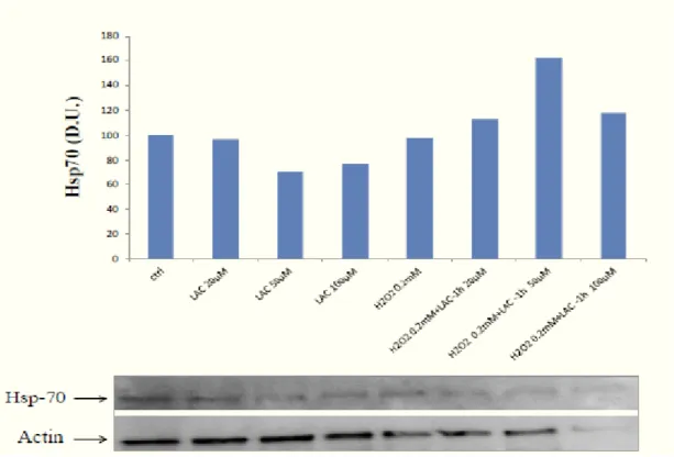
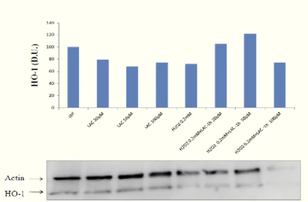
Antioxidant and Neuroblastoma cells
Recent evidences show that a minimum amount of free radicals, which until now have been considered only due to aging is a necessary condition for optimal cell function because it leads to a more efficient defense systems and increased cell survival (16). Bis(2-hydroxybenzylidene)acetone is a potent inducer of the phase 2 response through the Keap1-Nrf2-ARE pathway to study the protective effects of antioxidant molecules, including the HBB-2.
This double Michael reaction acceptor reacts directly with Keap1, the sensor protein for inducers, leading to enhanced transcription of phase 2 genes and protection against oxidant and electrophile toxicities.
Experimental evidence show that Bis(2-hydroxybenzylidene) is a potent chemoprotective agents, infact low concentrations (in the submicromolar range) of bis(2-hydroxybenzylidene)acetone markedly increased the activities of NAD(P)H:quinone acceptor oxidoreductase 1 (NQO1) and glutathione reductase, and the levels of total glutathione, three markers of the phase 2 response. In contrast, at high concentrations (in the micromolar range) the same compound caused G2/M cell cycle arrest and apoptosis (102).
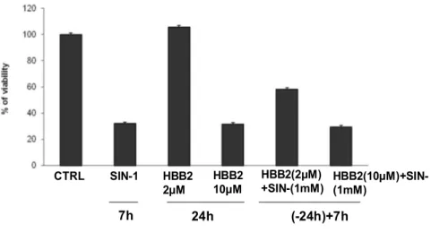
It was assumed the role of an antioxidant compound synthesis as the bis(2-hydroxy-benzilidene) acetone or HBB-2 as a possible modulator of vitagenes its use in vivo. It was also assumed its neuroprotective effect in human neuroblastoma cells exposed to nitrosative stress induced by SIN-1 to support the hypothesis that HBB may have anti-aging in the central nervous system.
The human neuroblastoma cells SH-SY5Y subclones neuroblasts are an extensively characterized cell line SK-N-SH (103,104) obtained in 1970 by a fabric of a metastatic neuroblastoma of a little girl of four years (105).
Neuroblastomas are tumors in childhood-onset originating from immature neuroblasts during the development of the peripheral nervous system (106).
The cell line SH-SY5Y cells are a cell population from neuroblast-like morphology, with a teardrop-shaped cell body (107) and small neurites, which grow to form small aggregates or clusters (105, 108) while a smaller part is in suspension, and represents the cells in mitosis. These cells are a well characterized model for the study of in vitro differentiation into a neuronallike phenotype inducible by various exogenous differentiating agents such as retinoic acid, phorbol esters, nerve growth factor (NGF),
25
and the membrane permeable analogue of cAMP, the cAMP-dibutiril (104, 109, 110). The differentiation of these cells is associated with the extension of long neuritis and can be quantified by morphological analysis of neuritogenesis (104,107).
The line SH-SY5Y is also used as a model for the study of neuronal cell death induced by oxidative stress associated with several chronic neurodegenerative diseases such as Alzheimer's disease, Alzheimer's and Huntington's (111). They are also used as a model for the dopaminergic neurotransmission and sympathetic study of Parkinson's disease (112).
Express adenosine A1 and A2A receptors and, in the presence of inhibitors of phosphodiesterase, increase levels of cAMP even in the absence of a receptor-mediated stimulation of adenylate cyclase evidence to suggest that the basal levels of cyclic nucleotides are subject to a high turnover (113). Other features of the cell line include the ability to convert glutamate into GABA, choline into acetylcholine, and the presence of dopamine-β-hydroxylase activity. Express μ opioid receptors and δ, in particular δ2 and the α2 adrenergic receptors (114, 110).
They are also considered an appropriate model for the study of σ receptors (113). The cells also express the calcium channels, and N-type and L-type muscarinic receptors (M3) and nicotinic receptors for neuropeptide Y (Y2 receptors) (112,115).
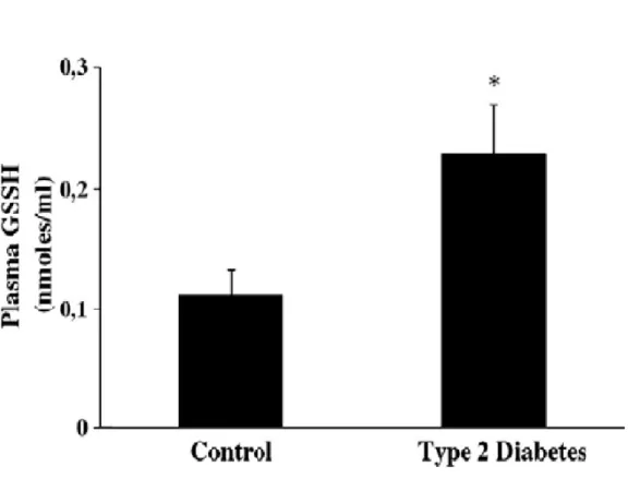
Oxidative stress and type II Diabetes
Oxidative stress is caused by an unbalance between a relative overload of oxidants and a depletion of antioxidants, and it is implicated in the pathogenesis of several chronic diseases, including atherosclerosis, ischaemia/reperfusion injury, chronic inflammatory diseases, renal failure and diabetes mellitus (116,117).
Several mechanisms seem to be involved in the genesis of oxidative stress in diabetic patients. Among these, glucose autoxidation, not enzymatic protein glycation as well as the formation of advanced-glycation end-products (AGEs) have been demonstrated in patients with diabetes and a direct relationship with the circulating blood glucose levels and glucose variability has been repeatedly demonstrated (116,118,119).
In particular, it has been shown that two subclasses of AGEs, such as N-ε(carboxymethyl)lysine and pentosidine, accumulate in various tissues of poorly controlled diabetic patients (120).
26
The interplay between oxidative stress and AGEs is very complex. In fact, reactive oxygen species (ROS) accelerate the formation of AGEs, which in turn, as glycated proteins, are also able to produce ROS via complex biochemical mechanisms (121,122).
In addition to the above mentioned AGEs and protein carbonyls, various other products derived from lipid peroxidation accumulate in biological fluids and tissues of diabetic patients. 4-hydroxy-2-nonenal (HNE) is considered an important marker of lipid peroxidation, but at the same time it is directly involved in both cytotoxicity and mutagenic activity. In fact, 4-HNE further reacts with protein residues, such as histidine, to generate stable Michael adducts (123,124).
Interestingly, HNE-modified proteins have been identified in the serum and in renal tissues of type 2 diabetic patients (123,125).
The free radical-based oxidation of arachidonic acid (126,127), is one of the most relevant biochemical pathways that generate isoprostanes. Increased levels of F2-isoprostanes can be found in the plasma or urine of patients affected by several chronic inflammatory or degenerative diseases, including diabetes, and are currently used as in vivo indicators of lipid peroxidation (116,128,129).
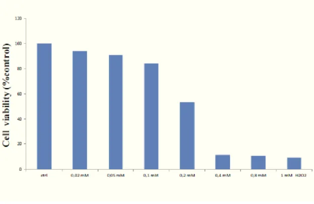
It is of interest that in addition to their well known role as in indicator of the oxidative stress status, several lines of evidences demonstrate that F2-isoprostanes are directly involved in vascular function and endothelial mediated platelet aggregation (130,131). It is hypothesized, in vitro, the role of oxidative stress in the pathogenesis and in clinical history of mellitus diabetes type 2, to evaluate the presence of systemic oxidative stress, glutathione status and cellular stress response in plasma and lymphocytes of patients with type 2 diabetes.
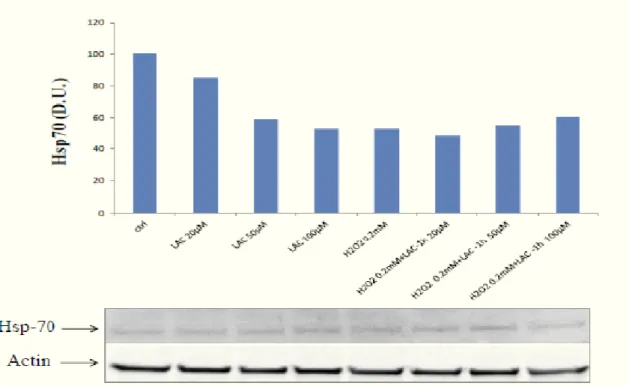
Eukaryotic cells, have developed various pathways to counteract oxidative stress-related damage. Among these stress, induced proteins, chaperones are essential to help the correct folding and maintenance of the proper conformation of other proteins and to promote cell survival after a large variety of environmental stresses (11). Therefore, normal chaperone function plays a pivotal role in the endogenous response of several tissues to an increased cellular stress, whereas altered chaperone function has been associated with the development of several diseases (132).
27
Diabetic nephropathy: effects of carnosine and cellular stress response
in podocyte cells
Diabetic nephropathy (DN) is one of the most severe complications of type 1 and type 2 diabetes and is the foremost cause for dialysis in the Western world (133).
Prime risk factors for developing DN are poor glycemic control and high blood pressure; yet, appropriate treatment of individual patients to minimize these risk factors can only delay the onset, but does not eliminate susceptibility to develop DN (134,135).
Recent experimental evidences have demonstrated that susceptibility to DN is strongly associated with a polymorphism in the CNDP1 gene (136). This gene encodes the serum carnosinase (CN-1) protein which degrades carnosine into �-alanine and histidine.
The (CTG)n polymorphism in the serum carnosinase (CN-1) gene affects CN-1 secretion (137). Since CN-1 is heavily glycosylated and glycosylation might influence protein secretion.
It is tested the role of N-glycosylation for CN-1 secretion and enzyme activity.
Furthermore has also been hypothesized whether CN-1 secretion is changed under hyperglycemic conditions.
Recently, is identified an allelic variant of human carnosinase 1 (CN1) that results in increased enzyme activity and is associated with susceptibility for diabetic nephropathy in humans. Investigations in diabetic (db/db) mice showed that carnosine ameliorates glucose metabolism effectively (138).
It is hypothesized renal carnosinase metabolism in db/db mice. Kidney CN1 activity increased with age and was significantly higher in diabetic mice compared to controls. Carnosine treatment largely prevents the alterations of renal carnosine metabolism. Susceptibility to diabetic nephropathy (DN) is strongly associated with a polymorphism in the CNDP1 gene, encoding the serum carnosinase (CN1) protein (139). The shortest allelic form (homozygous for the five-leucine allele, the so-called CNDP1 Mannheim) is more common in the absence of nephropathy and associated with lower serum carnosinase activities (140). CN1 is a dipeptidase which catalyses the hydrolysis of the dipeptides carnosine, anserine and homocarnosine (141). CN1 is present in different allosteric conformations (142). In mammals, two types of
L-28
carnosine-hydrolyzing enzymes (CN1 and CN2) have been identified. In humans, CN1 is expressed in various tissues and secreted from the liver into the blood, in rats CN1 expression is confined to the kidney (141).
Carnosine, anserine and homocarnosine are the most abundant histidine-containing dipeptides. They are widely distributed in mammals and tissue distributions and concentrations are species specific (143). All three peptides have antioxidant activity (144). Carnosine increases ischemia tolerance of neurons and hepatic cells by scavenging of reactive oxygen (145) species and acts as a natural inhibitor of the angiotensin converting enzyme (146). It restores erythrocyte deformability, inhibits protein glycation and cellular senescence and reduces the synthesis of matrix proteins such as fibronectin and collagen type VI of podocytes and mesangial cells (140). In the central nervous system, carnosine meets many criteria for a neurotransmitter (147). Recent studies, showed that carnosine influences glucose metabolism, however, mesangial expansion, as a sign of nephropathy, was not affected by carnosine treatment in diabetic mice (148).
In a previous study, we showed that the human serum histidine dipeptide concentrations are not correlated to CN1 activity (149). Since the absence of diabetic nephropathy is associated with low CN1 activity, these findings suggest that local effects in the kidney might be of importance. We therefore investigated renal histidine dipeptide metabolism and the impact of exogenous carnosine in db/db mice, as well as established model of diabetic nephropathy. Db/db mice are characterized by defective hypothalamic leptin signaling, persistent hyperphagia and obesity, high plasma leptin, glucose and insulin levels and progressive diabetic nephropathy.
Vitagenes and UCP proteins in Alzheimer’s disease
Alzheimer’s disease (AD) is a progressive neurodegenerative disorder and represents the most common cause of dementia in the elderly, accounting for 50-60% of all cases in Western world (150,151). The prevalence rates for AD rise exponentially with age, increasing markedly after 65 years.
AD is characterized by cognitive decline beginning usually with impairment of episodic memory, involving progressively all cognitive functions in the late stage
29
(152). Although some cases are familiar, sporadic AD is more common, affecting more than 15 million people worldwide (153).
The pathological hallmarks of AD are amyloid plaques, containing amyloid-β peptide, derived from the transmebrane amyloid precursor protein, and neurofibrillary tangles, composed of hyperphosforylated tau protein, in the medial temporal lobe structures and cortical areas of the brain together with neuronal death and synapses loss (154,155). Many approaches have been undertaken to understand AD, including Aβ aggregation, but the heterogeneity of the etiologic factors makes it difficult to define the clinically most important factors determining the onset and progression of the disease (156). Accumulation of Aβ characterizes AD as a protein misfolding disease, suggesting a pathogenic role for oxidative stress in protein clearance defect by the ubiquitin-proteasome system (157,158). In particular, misfolded Aβ is considered to be the key mediator of cellular oxidative stress in AD (159), and different evidences exist which indicate that oxidative stress is central to neurodegeneration in AD (160,161).
Consistently, increasing evidence indicates that factors such as oxidative stress and disturbed protein metabolism and their interaction in a vicious cycle are central to AD pathogenesis (162).
It is well known that living cells are continually challenged by conditions which cause acute or chronic stress.
Eukaryotic cells to adapt to environmental changes and survive different types of injuries, have evolved networks of different responses which detect and control diverse forms of stress (163). One of these responses, known as the heat shock response, has attracted a great interest as a universal fundamental mechanism necessary for cell survival under a wide variety of toxic conditions (8,3). Consistent with this, integrated survival responses exist in the brain, which are under control of redox regulated genes, called vitagenes, including heat shock proteins (Hsps), Sirtuins and Thioredoxin, that actively operate in detecting and controlling diverse forms of stress and neuronal injuries (3,164).
Sirtuins are a family of histone deacetylases that, in humans, includes at least seven members (silent information regulator two: SIRT 1-7) that exhibit different cellular and subcellular localizations and substrate specificities (165). The best studied sirtuin is SIRT-1, an NAD+ dependent enzyme that deacetylates several different protein
30
substrates involved in the regulation of cellular energy metabolism and redox state, thereby influencing cell survival and plasticity (15,166).
Thioredoxin (Trx), is a major redox control system, consisting of a 12 kD a redox active protein Trx, and a homodimeric selenoprotein called thioredoxin reductase (TrxR1). TrxR1 is a flavoprotein that catalyzes the NADPH-dependent reduction of oxidized thioredoxin protein. It is usually located in the cytosol, but it translocates into the nucleus in response to various stimuli associated with oxidative stress. Trx, thus, plays a central role in protecting against oxidative stress (167,168).
Uncoupling proteins (UCPs) are members of the super family of anion carrier proteins located in the inner membrane of mitochondria. These proteins have several hypothesized functions including thermogenesis in certain tissues, protection from reactive oxygen species (ROS), neuroprotection and export of fatty acids. UCPs influence the production of mitochondrial reactive oxygen species. In general, the available data indicate that UCP activity results in decreased superoxide and hydrogen peroxide production (169,170).
Oxidative stress and cellular stress response in glaucoma: implications
with Alzheimer’s disease
Glaucoma is a progressive optic neuropathy characterized by degeneration of neuronal tissue due to loss of retinal ganglion cells (RGCs), with accompanying compromission of visual field over time (171).
Research studies have demonstrated that RGC damage in glaucoma is not limited to the primary insulted neurons, but also involves neighboring neurons.
The increase in the prevalence of glaucoma with age is not accounted for only by the increase in ocular hypertension alone, being accompanied by an increase in the vulnerability of the optic nerve to the effects of glaucoma risk factors which increase as function of age. In particular, factors such as tissue hypoxia, disturbed protein metabolism and oxidative stress have been identified to interact in a vicious cycle underlying the pathogenesis of glaucoma (172,173), ultimately leading to apoptotic retina ganglion cell death (174,175, 176). In view of these considerations glaucoma can be viewed as a neurodegenerative disease which, similarly to other neurodegenerative pathologies, i.e., Alzheimer’s and Parkinson’s disease, where
31
irreversible functional deficit ensues as consequence of neuronal dysfunction and death. There is now a growing body of evidence demonstrating a link between AD and glaucoma.
Amyloid deposits, consisting of Aβ, which area characteristic feature of several neurodegenerative diseases such as Alzheimer’s (AD), mild cognitive impairment, and Parkinson’s disease (177) have been recently implicated in the pathogenesis of retinal damage, macular degeneration, and glaucoma (178). Accordingly, drugs designated to target β-amyloid (Aβ) has been found to reduce apoptotic degeneration of RGCs, asobserved in vitro and in vivo. Furthermore, the presence of increased levels of Aβ characterizes glaucoma as a protein misfolding disease, also suggesting a role for oxidative stress in the pathogenesis of retinal degenerative damage associated to glaucome.
Although oxidative stress has been recognized to play a critical role in the development and progression of glaucoma, yet, the exact mechanisms remain elusive. Oxidative stress can cause oxidative attack to DNA, proteins, and lipids, leading to DNA and protein modification, thus sustaining the pathophysiology of degenerative damage of RGCs (171). Relevant to protein misfolding, of emerging interest are heat shock proteins (HSPs), specialized molecular chaperones which mediate various cellular functions. HSPs are up regulated in response to conditions of stress in order to restore normal cell integrity (175). The heat shock response, an important component of vitagene family, contributes to establishing a cytoprotective state in a wide variety of human diseases. Vitagenes include, besides HSPs 70 and 32, the latter also called heme oxygenase-1 (HO-1), thioredoxin and sirtuins (179,180).
Several families of HSP have been implicated in neurodegenerative diseases and glaucomatous RGC apoptosis with increased levels of circulating autoantibodies to alpha-crystallins and HSP-27 and increased immunostaining of HSP-60, HSP-27 in RGCs and the retinal blood vessels in glaucoma patients (181,182).
In a rat glaucoma model, treatment with geranylgeranylacetone increases HSP-72 synthesis while reducing markedly RGC loss, possibly through interactions with different protein kinases, such as Akt kinase, and the inhibition of NF-kB. In this study we tested the hypothesis that there may be a causal relationship between AD and glaucoma that may be explained by systemic oxidative stress and dysregulation of cellular stress response.
32
Present work elucidated cellular stress response in peripheral cells in patients with glaucoma as com- pared to healthy volunteers, as control, in order to gain insight into the pathogenic mechanisms operating in the neurodegenerative damage associated with this disease and exploit the possible role of vitagenes in opening up new therapeutic targets for limiting the oxidative damage associated to degeneration occurring in glaucoma.
33
-2-AIM OF THE RESEARCH
Under normal conditions, the cell antioxidant defense systems operate very efficient there is a balance between pro-oxidant and antioxidant necessary for the maintenance of antioxidant defense system that, during normal cellular metabolism, is able to eliminate all free radicals that are generated.
In recent years, since oxidative stress has been considered the basis for some aspects of neurodegeneration, numerous experimental investigations have been conducted in order to reduce the effects of oxidative stress through the use of scavengers of free radicals (22). The cells are able to fight oxidative stress with many resources including bioactive molecules (glutathione, thioredoxin, flavonoids), lipoic acid, enzymes (heat shock proteins, superoxide dismutase, catalase, glutathione peroxidase, thioredoxin reductase, etc.) and redox sensitive transcription factor protein.
The heat shock proteins (Hsp) is one of the most studied active defense systems against oxidative damage. The heat shock response to contribute took office a cytoprotective state in a wide variety of human diseases such as inflammation, cancer, aging and neurodegenerative disorders.
Vitagenes encode for heat shock proteins (Hsp) Hsp70 (53), and heme oxygenase-1, the thioredoxin and the sirtuin protein systems, open new prospetive in medicine and pharmacology that molecules capable of activating these defense mechanisms are a potential target for novel cytoprotective strategies (183,184).
The aim of the present study are as follows:
1. Antioxidants and neuroblastoma cells 2. Oxidative stress and type II Diabetes
3. Diabetic nephropathy: effects of carnosine and cellular stress response in podocyte cells
4. Cellular stress response, sirtuins and UCP proteins in Alzheimer disease: role of vitagenes
5. Oxidative stress and cellular stress response in glaucoma: implications with Alzheimer’s disease