Molecular Medicine PhD
- cycle XXXII -
Academic Year 2018/2019
PLK1 and NOTCH1 at the interface of DNA
Damage Checkpoint and tolerance to genotoxic
stress
Supervisor: Candidate:
Prof. Claudio Talora Matteo Franchitto
Coordinator: (1240988)
Prof.ssa Isabella Screpanti
INDEX
Abstract...pag. 3
1. Introduction...pag. 4
1.1 Epidermis structure... 4
1.2 The Notch signalling... 7
1.3 Role of Notch1 in the skin cancer...12
1.4 The DNA Damage Stress...14
1.5 Dualism Notch1/Polo-Like Kinase-1 in the cell cycle...20
2. Aim of the work...pag.23
3. Results...pag.24
3.1 Plk1 as a central kinase involved in Arsenite-induced Notch1 downmodulation...243.2 Notch1 is a direct target of Plk1...26
3.3 Notch1 is a Substrate of Plk1 in the G2 phase of the Cell Cycle...27
3.4 Upon DNA Damage in G2, Notch1 Protects cells from apoptosis...28
3.5 Notch1 promotes inflammatory cytokine secretion in cancer cells that undergo growth-arrest in response to DNA damage...31
4. Figure Legends...pag.32
5. Discussion...pag.43
6. Experimental procedures...pag.46
7. References...pag.49
Abstract
Notch signalling plays a complex role in carcinogenesis and its signaling pathway has both tumor-suppressor and oncogenic components. We have shown that deregulation of NOTCH1 signalling is a crucial mechanism involved in Arsenic-induced keratinocyte transformation. We have used the Arsenic-induced transformation model to probe cellular and molecular mechanisms that control the duality of NOTCH1 signaling in malignant transformation. Here, to identify regulators that might control this dual activity of NOTCH1, we screened a chemical library targeting kinases and identified Polo-Like kinase 1 (PLK1) as one of the kinases involved in Arsenite-induced NOTCH1 downmodulation. We show that this critical correlation represents an important regulatory mechanism of NOTCH1 expression in the G2 phase of the cell cycle and in response to DNA damage in G2. Our results suggest that genotoxic stress causes a PLK1-dependent signalling response that antagonizes the involvement of NOTCH1 in the DNA damage checkpoint. Although, its precise contribution to the oncogenic phenotype of epithelial cancer remain unclear, we provide evidence that Notch signaling is altered but not abolished in SCC cells. Thus, it is also important to recognize that even in a single type of tumor, there is plasticity in Notch function and the dual role of NOTCH in cancer biology is undoubtedly complex and tumor type-independent.
1. INTRODUCTION
1.1 Epidermis structure
The epidermis is a thin layer of stratified squamous epithelium that rests on top of a basement membrane of extracellular matrix, which separates it and its appendages, including the hair follicles and sweat glands, from the underlying mesenchymally derived dermis (FIG. 1). The epidermis is tough and resilient, and is able to withstand the physical and chemical traumas of each passing day.
The epidermis is a self-renewing tissue: a single, adult skin stem cell has sufficient proliferative capacity to produce enough new epidermis to cover the body surface (Rochat A. et al, 1994). In the skin of furry mammals, slow-cycling, epithelial stem cells reside in a portion of the hair follicle known as the bulge (FIG. 1). These stem cells are multipotent and can give rise not only to epidermis, but also to hair follicles and sebaceous glands. Those stem cell progeny that exit the bulge and migrate upwards into the epidermis populate the innermost (basal) layer. The rate of proliferation and upwards migration is greatly accelerated when the skin is injured and wound healing is induced. In human skin, which has a meagre version of the hair coat of most mammals, the epidermis is much thicker and the basal layer harbours highly proliferative self-renewing cells. This has led scientists to wonder whether this subset of epidermal cells is similar or identical to the multipotent stem cells in the bulge of the hair follicle. Most researchers accept an universal definition of a stem cell as one that can divide to produce both daughter stem cells and cells that go on to differentiate. According to these criteria, a subset of basal epidermal cells (at least in human skin), as well as bulge cells, qualifies as stem cells. Whether stem cells in the basal layer have the capacity to generate hair follicles, as bulge stem cells do, remains unclear and is a topic of
considerable attention in the field. Most cells within the basal layer are the rapidly dividing progeny of stem cells (Jones P.H. et al, 1993; Jones P.H. et al, 1995). Referred to as transit amplifying, these cells undergo a limited number of divisions before they withdraw from the cell cycle commit to terminal differentiation, detach from the basement membrane and begin their trek towards the surface of the skin (Barrandon Y. et al, 1987). Cells that reach the body surface are
dead, enucleated, flattened cells (squames) that are subsequently sloughed and are continually
replaced by inner cells that move out-wards. So, the epidermis is in a constant state of dynamic equilibrium, replenishing itself every two weeks throughout life. A remarkable feature of adult, human epidermal stem cells is that they can be maintained and propagated in vitro in primary human keratinocyte cultures. But finding these stem cells and studying their properties presents a more difficult challenge, because such cultures contain a mixed population of cells. Nevertheless, stem-cell character in vitro has been correlated with the selective down-regulation of certain proteins, such as the transferrin receptor (Tani H. et al, 2000), 14-3-3σ (a nuclear-export protein), and the cytoskeletal keratins, K19and K15.
The epidermis is organized into a three dimensional lattice of tightly adhering cells. The cellular architecture of the epidermis is essential for its barrier and protective functions, and this architecture is perturbed in several human genetic skin disorders, including degenerative blistering diseases, such as epidermolysis bullosa simplex (EBS) and palmoplantar keratoderma, as well as in epithelial skin tumours and cancers. In the metabolically active layers of the epidermis, intercellular adhesion is accomplished by two types of intercellular junction: the desmosome and the adherens junction. Both of these intercellular junctions are essential for epithelial sheet formation (Green K.J. et al, 2000; Vasioukhin V. et al, 2000). Recently, it has been found that the core components of the adherens junction, its transmembrane anchor (e-cadherin) and its indirect link to the actin cytoskeleton are used in the first step of intercellular adhesion to draw epithelial cells together. In the second step, desmosomal cadherins and their indirect association with the keratin intermediate
filament cytoskeleton are required to clamp this work into place, so that redirected actin polymerization can then seal the membranes to make the adhering sheets of cells that form the epidermal barrier (Vasioukhin V. et al, 2000).
The epidermis is non-vascularized and receives its nutrients from blood vessels in the underlying dermis. The epidermis and dermis are separated by a basement membrane that is composed of extracellular matrix proteins, including collagen IV, fibronectin and laminin 5. Both the epidermis and the dermis contribute to the synthesis of basement membrane components, but the basal layer of the epidermis seems to be the sole manufacturer of collagen IV and laminin 5. The basal layer of epidermal cells both synthesize these components and adheres to them, and also polymerizes and/or organizes them into the basement membrane.
1.2 The Notch signalling
Development makes reiterative use of a surprisingly small set of essential molecular signals: the Wingless (Wg/Wnt), Hedgehog (Hh), Transforming Growth Factor-b(TGF-b), Receptor Tyrosine Kinase/Phosphotase (RTK/P) and Notch pathways (Gerhart, 1999). These five molecular archetypes act both individually and coordinately to interpret and transmit extrinsic signals as distinct cellular transcriptional responses. They mediate the establishment of polarity and body axes, coordinate pattern formation, and ultimately choreograph the morphogenesis of individual tissues. Recent advances have elucidated both the biochemical mechanisms regulating receptor activation and the molecular participants forming the intracellular signalling cascades of each of these pathways.
Notch signalling is a highly conserved process and plays an important role in the regulation of cellular growth, cell cycle arrest, and cellular development (Borggrefe T and Oswald F, 2009; Artavanis-Tsakonas S et al, 1999). The first mutant in NOTCH was isolated by Dexter in
Drosophila (1914), who described the lethality and the haploinsufficient wing notching that lends
the name to the gene. New alleles were identified by Morgan and Bridges and the collection allowed the studies of Poulson on the recessive neurogenic phenotype (Metz & Bridges 1917, Poulson 1939). Over the years, the analysis of these phenotypes eventually led to the discovery of different functions of Notch. Most importantly, in the last few years, it has uncovered three different Notch activities in development: lateral inhibition, boundary formation and cell fate assignation (Bray 1998). The first insights into the function and mode of action of Notch signalling came from studies in Drosophila Melanogaster and Caenorhabditis elegans (Greenwald 1985, Wharton et al, 1985, Yochem et al., 1988, Fehon et al., 1990) which allowed the discovery of a core set of molecules involved in Notch signalling and lead to the understanding of how they organize into a signalling pathway. In mammals, there are four Notch receptors (Notch1-4) and the abnormal
regulation of Notch signaling can promote cancer and other disorders (Bolos V et al, 2007; Cordle J et al, 2008). NOTCH1 regulates the expressions of target genes such as HES1, HES5, and HEY1, for the acceleration of cell growth (Allenspach EJ et al, 2002). Notch loci, first described in Drosophila nearly a century ago (for a historical perspective see Artavanis-Tsakonas et al.,1999),
encodes large (2500 aminoacids) and it is a Type I single transmembrane receptor involved in
signal transduction as a transcription regulator, which perceives signal transduction mediated by ligands (in mammals, Delta-like 1, 3, 4 and Jagged 1, 2) (Andersson ER et al, 2011). Notch consists of various domains that precisely regulate the function. The Notch receptor has 36-epidermal growth factor (EGF)- like repeats essential for the binding of ligands and there are three juxta-membrane repeats subjected to protelysis in the processing of Notch. Notch has also ankyrin repeats, a transactivation domain (TAD) involved in the transactivation of Notch, and a proline, glutamic acid, serine, and threonine (PEST)- degradation domain critical for the short half-life of Notch1 (Fiuza UM and Arias AM, 2007). During the maturation of Notch1 in the trans-Golgi, cleavage occurs by a furin-like convertase (S1 cleavage) for the transportation to the cell membrane (Logeat F et al, 1998). This cleavage separates Notch1 into two fragments, making a heterodimer. They move to the membrane and form the transmembrane protein complex. Then, cell-to-cell communication and proteolytic processing occurs when the extracellular domain of Notch1 is docked into the DSL domain of a ligand. Secondary hydrolysis is promoted by ligand binding, which cleaves the extracellular domain of the Notch1 receptor (Chillakuri CR et al, 2012; Mumm JS et al, 2000). The extracellular domain is subjected to secondary hydrolysis by ADAM (A Desintegrin And Metalloproteinase) protease and PS/y-secretase (De Strooper et al, 1999). The Notch1-ICD (NICD) fragment, which is produced by PS/y-secretase-mediated cleavage at site 3 (S3) within the transmembrane domain, translocates into the nucleus and interacts with CSL (CBF/recombining binding protein suppressor of Hairless [RBP-Jκ] in mammals, SuH [Suppressor of Hairless] in Drosophila melanogaster and Lag-1 in C. elegans) and MAML (mastermind-like) and functions as a transcriptional activator to regulate its target gene (Artavanis-Tsakonas et al,
1999; Mumm, JS and Kopan R, 2000), including members of the Hairy/enhancer-of-split (HES) family, the Hairy/enhancer-of-split related with YRPW motif (Hey) family, nuclear factor-kappa B (NF-κB), the vascular endothelial growth factor receptor (VEGF), cyclin D1, c-Myc, p21, p27, Akt, etc. (Guo et al, 2011). Elimination of PS function results in the Notch phenotype, which includes disruption of segmentation during the development of many kids of animals, demonstrating the importance of NICD generation (Selkoe et al, 2003). Activation of Notch receptors releases a quantum of signal in the form of NICD. Most Notch-mediated processes require a transient pulse of activity for instance, in developmental contexts where iterative activation of the Notch pathway is required. In some tissues, this could last a fraction of the cell cycle (Ambros, 1999; Bessho and Kageyama, 2003; Hirata et al., 2004). Even the few processes that require prolonged activation still seem to be sensitive to the activation “strength”, a yet to be defined aspect of Notch signalling. Given what we know about Notch biology, sustained Notch activation can be deleterious. Thus, in addition to the above-mentioned mechanisms that regulate NICD production, optimal signal strength is regulated in most cells by ensuring that NICD half-life is short. During the transcriptional activation process, NICD is phosphorylated on its PEST domain by the CDK8 kinase and targeted for proteasomal degradation by the E3 ubiquitin ligase Sel10/Fbw7 (Fryer et al., 2004; O'Neil etal., 2007; Thompson et al., 2007; Tsunematsu et al., 2004). This eliminates NICD, disassembles the ternary complex and resets the cell for the next round of signalling. Highlighting the importance of NICD turnover is the observation that C-terminal or PEST domain deletions or mutations that stabilize NICD can cause T-ALL in humans (Weng et al., 2004). Indeed, further studies on the T-ALL associated deletions identified additional conserved regulatory phosphorylation sites in Notch1 (Chiang et al., 2006). The kinase(s), phosphatase(s) and/or Ubiquitin ligases that target these sites remain to be identified. It is also unclear whether their regulatory mechanisms are coupled to transcriptional activation similar to CDK8 and FBW7.
Notch signalling involves a direct form of cell-cell communication. Direct cell-cell communication by Notch is employed for “lateral inhibition”, a process whereby negative cross-regulation between Notch and its ligand(s) in equipotent progenitors produces cells with divergent cell fates (Bray, 2006). Notch and its ligands can also be part of a positive feedback mechanism; here, Notch-dependent up-regulation of ligand expression and consequent reinforcement of Notch activation are involved in the coordination of differentiation along the same lineage. Both mechanisms are likely to operate in mammalian skin. The intensity of Notch signalling is crucial for cell fate decisions. For example, Notch haplo-insufficiency causes the "notched-wing" phenotipe in
Drosophila (Fehon et al, 1991).
Structure of Notch and its ligands. Notch ligands, Delta and Jagged/Serrate, are composed of a DSL region responsible for the interaction with the Notch receptor and several EGF repeats. Jagged/Serrate also contains an extracellular cystein-rich region. Notch is composed by up to 36 EGF-like repeats. EGF repeats 11 and 12 are sufficient to mediate the interaction between Notch and its ligands. Notch also contains a cysteine-rich region known as Lin-12 repeats in close proximity with heterodimerization domains that bind non-covalently extracellular Notch with membrane-tethered intracellular Notch. In its intracellular part, Notch has a region called RAM (RBPjk Associate Molecule) followed by repeated structural motifs named Ankyrin repeats (mediate the interaction between Notch and CBF1/Su(H)), a transactivation domain (TAD) and a PEST domain. The PEST domain is involved in the degradation of Notch. PM, plasma membrane. Citation: Journal of Endocrinology 194, 3; 10.1677/JOE-07-0242
Canonical Notch signalling pathway. Notch binding to ligand elicits several steps of cleavage. The first one at the S2 site is mediated by the proteases ADAM10 or by TACE (TNF-α-converting enzyme). This catalyzes the processing of Notch in the intramembranous S2 and S3 sites by the γ-secretase complex. Thus, Notch intracellular domain (NICD) is released and translocates into the nucleus where it dislodges repressors (co-R) associated with the DNA-binding CSL transcription factor. NICD and CSL form a ternary complex together with Mastermind (Mam) that recruits transcription factors activating target gene expression.
1.3 Role of Notch1 in the skin cancer
Cancer development is the combined result of deregulated stem cell populations and alterations in their surrounding environment. Cell and tissue homeostasis is achieved by a complex interplay of signaling pathways, coordinating behavior of a cell with its neighbors in a closely integrated fashion. Thus, the biological function of individual cell regulatory pathways can only be understood within the specific cells and tissues in which they operate. This is particularly relevant for Notch signalling, and its highly context-dependent function in different cell types and, within the same cell type, at different developmental stages and under normal versus pathological conditions. Notch signalling is essential for development and it is a type of cell-cell signaling that participates in a wide range of biological process from neurodegeneration to tumorigenesis (Artavanis-Tsakonas et al, 1999; Mumm JS and Kopan R, 2000). Alteration of Notch signalling has
been described as a major player in several human cancers (Palermo et al, 2014). Furthermore,
multiple lines of evidence indicate that Notch signalling is not exclusively oncogenic but can act as a tumor suppressor. In animal models, evidence for Notch signalling in mediating each of these roles has been established. Additionally, the NOTCH1 tumor suppressor role is also underlined by the loss or inactivating mutations of members of the Notch signaling pathway in human cancers, particularly in head and neck squamous cell carcinoma (HNSCC), the most common human
ectodermal cancer,
resulting in >300,000 deaths per year (Goon PK et al, 2009; Madan V et al,
2010). SCCs arise from renewable squamous epithelial cells that serve to create an
environmental
barrier in the skin,
esophagus, lung, and cervix. In HNSCC inactivating mutations of NOTCH1
were found in 10–15% of the tumors (Agrawal N et al, 2011; Dotto G.P., 2008; Nicolas M et al, 2003; Nowell CS and Radtke F, 2017; Rangarajan A et al, 2001; Wang et al, 2011). Interestingly, a subset of HNSCC tumors with Notch1 wild-type sequence exhibit Notch pathway copy number increase with activation of the downstream Notch targets HES1/HEY1 (Agrawal N et al, 2011;
Wang et al, 2011). Additionally, inhibition of NOTCH or HEY1 significantly decreased cell growth of primary tumor-derived cells, indicating their potential involvement in HNSCC development (Agrawal N et al, 2011; Wang N.J. et al, 2011; Sun W et al, 2014). We have recently reported that the clinical manifestation of Hailey–Hailey disease, a hereditary skin disease, is associated with NOTCH1 downmodulation as a consequence of increased oxidative stress (Biolcati G et al, 2014; Cialfi et al, 2010; Manca S et al, 2011). Much recent interest has focused on the role of ROS in normal and malignant cell biology (Jaramillo MC, Zhang DD, 2013; Naviaux RK, 2012). Organisms living in aerobic conditions are continuously subjected to ROS, and the response to ROS influences central cellular processes such as proliferation, apoptosis, and senescence, and elevated levels of ROS are associated with various human diseases including various cancers (Gorrini C et al, 2013). Of note, NOTCH1 contributes to leukemia-initiating cells’ maintenance through
regulation of ROS intracellular levels (Giambra V et al, 2012). The molecular regulation of the
dichotomous function of Notch signalling remains poorly understood. For this reason, we studied this dual activity of NOTCH1 in Arsenic-induced keratinocyte transformation thus providing a model to investigate the molecular aspects determining whether Notch signalling will be either oncogenic or tumor suppressive (Cialfi S et al, 2014). We observed that the mechanism is characterized by two phases: the first phase involves the downmodulation of NOTCH1 expression and the second phase involves the acquisition of resistance to arsenite induced-NOTCH1 downmodulation (Cialfi S et al, 2014). We found that maintenance of Notch1 expression supports metabolic activities to enhance cytoprotection against oxidative-stress that as a side effect may sustain cell proliferation and keratinocyte transformation, strengthening the hypothesis that tumor cell selection could favor partial, rather than complete inactivation of this signalling pathway (Cialfi S et al, 2014). To identify regulators that may influence the dichotomous NOTCH1 function, we screened a chemical library targeting human kinases and identified Polo-Like kinase-1 (PLK1) as one of the kinases involved in Arsenite-induced NOTCH1 downmodulation.
1.4 The DNA damage stress
One of the most well-known and widely reported aspect in cancer biology is the acquisition of genetic mutations that underlie cell transformation and tumor progression. From this perspective, cell transformation is a genetic process of tumor cells adapted to stressful environmental conditions; if to ‘cell adaptation’ can be conferred the Darwinian concept to respond to life’s needs for survival, the nature of what adaptation means for tumor cells is extremely elusive. Either physical or chemical environmental agents can cause DNA damage and consequently genetic mutations that promote cell transformation. Examples of physical agents promoting mutations are ionizing radiation, ultraviolet light present in sunlight which can promote the estimated rate of up to 10,000 DNA lesions per cell per day (Ciccia A., Elledge S.J, 2010; Hoeijmakers J.H, 2009); chemical agents such as benzo(a)pyrene B(a)P, 7,12-dimethylbenz[a]anthracene (DMBA), that generate DNA adducts, leading to mutations (Basu A.K, 2018). Beside exogenously, DNA damage can also occur endogenously as cells divide, with tens of thousands events every day in each single cell (Hoeijmakers J.H, 2009). Thus, DNA damage might potentially affect the function of central regulators of many biological processes, ultimately leading to cancer development. Additionally, infectious pathogens elicit an oncogenic spiral that is one of the causes of cancer development (Yasunaga J.I., Matsuoka M., 2018). If we assess the concept that ‘adaptation’ means the optimization of the phenotype whereby the organism acquires changes that increase its survival and reproductive success, when this concept is applied to cell transformation it remains extremely vague. Although this concept is suitable for viral carcinogenesis that hijacking cellular pathways promotes the survival and proliferation of infected cells, in a multicellular organism, cells do not need to adapt their phenotype to a non-permissive environment. Unquestionably, in multicellular organisms, cells are immersed in growth conditions favorable to their replication. However, there is an obvious difference in the relationship between adaptation and environment in unicellular versus
multicellular organisms. Life and replication in unicellular organisms are dependent on the conditions present in the environment and they survive if they are able to adapt to environmental changes. In sharp contrast, in multicellular organisms cell division is tightly regulated to control cell shape, tissue patterns, and morphogenesis (Rue P., Martinez Arias A., 2015), although cells are typically immersed in permissive environmental conditions. Preservation of the integrity of multicellular organisms relies on these extra layers of developmental control that function to restrain cellular proliferation that may change in response to environmental or intracellular stress signals. This implies that, as previously defined (Chigira M., 1993; Hersh E.M. et al, 1975) cancer cells arise from cells adapted to respond to holistic control system and the escape from these host defense mechanisms represents an important strategy for cell transformation. Genetic damage produced by either exogenous or endogenous mechanisms represents an ongoing threat to the cell. To preserve genome integrity, eukaryotic cells have evolved repair mechanisms specific for different types of DNA Damage. However, regardless of the type of damage a sophisticated surveillance mechanism, called DNA damage checkpoint, detects and signals its presence to the DNA repair machinery. DNA damage checkpoint has been functionally conserved throughout eukaryotic evolution, with most of the relevant players in the checkpoint response highly conserved from yeast to human (Harrison J.C., Haber J.E. et al 2006). Checkpoints are induced to delay cell cycle progression and to allow cells to repair damaged DNA (Figure 1). Once the damaged DNA is repaired, the checkpoint machinery triggers signals that will resume cell cycle progression (Bartek J., Lukas J., 2007). In cells, multiple pathways contribute to DNA repair, but independently of the specific pathway involved, three phase are traditionally identified: Sensing of damage, signal, and downstream effects (Figure 2). The sensor phase recognizes the damage and activates the signal transduction phase to select the appropriate repair pathway. For example, cells pose at least four independent mechanisms for repairing Double-Strand-Breaks (DSBs): Non-Homologous End-Joining (NHEJ), either classic-NHEJ or alternative-NHEJ, Homologous Recombination (HR), and
single-strand annealing (SSA) (Ciccia A., Elledge S.J, 2010; Bartek J., Lukas J., 2007; Chaudhury I. et al, 2016; Shaltiel I.A. et al, 2015).
Furthermore, highlighting the complexity of the DNA damage response, in mammals, at least four, in part, independent sensors can detect DSBs: Mre11-Rad50-Xrs2 (MRN), Poly ADP-Ribose polymerase(PARP), Ku70/Ku80 and Replication protein A (RPA) that binds single stranded DNA permitting the further processing of DSBs (Ciccia A., Elledge S.J, 2010; Cannan W.J., Pederson D.S., 2016). In the presence of DSBs, the activation of the DNA damage response and the mobilization of the repair proteins give rise to the formation of nuclear foci at the sites of damage. In yeast, the MRX-complex (Mre11-Rad50-Xrs2) is recruited at the site of DSBs (Baldo V. et al, 2012). Localization of MRX-complex to the damaged site is required to recruit and activate the protein kinase Tel1, which initiates DSBs signaling (Shaltiel I.A. et al, 2015; Syljuasen R.G., 2007). A similar mechanism is employed by MRN-complex in mammal cells (in which Nbs1 is the mammalian ortholog of Xrs2). MRN-complex orchestrates the cellular response to DBSs by physically interacting and activating the kinase Ataxia-Telangiectasia Mutated (ATM, the mammalian ortholog of Tel1). The signal is transduced by ATM that phosphorylates the histone variant Histone-2AX (H2AX) generating g-H2AX that promotes the recruitment of Mediator of Damage Checkpoint 1 (MDC1) protein at the site of damage. MDC1 amplifies the DNA-Damage Response (DDR) signal through the iterated recruitment of the MRN-ATM complex at the damage site that further phosphorylates adjacent H2AX molecules extending theγ-H2AXmark. Additionally, MDC1 functions as an interaction platform for other DDR components including chromatin remodelers and ubiquitin ligase complexes (Shaltiel I.A. et al, 2015; Syljuasen R.G., 2007). The recruitment of these factors is essential to create a more open and accessible chromatin conformation to facilitate access at sites of DNA lesions and to allow ubiquitin-mediated accumulation of DNA repair factors, which will ultimately contribute to DNA repair pathways (Shaltiel I.A. et al, 2015; Syljuasen R.G., 2007; Uckelmann M., Sixma T.K., 2017). An integral part of the DNA damage response is the parallel induction of repair mechanisms and reversible cell
cycle arrest that delays cell cycle progression to give cells time for DNA repair (Bartek J., Lukas J., 2007). The Checkpoint kinases 1 and 2 (Chk1 and Chk2) are key downstream effectors of DDR signalling as they promote cell cycle arrest. ATM/ATR phosphorylate and activate the Chk1 and/or Chk2 kinase (Bartek J., Lukas J, 2003). While Chk1 and Chk2 have overlapping substrate preferences, they contribute differentially to the maintenance of the cell cycle checkpoint and a central mechanism in the induction of the checkpoint-induced cell cycle arrest is the inhibition of cyclin-dependent kinase(s) (Cdk), where ATM and Chk2 are required to both stabilize and increase p53 DNA binding activity which in turn results in the induction of its several transcriptional targets, among which the Cdk-inhibitor protein p21waf1/cip (Harper J.W. et al, 1993; Harper J.W. et al, 1995).
The DNA Damage response in cancer cells is important. Cancer is often regarded as an asexual evolution in which cancer cells arise through the sequential acquisition of beneficial mutations that should confer an increased fitness to the adapted cells (Sprouffske K. et al, 2012; Taylor T.B., 2013; Taylor T.B. et al, 2017). Checkpoint adaptation serves as a mechanism by which cells become adapted to stressful conditions (Syljuasen R.G., 2007; Lupardus P.J., Cimprich K.A., 2004; Bender K. et al, 2018; Lewis C.W., Golsteyn R.M., 2016; Syljuasen R.G. et al, 2006; Swift L.H., Golsteyn R.M., 2014). In this process, the interaction between DNA repair pathways and cell cycle checkpoints determines cell fate decision and prevents neoplastic transformation. Preservation of integrity of multicellular organisms relies on these extra layers of developmental control. While the nature of what adaptation means for tumor cells in a multicellular organism remains puzzling, several observations indicate that the DNA Damage response may also affect the biology of the surrounding cellular microenvironment (Malaquin N. et al, 2015). In this process, the DNA damage response in cancer cells produces a paracrine signaling to induce changes in nearby microenvironment. However, DNA-damage response plays a crucial role, not only in cancers, but also in a wide variety of hereditary as well as non-genetic diseases (Cialfi S. et al, 2016; Horwitz E. et al, 2018; Milanese C. et al, 2018; Waller R. et al, 2018; Wang H. et al, 2018). A better
understanding of how the DDR-driven signals are regulated and received by the surrounding microenvironment could represent an opportunity to understand how the systemic homeostasis controls cell fitness.
1.5 Dualism Notch1/Polo-Like Kinase-1 in the cell cycle
Cell cycle transitions are driven by activation of a specific cyclin-dependent kinase (Cdk) in complex with its cyclin. Cyclin-dependent kinases (CDKs) are crucial regulators of the cell division cycle in eukaryotes (Murray AW, 2004; Pagano M and Jackson PK, 2004). Both S phase and mitosis require CDK activity; however, whereas during the G1/S transition Cdk1, Cdk2, Cdk3, Cdk4, and Cdk6 have apparently redundant functions, at G2/M, Cdk1 is nonredundant (Bashir T and Pagano M, 2005). Despite the number of CDKs that are active at G1/S, the amount of total CDK activity required for DNA replication to occur is significantly less than the sole Cdk1 activity necessary for entry into mitosis. Entry into mitosis requires active cyclin B-Cdk1 complexes (Morgan D.O., 1995). The activity of cyclin B-Cdk1 complexes is largely determined by the phosphorylation state of two residues in the ATP binding region of Cdk1. Phosphorylation of these conserved residues (Thr14 and Tyr15), by Myt1 and Wee1 respectively, inhibits ATP binding and blocks Cdk activity accordingly (Smits V.A. and Medema R.H., 2001). Removal of inhibitory phosphorylation is mediated by members of the Cdc25 dual specificity phosphatases, of which three isoforms are present in human cells: Cdc25A, Cdc25B, and Cdc25C (Donzelli M and Draetta G.F., 2003). Of these, Cdc25C and Cdc25B were shown to promote mitotic entry, by activating the cyclin B-Cdk1 complex. Cdc25B has been proposed to act as the “starter” phosphatase that initially activates cytoplasmic cyclin B-Cdk1 at the G2/M transition, while Cdc25C is thought to act in a positive feedback loop with cyclin B-Cdk1, important for full and irreversible activation of the complex. For one, activation of the DNA damage checkpoint causes downregulation of Cdc25A through ubiquitin-mediated proteolysis. In addition, Cdc25B and Cdc25C are phosphorylated by Chk1, Chk2, or p38 in response to unreplicated DNA and a variety of DNA damaging agents. However, instead of promoting degradation, DNA damage-induced phosphorylation of Cdc25C has also been shown to inhibit its phosphatase activity (Furnari B et al., 1997). Thus, dephosphorylation
of Thr14 and Tyr15 on Cdk1 is effectively blocked in response to DNA damage. The Polo-box of PLK1 was shown to bind specifically to Cdc25C (Elia et al., 2003) and it can phosphorylate and activate Cdc25C in vitro (Smits V.A. and Medema R.H., 2001) and mediate nuclear translocation of Cdc25C (Toyoshima-Morimoto F et al, 2002).
The Polo Kinase is an important regulator of cell division responsible for a wide number of functions: centrosome maturation, DNA replication, cytokinesis, mitotic entry (G2/M transition) and adaptation to persistent DNA damage (Archambault V et al, 2015; van Vugt M et al, 2004).
Ever since the discovery of Polo kinase in Drosophila in 1988, Plks have emerged as a family of kinases that perform several crucial functions in cell division (Petronczki M et al, 2008). Five members of the Plk family (Plk1–5) have been discovered in humans; all of which seem to have largely nonoverlapping functions during the cell cycle (van de Weerdt B.C.M and Medema R.H., 2006; Andrysik Z et al; 2010). Plks consist of a kinase domain and two polo box regions (with the exception of Plk4 and Plk5). The two polo box regions fold together to form a polo box domain (PBD), a functional domain that can bind phosphorylated peptides. Activation of PLK1 requires phosphorylation on a conserved threonine in the T-loop of the kinase domain (T210) in G2 phase by Aurora-A, in concert with its cofactor Bora (Bruinsma W et al, 2014; Jang Y.J. et al, 2002). The precise regulation of the Bora–Plk1 interaction is unclear, but is probably initiated by Cdk1-dependent phosphorylation of Bora (Chan E.H.Y. et al, 2008). This suggests that Cdk1 activity is required for the activation of PLK1 in G2 phase, whereas PLK1 activation in turn is required for the activation of cyclin B/Cdk1. This type of regulation is often seen in cell cycle transitions, which typically depend on such interlinked feedback and feed-forward loops to establish bistable systems that render cell cycle transitions irreversible (reviewed in Lindqvist A et al, 2009).
PLK1 is upregulated in many human tumors and this has been found to correlate with poor prognosis. However, it has to be noted that few mutations have been found in PLK1 and that the mutations that have been reported lead to destabilization of the protein rather than increased expression or activity. This suggests that PLK1 upregulation is an indirect consequence of increased
cell proliferation in tumors, rather than a causal role for PLK1 deregulation in tumorigenesis (Lens S.M.A et al, 2010). Nonetheless, its essential roles during the cell cycle have made PLK1 an interesting target for anticancer treatment (Lens S.M.A et al, 2010). Many of its interactions and substrates have been studied extensively (Archambault V and Glover D.M., 2009), and analysis of interaction partners of the polo box domain (PBD) of Plk1 shows that it interacts with proteins acting in a wide variety of processes, many of which are still unexplored (Lowery D.M. et al, 2007). We identified Notch1 as a novel direct target of PLK1 kinase activity. PLK1 inhibition reduced Arsenite-induced NOTCH1 downmodulation. Arsenic is known to have genotoxic and mutagenic effects; genotoxic stress causes proliferating cells to activate the DNA damage checkpoint, to assist DNA damage recovery by slowing cell cycle progression. Thus, to drive proliferation and transformation, cells must tolerate DNA damage and suppress the checkpoint response ((Wakida T et al, 2017) and reference there in). We report here that PLK1 promotes NOTCH1 downmodulation to the G2-M transition, conversely NOTCH1 remains active during a DNA damage-induced G2 arrest.
Our data show that NOTCH1 plays pleiotropic effects in DNA damage-arrested cells, and also in those contexts where NOTCH1 is known to play a tumor suppressor function, cancer cells might still be dependent on specific Notch1 signals to sustain their cancerous phenotype.
2. AIM OF THE WORK
Notch signalling is a highly conserved process and plays a key role in cell fate determination, cellular development, cellular self-renewal and, also, tumorigenesis. The canonical Notch pathway is mediated by the regulated intramembrane proteolysis pathway, in which Notch receptors undergo ligand-dependent sequential endoproteolysis via different enzymes, including PS/y-secretase. The NOTCH-1 ICD (NICD), which is produced by PS/y-secretase-mediated cleavage at site 3 (S3) within the transmembrane domain, translocates to the nucleus and promotes the transactivation of target genes. Alteration of Notch signalling has been described as a major player in several human cancers. Furthermore, multiple lines of evidence indicate that Notch signalling is not exclusively oncogenic but can act as a tumor suppressor, in a context-dependent manner. We study NOTCH1 in the skin context, where it is tumor-suppressor. For this fact, we use SCC022 and HaCaT cells to investigate the role of NOTCH1 in the tumoral and physiological context. We study the Polo-like Kinase 1 (PLK1), a kinase involved in G2/M transition, direct effector of the G2 DNA damage checkpoint and promoting arsenite-induced NOTCH1 downmodulation. For this reason, we investigate the correlation of axis PLK1/NOTCH1 in response to DNA damage and the effects on cell cycle progression.
3. RESULTS
3.1 Plk1 as a central kinase involved in Arsenite-induced Notch1 down-
modulation
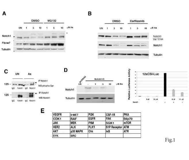
To explore the mechanisms that determine whether Notch signalling will be either oncogenic or tumor suppressive we used a well-defined in vitro model in which the non-tumorigenic human keratinocyte cell line (HaCaT) was acutely exposed to arsenic-trioxide (Arsenite). We previously demonstrated that loss of FBXW7 induction might contribute to acquire both resistance to Arsenite-induced NOTCH1 downmodulation and HaCaT transformation (Cialfi S. et al, 2014). Here we show that Arsenite stimulated the serine phosphorylation of Notch1 with the parallel decreased expression of NOTCH1 and upregulation of FBXW7 levels (Fig.1 A, B, C). Treatment of cells with the proteasome inhibitors prevented the decrease of Notch1 expression (Fig.1A, B). FBXW7 is a constituent of the SCF ubiquitin ligase complex (SKP1-CUL1-F box) that controls the degradation of Notch1. Substrate phosphorylation is required for FBXW7-‐mediated recognition (Davis R.J. et al, 2014; Koepp D.M. et al, 2001; Yada M. et al, 2004). Thus, we developed a luciferase assay to identify the kinase that would prime NOTCH1 for recognition by FBXW7. First, HaCaT cells were transiently transfected with an expression vector of NOTCH1-IC. At 36 hours after transfection, the cells were treated with Arsenite for the last 12 hours at the indicated concentrations (1-5-10µM ). Total cell lysates were collected and subjected to western blot analysis.
Arsenite treatment decreased the Notch1 level compared with the vehicle-treated control cells (Fig.1D) indicating that exogenous NOTCH1-IC is degraded similarly to the endogenous Notch1. Then, we used a 12xCSL-luciferase reporter vector responsive to Notch1 signalling and we
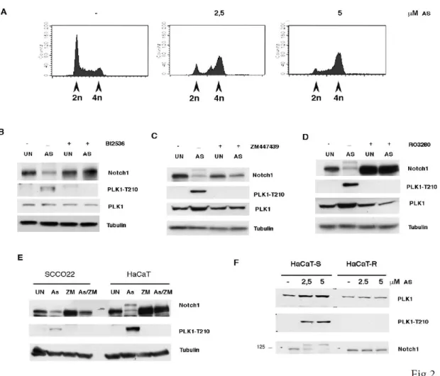
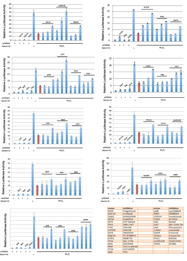
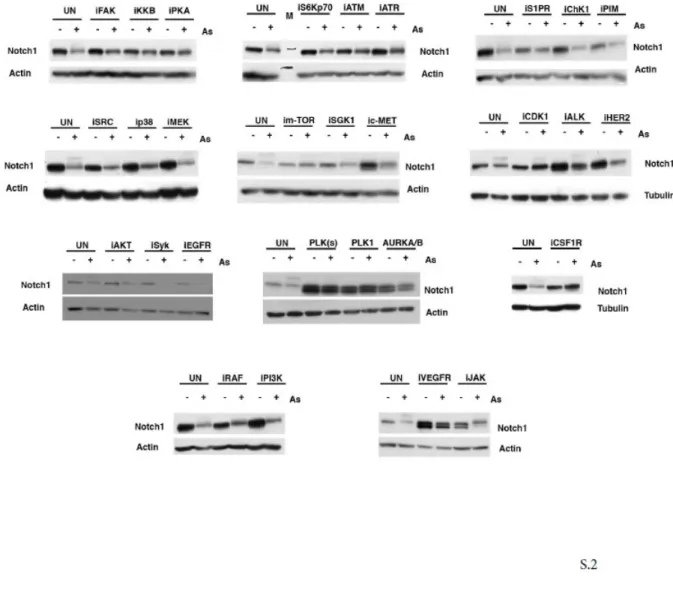
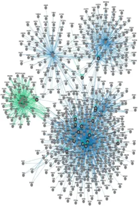
found that NOTCH1 transcriptional activity is strongly suppressed by Arsenite treatment (Fig.1D-right panel). This functional assay was used to screen a kinase inhibitor library of 378 small-molecule compounds. All compounds were screened in triplicate at 10mM in the presence of 5 µM Arsenite (data not shown). Those compounds showing at least a >50% recovery of luciferase activity were further tested by luciferase assay and western blot (Fig.S1 and S2). We identified 27 kinases able to rescue the NOTCH1 luciferase activity (Fig.1E). To understand the functional context of how the identified kinases might have an impact on Notch1, we performed a network analysis in which we investigated all possible direct and indirect interactions among them. For this purpose, the full Pathway-Commons database of reported protein interactions in Simple Interaction Format (SIF) was performed. This analysis resulted in a network comprising 611 proteins with 2263 interactions (Figure S3). The central component of the shortest path network was the protein Plk1. Plk1 is a pro-mitotic kinase, and its main function is to facilitate the mitotic process (Archambault V. et al, 2015; van Vugt M.A. et al, 2005). However, Plk1 also promotes cell cycle progression in cells under stress conditions, thus facilitating tolerance to genotoxic stress (Wakida T et al, 2017). Arsenic is known to have genotoxic and mutagenic effects and we observed that Arsenite treated cells were arrested in G2 ((Cialfi S. et al, 2014) and Fig.2A). Thus, we tested whether PLK1 activity might affect NOTCH1 expression following Arsenite treatment. PLK1 activation requires phosphorylation on a conserved threonine in the T-loop of the kinase domain (T210). PLK1 is first phosphorylated on T210 in G2 phase by the kinase Aurora-A, in concert with its cofactor Bora (Bruinsma W. et al, 2014; Yang Y.J. et al, 2002). Thus, to further characterize the pattern of T210 phosphorylation and Notch1 stability, HaCaT cells were treated with Arsenite and cultured in the presence or absence of Plk1 inhibitors. In agreement with the luciferase assay, accumulation of Notch1 protein upon Plk1-inhibitors treatment was observed in Arsenite untreated and treated HaCaT cultures as well as in SCC022, squamous cell carcinoma derived cell line (Fig.2B-D-E). We previously demonstrated that transformed keratinocytes acquire resistance to Arsenite-induced NOTCH1 downmodulation. Here, we found that PLK1 activation was not observed in
Arsenite-transformed keratinocytes after Arsenite treatment (Fig.2 F). This indicates that PLK1 activity might play a potential contribution at the early stages of arsenite carcinogenesis and that in Arsenite-transformed keratinocytes Plk1 is not longer required in response to Arsenite-treatment, as cells have acquired a molecular switch required for cellular adaptation to genotoxic stress, ( e.g metabolic adaptation) (Cialfi S. et al, 2014).
3.2 Notch1 is a direct target of Plk1
Analysis of the Notch1 C-terminal primary amino acid sequence by different computational platforms revealed the presence of multiple potential phosphorylation sites for the Plk1 consensus sequences (RXX[pS/pT]XRXXR). However, to narrow down the number of candidate motifs prior to experimental verification we analysed the Notch1 protein sequence by considering as putative candidate motifs only those identified via a high-stringency analysis and that can be recognized by both PhoshoNET and GPS-Polo 1.0 platforms. Two sites S1791 and S2349 were identified by these criteria (Supplemental Fig.4 A-B-C). Interestingly, both motifs are conserved across species and S1791 was found to be phosphorylated also in colon cancer cells (21). To confirm that Notch1 can be phosphorylated by Plk1, we performed an in vitro kinase assay using purified recombinant PLK1 and Notch1- IC fragment as substrate. As shown Fig.S4 D, the C-terminal Notch1 fragment was readily phosphorylated by Plk1. Additionally, when the two putative phosphorylation sites, S1791 and S2349 were replaced by Ala wild-type Notch1-IC but not the mutant was efficiently phosphorylated (Fig. S4E). To test whether the phosphorylation of Notch1-IC on the putative Plk1 phosphorylation sites determined the stability of Notch1-ICD cells expressing either wild-type Notch1-IC or mutants Notch1-IC-A1791/A2349 constructs were treated with Cycloheximide. At various time points thereafter, the transfected cells were lysed and the amounts of the Notch1
proteins were measured by Western blot analysis. We found that mutation of Ser 1791/2349 promotes Notch1-IC stabilization (Fig. S4 F).
3.3 Notch1 is a Substrate of Plk1 in the G2 phase of the Cell Cycle
To understand the functional significance of Plk1-mediated regulation of Notch1 we focused our attention to the Plk1/Notch1 expression during cell cycle. It is well known that in G2, Plk1 is activated to promote entry into mitosis (van Vugt M.A. et al, 2005 and reference there in). Thus, we sought to find the physiological conditions required to degrade Notch1 in the cell cycle context. To this purpose, we conducted synchronization experiments in HaCaT and SCCO22 human cells. A Hydroxyurea block and release was performed to synchronize the cells in G1/S, and the cell cycle profile was monitored. After the cells were released from the Hydroxyurea-induced G1/S block, the cells were harvested and subjected to a Western blotting analysis. The phosphorylation of Thr210 was observed strongly at the G2 phase of the cell cycle, a pattern inversely correlated with the Notch1 expression (Fig. 3 A and B). However, the inhibition of Plk1 by BI2536 induced the accumulation of Notch1 protein (Fig.3 C), confirming that Plk1 promotes Notch1 downmodulation during the cell cycle. Our data indicate that PLK1 phosphorylates and consequently destabilizes Notch1 in the G2>M transition. However, in order to be transformed, in cells under genotoxic stress the checkpoint response should be down-regulated to tolerate the cellular DNA damage stresses. Plk1 activation regulates the checkpoint activation and allows cells to grow under genotoxic stress (Furuya K. et al, 2010). Moreover, Plk1 is also known to be involved in promoting resistance to chemotherapeutic regimens with drugs such as doxorubicin (a DNA intercalating compound) (Gutteridge, R. E. et al, 2016). We found that under arsenite treatment Notch1 is continuously
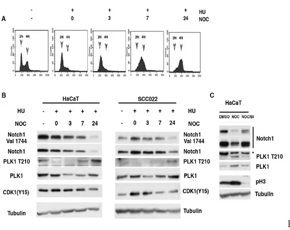
degraded and in this condition Plk1 is active (Fig. 1 and 2). Notably, a G2 phase-specific inactivation of Plk1 after DNA damage has been described, the reason for this inactivation is to promote cell cycle exit in order to avoid proliferation and entry in mitosis in the presence of damaged DNA. Thus, we investigated whether Plk1 targets Notch1 during G2 in response to DNA damage. To this end, both HaCaT and SCCO22 cells were synchronized at G1/S and then allowed to progress through the cell cycle. At 7 hrs after the release from G1/S (when cells were in G2), cells were pulsed with Doxorubicin for 1 hr to induce DNA damage and harvested 18 hrs after Doxorubicin release (Fig.4 A only HaCaT cells are shown). As expected, induction of DNA Damage results in decreased levels of Plk1 (Fig. 4B,C). Notably, when Plk1 was inactive, the expression of Notch1 was restored indicating that Notch1 expression is upregulated during G2-Damage checkpoint (Fig.4E).
3.4 Upon DNA Damage in G2, Notch1 Protects cells from apoptosis
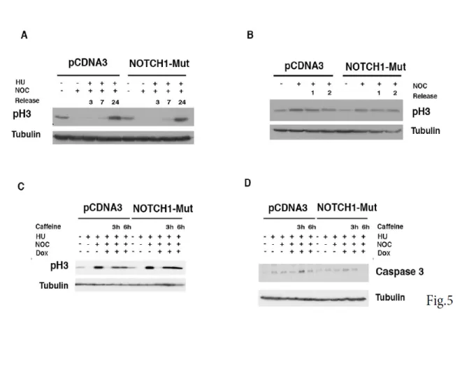
To unravel how Plk1 and Notch1 might functionally interact, we investigated whether NOTCH1 had a mitotic role. To this end, we made use of Ser 1791/2349 mutant Notch1-IC. SCCO22 cells were transfected with either empty vector or Notch1-IC Ser 1791/2349 mutant. Cells were synchronized at the G1 13 /S and released into the cell cycle; we didn’t observe any difference in cell cycle progression as phosphorylated Histone H3 (p-H3) showed the same kinetic during release (Fig. 5A) in both control and Notch1-IC Ser 1791/2349 mutant treated cells. Furthermore, no mitotic delay was detected in cells examined at either early time (1 and 2h) or at longer time after Nocodazole release (Fig. 5 B and data not show). We conclude that in this cellular context Notch1 does not alter the G2/M transition. Previous observations established that Plk1 plays a
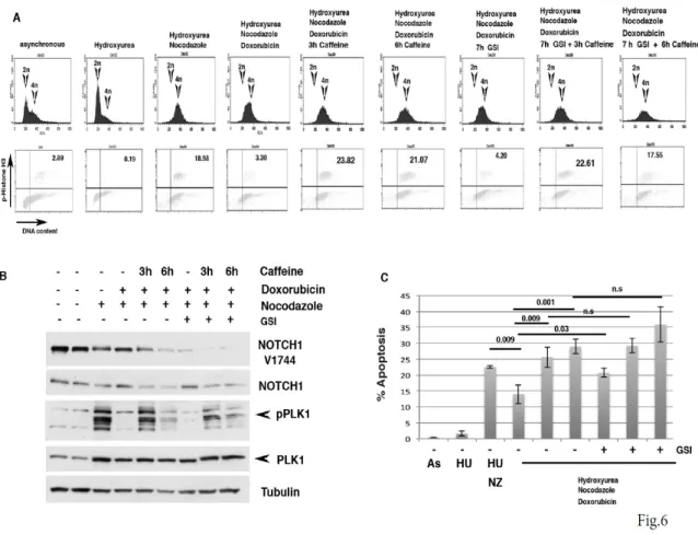
critical role in the G2 checkpoint recovery following DNA damage (van Vugt, M. A., et al, 2005; van Vugt, M. A., et al, 2004), and we found that Notch1 expression is upregulated during G2-Damage checkpoint (Fig.4). Thus, we evaluated whether Notch1 expression would alter recovery following DNA damage. To test this, cells were synchronized at the G1 /S and released into the cell cycle, after 6 hrs from release cells were treated with Doxorubicin to induce G2 damage checkpoint. Later cells were treated with caffeine to abrogate G2 checkpoint response. As expected, we detected an increase of phosphorylated Histone H3 (pH3) in empty vector treated cells after Caffeine addition (Figure 5C). Interestingly, Notch1-IC mut expression enhanced pH3 expression (Figure 5C). Treatment of cells with Caffeine abrogate G2 checkpoint but also promotes mitotic catastrophe and apoptosis (van Vugt, M. A., et al, 2005). Consistently, we found that in empty-vector treated cells Caffeine treatment induced caspase-3 activation, whose expression levels were reduced in Notch-IC mut treated cells (fig.5 D). Although, we observed a differential expression of the cleaved caspase-3 neither empty nor Notch1-IC mutant treated cells showed sign of apoptosis after caffeine addition (data not shown). The mechanism by which DNA-damaged cells escape from apoptosis during DNA-damage checkpoint is poorly understood. Therefore, we wondered whether the requirement of Notch1 during DNA-damage induced G2 checkpoint could be restricted to such an anti-apoptotic signaling. To test this, we designed an experimental set-up to examine if a cell cycle arrest/restart following a DNA damage-induced G2 arrest in HaCaT cells would be dependent on the function of Notch1. HaCaT immortalized cells were chosen because in this cellular context, conversely to SCCO22 cells, sustained DNA damage checkpoint promotes apoptosis. Thus, HaCaT cells released from a Hydroxyurea block were treated with doxorubicin at 7 hrs after release, a time at which the great majority of the cells had completed S-phase (Fig. 6A). Using this approach we were able to obtain a highly synchronous population of cells arrested at the G2 DNA damage checkpoint by doxorubicin (Fig.6A). Subsequently, we mimicked checkpoint silencing by addition of the checkpoint kinase inhibitor Caffeine and allowed the cells to enter mitosis in the presence of Nocodazole. Notably, Doxorubicin treatment of HaCaT cells resulted in lower mitotic index when
compared to control cells (Fig. 6 A lower panels, diagram 3-4). After 3-6 hrs of Caffeine treatment a significant fraction of cells entered mitosis as judged from phospho-Histone H3 staining (Fig.6 A lower panels). When cells entering in the G2-damage induced checkpoint were examined in more detail, a decrease in pPlk1 level and the appearance of Notch1 expression were observed (Fig.6 B lane 3-4). When we analyzed cell recovery from DNA damage induced arrest after doxorubicin treatment, we found that G2-arrested cells could be forced to enter mitosis following addition of caffeine. Interestingly, Caffeine treatment increased Plk1 expression, indicating that as previously shown Plk1 becomes essential for mitotic entry and recovery from a DNA damage-induced G2 arrest (van Vugt, M. A., et al, 2004). Consistent with a role for Plk1 in the control of Notch1 expression, we found that pPlk1 activation was paralled by Notch1 downmodulation when Caffeine was added to induce recovery from a DNA damage-induced G2 arrest (Fig 6B). Notably, Notch1 does not seem to be instrumental for achieving a DNA damage-induced arrest, since GSI-treated cells efficiently arrested in response to DNA damage (Fig. 6 A, 7th 31 diagram). Strikingly, when we examined the fate of the damaged cells that are in the DNA damage-induced G2 arrest or induced to enter mitosis by the addition of Caffeine in the presence of GSI, we found that cell viability was severely affected (Fig. 6 C). These results demonstrate that Notch1 protects cells from DNA damage-induced arrest and that Plk1-mediated degradation of Notch1 may be essential for recovery from a DNA damage-induced arrest.
3.5 Notch1 promotes inflammatory cytokine secretion in cancer cells that
undergo growth-arrest in response to DNA damage
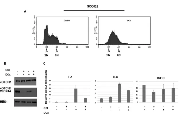
Induction of cell cycle arrest in response to DNA-damage represents a protective mechanism against harmful mutations but also promotes apoptosis (van Vugt, M.A. et al, 2005; van Vugt, M.A. et al, 2004). We found that Notch signalling protects immortalized HaCaT cells from DNA damage-induced apoptosis. Conversely, we observed that in the squamous cell carcinoma cell line, SCCO22, induction of DNA damage by doxorubicin treatment promotes a permanent cell cycle arrest with no sign of apoptosis (Fig. 7 and data not shown). In response to DNA damage, growth-arrested cancer cells also develop a secretory phenotype that alters tissue microenvironments and might stimulate tumor growth in vivo (Rodier, F. et al, 2009). Among the secreted factors, IL-6 and IL-8 are of particular interest. These cytokines have been shown to promote tumorigenesis by regulating processes associated with tumorigenesis raging from cancer metabolism to metastasis (Rodier F. et al, 2009; Colombo M. et al, 2018). Therefore, we wondered whether Notch1 during DNA-damage induced G2 checkpoint could be involved to such secretory signaling. To test this, SCCO22 cells were treated with Doxorubicin to induce G2 damage checkpoint (Fig. 7A). Later cells were treated with GSI to inhibit Notch signalling (Fig.7B,C). As expected, we detected an increase of IL-6 and IL-8 in Doxorubicin treated cells (Figure 7C). Interestingly, GSI treatment decreased both IL-6 and IL-8 expression (Figure 7C), but not TGFB1 that has been associated with the development of a secretory phenotype of cancer cells. Thus, these data support a model in which the epithelia cancer cells, SCCO22, use Notch signaling to support a secretory phenotype.
4. FIGURE LEGENDS
Figure 1. Decreased NOTCH1 levels in As2O3 2 treated keratinocytes. (A,B) HaCaT cells were untreated or treated with As2O3 (As).
Twenty-four hours (24 hrs) post-treatment, cells were either untreated or treated with MG132/Carfizomib for 5 hrs before collection; immunoblotting was performed with the indicated antibodies. (C) HaCaT cells were treated with As2O3 for 24 hrs before collection cell extract were immunoprecipitated using an antibody agaist NOTCH1 and immunoblotting was performed with the indicated antibodies. (D) HaCaT cells were transfected with either pCDNA3 or NOTCH1-IC (encoding the human Notch1-IC, 1757-2555). Thirty-six hours (36 h) post-transfection, cells were treated with As2O3 for 24 hrs before collection; immunoblotting was performed with the indicated antibodies. D- right panel, HaCaT cells were co-transfected with the NOTCH responsive promoter 12XCLS and the NOTCH1-IC plasmid then treated with increasing amounts of As2O3 (5 and 10µM) 12 hrs before collection. (E) Summary of the results of the screening experiments for several kinase inhibitors using the luciferase reporter as indicated in D and E.
Figure 2. Effects of PLK1 inhibition in As2O3 -treated cells. A) HaCaT cells were treated for 24 hr with the indicated amount of
Arsenite, then cells were collected and cell cycle analyzed by FACS. B, C, D) Immortalized HaCaT cells were treated with the indicated amount of As2O3 for 24 hrs; then cells were treated with plus/minus the indicated inhibitors for 24 hrs and analyzed by immunoblot with the indicated antibodies. (E) The indicated cell lines were treated with As2O3 for 24 hrs; then cells were treated with plus/minus 10 µM ZM447439 (ZM) for 24 hrs and analyzed by immunoblot with the indicated antibodies. (F) Immortalized (HaCaT-S) and As2O3 -transformed HaCaT cells (HaCaT-R) were treated with increasing amount of As2O3 for 24 hrs and analyzed by immunoblot with the indicated antibodies.
Figure 3. PLK1-Dependent Degradation of NOTCH1 at the G2-M transition. A,B) HaCaT and SCC022 cells were collected at the
indicated time points after release from G1/S, cell cycle analyzed by FACS (FACS profile is shown only for HaCaT cells) and cell lysates were immunoblotted with antibodies to the indicated proteins. C) HaCaT cells were treated for 16 hrs with Nocodazole to induce a mitotic block, and BI2536 added 8 hrs before harvesting. Prometaphase cells were then collected by shake-off and cell extracts were analyzed by immunoblotting with antibodies to the indicated proteins.
Figure 4. NOTCH1 expression in G2 DNA Damage Arrest. A,B) HaCaT cells were left untreated (diagram 1) or treated with
Hydroxyurea for 19 hrs. Alternatively, cells were released from the HU block and either untreated or treated after 7 hrs with Doxorubicin for 1 hr and subsequently grown in the presence of Nocodazole for 18 hrs. Following these treatments, cells were collected at the indicated time-points after release from G1/S, cell cycle analyzed by FACS (FACS profile is shown only for HaCaT cells) and cell lysates were immunoblotted with antibodies
Figure 5. Overexpression of NOTCH1 mutant unphosphorylable by PLK1 has not effect on cell cycle progression. (A) SCCO22
cells were transfected with, either control, empty-PCDNA3 vector, or A1791/A2391-NOTCH1-ICD mutant. The cells were synchronized with Hydroxyurea for 19 hrs. At the indicated time points after release, the cells were harvested and subjected to immunoblotting for the indicated proteins.
(B) Cells were treated as described for panel A, except that cells were trapped with Nocodazole for 14 h and then released. At the indicated time
points after release, the cells were harvested and analyzed with the indicated antibodies. (C,D) SCCO22 cells were transfected with either control, empty-PCDNA3 vector, or A1791/A2391-NOTCH1-ICD mutant. The cells were synchronized with Hydroxyurea for 19 hrs. Cells were released from the HU block and either untreated or treated after 7 hr with doxorubicin for 1 hr and subsequently grown in the presence of Nocodazole and Caffeine the last 3 and 6hrs. Cells were harvested and subjected to immunoblotting for the indicated proteins.
Figure 6. NOTCH1 expression in recovery from a G2 DNA Damage Arrest A) HaCaT cells were left untreated or treated with
Hydroxyurea (HU) for 19 hrs. Alternatively, cells were released after the HU block and 7 hr after release treated with Doxorubicin for 1 hr and subsequently grown in the presence of Nocodazole for 18 hrs. Following these treatments, Caffeine was added for indicated time periods to allow recovery from the checkpoint-induced arrest 3 and 6hr before harvesting the cells. DNA content and phospho-Histone H3 positivity were determined.
(B) Cells were treated as described under (A) and whole-cell lysate was used for Western blotting with the indicated antibodies (C). Cells were
Figure 7. NOTCH1-dependent increased expression of IL-6 and IL-8 during DNA-Damage induced growth arrest. SCCO22 cells
were treated with doxorubicin following and then either DMSO or GSI was added and cells maintained in culture for further 24 hrs. In (A) Cells were analyzed by FACS analysis. (B) Cells were treated as described in (A) and whole-cell lysate was used for Western blotting with the indicated antibodies. In (C) cells were treated as described for panel A and total RNA was used for qRT-PCR with the indicated probe.
Fig. S.1
Figure S1. A) HaCaT cells were co-transfected with the NOTCH responsive promoter 12XCLS and the NOTCH1-IC plasmid plus 5µM
As 2 2O3 and increasing amounts of the indicated inhibitors. 1, 5 10 µM. As control cells were co-transfected with the NOTCH responsive promoter 12XCLS and the pcDNA3 plasmid plus 5µM of the indicated inhibitors.
Figure S2. A) HaCaT cells were treated with either DMSO or 5µM As2O3 alone or in combination with the indicated inhibitors (10µM).
Fig. S.3
Figure S3. Pathway commons network visualizer (PCViz) (http://www.pathwaycommons.org) was used to detect functional interaction
among the kinases identified in the screening experiments.
Fig. S.4
Figure S4. Direct In Vitro phosphorylation of Notch1 by PLK1. A) Phosphorylation sites identified in NOTCH1-IC using KINEXUS
and GPS-Polo 1.0 platform. (B-C) Evolutionary conservation of Ser1791 and Ser 2439 in the Notch1-IC sequence, analysis was performed by Kinexus platform (http://www.phosphonet.ca) (D) GST-NOTCH1-IC 1754-2555 was incubated with or without PLK1 followed by in vitro kinase assays. Fractions of the same reactions were analyzed by 7.5% SDS-PAGE. Experiment was performed in two replicates R1 and R2. E) NOTCH1-ICD was immunoprecipitated from HaCaT cells transfected with either WT and NOTCH1-IC mutant protein and analyzed by in vitro kinase assays as indicated in D except that the assay reaction was analysed by spotting the reaction mixture onto strips of P81 paper and analyzed in a scintillation counter. F) HaCaT cells were transfected with plasmids expressing WT-NOTCH1-ICD and mutant A1791/A2391-NOTCH1-ICD for 24 h, then treated with 100µm cycloheximide for the indicated periods of time. The levels of NOTCH1 in the cell lysates were determined by Western blotting.
5. DISCUSSION
Notch1 activity plays pivotal roles in signalling for diverse cellular process, such as cell differentiation, stem cell renewal, proliferation and transformation (Artavanis-Tsakonas S. et al, 1999; Nowell C. S. and Radtke, F., 2017; Kopan, R. and Ilagan, M. X., 2009). Notch1 signalling has been reported to have a contradictory role in cell transformation (Palermo R. et al, 2014; Nowell, C. S. and Radtke, F., 2017). However, a widely accepted model implies that the impact of Notch1 signaling is highly context dependent and it can have opposite effects in different systems. We have used Arsenite-induced malignant transformation of a human epithelial cell line as an in vitro model to study the mechanisms that can result in Notch1 role and function alterations (Cialfi, S. et al, 2014). We previously demonstrated that whereas Arsenite-mediated apoptosis of immortalized keratinocytes was associated with Notch1 down-regulation, Arsenite-mediated transformation of these cells was characterized by increased Notch1 stability (Cialfi, S. et al, 2014). We found that Notch1 regulates cellular metabolism and apoptosis, which in turn differentially impact cell proliferation and cell transformation (Cialfi, S. et al, 2014). Consequently the cellular genetic/context may impinge on the antagonistic duality of Notch1 function. We presented evidence indicating that FBXW7 is required for the differential expression of Notch1 during Arsenite-mediated transformation; indicating that kinases and biochemical pathways could be involved in Notch1 phosphorylation in tumors. Given that Notch1 stability and signaling are controlled by its phosphorylation (Borggrefe, T. et al, 2016), the study of kinases that could be implicated in this post-translational modification could help to elucidate the mechanisms controlling Notch1 dichotomy in cancer development. In this study, the effects of 378 cellular kinase inhibitors on Notch1 transcriptional activity and protein stability after Arsenite-treatment were investigated. Our findings indicate that multiple kinases implicated in various cell signaling pathways can participate in these outcomes: FAK, IKKB, PKA, ATM, ATR, SRC, p38, m-TOR, GSK1, c-MET, CDK1,
ALK, PLK1, AURKA/B, CSF1R VEGFR and JAK. To understand how the identified kinases might have an impact on Notch1, we performed a network analysis to investigate all possible direct and indirect interactions among them. The central component of the shortest path network was the protein Plk1, which is a central regulator of cell division required for several events of mitosis and cytokinesis (Archambault, V. et al, 2015; van Vugt, M. A. and Medema, R. H., 2005). Whereas in non-damaged cells Plk1 pathways is involved in G2-M transition, Plk1 was shown to be a direct target of the G2 DNA damage checkpoint. Indeed in response to a wide range of DNA-damaging agents, Plk1 was shown to be catalytically inactivated. Moreover, this inhibition was shown to depend on functional ATM or ATR (van Vugt, M. A. and Medema, R. H., 2005). Such control of the cell cycle machinery may be critically important to prevent a premature restart of the cell cycle following genotoxic stress. However, in addition to being a target of the DNA damage checkpoint, Plk1 was also shown to regulate cell cycle progression after a damage-induced cell cycle arrest. In this context cells escape the DNA damage checkpoint arrest in a process called ‘adaptation’. Such a mechanism allows damaged cells to eventually divide and possibly survive and undergo transformation (van Vugt, M. A. and Medema, R. H., 2005; Wakida, T. et al, 2017). Consistent with the above observation we found that when challenged with Arsenite, cells were G2-arrested. The data presented here show that Notch1 is a novel substrate of Plk1. Additionally, we found that in an unperturbed cell cycle, Plk1 appears to be involved in Notch1 downmodulation at the mitotic entry. Interestingly, we observed an increase in the levels of T210-Plk1 expression, which indicates that Plk1 by facilitating tolerance to Arsenite-induced genotoxic stress might favor Arsenite-induce cell transformation. Notably, the coordination of this pathway becomes critical for both DNA-Damage checkpoint and mitotic entry in cells recovering from a DNA damage-induced arrest (Verma, N. et al, 2019). Although its exact involvement remains to be established, in Arsenite-induced transformation Notch1 represents a checkpoint mediator targeted by Plk1 in order to silence the DNA-damage checkpoint in a condition in which damage persists for long periods of time. Thus, Plk1 activation initiates an escape program from checkpoint-mediated arrest prior completion of
damage repair. Notch1 inactivation is part of the Plk1-associated adaptation program to DNA Damage that can result in enhanced cell death (e.g through mitotic catastrophe) but at the same time may allow the propagation of defects in the genome to the daughter cells that may contribute to cell transformation. Although, our observations necessitate further analysis to understand how deregulation of Notch pathway impacts on signaling that respond to DNA damage, we provide evidence that Notch signalling is altered but not abolished in SCC cells. We found that Notch signalling might contribute to the secretory phenotype of epithelial cancer cells. Thus,the dual role of Notch in cancer biology is undoubtedly complex and tumor type-independent. It is important to recognize that even in a single type of tumor, there is plasticity in Notch function that deserves greater attention. Potentially, Notch plasticity might be modulated and could represent a key determinant to switch on/off either the oncogenic or tumor suppressor function of Notch signalling in a single type of tumor.