doi: 10.3389/fcell.2018.00167
Edited by: Isaac Henry Bianco, University College London, United Kingdom Reviewed by: Filippo Del Bene, Institut Curie, France QueeLim Ch’ng, King’s College London, United Kingdom *Correspondence: Lucia Poggi [email protected] Matthias Carl [email protected] Specialty section: This article was submitted to Cell Growth and Division, a section of the journal Frontiers in Cell and Developmental Biology Received: 28 September 2018 Accepted: 23 November 2018 Published: 06 December 2018 Citation: Poggi L, Casarosa S and Carl M (2018) An Eye on the Wnt Inhibitory Factor Wif1. Front. Cell Dev. Biol. 6:167. doi: 10.3389/fcell.2018.00167
An Eye on the Wnt Inhibitory Factor
Wif1
Lucia Poggi
1* , Simona Casarosa
2and Matthias Carl
3*
1Laboratory of Molecular and Cellular Ophthalmology, Centre for Integrative Biology, University of Trento, Trento, Italy, 2Laboratory of Neural Development and Regeneration, Centre for Integrative Biology, University of Trento, Trento, Italy, 3Laboratory of Translational Neurogenetics, Centre for Integrative Biology, University of Trento, Trento, Italy
The coordinated interplay between extrinsic activating and repressing cell signaling
molecules is pivotal for embryonic development and subsequent tissue homeostasis.
This is well exemplified by studies on the evolutionarily conserved Wnt signaling
pathways. Tight temporal and spatial regulation of Wnt signaling activity is required
throughout lifetime, from maternal stages before gastrulation until and throughout
adulthood. Outside cells, the action of numerous Wnt ligands is counteracted and
fine-tuned by only a handful of well characterized secreted inhibitors, such as for
instance Dickkopf, secreted Frizzled Related Proteins and Cerberus. Here, we give an
overview of our current understanding of another secreted Wnt signaling antagonist,
the Wnt inhibitory factor Wif1. Wif1 can directly interact with various Wnt ligands and
inhibits their binding to membrane bound receptors. Epigenetic promoter methylation
of Wif1, leading to silencing of its transcription and concomitant up-regulation of
Wnt signaling, is a common feature during cancer progression. Furthermore, an
increasing number of reports describe Wif1 involvement in regulating processes during
embryonic development, which so far has not received as much attention. We will
summarize our knowledge on Wif1 function and its mode of action with a particular
focus on the zebrafish (Danio rerio). In addition, we highlight the potential of Wif1
research to understand and possibly influence mechanisms underlying eye diseases
and regeneration.
Keywords: Wif1, Wnt signaling, zebrafish, eye, retina, cancer
INTRODUCTION
Gene regulatory mechanisms facilitate the fundamental necessity that not all cells express and
activate all genes and signaling pathways at the same time. Even in the presence of pathway
components within a cell, the temporal control of signaling activity needs to be tightly controlled
during development and tissue homeostasis. This can occur outside the cells such that ligands
are prevented to bind receptors by secreted inhibitory molecules. These repressor proteins
can act by directly binding to the ligand or by interacting with their receptor. Studies of the
Wnt signaling cascades have been particularly informative as to the mechanisms of extrinsic
control of signal activation by either of the nineteen Wnt ligands in mammals and
twenty-six in teleosts (
Kawano and Kypta, 2003
;
Beretta et al., 2011
;
Cruciat and Niehrs, 2013
). The
secreted repressors interacting with Wnt ligand receptors that have been identified so far include
Dickkopf (Dkk) (
Glinka et al., 1998
;
Niehrs, 2006
), Sclerostin
(Sost)/Wise (Sostdc1) (
Itasaki et al., 2003
;
Li et al., 2005
;
Semenov
et al., 2005
) and Insulin growth factor binding protein 4 (Igfbp4)
(
Zhu et al., 2008
). Conversely, those that directly bind to Wnt
ligands comprise secreted Frizzled Related Proteins (sFRPs)
(
Hoang et al., 1996
), Cerberus (
Bouwmeester et al., 1996
) and
Wnt inhibitory factor 1 (Wif1) (
Hsieh et al., 1999
). Among these,
Wif1 has perhaps received the least recognition. Its function
is, however, implicated in various crucial processes during eye
development and homeostasis (
Hunter et al., 2004
;
Park et al.,
2014
), neurogenesis and axon extension (
Hunter et al., 2004
;
Nakaya et al., 2008
), lung and anorectal development (
Xu
et al., 2011
;
Ng et al., 2014
), tooth morphogenesis (
Lee et al.,
2015
), chondrogenesis (
Surmann-Schmitt et al., 2009
), stem cell
maintenance (
Ding et al., 2008
;
Nakatsu et al., 2011
), regeneration
and cancer (
Wissmann et al., 2003
;
Lim et al., 2017
; Figure 1).
The Wif1 protein structure exhibits intriguing features suggesting
that its full spectrum of action and importance has yet to be
uncovered. Its expression is largely conserved from teleosts to
human. This is a prerequisite for using genetically amenable
animal models such as the zebrafish for studying Wif1 function
in vivo to better understand the pathophysiology of Wif1 linked
human diseases needed for therapy development.
WIF1 CHARACTERISTICS
Wif1 was identified as an expressed sequence tag in the human
retina and was functionally described as a negative regulator of
canonical or Wnt/beta-catenin signaling in 1999 (
Hsieh et al.,
1999
). However, Wif1 is able to physically interact with both
canonical and non-canonical Wnt ligands such as Drosophila
wingless, and vertebrate Wnt3a, Wnt4, Wnt5a, Wnt7a, Wnt9a,
and Wnt11 (
Hsieh et al., 1999
;
Surmann-Schmitt et al., 2009
). In
addition, Wif1 binding to zebrafish glycoprotein Olfactomedin1
(
Nakaya et al., 2008
) and to connective tissue growth factor
(CTGF/CCN2) was reported
in vitro (
Surmann-Schmitt et al.,
2012
). The 379 amino acids (aa) Wif1 protein comprises an
N-terminal signal sequence, a unique and conserved 150 aa
Wif-domain, five epidermal growth factor (EGF)-like repeats, and
a 45 aa long hydrophilic tail (
Hsieh et al., 1999
; Figure 1).
Crystal structure analysis of the Wnt ligand binding
N-terminal
part of the Wif1 domain (Wif1WD) and its interaction with
Wnt3a revealed that the EGF domains of Wif1 are required
for Wnt ligand binding (
Malinauskas et al., 2011
). In addition,
EGF domains II–V have heparan sulfate proteoglycan (HSPG)
binding properties. HSPGs strengthen the interaction between
Wif1 and Wnt ligands (
Avanesov et al., 2012
), but they are also
known to modulate diffusion of morphogens with which they
interact such as Wnt proteins (
Panakova et al., 2005
;
Yan and Lin,
2009
). Thus, in addition to Wnt ligand binding, Wif1 might well
influence the generation of morphogen gradients and/or protect
cells within such gradients from signal cascade activation. This
feature might not be restricted to Wnt ligands. The Drosophila
Wif1 ortholog “Shifted” promotes hedgehog signaling (
Glise
et al., 2005
;
Gorfinkiel et al., 2005
), and this was hypothesized to
potentially occur also in vertebrates (
Avanesov et al., 2012
).
Intriguingly, a pocket for phospholipids was identified in
the Wif1WD domain (
Malinauskas et al., 2011
) binding
1,2-dipalmitoyl-phosphatidylcholine (DPPC). This suggested that
Wif1 shares similarities with lipoprotein particles that can
sequester Wnt3a-linked lipids (
Willert et al., 2003
;
Panakova
et al., 2005
;
Malinauskas et al., 2011
). However, since the Wif1WD
pocket already binds DPPC, the exchange with a Wnt
ligand-bound palmitoleoyl moiety would require energy in the aqueous
environment and would therefore rather be unlikely. It was
hypothesized that the lipid containing pocket in the Wif-domain
may provide conformational flexibility and thereby indirectly
contribute to expose the appropriate surface for ligand binding
(
Malinauskas et al., 2011
). Interestingly, also RYK-receptors
involved in non-canonical Wnt signaling contain Wif-domains,
raising the possibility that the lipid pocket could also be used
directly for the binding of moieties attached to Wnt ligands
(
Kawano and Kypta, 2003
;
Malinauskas and Jones, 2014
).
WIF1 EPIGENETICS AND CANCER
The Wnt pathway is well known for regulating cell stemness
in many organs and tissues including bone, intestine and skin
(
Steinhart and Angers, 2018
). In these and several other tissues,
deregulation of various Wnt pathway components has been
implicated in cancer occurrence and/or recurrence (
Tai et al.,
2015
;
Nusse and Clevers, 2017
). One of these components is Wif1,
which is downregulated in prostate, breast, lung and bladder
cancers, as shown by RNA microarray analysis (
Wissmann
et al., 2003
). Genomic studies identified a CpG island in the
human Wif1 promoter 1,5 kb upstream of the Wif1 gene
(
Reguart et al., 2004
). Methylation of CpG islands is one of
the major modes of inactivating tumor suppressor genes in
cancer (
Herman and Baylin, 2003
). Indeed, hypermethylation
of the Wif1 promoter, leading to Wif1 silencing (and thus
activation of Wnt/beta-catenin signaling), was shown to be
associated with various types of cancers such as lung cancer,
in particular non-small cell lung cancer (NSCLC) (
Wissmann
et al., 2003
;
Tan et al., 2013
;
Zheng et al., 2016
;
Guo et al.,
2017
), osteosarcoma formation (
Kansara et al., 2009
), colorectal
cancer (
Hu et al., 2018
), cervical cancer (
Ramachandran et al.,
2012
) and others (Figure 1). In NSCLC, Wif1 promoter
hypermethylation can be counteracted by microRNAs, which
negatively regulate DNA methyltransferases in a regulatory
feedback loop (
Tan et al., 2013
). The epigenetic silencing of
secreted Wnt pathway inhibitors related to cancer appears
not restricted to Wif1 but holds true also for most other
members such as Dkk1-3, Sost, Igfbp4 or sFRP1-5 (
Mazieres
et al., 2004
;
Aguilera et al., 2006
;
Roman-Gomez et al., 2007
;
Sato et al., 2007
;
Elston et al., 2008
;
Kongkham et al., 2010
;
Fellenberg et al., 2013
;
Gopal et al., 2013
). In addition, Wif1
function is required to prevent metastasis of cancer cells. Prostate
cancer (PCa) cells can invade bone tissue. In PCa cell lines,
Wif1 expression is spontaneously downregulated by promoter
hypermethylation. Restoring
Wif1 expression, however, leads to
a reduction in cell invasiveness and motility by upregulation
of epithelial markers (
Yee et al., 2010
). These studies are
encouraging for the development of cancer therapies. For
instance, targeted disruption or addition of CpG islands in
the Wif1 promoter using genome editing techniques would
be informative with respect to the resulting cell behaviors
in established Wnt cancer models. In parallel, effects at the
developmental level can be analyzed
in vivo in the physiological
environment of the zebrafish. Such complementing studies in
zebrafish would not only be interesting regarding the epigenetic
regulation of genes in general, but would at the same time give
important insights into potential side-effects when developing
therapies.
Expression profiling experiments have revealed that Wif1 is a
downstream target of Wnt/beta-catenin signaling suggesting that
Wif1 may act as a feedback inhibitor (
Wissmann et al., 2003
;
Reguart et al., 2004
;
Vaes et al., 2005
;
Boerboom et al., 2006
;
Zirn et al., 2006
;
Kansara et al., 2009
). Thus, Wif1 could be a
central player in the dynamic control of Wnt signaling through
a regulatory feedback mechanism. Wif1 also plays roles during
embryonic development and some evidences collected mainly in
mice and zebrafish implicate that Wif1 is similarly self-regulating
its own expression during developmental processes (
Diep et al.,
2004
;
Yin et al., 2012
). Such Wif1 regulatory feedback loops
can involve hedgehog (Hh) such that Hh positively regulates
Wif1 expression to inhibit Wnt signaling. In turn, Wnt signaling
maintains
Hh expression. This mechanism is important for swim
bladder development in the zebrafish (
Yin et al., 2012
; Figure 1).
Wif1 morpholino knockdown reduces cell proliferation resulting
in defective swim bladder development such that epithelium and
mesenchyme growth are inhibited, smooth muscle differentiation
is abolished and the organization of mesothelium is perturbed.
WIF1 EXPRESSION AND FUNCTION IN
EMBRYONIC DEVELOPMENT
Zebrafish
Wif1 starts to be expressed in the presumptive paraxial
mesoderm during late gastrulation (
Thisse and Thisse, 2005
).
During subsequent neurulation stages
Wif1 expression appears
largely similar in
Xenopus and zebrafish in the notochord, visceral
arches, nasal placodes, swim bladder/lung, otic vesicles, somites
(Xenopus), lateral line and corneal epithelium (zebrafish) and
discrete domains of the brain (
Hsieh et al., 1999
;
Thisse and
Thisse, 2005
;
Yin et al., 2012
;
Lush and Piotrowski, 2014
).
In zebrafish, the latter comprise the ventral midbrain and
developing dorsal diencephalon (
Thisse and Thisse, 2005
).
Similarly, in mammals and birds
Wif1 expression initiates
relatively late during development and is mainly restricted to
the brain, lung, retina, and cartilage (
Hsieh et al., 1999
;
Hunter
et al., 2004
;
Hu et al., 2008
;
Surmann-Schmitt et al., 2009
).
In adult mice,
Wif1 expression is retained in the heart and
lung and also in the brain and eye, albeit at lower levels
(
Hsieh et al., 1999
). The rather late onset of
Wif1 expression
might explain the subtle effects observed in
Wif1 knock out
mice, which exhibit accelerated development of
radiation-induced osteosarcomas but no recognizable morphological
malformations (
Kansara et al., 2009
). Only in more recent
years, mammalian Wif1 was additionally implicated in lung
development (
Xu et al., 2011
), tooth morphogenesis (
Lee et al.,
2015
) and anorectal development (
Ng et al., 2014
; Figure 1).
The subtle impact on embryonic development and/or
maintenance of embryonic structures caused by loss of Wif1
may rather be counterintuitive given its direct interaction with
at least six different canonical and non-canonical Wnt ligands
and several other proteins (
Nakaya et al., 2008
;
Surmann-Schmitt
et al., 2009, 2012
). Indeed, forced early ectopic expression
of
Wif1 mRNA in the ventral blastomeres of the Xenopus
embryo causes secondary axes typical for early inhibition of the
canonical Wnt signaling cascade (
Hsieh et al., 1999
). However,
Wif1 DNA overexpression leading to ectopic activation at later
developmental stages after mid-blastula-transition induces a mild
somite phenotype. Furthermore, so far no work has reported a
role for Wif1 function in non-canonical Wnt signaling during
embryonic development. Thus, Wif1 has the potential to regulate
fundamental early processes during axis formation similar to, for
instance, Dkk1 (
Glinka et al., 1998
). However, the onset of its
expression mainly after gastrulation implicates that the Wif1/Wnt
interaction may only fine-tune the spatial and temporal patterns
of Wnt activity (
Hsieh et al., 1999
).
Wif1 is discretely expressed
in cells of tissues, in which Wnt morphogen gradients are at
work. For instance, developing neuronal cells in the zebrafish
dorsal diencephalon show
Wif1 expression at developmental
times, when Wnt3a is active in and around the adjacent
mid-diencephalic organizing center to pattern the zebrafish thalamus
(
Thisse and Thisse, 2005
;
Mattes et al., 2012
). In contrast, cells
in the dorsal diencephalon anterior to the thalamus show Wnt
activity only later during development (
Hüsken et al., 2014
).
Thus, it is conceivable that particular cells in close vicinity to
the Wnt source are protected from premature Wnt signaling –
at least for a certain period of time. A Wif1/Wnt feedback
regulation for the temporal control of Wnt signaling activity
during neurogenesis would be a favorable mechanism to react to
dynamic changes within a morphogen gradient.
WIF1 IN STEM CELL PLASTICITY AND
AS THERAPEUTIC TARGET IN THE EYE
Enriched expression of
Wif1 is reported in the cornea of
the zebrafish (
Thisse and Thisse, 2005
), mouse (
Davis and
Piatigorsky, 2011
), monkey (
Ding et al., 2008
) and human
(
Nakatsu et al., 2011
) eye. Notably,
Wif1 is predominantly
expressed in the limbus of the cornea where limbal epithelial
stem cells (LESCs) are located (
Ding et al., 2008
;
Nakatsu
et al., 2011
). LESCs are important for the homeostasis and
wound healing capability of the corneal epithelium and hence
there is great interest in restoring LESC function in presently
untreatable pathologic conditions where corneal healing is
impaired (
Yazdanpanah et al., 2017
). The strong
Wif1 expression
in the limbal niche suggests a function in controlling the
quiescent state of LESCs under normal physiological conditions
(
Nakatsu et al., 2011
). Removal of Wif1 would allow activation of
Wnt signaling associated with the high proliferative behavior of
LESCs observed during wound healing and corneal regeneration
(
Nakatsu et al., 2011
). Despite the evolutionarily conserved
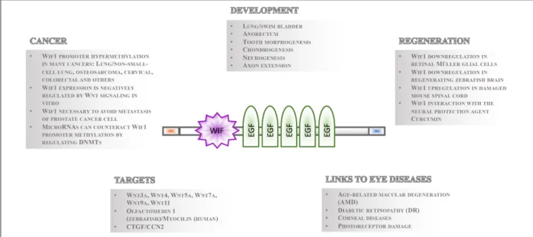
FIGURE 1 | Overview of Wif1 structure and function. The 379 aa Wif1 protein comprises an N-terminal signal sequence (orange box), a 150 aa Wif-domain, five epidermal growth factor (EGF)-like repeats, and a 45 aa long hydrophilic tail (blue box) at the C-terminus (Hsieh et al., 1999). Wif1 interacts with a number of proteins and has been linked to various processes in embryonic development, regeneration, cancer, and eye diseases as indicated.
Wif1 expression in the cornea and lens (
Thisse and Thisse,
2005
), surprisingly no studies on the role of zebrafish Wif1
have been reported. However, the combination of transgenes,
genetic mutants and
in vivo time-lapse imaging to visualize
dynamic processes under physiological conditions would almost
certainly help understanding more about the pathophysiology
of LESCs/Wif1/Wnt related diseases. For instance, inducible
systems to transiently activate Wif1 in injured wild type or
Wif1 mutant embryos, which carry additional transgenes like
anillin and signaling reporter to simultaneously highlight cell
proliferation (
Paolini et al., 2015
;
Cepero Malo et al., 2017
) and
Wnt signaling activity (
Moro et al., 2012
) would be an excellent
tool to study Wif1 dependent regeneration processes
in vivo. The
fact that cornea structure, development and maintenance appear
comparable between zebrafish and mammals (
Pan et al., 2013
;
Takamiya et al., 2015
) should encourage to exploit the zebrafish
as
in vivo model to understand the role of Wif1 in corneal
regeneration and homeostasis.
Wif1 is also expressed in early developing rod photoreceptors,
and in the interphotoreceptor matrix of the mature retina (
Hsieh
et al., 1999
;
Hunter et al., 2004
). Studies from Hsieh et al.
suggest that Wif1 plays a role as opposing binding partner
of Wnt4, providing a fine-tuning system for the regulation of
rod photoreceptor production during development (
Hsieh et al.,
1999
;
Hunter et al., 2004
). Dysregulated Wnt signaling has been
linked to the pathophysiology of neovascular age-related macular
degeneration (AMD) as well as diabetic retinopathy (DR) –
leading causes of blindness in adults (
Chen and Ma, 2017
).
Intriguingly, increased levels of
Wif1 were found in the aqueous
humor of patients with AMD (
Park et al., 2014
) as well as DR
(
Kim et al., 2007
), suggesting that Wif1 provides an attractive
candidate drug target to treat these eye disorders (Figure 1).
Furthermore, increasing levels of
Wif1 expression in the vitreous
humor correlate with degrees of photoreceptor damage (
Park
et al., 2014
). Alongside being a potential therapeutic target,
these studies highlight Wif1 as candidate biomarker of retinal
photoreceptor health and disease states.
Other Wif1 functions in the vertebrate eye involve its
interaction with members of the Olfactomedin protein family.
Within the zebrafish retina, the antagonistic interaction between
Wif1 and Olfactomedin 1 in the extracellular space may fine
tune retinal ganglion cell axon growth through modulation
of Wnt signaling (
Nakaya et al., 2008
). In the human eye,
Wif1 interaction with the Olfactomedin protein family member
myocilin appears critical for the regulation of the intraocular
pressure (IOP). High IOP in turn is a risk factor for glaucoma
and myocilin has been linked to more than 10% of juvenile onset
glaucoma cases, which result in the progressive degeneration
of the optic nerve (
Ortego et al., 1997
;
Kwon et al., 2009
;
Menaa et al., 2011
).
WIF1 REGULATION DURING
REGENERATION
Dynamic
changes
in
Wif1
expression
appear
tightly
associated to regenerative events in the brain and retina
(
Gonzalez-Fernandez et al., 2014
;
Lambert et al., 2016
;
Yao et al., 2016
;
Lim et al., 2017
). The capability to
regenerate lost or damaged neurons in response to injury
is a key feature of the fish central nervous system. In the
retina, this is achieved through activation and transient
asymmetric proliferation of retinal Müller glial cells - the
potential stem cells of the retina (
Nagashima et al., 2013
;
Lust et al., 2016
). Intriguingly, significant transcriptional
down-regulation of
Wif1 and concomitant activation of canonical
Wnt signaling was observed in transiently proliferating Müller
glial cells (
Yao et al., 2016
) and also during early stages of
zebrafish brain regeneration (
Lim et al., 2017
; Figure 1). In
contrast, damaging the mouse spinal cord appears to induce
up-regulation of
Wif1 and other Wnt antagonists (
Gonzalez-Fernandez et al., 2014
). These data suggest that Wnt signaling
after injury might be the key for
de novo neurogenesis, which
is inhibited in the mammalian central nervous system with
its limited regenerative capability (
Gonzalez-Fernandez et al.,
2014
and reviewed in
Lambert et al., 2016
). The different
molecular responses to injury in zebrafish and mammals with
respect to Wif1/Wnt signaling and regeneration open exciting
possibilities for the development of novel therapeutic approaches
to treat central nervous system injuries and possibly also
neurodegenerative disorders. Recently, Wif1 has been implicated
as potential molecular target of curcumin (
Tiwari et al., 2016
).
Curcumin is a natural polyphenol product derived from the
rhizome of the Indian spice turmeric (Curcuma longa) that
appears to provide neuroprotection in cellular and animal models
of neurodegenerative and neurological disorders (
Pluta et al.,
2015
). The capability of curcumin to promote adult neurogenesis,
neurite outgrowth and proliferation appears to occur through the
interaction with Wif1 and concomitant activation of canonical
Wnt signaling (
Tiwari et al., 2015
).
CONCLUDING REMARKS/OUTLOOK
In summary, the structure and mechanistic underlying Wif1
function is rather well described. Epigenetic silencing of the
Wif1 promoter is a common feature, frequently resulting
in cancer progression when uncontrolled. Wif1 appears
to fine-tune cellular processes, perhaps also temporally
controlling them, by dynamically regulating the delicate
balance of Wnt signaling via feedback loop activation.
Indeed, an increasing number of studies link Wif1/Wnt
signaling to different comparably subtle, yet important
processes in development and disease, particularly in the
eye. Furthermore, Wif1 downregulation and concomitant
upregulation of Wnt signaling has been connected to
regeneration processes in the zebrafish CNS, while spinal
cord lesions in mammals with limited regeneration potential
has the opposing effect on
Wif1 expression. Combining
the knowledge of the genetic and epigenetic feedback
regulation of Wif1 with the regeneration capacities of the
zebrafish nervous system has great potential to complement
and
aid
research
progress
on
neural
regeneration
in
mammals.
AUTHOR CONTRIBUTIONS
LP and MC had the original idea and outlined the review. All
authors contributed to writing the article.
ACKNOWLEDGMENTS
We would like to thank L. Guglielmi, A. Bühler and G. Covello
for their work on Wif1. We are grateful to the Centre for
Integrative Biology (CIBIO), University of Trento for financial
support.
REFERENCES
Aguilera, O., Fraga, M. F., Ballestar, E., Paz, M. F., Herranz, M., Espada, J., et al. (2006). Epigenetic inactivation of the Wnt antagonist DICKKOPF-1 (DKK-1) gene in human colorectal cancer.Oncogene 25, 4116–4121. doi: 10.1038/sj.onc. 1209439
Avanesov, A., Honeyager, S. M., Malicki, J., and Blair, S. S. (2012). The role of glypicans in Wnt inhibitory factor-1 activity and the structural basis of Wif1’s
effects on Wnt and hedgehog signaling.PLoS Genet. 8:e1002503. doi: 10.1371/
journal.pgen.1002503
Beretta, C. A., Brinkmann, I., and Carl, M. (2011). All four zebrafish Wnt7 genes are expressed during early brain development.Gene Expr. Patterns 11, 277–284. doi: 10.1016/j.gep.2011.01.004
Boerboom, D., White, L. D., Dalle, S., Courty, J., and Richards, J. S. (2006).
Dominant-stable beta-catenin expression causes cell fate alterations
and Wnt signaling antagonist expression in a murine granulosa cell
tumor model. Cancer Res. 66, 1964–1973. doi: 10.1158/0008-5472.
CAN-05-3493
Bouwmeester, T., Kim, S., Sasai, Y., Lu, B., and De Robertis, E. M. (1996). Cerberus is a head-inducing secreted factor expressed in the anterior
endoderm of Spemann’s organizer. Nature 382, 595–601. doi: 10.1038/
382595a0
Cepero Malo, M., Duchemin, A. L., Guglielmi, L., Patzel, E., Sel, S., Auffarth, G. U., et al. (2017). The zebrafish anillin-eGFP reporter marks late dividing retinal
precursors and stem cells entering neuronal lineages.PLoS One 12:e0170356.
doi: 10.1371/journal.pone.0170356
Chen, Q., and Ma, J. X. (2017). Canonical Wnt signaling in diabetic retinopathy. Vis. Res. 139, 47–58. doi: 10.1016/j.visres.2017.02.007
Cruciat, C. M., and Niehrs, C. (2013). Secreted and transmembrane wnt inhibitors
and activators. Cold Spring Harb. Perspect. Biol. 5:a015081. doi: 10.1101/
cshperspect.a015081
Davis, J., and Piatigorsky, J. (2011). Overexpression of Pax6 in mouse cornea directly alters corneal epithelial cells: changes in immune function, vascularization, and differentiation.Invest. Ophthalmol. Vis. Sci. 52, 4158–4168. doi: 10.1167/iovs.10-6726
Diep, D. B., Hoen, N., Backman, M., Machon, O., and Krauss, S. (2004). Characterisation of the Wnt antagonists and their response to conditionally activated Wnt signalling in the developing mouse forebrain. Brain Res. Dev. Brain Res. 153, 261–270. doi: 10.1016/j.devbrainres. 2004.09.008
Ding, Z., Dong, J., Liu, J., and Deng, S. X. (2008). Preferential gene expression in the limbus of the vervet monkey.Mol. Vis. 14, 2031–2041.
Elston, M. S., Gill, A. J., Conaglen, J. V., Clarkson, A., Shaw, J. M., Law, A. J., et al. (2008). Wnt pathway inhibitors are strongly down-regulated in pituitary
tumors.Endocrinology 149, 1235–1242. doi: 10.1210/en.2007-0542
Fellenberg, J., Sahr, H., Liu, L., Schonsiegel, F., Depeweg, D., Lehner, B., et al. (2013). Rescue of silenced UCHL1 and IGFBP4 expression suppresses clonogenicity of giant cell tumor-derived stromal cells.Cancer Lett. 336, 61–67. doi: 10.1016/j.canlet.2013.04.011
Glinka, A., Wu, W., Delius, H., Monaghan, A. P., Blumenstock, C., and Niehrs, C. (1998). Dickkopf-1 is a member of a new family of secreted proteins and
functions in head induction.Nature 391, 357–362. doi: 10.1038/34848
Glise, B., Miller, C. A., Crozatier, M., Halbisen, M. A., Wise, S., Olson, D. J., et al. (2005). Shifted, the Drosophila ortholog of Wnt inhibitory factor-1, controls the
distribution and movement of Hedgehog.Dev. Cell 8, 255–266. doi: 10.1016/j.
Gonzalez-Fernandez, C., Fernandez-Martos, C. M., Shields, S. D., Arenas, E., and Javier Rodriguez, F. (2014). Wnts are expressed in the spinal cord of adult mice and are differentially induced after injury.J. Neurotrauma 31, 565–581. doi: 10.1089/neu.2013.3067
Gopal, G., Raja, U. M., Shirley, S., Rajalekshmi, K. R., and Rajkumar, T. (2013). SOSTDC1 down-regulation of expression involves CpG methylation and is
a potential prognostic marker in gastric cancer.Cancer Genet. 206, 174–182.
doi: 10.1016/j.cancergen.2013.04.005
Gorfinkiel, N., Sierra, J., Callejo, A., Ibanez, C., and Guerrero, I. (2005). The Drosophila ortholog of the human Wnt inhibitor factor Shifted controls the
diffusion of lipid-modified Hedgehog. Dev. Cell 8, 241–253. doi: 10.1016/j.
devcel.2004.12.018
Guo, H., Zhou, S., Tan, L., Wu, X., Wu, Z., and Ran, R. (2017). Clinicopathological significance of WIF1 hypermethylation in NSCLC, a meta-analysis and literature review.Oncotarget 8, 2550–2557. doi: 10.18632/oncotarget.13707 Herman, J. G., and Baylin, S. B. (2003). Gene silencing in cancer in association
with promoter hypermethylation.N. Engl. J. Med. 349, 2042–2054. doi: 10.1056/ NEJMra023075
Hoang, B., Moos, M. Jr., Vukicevic, S., and Luyten, F. P. (1996). Primary structure and tissue distribution of FRZB, a novel protein related to Drosophila frizzled,
suggest a role in skeletal morphogenesis. J. Biol. Chem. 271, 26131–26137.
doi: 10.1074/jbc.271.42.26131
Hsieh, J.-C., Kodjabachian, L., Rebbert, M. L., Rattner, A., Smallwood, P. M., Harryman Samos, C., et al. (1999). A new secreted protein that binds to Wnt proteins and inhibits their activites.Nature 398, 431–436. doi: 10.1038/18899 Hu, H., Li, B., Zhou, C., Ying, X., Chen, M., Huang, T., et al. (2018). Diagnostic
value of WIF1 methylation for colorectal cancer: a meta-analysis.Oncotarget 9, 5378–5386. doi: 10.18632/oncotarget.23870
Hu, Y. A., Gu, X., Liu, J., Yang, Y., Yan, Y., and Zhao, C. (2008). Expression pattern
of Wnt inhibitor factor 1(Wif1) during the development in mouse CNS.Gene
Expr. Patterns 8, 515–522. doi: 10.1016/j.gep.2008.06.001
Hunter, D. D., Zhang, M., Ferguson, J. W., Koch, M., and Brunken, W. J. (2004). The extracellular matrix component WIF-1 is expressed during, and can modulate, retinal development.Mol. Cell. Neurosci. 27, 477–488. doi: 10.1016/j. mcn.2004.08.003
Hüsken, U., Stickney, H. L., Gestri, G., Bianco, I. H., Faro, A., Young, R. M., et al. (2014). Tcf7l2 is required for left-right asymmetric differentiation of habenular neurons.Curr. Biol. 24, 2217–2227. doi: 10.1016/j.cub.2014.08.006
Itasaki, N., Jones, C. M., Mercurio, S., Rowe, A., Domingos, P. M., Smith, J. C., et al. (2003). Wise, a context-dependent activator and inhibitor of Wnt signalling. Development 130, 4295–4305. doi: 10.1242/dev.00674
Kansara, M., Tsang, M., Kodjabachian, L., Sims, N. A., Trivett, M. K., Ehrich, M., et al. (2009). Wnt inhibitory factor 1 is epigenetically silenced in human osteosarcoma, and targeted disruption accelerates osteosarcomagenesis in mice. J. Clin. Invest. 119, 837–851. doi: 10.1172/JCI37175
Kawano, Y., and Kypta, R. (2003). Secreted antagonists of the Wnt signalling pathway.J. Cell Sci. 116, 2627–2634. doi: 10.1242/jcs.00623
Kim, T., Kim, S. J., Kim, K., Kang, U. B., Lee, C., Park, K. S., et al. (2007). Profiling of vitreous proteomes from proliferative diabetic retinopathy and nondiabetic
patients.Proteomics 7, 4203–4215. doi: 10.1002/pmic.200700745
Kongkham, P. N., Northcott, P. A., Croul, S. E., Smith, C. A., Taylor, M. D., and Rutka, J. T. (2010). The SFRP family of WNT inhibitors function as novel
tumor suppressor genes epigenetically silenced in medulloblastoma.Oncogene
29, 3017–3024. doi: 10.1038/onc.2010.32
Kwon, H. S., Lee, H. S., Ji, Y., Rubin, J. S., and Tomarev, S. I. (2009). Myocilin is a modulator of Wnt signaling.Mol. Cell. Biol. 29, 2139–2154. doi: 10.1128/mcb. 01274-08
Lambert, C., Cisternas, P., and Inestrosa, N. C. (2016). Role of Wnt signaling in
central nervous system injury.Mol. Neurobiol. 53, 2297–2311. doi: 10.1007/
s12035-015-9138-x
Lee, M. J., Kim, E. J., Li, L., and Jung, H. S. (2015). Roles of Wnt inhibitory factor 1 during tooth morphogenesis.Cell Tissue Res. 362, 61–68. doi: 10.1007/s00441-015-2170-3
Li, X., Zhang, Y., Kang, H., Liu, W., Liu, P., Zhang, J., et al. (2005). Sclerostin
binds to LRP5/6 and antagonizes canonical Wnt signaling.J. Biol. Chem. 280,
19883–19887. doi: 10.1074/jbc.M413274200
Lim, F. T., Ogawa, S., Smith, A. I., and Parhar, I. S. (2017). Proteomics identification of potential candidates involved in cell proliferation for early stage of brain
regeneration in the adult zebrafish.Zebrafish 14, 10–22. doi: 10.1089/zeb.2016. 1319
Lush, M. E., and Piotrowski, T. (2014). ErbB expressing Schwann cells control lateral line progenitor cells via non-cell-autonomous regulation of Wnt/beta-catenin.eLife 3:e01832. doi: 10.7554/eLife.01832
Lust, K., Sinn, R., Perez Saturnino, A., Centanin, L., and Wittbrodt, J. (2016). De novo neurogenesis by targeted expression of atoh7 to Muller glia cells. Development 143, 1874–1883. doi: 10.1242/dev.135905
Malinauskas, T., Aricescu, A. R., Lu, W., Siebold, C., and Jones, E. Y. (2011). Modular mechanism of Wnt signaling inhibition by Wnt inhibitory factor 1. Nat. Struct. Mol. Biol. 18, 886–893. doi: 10.1038/nsmb.2081
Malinauskas, T., and Jones, E. Y. (2014). Extracellular modulators of Wnt signalling.Curr. Opin. Struct. Biol. 29, 77–84. doi: 10.1016/j.sbi.2014.10.003 Mattes, B., Weber, S., Peres, J., Chen, Q., Davidson, G., Houart, C., et al. (2012).
Wnt3 and Wnt3a are required for induction of the mid-diencephalic organizer in the caudal forebrain.Neural Dev. 7:12. doi: 10.1186/1749-8104-7-12 Mazieres, J., He, B., You, L., Xu, Z., Lee, A. Y., Mikami, I., et al. (2004). Wnt
inhibitory factor-1 is silenced by promoter hypermethylation in human lung
cancer.Cancer Res. 64, 4717–4720. doi: 10.1158/0008-5472.can-04-1389
Menaa, F., Braghini, C. A., Vasconcellos, J. P., Menaa, B., Costa, V. P., Figueiredo, E. S., et al. (2011). Keeping an eye on myocilin: a complex molecule associated
with primary open-angle glaucoma susceptibility.Molecules 16, 5402–5421.
doi: 10.3390/molecules16075402
Moro, E., Ozhan-Kizil, G., Mongera, A., Beis, D., Wierzbicki, C., Young, R. M., et al. (2012). In vivo Wnt signaling tracing through a transgenic biosensor fish reveals novel activity domains.Dev. Biol. 366, 327–340. doi: 10.1016/j.ydbio. 2012.03.023
Nagashima, M., Barthel, L. K., and Raymond, P. A. (2013). A self-renewing division of zebrafish muller glial cells generates neuronal progenitors that require N-cadherin to regenerate retinal neurons.Development 140, 4510–4521. doi: 10.1242/dev.090738
Nakatsu, M. N., Ding, Z., Ng, M. Y., Truong, T. T., Yu, F., and Deng, S. X. (2011). Wnt/beta-catenin signaling regulates proliferation of human cornea epithelial stem/progenitor cells.Invest. Ophthalmol. Vis. Sci. 52, 4734–4741. doi: 10.1167/iovs.10-6486
Nakaya, N., Lee, H. S., Takada, Y., Tzchori, I., and Tomarev, S. I. (2008). Zebrafish
olfactomedin 1 regulates retinal axon elongationin vivo and is a modulator
of Wnt signaling pathway.J. Neurosci. 28, 7900–7910. doi: 10.1523/jneurosci. 0617-08.2008
Ng, R. C., Matsumaru, D., Ho, A. S., Garcia-Barcelo, M. M., Yuan, Z. W., Smith, D., et al. (2014). Dysregulation of Wnt inhibitory factor 1 (Wif1) expression resulted in aberrant Wnt-beta-catenin signaling and cell death of the cloaca
endoderm, and anorectal malformations.Cell Death Differ. 21, 978–989. doi:
10.1038/cdd.2014.20
Niehrs, C. (2006). Function and biological roles of the Dickkopf family of Wnt
modulators.Oncogene 25, 7469–7481. doi: 10.1038/sj.onc.1210054
Nusse, R., and Clevers, H. (2017). Wnt/beta-catenin signaling,
disease, and emerging therapeutic modalities. Cell 169, 985–999.
doi: 10.1016/j.cell.2017.05.016
Ortego, J., Escribano, J., and Coca-Prados, M. (1997). Cloning and characterization of subtracted cDNAs from a human ciliary body library encoding TIGR, a protein involved in juvenile open angle glaucoma with homology to myosin and
olfactomedin.FEBS Lett. 413, 349–353. doi: 10.1016/S0014-5793(97)00934-4
Pan, Y. A., Freundlich, T., Weissman, T. A., Schoppik, D., Wang, X. C., Zimmerman, S., et al. (2013). Zebrabow: multispectral cell labeling for cell
tracing and lineage analysis in zebrafish. Development 140, 2835–2846.
doi: 10.1242/dev.094631
Panakova, D., Sprong, H., Marois, E., Thiele, C., and Eaton, S. (2005). Lipoprotein particles are required for Hedgehog and Wingless signalling.Nature 435, 58–65. doi: 10.1038/nature03504
Paolini, A., Duchemin, A. L., Albadri, S., Patzel, E., Bornhorst, D., Gonzalez Avalos, P., et al. (2015). Asymmetric inheritance of the apical domain and self-renewal of retinal ganglion cell progenitors depend on Anillin function. Development 142, 832–839. doi: 10.1242/dev.118612
Park, K. H., Choi, A. J., Yoon, J., Lim, D., Woo, S. J., Park, S. J., et al. (2014). Wnt modulators in the aqueous humor are associated with outer retinal damage severity in patients with neovascular age-related macular degeneration.Invest. Ophthalmol. Vis. Sci. 55, 5522–5530. doi: 10.1167/iovs.14-14566
Pluta, R., Bogucka-Kocka, A., Ulamek-Koziol, M., Furmaga-Jablonska, W., Januszewski, S., Brzozowska, J., et al. (2015). Neurogenesis and neuroprotection in postischemic brain neurodegeneration with Alzheimer phenotype: is there a role for curcumin?Folia Neuropathol. 53, 89–99.
Ramachandran, I., Thavathiru, E., Ramalingam, S., Natarajan, G., Mills, W. K., Benbrook, D. M., et al. (2012). Wnt inhibitory factor 1 induces apoptosis and inhibits cervical cancer growth, invasion and angiogenesisin vivo. Oncogene 31, 2725–2737. doi: 10.1038/onc.2011.455
Reguart, N., He, B., Xu, Z., You, L., Lee, A. Y., Mazieres, J., et al. (2004). Cloning and
characterization of the promoter of human Wnt inhibitory factor-1.Biochem.
Biophys. Res. Commun. 323, 229–234. doi: 10.1016/j.bbrc.2004.08.075 Roman-Gomez, J., Jimenez-Velasco, A., Barrios, M., Prosper, F., Heiniger, A.,
Torres, A., et al. (2007). Poor prognosis in acute lymphoblastic leukemia may
relate to promoter hypermethylation of cancer-related genes.Leuk. Lymphoma
48, 1269–1282. doi: 10.1080/10428190701344899
Sato, H., Suzuki, H., Toyota, M., Nojima, M., Maruyama, R., Sasaki, S., et al. (2007). Frequent epigenetic inactivation of DICKKOPF family genes in human
gastrointestinal tumors. Carcinogenesis 28, 2459–2466. doi: 10.1093/carcin/
bgm178
Semenov, M., Tamai, K., and He, X. (2005). SOST is a ligand for LRP5/LRP6 and a Wnt signaling inhibitor.J. Biol. Chem. 280, 26770–26775. doi: 10.1074/jbc. M504308200
Steinhart, Z., and Angers, S. (2018). Wnt signaling in development and tissue
homeostasis.Development 145:dev146589. doi: 10.1242/dev.146589
Surmann-Schmitt, C., Sasaki, T., Hattori, T., Eitzinger, N., Schett, G., von der Mark, K., et al. (2012). The Wnt antagonist Wif-1 interacts with CTGF and inhibits CTGF activity.J. Cell. Physiol. 227, 2207–2216. doi: 10.1002/jcp.22957 Surmann-Schmitt, C., Widmann, N., Dietz, U., Saeger, B., Eitzinger, N.,
Nakamura, Y., et al. (2009). Wif-1 is expressed at cartilage-mesenchyme
interfaces and impedes Wnt3a-mediated inhibition of chondrogenesis.J. Cell
Sci. 122(Pt 20), 3627–3637. doi: 10.1242/jcs.048926
Tai, D., Wells, K., Arcaroli, J., Vanderbilt, C., Aisner, D. L., Messersmith, W. A., et al. (2015). Targeting the WNT signaling pathway in cancer therapeutics. Oncologist 20, 1189–1198. doi: 10.1634/theoncologist.2015-0057
Takamiya, M., Weger, B. D., Schindler, S., Beil, T., Yang, L., Armant, O., et al. (2015). Molecular description of eye defects in the zebrafish Pax6b mutant, sunrise, reveals a Pax6b-dependent genetic network in the developing anterior chamber. PLoS One 10:e0117645. doi: 10.1371/journal.pone.0117645
Tan, M., Wu, J., and Cai, Y. (2013). Suppression of Wnt signaling by the miR-29 family is mediated by demethylation of WIF-1 in non-small-cell lung cancer. Biochem. Biophys. Res. Commun. 438, 673–679. doi: 10.1016/j.bbrc.2013.07.123
Thisse, C., and Thisse, B. (2005). High Throughput Expression Analysis of
ZF-Models Consortium Clones. ZFIN Direct Data Submission. Available at: http://zfin.org
Tiwari, S. K., Agarwal, S., Seth, B., Yadav, A., Ray, R. S., Mishra, V. N., et al. (2015). Inhibitory effects of Bisphenol-A on neural stem cells proliferation and differentiation in the rat brain are dependent on Wnt/beta-Catenin pathway. Mol. Neurobiol. 52, 1735–1757. doi: 10.1007/s12035-014-8940-1
Tiwari, S. K., Agarwal, S., Tripathi, A., and Chaturvedi, R. K. (2016). Bisphenol-A mediated inhibition of hippocampal neurogenesis attenuated by curcumin via
canonical wnt pathway.Mol. Neurobiol. 53, 3010–3029. doi:
10.1007/s12035-015-9197-z
Vaes, B. L., Dechering, K. J., van Someren, E. P., Hendriks, J. M., van de Ven, C. J., Feijen, A., et al. (2005). Microarray analysis reveals expression regulation of Wnt antagonists in differentiating osteoblasts.Bone 36, 803–811. doi: 10.1016/ j.bone.2005.02.001
Willert, K., Brown, J. D., Danenberg, E., Duncan, A. W., Weissman, I. L., Reya, T., et al. (2003). Wnt proteins are lipid-modified and can act as stem cell growth factors.Nature 423, 448–452. doi: 10.1038/nature01611
Wissmann, C., Wild, P. J., Kaiser, S., Roepcke, S., Stoehr, R., Woenckhaus, M., et al. (2003). WIF1, a component of the Wnt pathway, is down-regulated in prostate, breast, lung, and bladder cancer.J. Pathol. 201, 204–212. doi: 10.1002/path. 1449
Xu, B., Chen, C., Chen, H., Zheng, S. G., Bringas, P. J., Xu, M., et al. (2011). Smad1 and its target gene Wif1 coordinate BMP and Wnt signaling activities to regulate
fetal lung development.Development 138, 925–935. doi: 10.1242/dev.062687
Yan, D., and Lin, X. (2009). Shaping morphogen gradients by proteoglycans.Cold
Spring Harb. Perspect. Biol. 1:a002493. doi: 10.1101/cshperspect.a002493 Yao, K., Qiu, S., Tian, L., Snider, W. D., Flannery, J. G., Schaffer, D. V., et al. (2016).
Wnt regulates proliferation and neurogenic potential of muller glial cells via a
Lin28/let-7 miRNA-dependent pathway in adult mammalian retinas.Cell Rep.
17, 165–178. doi: 10.1016/j.celrep.2016.08.078
Yazdanpanah, G., Jabbehdari, S., and Djalilian, A. R. (2017). Limbal and corneal epithelial homeostasis.Curr. Opin. Ophthalmol. 28, 348–354. doi: 10.1097/icu. 0000000000000378
Yee, D. S., Tang, Y., Li, X., Liu, Z., Guo, Y., Ghaffar, S., et al. (2010). The Wnt inhibitory factor 1 restoration in prostate cancer cells was associated with reduced tumor growth, decreased capacity of cell migration and invasion
and a reversal of epithelial to mesenchymal transition.Mol. Cancer 9:162.
doi: 10.1186/1476-4598-9-162
Yin, A., Korzh, V., and Gong, Z. (2012). Perturbation of zebrafish swimbladder
development by enhancing Wnt signaling in Wif1 morphants.BBA Mol. Cell
Res. 1832, 236–244. doi: 10.1016/j.bbamcr.2011.09.018
Zheng, Y., Li, X., Jiang, Y., Xu, Y., Song, B., Zhou, Q., et al. (2016). Promoter hypermethylation of Wnt inhibitory factor-1 in patients with
lung cancer: a systematic meta-analysis.Medicine 95:e5433. doi: 10.1097/md.
0000000000005433
Zhu, W., Shiojima, I., Ito, Y., Li, Z., Ikeda, H., Yoshida, M., et al. (2008). IGFBP-4 is an inhibitor of canonical Wnt signalling required for cardiogenesis.Nature 454, 345–349. doi: 10.1038/nature07027
Zirn, B., Samans, B., Wittmann, S., Pietsch, T., Leuschner, I., Graf, N., et al.
(2006). Target genes of the WNT/beta-catenin pathway in Wilms tumors.Genes
Chromosomes Cancer 45, 565–574. doi: 10.1002/gcc.20319
Conflict of Interest Statement: The authors declare that the research was conducted in the absence of any commercial or financial relationships that could be construed as a potential conflict of interest.
Copyright © 2018 Poggi, Casarosa and Carl. This is an open-access article distributed under the terms of the Creative Commons Attribution License (CC BY). The use, distribution or reproduction in other forums is permitted, provided the original author(s) and the copyright owner(s) are credited and that the original publication in this journal is cited, in accordance with accepted academic practice. No use, distribution or reproduction is permitted which does not comply with these terms.