This document is the accepted version of a published work that appeared in final
form in Pept Sci, after technical editing by the publisher. To access the final edited
and published work, see https://doi.org/10.1002/pep2.24041
From liposomes to cells: filling the gap between physicochemical and microbiological studies of the activity and selectivity of host‐defense peptides
Filippo Savini,a# Sara Bobone,a# Daniela Roversi,a© Maria Luisa Mangonib and Lorenzo Stellaa*
a) Department of Chemical Science and Technologies, University of Rome Tor Vergata, 00133, Rome, Italy. b) Laboratory affiliated to Pasteur Italia-Fondazione Cenci Bolognetti, Department of Biochemical Sciences,
Sapienza University of Rome, Rome, via degli Apuli, 9-00185, Italy; #These authors contributed equally to the work
©Current address: IRBM Science Park, 00040 Pomezia (RM), Italy.
* To whom correspondence should be addressed Prof. Lorenzo Stella
Department of Chemical Science and Technologies, University of Rome Tor Vergata,
00133, Rome, Italy. Tel. +39-0672594463
E-mail: [email protected]
ABSTRACT
Host-defense peptides (HPDs) are bactericidal and immunomodulatory molecules, part of the innate immune system of many organisms, including man. They kill bacteria mostly by perturbing their membranes, and for this reason they are a promising class of molecules to fight drug-resistant microbes. However, their success towards clinical application is still limited, partly due to many unanswered questions on their activity and function. Our current understanding of HDPs has been reached by two parallel, but largely independent, approaches: microbiological studies on HDP effects on cells, and physicochemical investigations on model membranes. All current models for the mechanisms of HDP membrane perturbation and cell selectivity were derived from the latter kind of studies, but their relevance for real cells still had to be demonstrated. In the last few years, several studies led to quantitative insights into HDP behavior directly in cells: membrane-binding and peptide-induced pores in bacteria and liposomes were compared; the number of cell-bound peptide molecules needed to kill a bacterium was determined; the variation of peptide activity and toxicity with the density of cells was characterized; selectivity was examined in a mixture of target and host cells; the sequence of events leading to bacterial death was observed in real time by microscopy on single cells. Overall, these approaches led to a new understanding of HDPs that will be helpful for their development into effective antibiotic drugs.
Keywords:
Antimicrobial peptides, fluorescence spectroscopy, microscopy, drug-resistant bacteria, model membranes.
Graphical abstract
1) Introduction
The emergence and diffusion of pathogenic bacteria endowed with multidrug resistance is a dramatic threat to global health and safety.1 Several infectious diseases, now easily treatable, could turn deadly again, and milestones in the development of the medical science, such as surgery, transplants, anticancer chemotherapy, could become unusable, as they rely on the effectiveness of antibiotics. This situation is worsened by the reduced interest of pharmaceutical companies in the development of new antibiotics, due to their low profitability.2-3 For these reasons, new drugs against resistant bacteria are severely needed.
Host-defense peptides (HDPs) are considered promising lead compounds for the development of a new class of antibiotic molecules.4 They constitute a fundamental component of the innate immune defense of many organisms, including humans, and have a wide spectrum antibacterial activity. HDPs are highly heterogeneous in length, amino acid composition and secondary structure, but most of them are characterized by a short sequence (10 to 50 amino acids), with a high content of both hydrophobic and basic residues and a net positive charge, generally ranging from +2 to +9.5 HDPs have many different secondary structures, but the best characterized class comprises cationic peptides that attain a helical conformation after membrane binding, with an amphipathic spatial arrangement of the side chains.6 Thanks to these properties, they are able to interact with the negatively charged bacterial membranes, leading to the perturbation of their permeability, through the formation of pores or bilayer defects.7 Since no association to specific receptors or proteins is involved in this process, the development of resistance is particularly unlikely.8-9 Furthermore, HDPs are able to discriminate between host and target cells, thus exhibiting limited toxicity against eukaryotic cells. Selectivity is presumably determined by the difference in lipid composition of the membranes of the two cell types, as studies on liposomes show a higher affinity of HDPs for bilayers mimicking bacterial membranes.6
Although different models have been proposed to describe the process of membrane perturbation by HDPs, most of them involve peptide binding to the bilayer surface and the formation of channels, pores or bilayer defects when a threshold of membrane-bound peptide is reached (see Section 3).7
HDPs were originally discovered for their direct killing activity in vitro, and are currently isolated and screened based on this property. For these reasons, they are commonly called also antimicrobial peptides. However, more recently they have been demonstrated to modulate the immune response in vivo, e.g. by enhancing the recruitment of immune cells to infection sites,10 and therefore they have been renamed HDPs. Which of these functions is more important in vivo is currently debated.11
Research on HDPs is now decades long, and thousands of different sequences have been identified.5 A host of physicochemical techniques and membrane models has been applied to their characterization.12 However, notwithstanding the very promising properties of HDPs, there has been no real breakthrough in their clinical application, although several peptides are now in clinical trials. 13 In our opinion, one of the reasons for this lack of success is that several very basic aspects of their function are still poorly understood.
Biophysical techniques can provide very detailed information on the interaction of HDPs with membranes, and most of our knowledge of the molecular details of the pore formation process comes from this kind of studies. However, these approaches are typically applied to model systems, such as planar bilayers, liposomes or micelles. Until recently, it was unknown if models such as the “carpet” mechanism were relevant also for the activity of HDPs in real bacteria. This lack of information left also several other important questions unanswered: is the high membrane coverage required to create pores in liposomes needed also to perturb bacterial membranes? Can such high membrane-bound concentrations be achieved under realistic, in vivo conditions? If not, is it possible that other activities, rather than direct bacterial killing, prevail in the organism? Another problem is related to selectivity: liposome studies usually indicate that HDPs have different affinities for bilayers mimicking the membranes of bacteria or eukaryotes, but peptide affinity towards the two types of cells was essentially unexplored. Furthermore, until recently it was totally unknown how the activity and the toxicity of HDPs depends on the density of target and host cells, as antimicrobial activity and toxicity assays are usually carried out independently, using standardized, fixed cell densities, which are not necessarily representative of the cell concentrations present at a typical infection site. It has even been proposed that the peptide selectivity observed in vitro is just a result of the conditions used in these assays.14 Retention of activity in the body and lack of selectivity are two essential requirements for the clinical application of HDPs.
In the last few years, some groups,15-19 including ours,20-21 have started trying to extend the powerful physicochemical approaches, normally applied to model membranes, to quantitative experiments on real cells, in order to tackle the questions discussed above. This review is devoted to the recent studies striving to fill the gap between microbiological and biophysical studies of HDPs. Section 2 and 3 provide brief overviews of the current microbiological and physicochemical approaches used to study HDPs and of their respective limitations. Section 4 focuses on the recent studies trying to bridge the realms of model systems and real cells. Finally, Section 5 describes recent quantitative single cells microscopic studies. A review on a similar topic was published at the beginning of 2015,16 but the scope was different from that of the present article, and this field is progressing so rapidly that an update is warranted. Due to space limitations, we will not discuss the recent developments in the application of circular dichroism and NMR to studies of peptide/cell interaction, as these methods have been reviewed elsewhere.22-23
Overall, the studies considered in this review clarified several key points of the mechanism of bacterial killing by HDPs, and provided novel insights to understand their function in vivo and to screen and design new molecules for therapeutic applications. However, this new approach is just at the beginning and the results collected so far provoked several new questions, opening a new window on the fascinating world of HDPs.
2. The microbiological perspective
Different in vitro microbiological assays are commonly used to measure the antibacterial activity of antibiotic drugs,24 including HDPs.25 Those that are currently more used are determinations of the minimum inhibitory concentration (MIC) or of the minimum bactericidal concentration (MBC) by the broth dilution method.
MIC is defined as the lowest concentration of antimicrobial agent that inhibits the visible growth of a microorganism, in a liquid growth medium containing serial two-fold dilution of the antimicrobial agent.26 The main drawback of this approach is that it does not allow a discrimination between bactericidal and bacteriostatic effects, i.e. microbial killing or a mere impediment of bacterial growth. By contrast, MBC is defined as the minimal drug dosage killing at least 99.9% of the original bacterial cells.27 After incubation in the broth solutions containing the antibiotic compounds, samples can be sub-cultured on agar plates to count the number of surviving cells, i.e. those able to form a new, growing bacterial colony (colony forming units, or CFUs).24
A shortcoming of these microbiological assays is that they refer to the total concentration of the active molecule in the sample, rather than to the fraction of the antimicrobial that is actually interacting with cells. This limitation is particularly relevant for HDPs, since they must associate to the bacterial membranes to exert their action. The fraction of membrane-bound peptide obviously depends on the concentration of bacterial cells in the sample. However, cell density varies during the MIC assays, which require bacterial growth. In addition, the recommended value for the initial bacterial cell density (inoculum) is 5 x10⁵ CFU/mL for broth dilution assays.28 This choice was dictated by purely practical criteria related to the clinical practice: the inoculum is high enough to avoid false susceptibility results and to provide statistically satisfactory data for determining the MBC, and low enough to prevent false resistance findings.26 However, the particular inoculum used in the assays has no special significance with respect to the bacterial cell-densities that can be found in clinically relevant infections in vivo, which range from 1 CFU/mL (in blood stream infections) to 109 CFU/mL (in soft tissue or peritoneal infections).29-30 Finally, it should be considered that under commonly used assay conditions the available peptide concentration is reduced by association to components of the growth medium.31 This effect can reduce peptide activity by more than one order of magnitude. An additional reduction in the available peptide concentration can be caused by the adsorption of the amphipathic HDPs to the surface of the container where the assay is performed.25-26 For all these reasons, the commonly used microbiological assays for HDP activity do not provide indications on the quantity of peptide associated to the bacterial cells when killing occurs, as significant amounts of peptide can remain in solution or can be sequestered by medium components or be adsorbed to the container walls. Furthermore, the number of bacterial cells in the sample is ill-defined, as it varies during the assay. Therefore, MIC and MBC values depend significantly on the conditions used in the assay, which, however standardized, might be very different from what is encountered in vivo.
In addition to determining the active concentrations of HDPs, microbiological studies have been devoted to understand their mechanism of bacterial killing. These investigations clearly indicated that the bacterial membranes are the main target of HDPs, and that association with specific proteins is not involved in peptide interaction with microbial cells. In Gram-negative bacteria the cell envelope consists of two membranes: the inner, cytoplasmic membrane (CM) is composed of a phospholipid bilayer, whereas the outer membrane (OM) is asymmetric, with the outer leaflet mainly composed of Lipopolysaccharides(LPS), and the inner one comprising phospholipids such as phosphatidylethanolamine, phosphatidylglycerol and cardiolipin. Between the two membranes is the periplasmic space, which contains a peptidoglycan layer.32 Gram-positive bacteria lack an OM but are surrounded by layers of peptidoglycan many times thicker than is found in the Gram-negatives. Threading through these layers of peptidoglycan are long anionic polymers, called teichoic acids. In addition, all cell membranes contain a high proportion of proteins. Several assays are available to study peptide effects on the cell membranes. 1-N-phenylnaphthylamine (NPN, MW of ~ 200 Da) is a fluorescent dye commonly used to assay perturbation of the OM of Gram-negative bacteria, since its fluorescence increases significantly after damage to the OM enhances its membrane uptake.33 CM perturbation can be determined by assays employing Sytox Green (SG) or propidium iodide (PI), membrane-impermeant nucleic acid stains (MW ~ 600 Da) whose fluorescence intensity rapidly increases upon binding to nucleic acids of cells whose cytoplasmic (inner) membrane has been injured.34 However, to detect larger disruptions of the CM, the leakage of bulky cytosolic components such as the enzyme beta-galactosidase, can be monitored photometrically in the bacterial culture supernatant, after addition of the specific chromogenic substrate 2-nitrophenyl β-D-galactoside (ONPG).34-35 These assays demonstrate that the bacterial membranes are damaged by HDPs, and that, as a consequence, the transmembrane gradients are dissipated.36-37 Usually, correlation between membrane perturbation and killing is observed, indicating that disruption of membrane integrity is the main bactericidal mechanism. However, for some peptides it has been suggested that cytoplasmic-membrane permeabilization in itself is not the killing mechanism, but it may be necessary for reaching an intracellular target.38
Interestingly, in most cases, the bactericidal activity is the same for natural HDPs and for their all-D enantiomers, ruling out chiral interactions with proteins. Although this finding is often mentioned, a comprehensive review of data on enantiomeric peptides has not been reported in the literature. In Figure 1 we collected data from multiple studies.39-46 Overall, these results strongly support the conclusion that chirality does not affect activity. Also in this case, exceptions do exist, but they are considered an indication that the mechanism of action of some specific HDPs is not based on membrane perturbation.47, 48
Perturbation of bacterial membranes as the main mechanism of bacterial killing by HDPs is demonstrated also by microscopic studies.49 Even if real time imaging is possible (see Section 8), currently most studies observe cells at a specific time point after treatment with HDPs.50 The use of specific fluorophores allows the assessment of membrane integrity and of cell viability within individual cells.51 Confocal microscopy with
fluorescently-labeled HDPs has been employed to investigate their location in bacteria, showing for instance that magainin concentrates on the cell envelope, while some peptides, such as buforin, act on intracellular targets.52 The higher resolution of electron and scanning probe microscopies allows direct visualization of the effects caused by HDPs on the surface of bacterial cells. In most cases, these studies confirmed the bacterial envelope as the main target, showing roughening of the microbial surface, formation of blebs and local disruptions, and cell disintegration at high peptide concentrations. 34, 37-38, 53-64 An interesting observation is the preferential disruption of bacterial apical ends.61 This finding suggests that these regions, enriched in the negatively charged lipid cardiolipin, are specific targets for HDPs.61, 65
3. The physicochemical approach: vesicle leakage and water‐membrane partition studies.
In comparison to microbiological assays, biophysical studies on the activity of HDPs strive for a better control of the experimental system at the considerable cost of oversimplifications. Similarly to a good old joke on physicists,66 bacteria are approximated as spheres (a vesicle formed by a simple lipid bilayer), and activity is determined by the peptide ability to cause release of liposome contents.
Studies on liposomes imply that the bacterial membrane is the sole target of HDPs and that membrane proteins and other non-lipidic components do not play a significant role in the pore-formation process. These hypotheses are based on and supported by the evidences discussed above. However, systematic, quantitative studies of correlation between activity on model membranes and against bacteria are sparse, and their results contradictory.67-69 In addition, the lipid composition of the vesicles used in such experiments (typically a mixture of two phospholipids recapitulating the fluidity and electrostatic charge of bacterial membranes) is usually strongly simplified with respect to the real biological structures. By contrast, the bacterial cell envelope is actually a complex multilayered structure.70
The severe simplifications of the experimental system implied in liposome studies are balanced by the amount of information that can be gathered. The most common experimental system is represented by bulk experiments on suspensions of so-called large unilamellar vesicles (LUVs), with diameters of the order of 100 nm. By entrapping fluorescent dyes inside the liposomes, it is possible to follow peptide-induced pore formation by measuring their release,71 and by using molecules of different sizes the pore dimensions can be determined.72 In addition, it is possible to study peptide-induced changes in membrane order and fluidity, lipid domains, lipid translocation across the two leaflets (flip-flop), vesicle aggregation and fusion, etc. 72-75
Peptide association to the membranes can be easily followed quantitatively by spectroscopic (fluorescence, CD) or calorimetric methods.72, 76In a typical experiment, the peptide is titrated with increasing concentrations of vesicles, and the fraction of bilayer-associated peptide fM is determined for each
experimental point from the spectroscopic signal S through the following equation:72, 77-79
fM=(S-SW)/(SM-SW) (1
Here Si is the signal measured (under the same experimental conditions) with all molecules in state i, i.e. free
in solution (W) or membrane bound (M).
The simplest model describing how fM depends on the lipid concentration is as a partition between two
immiscible phases (water and membrane).77 In this case, it can be shown that the following hyperbolic equation holds:78
𝑓𝑓𝑀𝑀=𝐾𝐾+[𝐿𝐿][𝐿𝐿] (2
However, this model is based on the hypothesis of an ideal behavior. Significant deviations are expected as soon as the peptide concentration in the membrane increases, or in cases in which the two-states hypothesis breaks down, e.g. when peptides aggregate. The equation reported above has a striking property that can be exploited to test its adequateness in the case under study: it does not depend on peptide concentration. Therefore, two curves measured at different peptide concentrations should overlap. Otherwise, more complex models should be employed.78
Using these approaches, it has been possible to determine the fraction of the total peptide concentration that is actually bound to the lipid bilayers under the conditions used in the leakage assays. Coupling these data to the peptide-induced leakage experiments, it is easy to obtain the threshold concentration of peptide (per lipid or per vesicle) that is needed to cause membrane perturbation. Such studies generally showed that membrane-bound peptide concentrations corresponding to an almost complete degree of coverage of the artificial membrane (lipid to membrane-bound peptide ratios in the order of 10:1) are necessary to permeabilize the bilayer.80 Furthermore, studies of peptide-membrane association have provided a possible explanation for the selectivity of HDPs: cationic peptides usually have a higher affinity for the negatively charged lipids present in the membranes of bacteria, than for the globally neutral character of the outer leaflet of eukaryotic membranes.6, 81-83
In addition to bulk spectroscopic studies on LUVs, several mechanistic details can be derived from microscopy experiments on single giant unilamellar vesicles (GUVs), with a diameter in the 10 µm range. GUVs can be analyzed one at a time, thus evidencing possible heterogeneities in the system (e.g. peptide distribution, peptide effects on the membrane) by avoiding averaging on a sample with a large number of vesicles. For instance, we observed that peptide distribution among the vesicles in the sample can be strongly inhomogeneous.84 Leakage studies performed by following dye entry or exit from the GUVs showed that usually peptides (e.g. maculatin, magainin, melittin, PMAP-23, BAX-α5,)85 form pores without a total disruption of the vesicle membrane. Dye leakage starts only after an initial lag phase, which is different for each GUV, showing that pore formation is a stochastic event, favored by peptide binding but triggered by thermal fluctuations.86-89
Several other aspects of peptide/membrane system can be elucidated by exploiting multiple spectroscopic techniques: IR absorption,73 fluorescence,72 circular dichroism,90-91 EPR,74 NMR.75, 92-93 For instance, it is possible to determine peptide conformation, aggregation state, depth of insertion, orientation
in the membrane and distribution between the two leaflets of the bilayer. Studies from our group have demonstrated that many of these phenomena modulate the final peptide membrane-perturbing activity: peptide aggregation in water can compete with membrane binding and modulate peptide activity and selectivity; conformational equilibria can modulate the effective peptide hydrophobicity and therefore its tendency to associate to different membranes; peptide orientation and aggregation in the bilayer determine the structure of the pores.6, 71, 78, 84, 94-101
Overall, the great amount of data collected by biophysical studies of HDPs has led, among other things, to a better understanding of the mechanism of pore formation. For instance, many cationic peptides have been demonstrated to perturb model membranes according to the Shai, Matsuzaki and Huang model.7, 98 This mechanism has also been called the “carpet” model, because it is based on high peptide accumulation on the membrane surface, causing a disruption of lipid packing: when the peptide reaches the membrane it inserts in the head-group region of the outer leaflet, causing a disruption in lipid packing and a stress in the membrane due to the tension asymmetry between the two layers. When a threshold peptide/lipid ratio is reached (see above), the stress is released by the formation of pores or defects, which cause leakage.
4. Connecting the two worlds
Due to the lack of information on peptide binding to real cells, until a few years ago it was still unknown whether bacterial killing requires the same degree of membrane coverage observed for liposome permeabilization, and whether the mechanistic models of pore formation developed from biophysical studies on liposomes are relevant for the perturbation of bacterial membranes. The high membrane coverage required to display the pore-forming activity of HDPs in model membranes might be difficult to achieve in living systems, and other functions, such as immunomodulation, might prevail.
In 2009, a seminal work by Miguel Castanho and coworkers80 provided a new impulse to the field, by suggesting a link between the biophysical water-membrane partition experiments performed on artificial vesicles and the microbiological activity investigations. This study, and others,68, 102 have reported a good correlation between in vitro bactericidal activities and vesicle leakage studies, even though, as is often the case for HDPs, this finding is not general.67 Based on these observations, Melo and Castanho assumed that the partition constants determined in peptide-liposome association studies were valid also for the interaction of HDPs with bacterial cell membranes. Then, by using the density of cells used in the microbiological studies and by estimating the number of lipids per bacterial cell, the authors evaluated the lipid concentration and thus the fraction of cell-bound peptides in the MIC experiments. Finally, from the total peptide concentration, they assessed the amount of peptide bound per cell-membrane lipid needed to cause killing. In this way, they obtained threshold values of 3-30 lipids per bound peptide. These numbers are comparable to those observed in experiments with liposomes, and correspond to an extremely high coverage of the bacterial membrane.
This work, although very insightful, was based on strong assumptions, whose correctness was far from granted a priori: i) that peptides associate to bacterial membranes only, and not to other components; ii) that peptide association to bacterial cells is an ideal water-membrane partition equilibrium, and thus, among other things, it does not depend on peptide concentration.
In order to test these hypotheses, and based on our previous experience on studies of water/membrane partition and of other equilibria affecting peptide activity, we decided to measure peptide association to real, live bacteria, and to determine bacterial killing under exactly the same, well controlled conditions.
4.1. Peptide binding to bacterial cells
By searching the literature, we realized that a few studies on peptide association to cells had actually been performed before the articles by Castanho’s group, even if their consequences had probably not been fully appreciated. These investigations were based on incubation of the peptides with cells, separation by centrifugation and determination of the free (or bound) concentration (by using radioactively labeled analogues or by HPLC). As soon as 1988, Bruce Merrifield and his group103 measured binding of cecropin A to Escherichia coli, Bacillus megaterium and Bacillus thuringiensis cells and to erythrocytes. In the words of the authors, the separation method outlined above suffered from peptide-induced disruption of the cells, causing the release of peptide-containing debris that could not be efficiently pelleted, leading to an underestimation of the cell-bound peptide fraction. In any case, the authors estimated that 5 × 10⁵ to 2 × 10⁷ peptides per cell were needed to cause the death of 50% of the bacteria. In 2002, Selsted and coworkers104 measured binding of Rhesus theta defensin to E. coli, by incubation and separation by centrifugation (but quantifying the free peptide by HPLC). Their data indicate that, at peptide concentrations slightly above the MIC, approximately 3 × 10⁸ peptides are bound per bacterial cell.17 Another study by Albrecht et al.,105 in the same year suggested that several millions of Protegrin-1 molecules are bound to a bacterial cell (Pseudomonas aeruginosa and Burkholderia cepacia) at concentrations around the MIC. However, in that investigation the conditions for the bacterial killing assays (radial diffusion) were distinctly different from those used in the binding experiments, and therefore a comparison between the two datasets is questionable. To the best of our knowledge, these three studies were the only ones in which a quantitative determination of the cell-bound peptide concentration needed to cause killing was attempted. In addition to the limitations listed above, all these investigations studied a single cell density, varying peptide concentration.
In our case, we selected fluorescence spectroscopy to assess peptide binding to cells, building on our expertise on this technique. As indicated in section 3, the high sensitivity of this approach allows quantification of binding phenomena without the need for separation steps, in a wide range of peptide concentrations. Large spectral variations were expected only after association to the apolar membrane environment, rather than to other cellular components, and therefore the technique could report mostly on
the cell-membrane associated peptide, rather than on the total cell-bound peptide. On the other hand, in principle serious spectral deformations could be expected due to the absorption and scattering caused by the bacterial cells; however, control experiments showed that this was not the case. Finally, by appropriately choosing a fluorophore, no significant background signal from the bacterial cells was to be expected. The dye might perturb peptide behavior, but this was of no great consequence, since both the killing and the binding experiments were performed with the labeled peptide.
As a test case, we chose E. coli cells and the antimicrobial peptide PMAP-23, which we had previously extensively characterized.94 This cathelicidin is 23 residues long (RIIDLLWRVRRPQKPKFVTVWV), cationic, and amphipathic, and becomes helical when it binds to membranes. The peptide was labeled at its N-terminus with dansyl (5-(dimethylamino)naphthalene-1-sulfonyl.20 To have a well-defined number of cells, we used an MBC assay in a minimal medium where bacteria remained vital but did not multiply, maintaining a constant number of live cells [(4.5 ± 0.5) × 108 cells/mL] for the duration of our experiments. Under these conditions, the MBC was 10 µM (Figure 2a). We determined peptide binding to bacterial membranes under the same conditions, by titrating the peptide with increasing concentrations of E. coli cells (Figure 2b). From these data, we obtained the fraction of membrane-bound peptide, according to the treatment described in Section 3 for experiments with liposomes (Figure 2c).
This simple experiment provided several pieces of information, with practical and theoretical implications, and spawning many new questions.
• The number of bound peptide molecules per cell needed to cause killing is ∼ 107.
• By considering the peptide size and the dimensions of an E. coli cell, this order of magnitude represents an extremely high coverage of the bacterial membranes, with a bound peptide/lipid ratio of 1:4.20 These orders of magnitude are comparable to those related to pore formation in model membranes, and consistent with the few available previous estimates obtained in bacteria (see above). They are perfectly consistent with the carpet model, suggesting that this mechanism is probably indicative also of what happens in real bacteria. On the other hand, our results were rather surprising, and we wondered how general they were, or whether we were missing something. Recently, also William Wimley and his group performed peptide/bacteria association studies with the artificial antimicrobial peptide ARVA-D.17 Using a separation approach, they estimated that ∼ 108 peptides must bind per cell to achieve sterilization. Also this number is comparable with ours, considering that ARVA is 12 residues long (while DNS-PMAP23 comprises 23 amino acids), that Wimley and coworkers used a more stringent criterion for the antimicrobial activity (total sterilization versus 99.9% killing) and that their assay measures the total cell-bound peptide, while changes in the fluorescence spectra are likely mostly due to peptides associated to membranes.
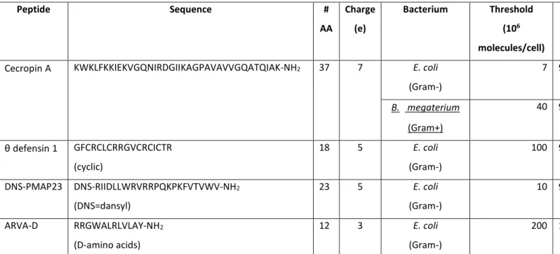
Table 1 summarizes all the data collected so far on the threshold of bound peptide molecules per cell needed to cause bacterial killing.
As pointed out by Wimley,17 the fact that killing is caused by massive peptide accumulation on bacteria could be one of the reasons why resistance to AMPs and mimics does not arise as easily as that to chemical antibiotics. However, these findings also rise several questions:
• where do all these peptide molecules accumulate?
• Do they bind mostly to membranes or to components of the cell wall, or to theLPS of the OM of Gram-negative bacteria?
• Do they interact with intracellular molecules as well?
Simple geometric considerations 17, 20, 103 show that the observed peptide/cell ratios are even higher than complete coverage of the membrane surface (even considering the two membranes, and the four leaflet of an E. coli cell). It should be considered that, due to roughness at the molecular scale, the geometric surface of the bacterium might be significantly smaller than the real surface area available for binding.106 Furthermore, binding could take place in a multilayer arrangement, or association to other components of the bacterial cell might be taking place. Our experiments were performed by measuring changes in fluorescence intensity that is likely sensitive mostly to insertion in the bacterial membrane, while all other studies determined the total cell bound peptide concentration by separation techniques. The values obtained with the two approaches (even though not on the same system) are not very different, considering also that they represent order of magnitude estimates, and could indicate that membrane binding is favored with respect to other interactions. Indeed, a recent study suggests that PMAP-23 has no significant affinity for LPS.107 When we compared our cell-binding data with those obtained with liposomes formed with an E. coli lipid extract, we observed a surprisingly good agreement (reporting the two curves as a function of total lipid concentration).20 These data indicate that, despite their simplicity, liposomes are a good model to study peptide association to bacterial lipids. In addition, they further support the idea that our results mainly refer to peptide association to cell membranes (rather than to other cell components), in agreement with one of Castanho’s hypotheses.
In some cases binding experiments were performed with a fixed cell density and variable peptide concentrations (Merrifield and Selsted), while in others the peptide concentration was fixed and the cell density was varied (Wimley and us). In any case, some of the data points very likely correspond to conditions where the bacteria are killed (and thus likely permeabilized), while in other cases the peptide might not be sufficient to lyse the cells. Therefore, the actual accessible binding targets could vary along the experiments, and also with time (during the membrane poration process). In our case, we did not observe any changes in the fluorescence after a few minutes, but ongoing experiments in our laboratory and studies by other groups with different cells or peptides are indicating that this situation is not general.15, 22, 108 Further studies are definitely needed to determine the location of peptide molecules in the cells, and the kinetics of distribution among the different sites.
Finally, it is worth mentioning that the binding curves we obtained deviated only slightly from the behavior predicted from an ideal partition. In particular, in our case the curves obtained for peptide concentrations of 1 and 10 μM were rather similar,20 approximating the peptide concentration independence expected for an ideal behavior. Overall, our data showed that the hypotheses put forward by Castanho’s group were justified, at least approximately. However, this finding is not general, as the binding data from Wimley’s group17 showed a strong dependence of the isotherms on concentration (for a different peptide).
4.2. Cell-density dependence of the activity (inoculum effect)
The so-called “inoculum effect” is a well-known dependence of the MIC of traditional antibiotics on the size of the bacterial inoculum in the growth medium109. Surprisingly, only very few studies investigated this phenomenon for HDPs, without discussing the origin of the observed behavior. Levinson et al.,110 reported that the bactericidal activity of magainins against P. aeruginosa was inoculum dependent above 3x10⁵ cells/ml, but it did not vary if the inoculum cell density was reduced below this value. Similarly, Jones et al.111 observed an inoculum effect for lactoferricin B against E. coli, with a plateau at inoculum densities below 106 cells/ml (Figure 3).
After performing the peptide/cell association studies discussed above, we soon realized that our data allowed a prediction of the dependency of the active concentration (i.e. the MBC, in our case) on the density of bacterial cells. Those data provided the threshold of DNS-PMAP23 molecules that must (on average) associate to each cell to kill it (TB=1.1x10⁷) and the fraction of total peptide that is cell/bound (as a function
of bacterial cell density). With this information, it is possible to calculate how the total peptide concentration needed for killing (i.e. the MBC) depends on cell-density21, assuming that this partition curve does not depend strongly on peptide concentration (see above).
Based on these theoretical considerations, we derived the following behavior for the MBC:
𝑀𝑀𝑀𝑀𝑀𝑀 = �1 + [𝑀𝑀𝐵𝐵𝐵𝐵𝐵𝐵𝐵𝐵𝐵𝐵𝐵𝐵𝐵𝐵] 𝐾𝐾𝑎𝑎𝑎𝑎𝑎𝑎.⁄ 𝐵𝐵 �𝑀𝑀𝑀𝑀𝑀𝑀𝑚𝑚𝑚𝑚𝑚𝑚. (3 with
𝑀𝑀𝑀𝑀𝑀𝑀𝑚𝑚𝑚𝑚𝑚𝑚. = 𝐾𝐾𝑎𝑎𝑎𝑎𝑎𝑎.𝐵𝐵 10 3
𝑁𝑁𝐴𝐴𝑇𝑇𝐵𝐵 (4
Here NA is Avogadro’s constant, MBC and MBCmin. are expressed in moles/l, 𝐾𝐾𝑎𝑎𝑎𝑎𝑎𝑎.𝐵𝐵 (the apparent partition
constant for cell association) and the bacterial cell density ([Bacteria]) are reported in cells/ml and TB is in
molecules per cell.
This Equation predicts a linear decrease in the MBC with decreasing cell density. However, it also foresees that even when the cell-density becomes extremely low, the MBC does not decrease below a limiting value, equal to MBCmin. This behavior can be understood based on the peptide/cell association equilibrium. At cell densities significantly higher than the apparent partition constant, all the peptide in the sample is associated
to bacteria. Therefore, more cells need more peptide molecules to kill them. However, in the low cell-density regime (Bacteria]<<𝐾𝐾𝑎𝑎𝑎𝑎𝑎𝑎.𝐵𝐵 ), most of the peptide will stay free in solution. In this interval, the partition equilibrium can be approximated by a linear behavior (such as a Langmuir binding isotherm or a Michaelis-Menten enzyme kinetics), and therefore the fraction of cell-bound peptide molecules decreases proportionally to [Bacteria]. As a consequence, the two effects (less cells to kill, and a lower fraction of cell-bound peptide) cancel each other, and the total peptide concentration needed in the sample to kill the bacteria remains constant.
A similar theoretical prediction (i.e. a linear dependence of MBC, with a nonzero intercept) had been recently reported for the trend of peptide membrane-perturbing activity in liposomes with the concentration of vesicles,112 based on a complex model of several aspects of the peptide and lipid bilayer behavior at the molecular level. By contrast, the present treatment shows that the predicted trend simply arises from a close to ideal partition equilibrium, without the need of any assumptions on molecular events.
Figure 3 reports the MBC values measured for DNS-PMAP23 in the presence of different E. coli cell-densities.21 The behavior predicted above was actually observed, with the MBC never decreasing below 3 μM. The agreement between the experimental data and the predicted curve (which is not a fit, but a calculation based on the values of TB and 𝐾𝐾𝑎𝑎𝑎𝑎𝑎𝑎.𝐵𝐵 determined previously) was acceptable even quantitatively.
Interestingly, approximately at the same time of our study, another detailed and quantitative investigation on the inoculum effect for the activity of pexiganan against E. coli was reported by the group of Wilson Poon.113 Also in this study, a plateau of the active concentration (MIC, rather than MBC in this case) was observed at low cell densities. These data are reported in Figure 3, together to those of Jones111 and to ours. The coincidence of the trends in the three datasets is striking.
The discussion reported above shows that this experimental trend can be explained (and actually predicted) based on the peptide/cell association equilibrium. However, other interpretations are possible. For instance, also the group of Sattar Taheri-Araghi,114-115 recently observed a cell-density dependence of antimicrobial activity (MIC) and performed single-cell microscopy experiments suggesting that the effect is not due to an equilibrium binding behavior, but rather to irreversible peptide sequestration by dead bacterial cells. A similar interpretation was supported by the work of Poon and coworkers.113 Also in this case, several experiments, including single cell studies, indicated that sub-MIC concentrations cause the death of a fraction of the bacterial cells, leaving the growth of the others unaffected, as the peptide is sequestered by the killed bacteria, possibly due to peptide binding to intracellular targets, or to the action of proteolytic enzymes released from the lysed cells.
Whatever the origin of the observed behavior, the indication that micromolar total peptide concentrations are necessary to kill the bacteria, even when they are present at low cell counts, might have significant practical consequences. Regarding the possible clinical application of HDPs or petptidomimetic molecules inspired by them, reaching such relatively high concentrations in vivo by systemic administration
might prove problematic. On the other hand, concerning the physiological function of HDPs, it should be considered that some of these peptides can naturally reach concentrations that are even significantly higher than micromolar. For instance, this can happen in the granules of leukocytes, in the immediate vicinity of degranulating phagocytes, at the bottom of intestinal crypts,10 in the hemolymph of insects after a bacterial infection,116 or on the skin of some frogs.117 In addition, it is important to note that multiple HDPs are normally present in the organism, and they often act at the same time and can exhibit synergism.75. 118 However, in other cases, where the physiological concentrations of HDPs are lower than micromolar, other functions, such as immunomodulation, might be more important than direct bacterial killing.10 Finally, the finding that HDP active concentrations exhibit a plateau at low cell densities might provide a rationale for some observations on the production of these peptides in insects immune-challenged with bacteria: in some cases, the up-regulation of HDP genes is not dose-dependent and the expression and synthesis of these peptides is maintained for a long time, independent of the number of bacteria in the haemolymph.119-120
4.3. Peptide association to eukaryotic cells and selectivity
In addition to peptide association to bacterial cells, interaction with host cells is also very important for the function of HDPs in vivo. As discussed above, in model membrane studies a significantly higher affinity is usually observed for liposomes mimicking bacterial membranes, compared to vesicles representing the composition of erythrocyte membranes.6 Moreover, in vitro microbiological tests of peptide activity and toxicity usually show that much higher peptide concentrations are needed for damaging human cells than to kill bacteria. The common interpretation of these data is that HDPs are selective, thanks to their higher affinity for the target cells. However, it has been noted14 that the experimental conditions for the microbiological assays are rather different (typically, 5 × 10⁵ - 1 × 10⁶ cells/mL in MIC assays, and 5 × 10⁸ cells/mL in tests of hemolytic activity) and that red blood cells (RBCs) are approximately 10 times bigger than bacteria. More importantly, activity assays are performed separately on the two cells population, while these coexist in a realistic condition of infection. Therefore, it has been questioned if the selectivity of HDPs observed in biophysical and in vitro studies is real, or a simple consequence of the experimental conditions used in the two assays.14 In order to tackle these aspects, we investigated the cell-density dependence of the hemolytic activity of the DNS-PMAP23 peptide, developing a protocol that allowed us to vary the erythrocyte concentration of 4 orders of magnitudes. The observed trend in hemolytic activity was very similar to our findings for bacteria, with an approximately linear increase with increasing cell densities, and with a limiting value at low cell counts (< 107 cells/mL).21
Unfortunately, the high absorbance of the heme group prevented us from determining peptide binding to RBCs with the same spectroscopic approach used for bacteria (see above). However, two other studies have determined peptide/erythrocyte interaction directly, by using the separation approaches described above.17, 103 While cecropin A showed very little affinity for RBCs,103 the ratio of affinities of the artificial
peptide ARVA-D for bacteria and erythrocytes depended on peptide concentration.17 In any case, our finding that both antimicrobial activity and toxicity depend on the density of cells implies that the effective selectivity depends on the concentration of the two cell populations (bacteria and host cells). How much peptide will bind to a type of cell or to the other will depend on the respective partition constant, but also on the concentration of cells of each cell type. The final effects on the cells will depend on the fact that the respective threshold of bound peptide needed for membrane perturbation is reached or not.21, 112 This discussion, based on simple partition equilibria, leads also to the prediction that when both cell types are present at the same time, the antimicrobial activity and/or the toxicity of the peptides could be inhibited by peptide sequestration of a fraction of the peptide molecules due to binding to the other cell population. To test this hypothesis, both our group21 and Wimley’s17 simultaneously developed novel assays on mixed populations of bacteria and RBCs. We studied both bacterial killing and RBC lysis, while Starr et al. measured the antibacterial activity only. On the other hand, we performed experiments by adding the peptide to the mixture of the two cell types, while Wimley’s group studied also the effects of order of addition of RBCs and peptides to the bacterial culture. Rather surprisingly, both studies showed that when peptides were added to a mixture of bacteria and RBCs with a large excess of erythrocytes (10-104 times), no inhibition of the antimicrobial activity was observed, in dramatic contrast to the predictions of the simple partition equilibria discussed above, where sequestration of a significant peptide fraction due to erythrocyte binding would be expected (Figure 4). A further indication that an equilibrium model is not adequate to represent peptide interaction with a mixture of cells came from the observation that the results of the experiment depend on the order of addition of the different components. Wimley reported a significant inhibition of the antimicrobial activity when the peptide was incubated with RBCs first, and then both were added to the bacterial culture.17 A possible interpretation of this finding is peptide degradation by erythrocyte proteases,121 but inhibition caused by preincubation with RBCs was observed also for an all-D peptide.17
Overall, these data indicate that non-equilibrium phenomena, possible kinetic effects or irreversible binding, are determinant in the interaction of HDPs with target and host cells. Studies in this area are just at the beginning, and further experiments will be essential to clarify this important point. Whatever the mechanism underlying the findings reported above, these studies provide support for a direct bactericidal function and significant selectivity of HDPs under realistic conditions. Therefore, they also bode well for possible therapeutic applications, at least for topical treatments. Indeed, several reports exist regarding the efficacy of HDPs in vivo122 and their ability to concentrate at sites of infection.123 However, the results of Starr et al. on peptide inhibition by preincubation with RBCs support the view that systemic administration might be faced with significant hurdles (sequestration by blood components, proteolytic degradation, rapid clearance, etc.). Finally, the results summarized in this section strongly suggest the adoption of mixed cell population assays (such as those developed in the studies reported above) as the new standard for the screening and evaluation of HDPs.
5. Quantitative single‐cell microscopic studies
Recently, the group of James Weisshaar developed a novel approach employing time-lapse wide field imaging.18 Their method enables direct, simultaneous observation of multiple HDP-induced effects on single bacteria cells as a function of time, with a resolution in the seconds range. The peptide is often labeled with a fluorophore to visualize its distribution, and quantify its concentration. A green fluorescent protein (GFP), expressed and translocated to the periplasmic space by a mutant E. coli strain, allows the detection of OM perturbation, by the observation of GFP release. The fluorescence of the particular GFP employed is pH sensitive, so that even the formation of small pores allowing the passage of protons can be detected by visualizing intensity changes. Perturbation of the CM can be observed thanks to a membrane-impermeant DNA-binding probe (e.g. SG) added to the external medium. In the case of Gram-positive bacteria such as B. subtilis, cells producing a GFP in the cytosol were employed. Finally, acquisition of phase-contrast images enables the measurement of cell size, thus allowing the quantification of cell growth.
Several HDPs have now been studied by this approach: LL-37124-128, cecropin A 126, 128-129, the hybrid peptide CM15,127 alamethicin,130 melittin,128 indolicidin,128 and even artificial antimicrobial polymers.131 Overall, the picture resulting from these studies is rather complex, showing that each peptide behaves in a somewhat different way, even though several common features are present.
In the case of Gram-negative bacteria, peptides bind to the OM and the whole cell becomes uniformly coated shortly after addition.124 Then peptides translocate across the OM;124, 129 this event takes place locally, preferentially at curved regions of the cell, i.e. the septum of dividing cells, or the end caps of single cells. This finding (which has been reported also in AFM studies) might be related to a higher concentration of negatively charged lipids, such as cardiolipin, in those regions. Importantly, when the peptide gains access to the periplasmic space, cell growth halts. Successively, the OM is permeabilized to large solutes at sites where the peptide is concentrated, allowing the leakage of GFP outside the cell. A lag time of a few minutes is observed between peptide addition and this event, varying significantly from cell to cell, with septating cells attacked first, and some peptides (e.g. cecropin A) acting faster than others (e.g. fluorescently labelled LL-37). After a further lag time lasting from seconds (cecropin A) to several minutes (LL-37) the CM is permeabilized and SG can enter the cytosol. This event is again local, and pores are stable and situated at cell regions of high membrane curvature and cardiolipin content (septa or endcaps). However, while LL-37 perturbs the CM in the same cell region of OM permeabilization, cecropin A surprisingly causes CM permeability at a different site. There are also cases (CM15, unlabeled LL-37, artificial polymers) where the peptides apparently reach the CM without creating large pores in the OM, and permeabilize the cytoplasmic bilayer first, so that GFP is observed to enter the cytosol, rather than leaving the cell envelope. Interestingly, in addition to OM permeabilization, several other effects might participate to halting cell growth: LL-37, CM-15, melittin and artificial polymers have been shown to cause oxidative stress once they reach the periplasm,
while cecropin A and indolicidin do not.18, 127-128 This process contributes significantly to antimicrobial activity, as MICs of peptides causing oxidative stress increase several times for bacteria growing under anaerobic conditions. By contrast, while cecropin A perturbs nucleoid organization once it reaches the cytoplasm, LL-37 does not.126
It is worth mentioning that the kinetics of E. coli attack by the hybrid HPD CM15 has been studied independently also by high-speed AFM.60 The observable peptide-induced symptom was an increase in surface roughness. In agreement with Weisshaar’s findings, this effect took place after a lag phase, that varied from cell to cell, but completed in less than a minute once started. By coupling optical microscopy imaging with high-speed AFM and performing the live/dead assay on the same sample, damage to the cell surface was demonstrated to be strongly correlated with bacterial death.
LL-37 and alamethicin have been studied also with the Gram-positive bacterium B. subtilis.125, 130 In the case of the cationic HDP LL-37, the mechanism of membrane perturbation depended on peptide concentration. At 2 μM, a decrease in growth rate occurred without membrane permeabilization, but the bacteria kept increasing their size, although at a slower pace. The original rate could be recovered by removing the peptide. At 4 μM and above, abrupt permeabilization took place, together with cell shrinkage. In the latter case, pores were local and stable over time, and growth was not recovered after peptide removal. The hydrophobic peptide alamethicin has been demonstrated to form pores in artificial membranes through a mechanism called “barrel-stave”, different from the carpet model.97 In this case, growth halting always preceded CM permeabilization, even to the passage of protons (by 2-3 minutes). Successively, cells were observed to shrink (approximately 10 minutes after peptide addition), SG could get into the cytosol and only eventually cytosolic GFP was released, followed by other cell components, as shown by the loss of phase contrast. Differently from experiments on cationic HDPs, in the case of alamethicin pores were not localized, and grew bigger over time.
Overall, Weisshar’s studies, thanks to their time and space resolution, provide a completely new view of the mechanism of bacterial attack by HDPs. First of all, they indicate that bacteria start “suffering” well before the CM is porated. Unfortunately, these experiments do not provide direct indications on bacterial death, but only on inhibition of single cell growth. In some cases, cells that had stopped increasing their length could start growing again after the peptide was removed. Therefore, formation of pores in the CM cannot be ruled out as the mechanism of killing. Indeed, in all cases the cytosolic bilayer eventually became permeable. Another important point is that, when peptides enter the periplasm of E. coli, they start several cellular events, including oxidative stress, which could contribute to cell damage. An additional interesting result of Weisshaar’s studies is a clear definition of the time-scale of peptide interaction with cells. In the case of Gram-negative bacteria, effects start to be seen after seconds to minutes, and the whole process is completed in a time going from a few minutes to approximately half an hour (in the case of LL-37). Finally, HDPs were shown to cause membrane permeability only at well-defined locations in the cell (with the single
exception of alamethicin). This finding is surprising, in view of the high peptide accumulation needed to cause bacterial killing.20 However, it is conceivable that pores start forming at a specific site, and that this is sufficient to cause the leakage of fluorescing molecules observed in the microscopy experiments. In any case, further experiments are warranted to better clarify the many new insights provided by these studies.
Regarding peptide-induced permeability, it is worth mentioning that recently Huey Huang and coworkers quantitatively compared pore formation in GUVs and in E. coli spheroplasts, i.e. cells from which the cell wall has been removed.19, 132 In both systems, the addition of LL-37, melittin or alamethicin caused membrane permeability to fluorescent dyes (calcein or fluorescently labeled dextran with average molecular weight 4000 Da), after a lag phase that was different for each individual system. In addition, the values of peptide-induced permeability were in very good agreement.19
6. Summary and outlook
The last five years have witnessed the blossoming of studies applying the quantitative methods of biophysical chemistry, normally used with model membranes, to investigations of the interaction of HDPs with real cells. These approaches have provided several new insights:
• Peptide binding to bacterial membranes and to liposomes seems to be comparable (when considering the lipid concentration).20
• A high coverage of the bacterial surface is needed for killing.17, 20
• Both the activity and the toxicity of HDPs depend on the concentration of cells (inoculum effect), but a minimum, threshold total concentration (in the μM range) seems to be required, even at very low cell counts.21, 113
• The selectivity of HDPs is real: when HDPs are added to a mixture of bacteria and host cells, with a large excess of the latter, the antimicrobial activity is not significantly inhibited.17, 21 However, preincubation with host cells significantly reduces activity.17
• Bacterial membranes are usually made permeable at specific sites, coinciding with regions of high curvature (septa or endcaps).18
• The properties of equilibrium pores formed in cells are comparable with those observed in model membranes.19
• Bacterial growth is often inhibited after perturbation of the OM but before the formation of pores in the CM.20
At the same time, several new questions have arisen from these findings: • How general are the behaviors now observed only in a few systems?
• Can the peptide concentrations needed for direct antimicrobial activity be reached in vivo? • Where are the large amounts of peptide molecules needed for killing located in the bacterial cell? • What are the kinetic steps of HDP binding to target and host cells?
• What are the mechanisms inhibiting bacterial growth before the formation of pores in the cytosolic membrane?
When quantitative comparisons have been performed on HDP interaction with liposomes and bacteria (e.g. regarding the binding affinity,20 or the properties of equilibrium pores19) a surprisingly good agreement has been observed. In addition, the data discussed in Section 2 indicate that the models of pore formation developed from liposome studies are relevant also for the perturbation of bacterial membranes. Therefore, model membranes will remain a valuable tool in future studies of the mechanism of membrane perturbation by HDPs. At the same time, the studies presented here indicate that quantitative studies of HDP interaction with real cells can open a completely new window on our understanding of these systems. The data collected so far indicate that some properties might be shared by several HDPs, but also that each peptide has some peculiarities. Therefore, studies on a larger number of peptides, and on several different bacteria are warranted to strengthen and expand the scope of the current findings.
Confirmation that a micromolar threshold for bactericidal activity is needed even at very low cell densities, would represent an important finding, It has been reported that in some cases immunomodulation takes place in vitro at concentrations lower than those needed for bacterial killing.10-11 It is therefore essential to clarify which is the predominant mechanism of action of HDPs: today most studies screening HDPs, or isolating new HDP molecules, are performed focusing on the direct antibacterial activity. However, these investigations might be on a completely wrong path if immunomodulation was the predominant mechanism by which these molecules protect our organism.10-11, 133
Observation of the inoculum effect (which was well known for traditional antibiotics) suggests that studies of the cell-density dependence of HDP activity and selectivity should become a standard part of peptide characterization. Similarly, the finding that under certain conditions host cells can inhibit peptide activity indicates that assays with co-cultures of eukaryotic and bacterial cells should be adopted.
From a technical point of view, real-time single-cell microscopic approaches can easily become increasingly powerful by exploiting multiple specific markers of different processes, and increasing the number of channels that are acquired during an experiment. Recently, the application of microfluidic techniques to microbiological studies of bacteria has allowed an unprecedented control on the growth conditions, and the parallel observation of up to 105 live cells in real-time in a single experiment.134-135 We expect that these technical advances will provide extremely powerful new tools for the characterization of HDPs.
The novel findings described in this review and the future answers to the new questions that have arisen are likely to lead to an important step forward in our understanding of HDPs and in their development for therapeutic purposes.
ACKNOWLEDGEMENTS
This work was supported by MIUR (grant PRIN 20157WW5EH_007) and University of Rome Tor Vergata (Consolidate the Foundations GRANT, AMPSA). The illustration of a cross-section of a small portion of an Escherichia coli cell in the graphical abstract is by David S. Goodsell, the Scripps Research Institute (http://mgl.scripps.edu/people/goodsell/illustration/public).
L. S. would like to thank Prof. Basilio Pispisa for originally introducing him to the field of peptide/membrane interactions by convincing him that complexity should not always be avoided.
REFERENCES
1. Laxminarayan, R.; Duse, A.; Wattal, C.; Zaidi, A. K.; Wertheim, H. F.; Sumpradit, N.; Vlieghe, E.; Hara, G. L.; Gould, I. M.; Goossens, H.; Greko, C.; So, A. D.; Bigdeli, M.; Tomson, G.; Woodhouse, W.; Ombaka, E.; Peralta, A. Q.; Qamar, F. N.; Mir, F.; Kariuki, S.; Bhutta, Z. A.; Coates, A.; Bergstrom, R.; Wright, G. D.; Brown, E. D.; Cars, O., Antibiotic resistance-the need for global solutions. Lancet Infect Dis 2013, 13 (12), 1057-98.
2. Butler, M. S.; Blaskovich, M. A.; Cooper, M. A., Antibiotics in the clinical pipeline at the end of 2015. J Antibiot (Tokyo) 2017, 70 (1), 3-24.
3. Cooper, M. A.; Shlaes, D., Fix the antibiotics pipeline. Nature 2011, 472 (7341), 32.
4. Li, J.; Koh, J. J.; Liu, S.; Lakshminarayanan, R.; Verma, C. S.; Beuerman, R. W., Membrane Active Antimicrobial Peptides: Translating Mechanistic Insights to Design. Front Neurosci 2017, 11, 73.
5. Wang, G.; Li, X.; Wang, Z., APD3: the antimicrobial peptide database as a tool for research and education. Nucleic Acids Res. 2016, 44 (D1), D1087-93.
6. Bobone, S.; Bocchinfuso, G.; Park, Y.; Palleschi, A.; Hahm, K. S.; Stella, L., The importance of being kinked: role of Pro residues in the selectivity of the helical antimicrobial peptide P5. J. Pept. Sci. 2013, 19
(12), 758-69.
7. Lee, T. H.; Hall, K. N.; Aguilar, M. I., Antimicrobial peptide structure and mechanism of action: a focus on the role of membrane structure. Curr. Top. Med. Chem. 2016, 16 (1), 25-39.
8. Wimley, W. C., Describing the mechanism of antimicrobial peptide action with the interfacial activity model. ACS Chem Biol 2010, 5 (10), 905-17.
9. Fox, J. L., Antimicrobial peptides stage a comeback. Nat. Biotechnol. 2013, 31 (5), 379-82.
10. Hancock, R. E.; Haney, E. F.; Gill, E. E., The immunology of host defence peptides: beyond antimicrobial activity. Nat Rev Immunol 2016, 16 (5), 321-34.
11. Hilchie, A. L.; Wuerth, K.; Hancock, R. E., Immune modulation by multifaceted cationic host defense (antimicrobial) peptides. Nat Chem Biol 2013, 9 (12), 761-8.
12. Hansen, P. R., Antimicrobial peptides: methods and protocols. Springer: 2017; Vol. 1548. 13. Mishra, B.; Reiling, S.; Zarena, D.; Wang, G., Host defense antimicrobial peptides as antibiotics: design and application strategies. Curr. Opin. Chem. Biol. 2017, 38, 87-96.
14. Matsuzaki, K., Control of cell selectivity of antimicrobial peptides. Biochim. Biophys. Acta 2009,
1788 (8), 1687-92.
15. Avitabile, C.; D'Andrea, L. D.; Romanelli, A., Circular Dichroism studies on the interactions of antimicrobial peptides with bacterial cells. Sci Rep 2014, 4, 4293.
16. Freire, J. M.; Gaspar, D.; Veiga, A. S.; Castanho, M. A., Shifting gear in antimicrobial and anticancer peptides biophysical studies: from vesicles to cells. J. Pept. Sci. 2015, 21 (3), 178-85.
17. Starr, C. G.; He, J.; Wimley, W. C., Host Cell Interactions Are a Significant Barrier to the Clinical Utility of Peptide Antibiotics. ACS Chem Biol 2016, 11 (12), 3391-3399.
18. Choi, H.; Rangarajan, N.; Weisshaar, J. C., Lights, Camera, Action! Antimicrobial Peptide Mechanisms Imaged in Space and Time. Trends Microbiol 2016, 24 (2), 111-22.
19. Faust, J. E.; Yang, P. Y.; Huang, H. W., Action of Antimicrobial Peptides on Bacterial and Lipid Membranes: A Direct Comparison. Biophys. J. 2017, 112 (8), 1663-1672.
20. Roversi, D.; Luca, V.; Aureli, S.; Park, Y.; Mangoni, M. L.; Stella, L., How many antimicrobial peptide molecules kill a bacterium? The case of PMAP-23. ACS Chem Biol 2014, 9 (9), 2003-7.
21. Savini, F.; Luca, V.; Bocedi, A.; Massoud, R.; Park, Y.; Mangoni, M. L.; Stella, L., Cell-Density Dependence of Host-Defense Peptide Activity and Selectivity in the Presence of Host Cells. ACS Chem Biol
2017, 12 (1), 52-56.
22. Avitabile, C.; D'Andrea, L. D.; Romanelli, A., Studying the Interaction of Magainin 2 and Cecropin A with E. coli Bacterial Cells Using Circular Dichroism. Methods Mol Biol 2017, 1548, 247-253.
23. Booth, V.; Warschawski, D. E.; Santisteban, N. P.; Laadhari, M.; Marcotte, I., Recent progress on the application of 2H solid-state NMR to probe the interaction of antimicrobial peptides with intact bacteria. Biochim. Biophys. Acta 2017.
24. Balouiri, M.; Sadiki, M.; Ibnsouda, S. K., Methods for in vitro evaluating antimicrobial activity: A review. Journal of Pharmaceutical Analysis 2016, 6 (2), 71-79.
25. Sanchez-Gomez, S.; Lamata, M.; Leiva, J.; Blondelle, S. E.; Jerala, R.; Andra, J.; Brandenburg, K.; Lohner, K.; Moriyon, I.; Martinez-de-Tejada, G., Comparative analysis of selected methods for the
assessment of antimicrobial and membrane-permeabilizing activity: a case study for lactoferricin derived peptides. BMC Microbiol 2008, 8, 196.
26. Wiegand, I.; Hilpert, K.; Hancock, R. E., Agar and broth dilution methods to determine the minimal inhibitory concentration (MIC) of antimicrobial substances. Nat Protoc 2008, 3 (2), 163-75.
27. Lorian, V., Antibiotics in Laboratory Medicine. 5th edition. , . 5th ed.; Lippincott Williams & Wilkins: Philadelphia, PA, USA, 2005.
28. Patel, J. B. C. I., F.R.; Bradford, P.A.; Eliopulos, G.M.; Hindler, J.A.; Jenkins, S.G.; Lewis II, J. S.; Limbago, B.; Miller, L.A.; Nicolau, D.P.; Powell, D.P.; Swenson, J.M.; Traczewski, M.M.; Turnidge, J.D.;
Weistein, M.P.; Zimmer, B.L., Methods for Dilution Antimicrobial Susceptibility Tests for Bacteria that Grow Aerobically, Approved Standard, 10th ed. In M07-A10, Institute, C. a. L. S., Ed. 2012.
29. Konig, C.; Simmen, H. P.; Blaser, J., Bacterial concentrations in pus and infected peritoneal fluid--implications for bactericidal activity of antibiotics. J. Antimicrob. Chemother. 1998, 42 (2), 227-32.
30. Kang, D. K.; Ali, M. M.; Zhang, K.; Huang, S. S.; Peterson, E.; Digman, M. A.; Gratton, E.; Zhao, W., Rapid detection of single bacteria in unprocessed blood using Integrated Comprehensive Droplet Digital Detection. Nat Commun 2014, 5, 5427.
31. Zelezetsky, I.; Pontillo, A.; Puzzi, L.; Antcheva, N.; Segat, L.; Pacor, S.; Crovella, S.; Tossi, A., Evolution of the primate cathelicidin. Correlation between structural variations and antimicrobial activity. J. Biol. Chem. 2006, 281 (29), 19861-71.
32. Schwechheimer, C.; Kuehn, M. J., Outer-membrane vesicles from Gram-negative bacteria: biogenesis and functions. Nat Rev Microbiol 2015, 13 (10), 605-19.
33. Helander, I. M.; Mattila-Sandholm, T., Fluorometric assessment of gram-negative bacterial permeabilization. J Appl Microbiol 2000, 88 (2), 213-9.
34. Luca, V.; Stringaro, A.; Colone, M.; Pini, A.; Mangoni, M. L., Esculentin(1-21), an amphibian skin membrane-active peptide with potent activity on both planktonic and biofilm cells of the bacterial pathogen Pseudomonas aeruginosa. Cell Mol Life Sci 2013, 70 (15), 2773-86.
35. Marcellini, L.; Borro, M.; Gentile, G.; Rinaldi, A. C.; Stella, L.; Aimola, P.; Barra, D.; Mangoni, M. L., Esculentin-1b(1-18)--a membrane-active antimicrobial peptide that synergizes with antibiotics and modifies the expression level of a limited number of proteins in Escherichia coli. FEBS J 2009, 276 (19), 5647-64.
36. Arcidiacono, S.; Soares, J. W.; Meehan, A. M.; Marek, P.; Kirby, R., Membrane permeability and antimicrobial kinetics of cecropin P1 against Escherichia coli. J. Pept. Sci. 2009, 15 (6), 398-403.
37. Hartmann, M.; Berditsch, M.; Hawecker, J.; Ardakani, M. F.; Gerthsen, D.; Ulrich, A. S., Damage of the bacterial cell envelope by antimicrobial peptides gramicidin S and PGLa as revealed by transmission and scanning electron microscopy. Antimicrob. Agents Chemother. 2010, 54 (8), 3132-42.
38. Friedrich, C. L.; Moyles, D.; Beveridge, T. J.; Hancock, R. E., Antibacterial action of structurally diverse cationic peptides on gram-positive bacteria. Antimicrob. Agents Chemother. 2000, 44 (8), 2086-92.
39. Bessalle, R.; Kapitkovsky, A.; Gorea, A.; Shalit, I.; Fridkin, M., All-D-magainin: chirality, antimicrobial activity and proteolytic resistance. FEBS Lett. 1990, 274 (1-2), 151-5.
40. Wade, D.; Boman, A.; Wahlin, B.; Drain, C. M.; Andreu, D.; Boman, H. G.; Merrifield, R. B., All-D amino acid-containing channel-forming antibiotic peptides. Proc Natl Acad Sci U S A 1990, 87 (12), 4761-5.