Universit`a Politecnica delle Marche
Scuola di Dottorato di Ricerca in Scienze dell’Ingegneria Curriculum in Ingegneria Informatica, Gestionale e dell’Automazione
An infrastructure for
decision-making to support
neonatal clinical care and research
Ph.D. Dissertation of:
Annalisa Cenci
Advisor:
Prof. Primo Zingaretti
Curriculum Supervisor: Prof. Francesco Piazza
Scuola di Dottorato di Ricerca in Scienze dell’Ingegneria Facolt`a di Ingegneria
Ringraziamenti
Il primo ringraziamento va, senza dubbio, ai miei genitori, i due punti di riferimento della mia vita, senza i quali non sarei arrivata dove sono ora. Grazie perch´e posso sempre contare su di voi, perch´e sapete essermi vicini nei momenti difficili e gioire per i miei traguardi.
Un grazie enorme anche a mia sorella e a mio fratello, su cui posso sempre contare, e alla famiglia che si allarga..
Un grazie speciale anche a te Italo, perch´e con te posso essere sempre me stessa, perch´e mi capisci e mi sei vicino pi`u di chiunque altro, anche nei momenti pi`u stressanti, che tanto spesso sono capitati in questi anni di dottorato. Grazie per tutto!
Altro tipo di ringraziamento, ma non meno importante e sincero, va ai pro-fessori Primo Zingaretti ed Emanuele Frontoni, che mi hanno dato la possi-bilit`a di intraprendere questo stimolante percorso di ricerca. Grazie a Primo per i suoi preziosi consigli che mi hanno spronato a fare sempre meglio e ad Emanuele per la passione che mette nel suo lavoro e che cerca di trasmetterci quotidianamente.
Ringrazio tutti i membri del VRAI, del DII e dell’Universit`a che ho avuto il piacere di conoscere in questi anni, Rama, Michele, Rocco, Chiara, Mirco, Roberto e Roberto, Gloria. Grazie per avermi dato una mano quando potevate o anche solo per avermi allietato la giornata con una battuta.
Un grazie particolarmente sentito va ai miei due compagni di lavori, viaggi, avventure, chiacchiere e risate, che in questi tre anni da colleghi si sono trasfor-mati in amici fidati su cui so che potr`o contare anche in futuro.
Un grazie anche al Prof. Virgilio Paolo Carnielli, al personale del reparto della UTIN dell’ospedale pediatrico “G. Salesi“ di Ancona e alla JEF srl senza la cui collaborazione questo lavoro di dottorato non sarebbe stato possibile.
A tal proposito mi sento di ringraziare tutte le persone della JEF con cui mi trovo molto bene e da cui sto imparando molto. Grazie a Moira, Bianca, Luca, Andrea, Marco, Salvatore e a tutto il team di sviluppo.
Anche qui un grazie particolare, che viene dal cuore, va a te Ilaria. 7 anni fa ti ho conosciuta come mia correlatrice. Da correlatrice sei passata a collega e, poi, a molto pi`u che collega. Ormai se per me un’amica vera e leale. Posso dire, mai stata pi`u sincera, che se non fosse stato per te non sarei arrivata fin qui. Mi hai sempre spronato a continuare e ad andare avanti, credendo in me
il tuo perenne buonumore. Grazie!
Ultimo, ma non per importanza, un grazie alle mie amiche di sempre, Mari, Vale, Anna e Marti; grazie perch´e, nonostante lo studio, il lavoro e i mille impegni che ci tengono lontane, siete sempre l`ı pronte ad accogliermi a braccia aperte, a farmi divertire e a farmi sentire a casa come se ci vedessimo ancora tutti i giorni!
E per finire un grazie davvero sentito a tutti coloro, amici e parenti, che, anche se non ho citato, mi hanno aiutato ad arrivare a questo importante traguardo.
Ancona, October 2017
Sommario
I dispositivi medici, in genere, sono unit`a individuali, per cui una tipica sala ospedaliera in un reparto di terapia intensiva ospita un gran numero di dispositivi stand-alone, ognuno con una propria interfaccia utente.
Le culle dei neonati pretermine presso l’Unit`a di Terapia Intensiva Neona-tale (UTIN) dell’Ospedale Pediatrico “G. Salesi“ di Ancona sono circondati da numerosi dispositivi per il monitoraggio, la diagnosi e il trattamento di diverse malattie e tutti questi dispositivi forniscono un’enorme quantit`a di dati che, fino ad ora, venivano visualizzati solo su monitor e periodicamente trascritti in una cartella clinica cartacea. Le note manuali, quindi, venivano regolarmente, ma non immediatamente, trascritte in un foglio elettronico sul PC dell’unit`a neonatale con il rischio di errori e dimenticanze.
In questo contesto, i medici hanno espresso la necessit`a di raccogliere auto-maticamente i dati da tutti questi dispositivi per garantire che non venissero trascurati dettagli importanti per la cura del paziente, essendo consapevoli che l’automazione di questo processo pu`o facilitare e migliorare l’implementazione delle procedure della loro pratica clinica quotidiana.
Alla luce di quanto espresso, l’obiettivo di questa tesi `e quello di permet-tere l’interfacciamento e l’integrazione delle strumentazioni biomediche della UTIN in un’unica infrastruttura cloud, che consenta la comunicazione tra di-versi dispositivi medici e un unico database. L’architettura proposta consente l’automatizzazione del processo di raccolta, trasmissione, memorizzazione, elab-orazione e disponibilit`a dei dati dei dispositivi per il personale medico. Questa `e garantita dalla realizzazione di un’interfaccia web che supera le funzionalit`a di una semplice Cartella Clinica Elettronica (CCE), grazie allo sviluppo di moduli clinici innovativi. Essi contengono applicazioni, funzionalit`a, tecniche di estrazione e analisi dei dati e algoritmi decisionali, che forniscono in output consigli, reminder, allarmi, e importanti indicatori, in grado di supportare i medici nella previsione e nella diagnosi di malattie come l’ittero neonatale o le disabilit`a motorie, nel monitoraggio dei parametri fisiologici e di sviluppo, come la frequenza respiratoria e i parametri di crescita, e nelle decisioni da prendere riguardo problemi clinici, come gli apporti nutrizionali giornalieri e i follow-up.
Tutte le soluzioni sopra descritte sono state validate attraverso una serie di esperimenti condotti nella UTIN sotto l’attenta supervisione del primario del
Abstract
Medical devices, generally, have been unique units, so that a typical hospital room in an intensive care ward hosts a big number of stand-alone devices, each one with its own user interface.
Preterm infants’ cribs in Neonatal Intensive Care Unit (NICU) of “Women’s and Children’s Hospital G. Salesi” of Ancona are surrounded by many devices for the monitoring, diagnosis and treatment of several diseases and all these devices provide a huge amount of data that, until now, was only displayed on monitors and periodically transcribed in a paper medical record. Then manual notes were regularly, but not immediately, transcribed in an electronic sheet on the PC of the neonatal unit with the risk of errors and forgetfulnesses. In this context, physicians have expressed the need to automatically gather data from all these devices to ensure that no important details for patient care were overlooked, as they are aware that the automation of this process could facilitate and improve the implementation of the procedures of their daily clinical practice.
In the light of the above, the objective of this thesis is to allow the interfac-ing and integration of biomedical instrumentations of the NICU into a sinterfac-ingle cloud-based infrastructure that enables the communication between different medical devices and a unique database. The proposed architecture permits the automation of the process of device data collection, transmission, storage, processing and availability for medical staff, that is guaranteed through the implementation of a web interface that exceeds the functionalities of a simple Electronic Medical Record (EMR), thanks to the development of innovative clinical tools. They contain applications, functionalities, data extraction and analysis techniques and decision making algorithms, which provide in output advices, reminders, alarms, and important indicators that can support physi-cians in predicting and diagnosing diseases, such as neonatal jaundice or motor disabilities, in monitoring physiological and developmental parameters, such as respiratory rate and growth parameters, and in making decisions about clinical problems, such as daily nutritional intakes and follow-ups.
All the solutions described above have been validated through a series of experiments conducted in the NICU under the careful supervision of the Head Physician, Prof. V. P. Carnielli.
Contents
1 Introduction 1
1.1 Context . . . 1
1.2 Objectives and main contributions . . . 4
1.3 Structure of the thesis . . . 6
2 State of the art 9 2.1 Health Information System Infrastructures . . . 10
2.2 Electronic Medical Records and Electronic Health Records . . . 12
2.3 Evidence-Based Medicine and Decision Support . . . 16
3 A new infrastructure for neonatal care 19 3.1 SINC cloud-based healthcare architecture . . . 20
3.1.1 Data Platform . . . 22
3.1.2 Cyber Security . . . 23
3.2 Medical Device Network . . . 24
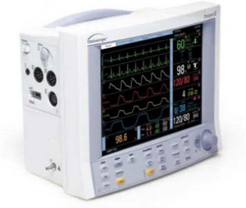
3.2.1 Patient Monitor . . . 24
3.2.1.1 Communication Protocol . . . 25
3.2.1.2 Patient Monitor Data . . . 27
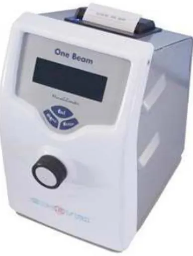
3.2.2 Bilirubinometer . . . 28
3.2.2.1 Communication Protocol . . . 29
3.2.3 Transcutaneous Bilirubinometer . . . 31
3.2.3.1 Communication Protocol . . . 32
3.3 The end-user web interface . . . 34
3.3.1 Authentications, Authorizations and Privacy Policy . . 35
3.3.2 Applications, Tools and Services . . . 36
3.3.2.1 Clinical Tools . . . 37 3.3.2.1.1 Anagraphic Tool . . . 37 3.3.2.1.2 Growth Tool . . . 38 3.3.2.1.3 Nutrition Tool . . . 40 3.3.2.1.4 Bayley Tool . . . 43 4 Decision-Making Tools 47 4.1 Bilirubin Tool . . . 47 4.1.1 Related Works . . . 49 4.1.2 SIN Guidelines . . . 51
4.1.3 Scenario . . . 55
4.1.4 Data Analysis: Bilirubin Decision-Making Algorithm . . 57
4.2 Activities Monitoring Tools . . . 61
4.2.1 The physical architecture . . . 61
4.2.2 Respiration Tool . . . 62
4.2.2.1 Related Works . . . 63
4.2.2.2 Data Analysis: Respiratory Rate Algorithm . . 64
4.2.3 Movement Tool . . . 65
4.2.3.1 Related Works . . . 68
4.2.3.2 Data Analysis . . . 69
4.2.3.2.1 Acquisition and Movements Detection Algorithm . . . 70
4.2.3.2.2 Data processing . . . 71
KPIs Extraction . . . 73
Markov Chain Model . . . 74
4.2.3.2.3 Extremities Tracking Algorithm . . . 75
4.2.4 Infant Semantic Segmentation . . . 77
4.2.4.1 Related Works . . . 78
4.2.4.2 Convolutional Neural Networks . . . 78
4.2.4.2.1 U-Nets . . . 78
4.2.4.2.2 SegNet . . . 80
4.2.4.2.3 ResNet . . . 81
4.2.4.2.4 FractalNet . . . 81
4.2.4.3 Adopted Metrics . . . 82
5 Results and discussion 85 5.1 SINC cloud-based healthcare architecture evaluation . . . 85
5.1.1 The web interface usability test . . . 86
5.2 Bilirubin Tool . . . 87
5.2.1 Data Analysis Results . . . 89
5.2.2 Discussion . . . 91
5.3 Respiration Tool . . . 93
5.3.1 Data Analysis Results . . . 94
5.3.2 Discussion . . . 96
5.4 Movement Tool . . . 96
5.4.1 Data Analysis Results . . . 100
5.4.1.1 MIA (Motion Infant Analysis) dataset . . . 100
5.4.1.2 KPIs and MC Transition Matrix . . . 101
5.4.2 Discussion . . . 107
5.5 Semantic Segmentation . . . 108
Contents
5.5.2 Data Analysis Results . . . 112
5.5.2.1 Quantitative Evaluation . . . 112
5.5.2.2 Qualitative Evaluation . . . 115
5.5.3 Discussion . . . 115
6 Conclusions and Future Works 119 6.1 Discussion . . . 119
6.2 Thesis contributions . . . 121
6.3 Future Works . . . 123
Appendices 125
List of Figures
3.1 The current scenario. . . 19
3.2 The cloud-based healthcare infrastructure. . . 20
3.3 The patient monitor: Datascope Passport 2. . . 24
3.4 Time trend of the parameters extracted from the patient monitor. 27 3.5 The bilirubinometer: Ginevri One Beam. . . 28
3.6 The bilirubinometer driver interface developed in Python for Ginevri One Beam input. . . 31
3.7 The transcutaneous bilirubinometer: Dr¨ager JM-105. . . 31
3.8 SINC authentication. . . 35
3.9 SINC home. . . 36
3.10 SINC “Anagraphic Tool” - Patient’s registry. . . 39
3.11 SINC “Growth Tool” interface - Patient’s growth data. . . 39
3.12 SINC “Growth Tool” interface - Patient’s growth parameters. . 40
3.13 SINC “Growth Tool” interface - Patient’s growth graphs. . . . 41
3.14 SINC “Nutrition Tool” interface - Patient’s nutrition adminis-tration. . . 43
3.15 SINC “Bayley Tool” - Automated Bayley test interface. . . 44
4.1 TcB hour-specific percentile nomogram. . . 53
4.2 TSB hour-specific percentile nomogram. . . 53
4.3 Nomogram for PT. Total bilirubin is plotted against PA in hours. The groups of GA are represented with lines of different colours. 54 4.4 Nomogram for BET. Total bilirubin is plotted against PA in hours. The groups of GA are represented with lines of different colours. . . 55
4.5 Nomogram for BET in case of haemolytic disease. Total biliru-bin is plotted against PA in hours. . . 55
4.6 Bilirubin Decision-Making workflow diagram. . . 60
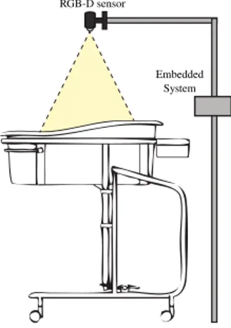
4.7 Representation of the configuration scheme of the system. . . . 61
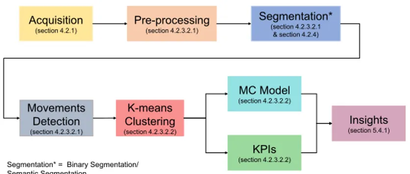
4.8 Followed pattern for preterm infants’ movements analysis. . . . 70
4.9 In figure 4.9a a depth frame from RGB-D sensor is shown. Fig-ure 4.9b is the result of clustering process; it highlights 4 clusters that correspond to the 4 infant’s limbs. . . 72
4.10 Different movements states in which infant can be in. Red rounds indicate the “in movement” limbs. . . 75 4.11 Longest path approach steps. . . 76 4.12 Illustration of the U-Net architecture. Image taken from [1]. . . 79 4.13 Illustration of the cascade of vanilla U-Net and shape
regular-ization networks. Image taken from [2]. . . 79 4.14 Illustration of implemented modified U-Net architecture. . . 80 4.15 Illustration of the SegNet architecture. Image taken from [3]. . 80 4.16 Residual Unit. Image taken from [4]. . . 81 4.17 Illustration of the FractalNet architecture. Image taken from [5]. 82 5.1 SINC “Bilirubin Tool” interface. . . 88 5.2 SINC “Bilirubin Tool” interface - Risk Factors. . . 89 5.3 Screenshot of interface developed with the Qt Framework while
it is running on Cubieboard2. . . 94 5.4 Example of respiration signal measured from RGB-D camera
(blu line) and of the filtered signal (red line) on a time window of 10s. . . 95 5.5 Scatter plot and regression line of RR mean values. . . 95 5.6 “Movement Tool” graphic interface. . . 97 5.7 “Colour-map” of the movements extracted from the software. . 97 5.8 “Movement Tool” graphic interface with velocity information. . 98 5.9 “Movement Tool” interface screenshots that represent a sequence
of fast movement 5.9a, slow movement 5.9b and the absence of movement 5.9c during a real-time acquisition. . . 99 5.10 “Movement Tool” graphic interface with extremities tracking. . 100 5.11 States activation sequence during time. . . 101 5.12 Transition matrix P of infant’s movements. . . 104 5.13 Temporal evolution of P (i|i) probabilities related to each state. 104 5.14 Transition matrix P of infant’s movements during different
pe-riods of the day. . . 106 5.15 Example of a PIDS Dataset positive instance. It consists of 16
bit original depth image (5.15a), 8 bit scaled depth image (5.15b) and the corresponding ground truth (5.15c). . . 110 5.16 Example of a PIDS Dataset negative instance. It consists of 3
images where the infant is not present in the frame: a 16 bit original depth image (5.16a), a 8 bit scaled depth image (5.16b) and the corresponding ground truth (5.16c), a completely black mask. . . 110
List of Figures 5.17 Example of a PIDS Dataset partial instance. It consists of 3
images where the infant is partially visible in the frame: a 16 bit original depth image (5.17a), a 8 bit scaled depth image (5.17b) and the corresponding ground truth (5.17c). Figure 5.17d is the corresponding RGB image used to facilitate ground truth annotation. . . 110 5.18 Example of a PIDS Dataset decentralised infant’s instance. . . 111 5.19 Example of a PIDS Dataset vertically positioned infant’s instance.111 5.20 Example of a PIDS Dataset instance acquired when lights are
very low. . . 112 5.21 Jaccard index trends over the 200 epochs for each CNN
List of Tables
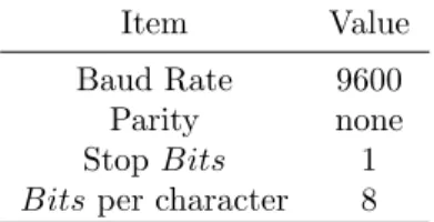
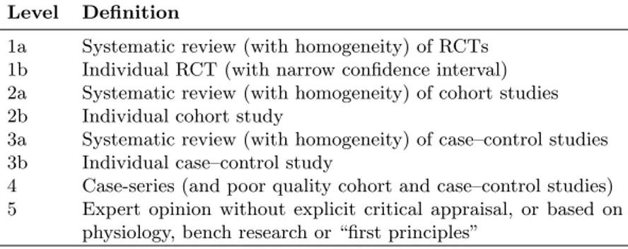
3.1 Bedside-to-VISA protocol technical specifications. . . 26 3.2 Bedside-to-VISA protocol string. . . 26 3.3 Communication protocol of Ginevri One Beam. . . 30 3.4 Communication string of Ginevri One Beam. . . 30 3.5 HL7 Protocol . . . 34 4.1 Evidence hierarchy from the centre for evidence-based-medicine. 52 4.2 Infants’ characteristics (Gestational Age and Weight). . . 64 5.1 Database Traffic. . . 85 5.2 Database Connections. . . 85 5.3 Query Statistics. . . 85 5.4 Mean RR values measured by RGB-D camera and patient monitor. 95 5.5 Data results and KPIs. . . 103 5.6 PIDS Dataset infants characteristics (Gestational Age and Weight).109 5.7 PIDS Dataset acquisition sessions and relative instances. . . 112 5.8 Jaccard and Dice indices of different CNN architectures. . . 113 5.9 Semantic segmentation results of different ConvNet architectures. 114 5.10 Jaccard Index results. . . 117 5.11 Qualitative result of prediction. . . 118 1 U-Net Indexes results. . . 128 2 U-Net2 Indexes results. . . 129 3 U-Net3 Indexes results. . . 130 4 ResNet Indexes results. . . 131 5 SegNet Indexes results. . . 132 6 FractalNet Indexes results. . . 133
Chapter 1
Introduction
1.1 Context
Technologic advances in neonatal intensive care and their impact on short term and long-term outcomes have been the major focus of neonatal clinical research. Neonatal mortality reached an unsurpassable minimum by the late 1990s. How-ever, this has been associated with high rates of neonatal morbidity and neu-rodevelopmental impairment.
Preterm birth is defined as all births before 37 completed weeks of gestation or fewer than 259 days since the first day of a woman’s last menstrual period [6]. Preterm births can be further sub-divided based on gestational age (GA): extremely preterm (<28 weeks), very preterm (28 - <32 weeks) and moderate preterm (32 - <37 completed weeks of gestation). Moderate preterm births may be further split to focus on late preterm birth (34 - <37 completed weeks).
Every year there are more than 15 million worldwide preterm births, that is more than 1 in 10 babies, and the number of cases continues to rise.
Prematurity is the leading cause of newborn deaths (babies in the first 4 weeks of life) and now the second leading cause of death after pneumonia in children under five years of age. It was responsible for nearly 1 million deaths in 2015. Three-quarters of them could be saved with current, cost-effective inter-ventions. Many of the surviving infants face a lifetime of significant disability, including learning and motor disabilities.
Preterm birth affects the anatomical and functional development of all or-gans, inversely with GA, as well as the acquisition of skills for survival in an extra-uterine environment.
In the case of preterm infants and low weight infants, the risk of mortality and morbidity increases; typical complications linked to prematurity occur in infants of very low GA (between 23 and 28 weeks) and weighting less than 1 kg.
Across 184 countries, the rate of preterm birth ranges from 5% to 18% of babies born; 7-9% of pregnancies do not reach the normal 40-week gestation period; 1% of newborn babies come to light before the 32nd week of gestation
and 0.5% even before the 28thweek.
In Italy, preterm births represent the 6.9% of the total birth rate; among the 40000 premature births born every year, 5600 have a body weight less than 1500 g. The care of these young patients, who would not be able to survive independently, require highly specialized staff and cutting-edge technologies. In the last five years, the development of biomedical technologies for intensive neonatal care has allowed the survival of patients born before the 23rd week
of gestation and of a weight at birth less than 400 g. To date, the survival of preterm infants has even reached more than 90%.
The sooner babies are born, the less prepared their bodies are for the outside world. They need special care to overcome the following challenges:
- breathing: many preterm babies start breathing on their own when they are born, but others need to be resuscitated. If the lungs are not fully developed and lack surfactant (a substance that helps keep the lungs expanded), preterm babies may have difficulty breathing. Sometimes, premature babies that start off breathing are not strong enough to con-tinue on their own. They exhaust themselves and may stop breathing (apnoea).
- brain: Preterm babies are at risk of bleeding in the brain, during birth and in the first few days after birth; about 1 in 5 babies weighting less than 2kg have this problem. Preterm babies can also have brain injuries from a lack of oxygen. Bleeding or lack of oxygen to the brain can result in cerebral palsy, developmental delays, motor impairments and learning difficulties.
- neonatal jaundice: it is caused by an accumulation of bilirubin (redish-yel-low pigment present in the bile and produced by haemoglobin catabolism) in the subcutaneous tissue and in the sclere; when bilirubin exceeds the limit values, the newborn is placed under a UV light lamp that promotes the elimination of bilirubin from the blood to the intestine.
- cardiovascular difficulty: there is often a delayed adaptation of the circu-latory system to post-natal life.
- feeding: Preterm babies can have trouble feeding because the coordi-nated suck and swallow reflex is not yet fully developed. They may need additional support for feeding. They often experience gastrointestinal difficulties and reduced food tolerances, so that additional support for feeding is needed: total parenteral nutrition (i.e., venous feeding) or gav-age feeding (milk administration by means of an orogastric tube, which goes directly from the mouth to the stomach) is often used.
- infections: Severe infections are more common among preterm babies. Their immune systems are not yet fully developed, and they have a higher risk of dying if they get an infection.
1.1 Context - staying warm: Preterm babies lose body heat more easily, putting them at risk of life-threatening hypothermia. They need extra energy and care to stay warm and grow.
- eyes: Preterm babies’ eyes are not ready for the outside world. They can be damaged by abnormal growth of blood vessels in the retina. The condition is usually more severe in very premature babies and if they are given a too-high level of oxygen. This can result in visual impairment or blindness.
As said, preterm babies are at risk of developing disabilities that will affect them for their entire lives, strongly depending on how early they were born, the quality of care they received during and around birth and the days and weeks that follow.
Hospitals with neonatal intensive care units can provide specialized care for preterm infants with serious health problems. They have special equipment and specially trained doctors and nurses who provide around-the-clock care for preterm babies who need extra support to keep warm, to breathe and to be fed, or who are very sick.
Preterm infants are housed in incubators or heated cribs, which provide temperature control and recreate the maternal uterine environment. Sometimes a “nest” with sheets is made inside the incubator to increase the sense of containment enjoyed by the baby in his mother’s womb.
Continuous monitoring of the vital functions of the small patient is required, which will then be connected to multiple probes and devices that generate alarms whenever the limit levels set by the staff of the neonatal intensive care ward are exceeded.
The hospital ward taken into consideration in this thesis is the Neonatal Intensive Care Unit (NICU) of the Women’s and Children’s Hospital “G.Salesi” in Ancona.
Here, infants’ cribs are surrounded by many devices for the monitoring, di-agnosis and treatment of the above described diseases and all these devices provide a huge amount of data that was only displayed on monitors. A mem-ber of the medical staff, usually the nurse on duty, periodically transcribed these data in a paper medical record. Then the manual notes were regularly, but not immediately, transcribed by nurses in an electronic sheet on the PC of the neonatal unit with the risk of errors and forgetfulnesses.
In this context, physicians have expressed the need to automatically gather data from all these devices in order to ensure that no important details for patient care were overlooked, as they are aware that the automation of this process can facilitate and improve the implementation of the procedures of their daily clinical practice. In this sense they have also contributed to the development of innovative systems, methods and algorithms for the study of
some important healthcare problems related to preterm infants.
1.2 Objectives and main contributions
In the light of the above, the first objective of this thesis was to allow the inter-facing and integration of biomedical instrumentation in the neonatal intensive care ward into a single cloud infrastructure that allowed the digital storage of all data from the various instruments.
In order to carry out optimal care processes, in fact, a maximum correlation and integration between all the physiological data collected by the instrumen-tation connected to the infant was required. Often the critical clinical situation is caused by a combination of pathologies, therefore the interactions between several physiological parameters must be studied.
In particular, the developed architecture was tested with the integration of three different type of instruments located in the NICU of the “Women’s and Children’s Hospital G.Salesi”:
- Patient monitor; - Bilirubinometer;
- Transcutaneous bilirubinometer.
Medical communication protocols are used to enable the communication and data transfer between the above mentioned infant’s monitoring devices and the unique database of the platform. For this purpose, Python drivers for input devices are being developed in order to communicate with the devices and acquire data.
Then, a process of data collection from different instrumentation was carried out by standardising the data and by forwarding them to the cloud, through import Web Services. A system built with these characteristics guarantees the interoperability property of the collected data allowing on-line sharing of the data.
At this point, another objective achieved by this thesis was to provide the medical staff with a valid tool to visualize this enormous amount of data. Data visualization plays a fundamental role in the interpretation of data. The visu-alization does not simply have an aesthetic purpose of presenting the results, but gives access to information and interpretations that otherwise would re-main latent. For this reason, a web interface has been designed and developed based on specifications given by healthcare personnel and medical guidelines. It acts like a web-portal and works as an application on Mobile Devices and Desktop Workstations, where medical staff members can access all available information.
Moreover, it is equipped with the main characteristics of the patient clinical record, but it exceeds the functionalities of a simple Electronic Medical Record
1.2 Objectives and main contributions (EMR), thanks to the development of innovative clinical tools, which contain functionalities, data analysis techniques and decision-making algorithms, useful to speed up and facilitate the work of doctors. These tools allow them to follow guidelines of daily clinical practice more closely, by automating steps and medical procedures, with the aim of improving patient care.
The processing of the data displayed on these clinical tools is, in fact, the result of decision-making systems, whose development and implementation is a further objective of this thesis in order to support the early diagnosis of diseases.
In particular, an algorithm has been developed to support the diagnosis of neonatal jaundice and the management of therapies to teat the disease. It consists in an evidence-based automated decision support system (DSS) for guideline based care to facilitate the implementation of clinical practice guide-lines for neonatal jaundice.
Another proposed algorithm supports the physician in fluid administration within nutrition task, another allows the automation of a clinical test used in follow-ups to assess the development of preterm infants.
Worthy of note is the implementation of an algorithm deployed to analyse preterm infants’ respiratory rate, which is able to activate an alarm signal when respiratory rate values go out of the physiological range, and of a method that, through computer vision techniques and machine learning algorithms, is able to detect infant’s movements in real-time from depth stream and to extract from the sequence of depth images, collected by an RGB-D sensor, some important features and indicators that can be used by clinicians to objectively study infants’ movements during their development.
The validation of these algorithms was made possible only thanks to the creation of a new device, another objective achieved in this work. It is a novel non-contact and non-invasive RGB-D system that consists in an RGB-D camera and in an embedded board, where the algorithms used to extract and analyse data from the depth images acquired by the camera are run in real-time.
All the above described solutions and algorithms have been validated through a series of experiments, conducted in the NICU of “G.Salesi” Hospital under the careful supervision of the Head Physician, Prof. V. P. Carnielli, which have also led to the creation of two datasets.
The work of this thesis is culminated in the funding of a regional project, named SINC, made in collaboration with the “Women’s and Children’s Hos-pital G. Salesi” of Ancona, as the coordinator of all the regional Neonatal Intensive Care Units, and where the Head Physician, Prof. V. P. Carnielli, a neonatologist of international fame, is the scientific director and validator of technological solutions, the Departments of Information Engineering and of In-dustrial Engineering and Mathematical Sciences of the Polytechnic University
of Marche and four local companies. SINC “System Improvement for Neonatal Care” project started in January 2017 and will last three years.
The objective of the SINC project is to develop an innovative integrated system that allows an enrichment of the instrumentation available for neona-tology and that will be able to automatically communicate the data to a cloud in order to guarantee the distribution of information and protocols of care on the territory, creating a new regional organizational model, on the basis of that developed in this thesis for the NICU of “G. Salesi” Hospital.
One of the project’s objectives is to manage a cloud infrastructure that makes services and utilities available throughout the region and that is able to inte-grate monitoring data collected by medical devices to facilitate patient diag-nosis and treatment. The aim is to create and experiment a new hospital and territorial model for the integrated management of neonatal care and thera-pies between the various levels of hospital specialization in the Marche Region, which will be supported by distributed data management systems.
1.3 Structure of the thesis
The thesis is organized in six Chapters, which describe the design and devel-opment of a healthcare infrastructure that has allowed the interconnection of several biomedical devices in a NICU and the communication of data coming from these instruments to a single DB, which is exploited in order to permit the automatization of the process of device data collection, transmission, storage, processing and availability for medical staff.
Chapter 2 reviews the state-of-the-art about the main topics addressed. It discusses the results achieved in the creation of distributed healthcare infras-tructures for the integration of medical devices in hospital wards and the prob-lems still outstanding in this regard, and then goes into more detail on the current situation regarding the development and use of Electronic Medical and Health Records, Evidence-Based Medicine and Decision Support Systems in general and particularly in Italy and in the Marche region.
Chapter 3 describes the proposed cloud-based infrastructure, that allows the communication between the three different type of medical devices of the NICU of the “Women’s and Children’s Hospital G.Salesi” through communication protocols that are thoroughly described, and the developed web-based platform, where data coming from these instruments can be displayed and analysed.
The functionalities of the implemented tools of the platform are briefly de-scribed in this Chapter, while in Chapter 4 the attention is focused on three of these tools, which have seen the application of decision-making algorithms and computer vision techniques applied with the aim of support physicians in everyday clinical practice.
1.3 Structure of the thesis Chapter 5, then, discusses the results related to the analysis of the data collected by the various instruments and stored in the various platform tools, which are processed using the algorithms described in the previous chapter through a series of experiments conducted within the NICU.
Finally, conclusions are reported in Chapter 6, as well as some future research directions.
Chapter 2
State of the art
Medical devices generally are monolithic units so that a typical hospital room in an intensive care ward hosts a number of stand-alone devices, each one with its own user interface, its own proprietary software, its own display and often its own built-in computer. Graphical user interfaces are all different and non-uniform and this can create confusion and misunderstandings among the healthcare personnel who has to use all the devices simultaneously.
Moreover, the devices are physically separated blocks and they can also be located on different bed sides so that the medical staff has to go from one device to another to use it, e.g., to see data on device display, to change device settings and to set up new vital parameters or new alarm values. In this way, the staff has to deal with different interfaces implemented on devices located in different places, which contributes to create mental confusion and poor organization and makes it more difficult for the physicians to rapidly take stock of the situation on the patient’s state of health. Furthermore, in a hospital ward there are many figures with different roles, e.g., the head physician, specialists in various fields and nurses, and each of them needs a tailored user interface suited to his own expertise and that shows him device data and information in the most suitable way to make him perform his job well and quickly.
Finally, around patient’s bed there are devices for the monitoring, the di-agnosis and the treatment of different diseases and it would be appropriate to bring together data coming from all these devices to ensure that the physician could have a general idea about the patient’s health conditions in order to not neglect any important detail for the patient’s care. On the contrary, data are usually saved separately for each device and they are not always sent to the pa-tient Electronic Medical Record (EMR), if it exists, so that it becomes difficult to search and find them, to create correlations between them and to make them available in real time to the clinician when he is visiting the patient, whereas this would bring great benefits to medical research [7].
Nowadays many devices already have some connection mechanisms that use serial ports, Ethernet, IEEE 802.11 or Bluetooth wireless, but they are typi-cally used to unidirectionally log data and events from these devices towards
the computer, permitting the reading of the measured value on a PC display. However, this is no longer sufficient: as demand increases for better health-care paradigms, it is evident that there is the need of having integrated and cooperating (interoperable) medical devices. Thus, the goal is to go beyond the simple connectivity to move towards a more complex device integration that offers the possibility to stream device data directly into electronic med-ical records (EMRs), to create a database for further statistic investigations of data so collected and to integrate the information derived from multiple medical devices into a single customised display.
In this thesis, a cloud computing solution is proposed for the creation of a cloud infrastructure that exceeds the simple EMR. A cloud-based DBaaS (Database-as-a-Service) is created with the abilities of storing and coopera-tively sharing medical data based on the HL7 message required for interoper-ability of medical information of different types. A database is created serving as a data consumer which allows the integration between device data, EMR data, personal patient’s data, patient’s medical history and so on. This per-mits to aggregate device data in one place, making easier the recorded data maintaining. This facilitates data searching in order to allow the doctor to make faster diagnosis, by having all relevant information available. Moreover, the identification of statistical trends and patterns is simplified by using data mining and machine learning techniques that can lead to the discovery of cor-relations between collected data and illnesses, which otherwise would not have been possible to observe by clinicians [8] and that could be an useful tool to support a long-period management of a university hospital [9].
2.1 Health Information System Infrastructures
Health Information Systems (HISs) can be described as the interaction be-tween individuals, processes and technologies used to support fundamental in-formation operations, management and availability, with the aim to improve healthcare services [10].
They provide information and enhance the knowledge creation in health care environments through data processing [11]. In particular, they are considered mechanisms for storing, processing, analysing and transmitting information required for planning, organization, execution and evaluation of health services. The above depicted scenario presents the opportunity to create an integrated cloud-based network that automates the process that begins with data collec-tion from multiple devices and ends with informacollec-tion accessibility from medical staff. To achieve this goal diverse cut-edge technologies were studied.
In the past, the healthcare information systems have been extensively im-plemented with traditional Management Information System (MIS) mode, but
2.1 Health Information System Infrastructures this does not comply with the current requirements of healthcare system. Lee et al. in [12] propose a service-oriented architecture (SOA)-based system that permits the integration of data from personal health devices and the creation of tailored users interfaces, but this work does not provide concurrency or con-stant and reliable service capabilities. Kulkarni and Ozturk in [13] implement a system called mPHASiS that leads to an end-to-end solution. Nonetheless, it is based on the Java programming language that makes the system less efficient for reasons of performance of Java Virtual Machine (JVM).
The proposed solution is based on the idea of cloud computing with which it is possible to provide customers with pervasive, affordable and on-demand services. Cloud computing owns the characteristics of parallel computing, dis-tributed computing, and grid computing, but it improves the user experiences over the Internet compared to these. Lots of applications are moved towards a cloud platform because it is able to supply services for diverse application scenarios [14, 15]. Nowadays, also lots of works on medical information systems are based on cloud services [16, 17]. Cloud infrastructure can provide the flexi-bility and scalaflexi-bility of service-oriented systems unlike the transaction-oriented MIS mode.
The proposed model provides support to ad hoc integration of diverse med-ical devices, distributed processing, and open accessibility. A cloud-based in-frastructure is presented where medical devices are plugged in and begin to cooperate, i.e. to collect and transmit data. The computer resources achiev-able with this environment are set up to receive, store, process, and distribute the information. The method described in this thesis permits the creation of an ad hoc integration network of different medical devices and of a distributed data processing.
The main requirement of the optimal solution is that it has to quickly imple-ment the methods to collect process and distribute infant’s vital data, from the crib to remote accessibility, by integrating all different medical devices [18, 19]. Moreover, it has to be flexible and extensible, i.e., it has to hold up diverse medical devices in different numbers that can be added to the system in different times [20]. Then, the proposed system has to guarantee the security and the safety of shared data, by ensuring strong access control and data encryption [21, 22].
To ensure data interoperability [23] it is necessary to realise data extrac-tion process from various instruments, normalising the database according to a record format defined in the XML language and specifically to the logic re-lated to language HL7 [24]. A database made with these characteristics enables online sharing of encrypted healthcare data, reported in a standard language, allowing a complete interoperability of shared data.
operational conditions, by ensuring its reliability [25] and it has to be scalable and compatible with the use in large healthcare environments and with the aggregation of different institutions [26].
In King et al. [27] they discuss about all these main problems and about their possible solutions with respect to the creation of a distributed architec-ture for the integration of medical devices in a hospital ward. They discuss about interoperability of data collected from devices, displays and databases of different vendors and about standardisation of device data streams and their reliability, security and safety, also in terms of privacy. However, the work is not concentrated on concrete solutions, leaving room to other works for putting into practise the proposed ideas.
2.2 Electronic Medical Records and Electronic
Health Records
All health information systems, as well as many other technologies, can be interfaced with a systematic collection of information: the electronic medical record (EMR).
The medical record is the tool used to manage patient’s clinical data, which are collected during meetings with healthcare professionals for prevention pur-poses, or when episodes of disease occur.
EMR is therefore the electronic evolution of the classic paper-based medical record, which has lost its effectiveness because the continuous improvement of the treatment process has led the paper version to become increasingly volumi-nous and easily damageable, with a wide variety of sources from which to obtain documents and information, and has made it more difficult to consult and find the necessary information for healthcare professionals in a timely manner [28]. Moreover, handwritten medical records may be lowly legible, and this can contribute to medical errors. Some pre-printed forms and the standardization of abbreviations were stimulated to improve reliability of paper-based medical records, but without remarkable results.
Thus, EMR may help with the standardization of digitized forms, terminol-ogy and data input in order to facilitate the collection of data for epidemiolterminol-ogy and clinical studies [29].
The EMR can enable information to be used and shared over secure net-works through digital formatting; track care (e.g. prescriptions) and outcomes (e.g. blood pressure); trigger warnings and reminders, send and receive orders, reports, and results; decrease billing processing time and create more accurate billing system; enable information to move electronically between organiza-tions [30].
2.2 Electronic Medical Records and Electronic Health Records As regards interoperability, EMRs would facilitate the coordination of health-care transfer in non-affiliated healthhealth-care structures [31]. A lot of information is conveyed in the EMR which can include laboratory results, radiology results, pharmacy orders, medical prescription, discharge/transfer orders and any other data from auxiliary services. In this way, some EMR-based systems are capable of automatically monitoring clinical events, by analysing patient data to pre-dict, detect and potentially prevent adverse events. Furthermore, data from an EMR can be used for statistics on issues such as quality improvement, physi-cians’ self-audit and resource management.
The EMR, actually, is the patient record created by providers for specific encounters in hospitals and ambulatory environments and it is limited to a single healthcare structure. It can serve as a data source for the electronic health record (EHR). The terms EHR and EMR, in fact, have often been used interchangeably, although differences between the models are now being defined [32, 33].
The EHR is a more comprehensive collection of the electronic health in-formation of individual patients or populations. It is a more comprehensive form involving integration and network access of data from different clinical applications and heterogeneous systems.
According to the definition of the International Organization for Standard-ization (ISO) the EHR is a repository of patient data in digital form, stored and exchanged securely, and accessible by multiple authorized users [34]. It con-tains retrospective, concurrent, and prospective information and its primary purpose is to support continuing, efficient and quality integrated health care.
EHR should contain, for each patient, all information resulting from their in-teraction with different healthcare professionals, hospital wards and structures in order to facilitate an integrated vision of patient data [35]. It grows over time: it self-feeds, preserves and organises the complete collection of personal health history.
Main requirements of an EHR include the definition of a flexible data struc-ture to allocate information in each patient’s profile, of a clinical information registry concerning subsequent consultations and prescriptions, and of a repos-itory of all medical and clinical history individualized and complete [36].
A system that manages the EHR must be able to meet three main demands: - the integration of data from different sources (e.g. hospitals, private
clinics, health care professionals); - the correct acquisition of data; - support for clinical decisions.
These interlinked characteristics within the EHR permit to achieve the goal pursued by the system: patient care. In order to achieve this goal, however, the EHR must also have interoperability characteristics, since processed data come
from different sources and are produced autonomously [37]. It must ensure the integration between different types of information (e.g. clinical, financial, administrative data) and not be limited in geographical terms, because it must link the various access points of users (general practitioner, physicians, health-care professionals, and patients) and the different healthhealth-care structures [38]. It should also ensure the integration of data provided by the various EMRs to obtain what IHE (Integrating the Healthcare Enterprise) calls a “longitudinal” view of the health status of individual patients.
In Italy, EMRs are quite widespread, but only within individual hospital wards and they are used more administratively than clinically.
According to a survey [39, 40] conducted in 2016 by the Healthcare Infor-mation and Management Systems Society (HIMSS), that has created the EMR Adoption Model (EMRAM), an eight-stage model that permits to assess the quality of the adopted EMRs on a scale from 0 to 7, in Italy the 6thlevel EMR
covers only the 2,2% out of a total of 135 EMRs evaluated by HIMMS, while there are not 7th level EMR, compared to the 8,6% and 2,9%, respectively, in
the Netherlands (total = 35) and the 32,7% and 6,1% in the USA (total = 5478). The majority of the italian healthcare structures are placed on stage 5 (34,8%) and 2 (34,1%) and the 6,7% still occupy stage 0 compared to 0% in the Netherlands and to 1,6% in USA.
There are many reasons for this situation. In fact, even if the EMR spread requires infrastructure investments, which are still lacking, the painful point is the lack of confidence, if not resistance, that healthcare workers show to-wards the EMR. On the one hand, a cultural and “self-protection” resistance to the transparency of the data that the EMR sets in motion, and on the other hand - even more significant - a technological approach that does not make the EMR confidant with the doctor, especially if it is in situations of urgency and speed. Doctors often denounce the adoption of complicated and redun-dant technological procedures, heterogeneous levels of integration, overlapping programs, excessive access and safety filters, which make “complex” the use of the EMRs [41, 42, 43, 44].
In any case, even Italy is increasingly moving towards the digitalization of the medical record, especially after the enactment of the Article 12 of Act 221/2012 that officially introduced the EHR, called “Fascicolo Sanitario Elettronico”, defined as “the set of data and digital documents relating to health and socio-medical information produced by current and past clinical events concerning the patient” [45]. Under Article 12.2, the EHR can be used for a triple purpose:
a prevention, diagnosis, treatment and rehabilitation; b study and scientific research in the medical context;
c health planning, verification of the quality of care and evaluation of healthcare.
2.2 Electronic Medical Records and Electronic Health Records Moreover, the n. 237 of October 2017 of the Official Gazette contains the publication of Circular n. 4, “Documento di progetto dell’Infrastruttura Nazionale per l’Interoperabilit`a dei Fascicoli Sanitari Elettronici (art.12 - comma 15-ter – D.L. 179/2012))”, available in the transparency section of the website of the Italian Agency for the Digital Agenda (AgID) [46], where EHR charac-teristics, its objectives and structure are illustrated in the light of the latest legislative changes affecting it.
The results of Government and regions efforts to promote the EHR can be seen in the following percentages: in 2015, the 40% of reports and the 9% of medical records were dematerialised; according to Federfarma, the 72% of med-ical prescriptions were dematerialized, up sharply from 26% in December 2014; the 16% of the reports were delivered online to the citizen, while bookings and payments made via the web were 12% and 8% respectively. The share of those who have heard about EHR (32%) doubled compared with the previous year, even if only 5% have actually used it, considering that at the moment, accord-ing to AgID data of April 2016, only six italian regions have an EHR already active and operational (Emilia-Romagna, Lombardy, Tuscany, Sardinia, and Valle d’Aosta), while in 11 others, including Marche Region, regional EHRs are being implemented.
As mentioned above, the Marche Region is also making progress in dissemi-nating the guidelines concerning the regional EHR infrastructure, with the aim of illustrating all the integration services envisaged in this area, in order to facilitate future suppliers of systems that will interact with the regional EHR in the implementation of integration interfaces [47].
On the basis of this document and those mentioned above, a web-based solution was therefore designed and developed, which allows the integration of data from different devices with those from the medical record. This solution was achieved by considering and preparing the communication with the regional EHR.
The final and future objective is, in fact, to go towards the creation of a network that includes all neonatologies of the Marche region, favouring the sharing of health data and the coordination of their flows, in order to facili-tate transfers between the different hospital structures, which may take place after a first analysis of the patient’s vital signals has already been made by the reception centre, and, vice versa, to allow the continuity of monitoring of the newborn once from the central hospital (“G. Salesi”) is transferred to a neonatology that is closer to his parents’ residence to ends there the last days of hospitalization.
The database created is also available for a “transversal” interoperability with all the colleagues who have need of continuity of care with respect to the same patient, and “vertical” interoperability with other databases and
Na-tional Health Service (NHS) operators. All services are backed by a guarantee for the highest security of data, encrypted both during transfer and storage, and access, in compliance with current legislations in terms of security and pri-vacy, is strictly permitted only to those who have the appropriate permissions. Users can remotely access patient data, using both the PC and mobile devices (smartphones and tablets).
2.3 Evidence-Based Medicine and Decision Support
Evidence-based medicine (EBM) aims to improve quality of care reducing the wide healthcare practice variations, and the inefficiencies, dangers, and in-equalities that have resulted from nonoptimal patient care as pointed out in “Crossing the quality chasm” by the Institute of Medicine [48].
EBM originated in 1992, from a series of studies started more than 10 years earlier at the Department of Clinical Epidemiology and Biostatistics of the Canadian McMaster University for the best use of scientific literature for med-ical updating.
From these roots, the EBM has developed the concept that “evidence” must play a leading role in therapeutic decisions, meaning with the term “evidence” up-to-date and methodologically valid information from medical literature. Originally defined as “a new emerging paradigm for medical practice”, the EBM received, four years later, a more cautious definition: EBM is “the conscien-tious, judicious and explicit use of current best evidence in making decisions about the care of individual patients”, then recognizing the need to integrate “evidence” with individual clinical skills of the healthcare professional (“exper-tise”) [49].
EBM has developed in two areas of application: public health macro-de-cisions or related to homogeneous population groups and individual patient medical practice.
EBM use has been approved by international healthcare organizations such as the WHO and the Institute of Medicine. but it has resulted to be far from easy to implement. About thirty years after its birth, it has so far found little application, especially in Italian clinical information systems. While there are several free or pay websites where clinical evidence and guidelines can be found and used, there has been no software integration between these sources and patient clinical data.
One of the tools used for the practice of EBM is Clinical Decision Sup-port defined as “providing clinicians or patients with computer-generated clini-cal knowledge and patient-related information, intelligently filtered or presented at appropriate times, to enhance patient care” [50], where clinical knowledge means, for example, the evidence contained in up-to-date guideline
recommen-2.3 Evidence-Based Medicine and Decision Support dations.
Clinical Decision Support Systems (CDSSs) are interactive software, very important within the medical industry, designed to assist doctors and other healthcare professionals (e.g. nurses) in the formulation of diagnosis, treat-ment decisions and managetreat-ment of therapies, through the collection and the elaboration of information coming from several sources.
In fact, knowledge-based CDSSs apply rules to data to be analysed by models or other methods via an inferential motor and provide results to the end user.
There are many different clinical applications in which CDSSs are used. They can be used to warn changes in patients’ condition from patient monitor data analysis [51], to help the evaluation of the clinical guidelines used to assist physicians in finding alternative treatment and diagnosis [52], or to identify similar factors between different cases that lead to complications [53].
A CDSS is used in a diabetic clinic to determine if a diabetics treatment would be safe given patient history, laboratory test results and known allergies and pathologies of a diabetic patient [54].
Generally, when a patient’s case is complex or the healthcare professional who has to make the diagnosis is inexperienced, a CDSS can support to decide which is the therapeutic treatment to be suggested based on up-to-date treatment guidelines.
CDSSs are also used extensively in Neonatal Intensive Care Units (NICUs), given the very critical conditions in which small patients live and the many ethical problems associated with them.
In [55] a prototype of integrated eXtended Markup Language (XML)-based CDSS is implemented in a NICU, with the aim to evaluate the effectiveness of real-time decision support and of the longterm impacts on physician decision-making. In [56] it is used to define, detect and generate clinical alerts from collected medical device data.
In [57] a decision-making support system based on specific outcome informa-tion for individual NICU patients is used to provide support for parents and healthcare professionals in ethically determining and deciding which treatments to perform.
In [58] a web services-based infrastructure to support CDSSs for processing multi-domain medical data from obstetrics, perinatal and neonatal hospital wards is proposed. It is used to reduce medical errors, to assist physicians in decision-making process, to improve patient care also by the optimization of alert detection systems.
In Italy, CDSSs are still not very widespread, while in the rest of Europe and North America they represent a growing phenomenon.
The report “Clinical Decision Support Systems Market by Component (Ser-vices, Software), Delivery Mode, Product Type (Integrated, Standalone), Model
(Knowledge-Based), Type (Therapeutic, Diagnostic), User Interactivity (Ac-tive, Passive), Application - Forecasts to 2021”, made by MarketsandMar-kets [59], studies the global clinical decision support systems market over the forecast period of 2016 to 2021. This global market is expected to reach USD 1.519,2 Million by 2021 from USD 856,3 Million in 2016, at a CAGR of 12.2%. The cause of this lack of diffusion in italian healthcare system is due to physicians’ aversion and to a great distrust of technology and CDSSs use as underlined in [60], but also to the lack of a passage in the current legislation (which is instead foreseen in many European and extra-European health sys-tems, starting from Belgium, the Netherlands and the United Kingdom) or which “the practice of the health professional” is explicitly encouraged to use a scientifically based CDSS (guidelines and good practices).
Despite this, having ascertained the excellent results deriving from the use of CDSSs in clinical practice, they are beginning to spread also in Italy in various fields of clinical application [61, 62, 63].
In fact, the results of the study conducted in [60] suggest that the CDSSs adoption path is marked by a progressive evolution of clinicians’ perceptions towards the system. A trend is emerged which moves from positions that are adverse to the adoption of CDSSs - characterized by strong resistances and a very little “perceived mastery” - to an hoped optimal situation, in which CDSSs are seen as a working tool at the service of clinicians, integrable (and not interchangeable) with their skills, adaptable to their needs and to the local organizational specificities.
In this thesis, decision-making models and algorithms are applied in the NICU environment to preterm infants’ data in order to generate alarms from the analysis of medical devices data, to help physicians in the administration of nutrients and in the diagnosis and treatment of neonatal jaundice, and to support the prediction of neuromotor disabilities from depth images as shown in next chapters.
Chapter 3
A new infrastructure for neonatal
care
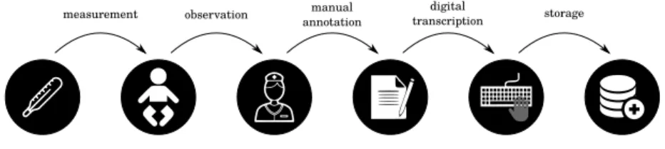
measurement observation manual annotation
digital
transcription storage
Figure 3.1: The current scenario.
The hospital ward that is taken into consideration for testing the new in-frastructure is the Neonatal Intensive Care Unit (NICU) of the “Women’s and Children’s Hospital G.Salesi” in Ancona, where preterm infants are taken into care [64]. As said, preterm birth affects the anatomical and functional devel-opment of all organs, inversely with GA. The degree of organs immaturity is a critical aspect for these babies, which leads them to have many health prob-lems. For this reason, the primary physiological functions of these babies have to be continuously monitored and often infants have to undergo major treat-ments and therapies. Hence, infants’ cribs are surrounded by lots of devices for the monitoring, the diagnosis and the treatment of different diseases. It is desirable that all these devices communicate between each other and with a unique database in order to create an integrated device network.
Nowadays, on the contrary, the majority of these devices only provide the ability to read data on their digital displays. Consequently, a member of the medical staff, generally the nurse in charge, has to collect infant’s data at bedside by writing them down to a paper medical chart on a regular basis. Then manual notes are regularly, but not immediately, transcribed by nurses in a PC of the Neonatal Unit. After that, data are transmitted to a local server that stores and organizes them together with the patient data collected in the EMR. Only at this moment, the medical staff can access data through an interface application. Focusing on the problem of infants’ vital data collection,
3.1 SINC cloud-based healthcare architecture face, and with software to collect, encode, encrypt and transmit data through configurable communication channels to be stored, forming the local interface. Data are picked up through applications of extraction, specifically concerted for every different type of devices, which use medical communication protocols or proprietary software to enable the direct data communication and transfer. After extracting and standardising into XML, through Web Service of im-portation, data are carried to the cloud database, where they are aggregated.
For security reasons, both during the import process and the storage phase in the aggregate DB, data are encrypted and accessed, using “strong” creden-tials as well as other medical authorities (or other health professions, as far as relevant information about an individual is concerned) authorized by the physician.
The intrinsic interoperability, obtained by using different device types and vendors and language independent XML technologies and the widespread HTTP as a transport, implies that devices can communicate each other and with the EHR by using Web Services [65].
Web Services, in fact, are web application components that permit the in-teroperability between different technologies that work in a same network. These services hide their internal complexities such as data types and busi-ness logic from their users, but expose their programming interfaces and their locations using UDDI (Universal Description, Discovery, and Integration) pro-tocols. They make available a software interface, described in a format which can be automatically processed, which permits the interaction between other systems and the Web Service itself by activating the functions available on the interface (services or requests of remote procedures) through special ”mes-sages” of request. Such request messages are included in an “envelope” that is formatted according to the standard XML, encapsulated and transported via Web protocol. In our case, the XML-based language for describing Web services application software interface is Web Services Description Language (WSDL), which is recommended by the World Wide Web Consortium (W3C). These interfaces, publicly exposed, guarantee the procedures of import data and access to the database by different authorized medical staff members for respective allowed functions. The XML-based protocol for accessing Web Ser-vices is Simple Object Access Protocol (SOAP), which is always recommended by W3C and the Web protocol on which SOAP operates is HTTP, the most commonly used and the only one that was standardised by the W3C.
Web services act like a broker between local medical devices and remote services. They are able to capture collected data from devices and to send them to suitable storage service hosted on cloud. They also allow the local storage for pre-processing, e.g., they allow data to be aggregated or simply analysed before the transfer to the main system.
On the other hand, Web Services work as an access point. Users through a web interface that acts like an application that works on Mobile Devices and Desktop Workstations, with which the hospital ward is equipped, can interact with data on the cloud database. Web Services receive requests from that interface to recover data from the cloud database.
The function of the Utility Computing Provider is to provide logical and physical infrastructure for data storage, processing and delivery services.
Its main components are the Platform front end interface, the Cloud Storage and the DBaaS (Database-as-a-Service). The platform front end interface is able to allow the management of the storage contents and to directly communi-cate with users. The interface can be a web client or a standalone application. The cloud storage is the model of data storage in which the digital data is stored in logical pools and it is based on highly virtualized infrastructure. Database-as-a-service [66] is one of cloud computing secondary service models and a key component of XaaS. This may be considered a subspecialty of the bigger Soft-ware-as-a-service model. In essence, DBaaS is a cloud database, which is a database that typically runs on a cloud computing platform. It is a cloud com-puting service model with which there is no need to physically launch a virtual machine instance for the database so that in such a configuration, application users do not have to install and maintain the database themselves. All of the administrative tasks, installation and maintenance are taken care of by the database service provider and application owners pay according to their usage.
3.1.1 Data Platform
The relational database for the storage and management of data is configured according to a structure optimized in terms of performance and zero data re-dundancy.
As DBaaS the Amazon Relational Database (Amazon RDS) for MySQL is used, since it makes it easy to set up, operate, and scale MySQL deployments in the cloud. The use of this DBaaS provides to the system all the features that we have discussed in section 2.1, which are the main requirements to obtain an efficient cloud-based healthcare infrastructure for medical device integration. It makes it easy to go from project conception to deployment. It is possible to access the capabilities of a production-ready relational database in minutes and without the need for infrastructure provisioning, and for installing and maintaining database software. It is possible to scale database compute and storage resources often with no downtime: the engine will automatically grow the size of database volume as database storage needs grow. It enhances relia-bility for critical production databases, including automated backups, database snapshots and automatic host replacement.
3.1 SINC cloud-based healthcare architecture For security reasons, it allows data encryption both during the transit, with the use of SSL, and during the cloud storage phase. Also backups, read replicas and snapshots are encrypted. Moreover, data are accessible only by authorized medical staff members by using strong authorization. There is also the possi-bility to run database instances in a virtual private network.
3.1.2 Cyber Security
The legislation about the protection of personal data (Legislative Decree no. 30 June 2003 n. 196) provides (art. 31) a general safety requirement under which the personal data object of treatment should be kept and controlled, also in relation to acquired knowledge with the technical progress, the nature of the data and the specific characteristics of the treatment, so as to minimize, through the adoption of suitable and preventive security measures, the risk of destruction or loss, even accidental, of the data, unauthorized access or treatment not allowed or inconsistent with the purposes for collection.
The obligation to adopt the “minimum security measures” (arts. 33 et seq.) is also provided. In particular, (pursuant to art. 34) the treatment of personal data carried out with electronics instruments is allowed only if the following measures are adopted:
- computer authentication;
- adoption of procedures for the management of authentication credentials; - use of a system of authorization;
- periodic update of the identification of treatment allowed to individual officers and employees for management or maintenance of electronic in-struments;
- protection of electronic tools and data against illegal treatment of data, unauthorized access and certain computer programs;
- adoption of procedures for storing backups, restoring access data and systems;
- adoption of encryption techniques or identification codes for certain treat-ments of data disclosing health status or sexual life performed by health organizations.
Under these conditions, in Nu.Sa. data are subjected to protection with a “strong” encryption system that guarantees high degree of security; the as-sumption is that only the rightful owner - the licensed doctor - has access to the data of his competence through an authentication system (SSO Single Sign On) described below. The cryptography algorithm is held by a third party, the Polytechnic University of Marche.