Università degli Studi di Catania
Facoltà di Medicina e Chirurgia
Dottorato di ricerca in Neurobiologia
Sede Amministrativa: Università di Catania Sede Consociata: Università di Roma “La Sapienza”
“Neuroanatomical structural correlates of
non-motor symptoms in patients with movement
disorders”
Dott.ssa Francesca Imperiale
Tutor: Prof. Giuseppe Meco
Indice
CHAPTER I... 5
1. 1 Parkinson’s disease... 5
1.2 Epidemiology of PD ... 5
1.3 Criteria for diagnosis ... 6
1.4 Motor symptoms of PD ... 8
1.5 Non Motor symptoms of PD ... 11
1.6 Risk factors for PD... 12
1.7 Neurobiological mechanisms of PD ... 15
CHAPTER II... 20
2.1 ICD- Definitions and phenomenology... 20
2.2.1 Pathological Gambling... 21
2.2.2 Hypersexuality... 22
2.2.3 Compulsive buying ... 22
2.2.4 Compulsive eating ... 23
2.3 Epidemiology of ICD ... 23
2.4 Pathophysiology of Addiction Processes ... 25
2.5 Psychiatric disorders in ICD patients ... 30
2.5.1 Depression... 30
2.5.2 Apathy... 35
2.5.3Anhedonia………..32
CHARPTER III ... 33
1. Introduction ... 39
2.1 Patients and Methods ... 42
2.2 Neurological, psychopathological and
neuropsychological evaluation ………44
2.3 Image acquisition and processing... 46
3 Statistical analisis ……….42
3.1 Neurological, neuropsychological and
psychopathological data……….42
3.2 Neuroimaging data ………...…….42
4. Results... 50
5. Discussion... 57
REFERENCES……….62
ABBREVIATION………..83
CHAPTER I
Involuntary tremulous motion, with lessened muscular power, in parts not in action and even when supported; with a propensity to bend the trunk forward and to pass from a walking to a running pace: the senses and intellects being uninjured. (Parkinson 2002)
1. 1 Parkinson’s disease
In 1817 James Parkinson, an English medical doctor, published his essay reporting six cases of paralysis agitans and, for the first time, described the disease as “involuntary tremulous motion, with lessened muscular power, in parts not in action and even when supported; with a propensity to bend the trunk forwards, and to pass from a walking to a running pace: the senses and intellect being uninjured“ (Parkinson 2002). In his publication, entitled “An Essay on the Shaking Palsy”, James Parkinson described the characteristic resting tremor, unusual posture and gait, paralysis and diminished muscle strength, and how the disease progresses over time (Parkinson 2002). Further neurologists, Trousseau, Gowers, Erb, and most notably Jean-Martin Charcot, between 1868 and 1881 were a landmark in the understanding of this disease (Lees 2007). In particular, Charcot has explained the difference between rigidity, weakness and bradykinesia and has also championed the renaming of the disease in honor of James Parkinson (Lees 2007). Over nearly two centuries, the Parkinson ’s disease (PD) description was been limited to selected clinical
settings and several population-based epidemiologic studies were published (Gudmundsson 1967).
Nowadays, Parkinson’s disease (PD) is defined as neurodegenerative disorder due to degeneration of neurons in the pars compacta of substantia nigra (SNc) without an identifiable cause.
1.2 Epidemiology of PD
PD is the second most common neurological disorder affecting disability after stroke. The aetiology of PD is unknown; therefore, reliable data on its incidence are particularly interesting.
The disease is found in all ethnic groups but with differences in prevalence (Alves, Forsaa et al. 2008). Prevalence and incidence of PD in European countries are estimated at approximately 108 to 257/100,000 and 11 to 19/100,000 per year, respectively, but they varied from country to country (von Campenhausen, Bornschein et al. 2005). The prevalence in Asia countries is slightly lower, all-age prevalence varied from 51.3 to 176.9/100,000 persons and the incidence from 6.7 to 8.7 per 100 000 persons per year (Muangpaisan, Hori et al. 2009). However, some data suggests that, at least in parts of the USA and Japan, the incidence has not changed significantly over the past few decades (Twelves, Perkins et al. 2003). The PD is generally considered a disease of late middle age with the average age of onset at around 60 years. There are cases of "early-onset" PD with patients developing symptoms prior to the age of 40(Van Den Eeden,
Tanner et al. 2003). The best epidemiological datasets, currently available, consistently suggest that men have a greater risk for developing PD than women. The rate for men (19.0 per 100,000, 95% CI: 16.1, 21.8) was 91% higher than that for women (9.9 per 100,000, 95% CI: 7.6, 12.2) (Alves, Forsaa et al. 2008).
This increased risk in men may reflect biological differences between men and women, such as the effects of sex hormones or X-chromosome-linked susceptibility genes (Wooten, Currie et al. 2004).
1.3 Criteria for diagnosis
PD is characterized by progressive loss of muscle control, which leads to trembling at rest of the limbs and head, stiffness, slowness, and impaired balance. As symptoms worsen, it may become difficult to walk, talk, and complete simple tasks. According to Hughes et al.’s criteria (Hughes, Daniel et al. 2001), the diagnosis of PD require the presence of bradykinesia (slowness of initiation of voluntary movement with progressive reduction in speed and amplitude of repetitive actions) and at least one of the following signs: muscular rigidity, 4-6 Hz resting tremor, postural instability not caused by primary visual, vestibular, cerebellar, or proprioceptive dysfunction.
A broad spectrum of non-motor symptoms, associated to motor signs, complicates the clinical picture of disease, encompassing neuropsychiatric, sleep, autonomic, and sensory disorders (Chaudhuri, Healy et al. 2006). While some problems such as
dementia and psychosis typically appear late in the disease course, non-motor symptoms are common across all stages and may be related to treatment by dopaminergic drugs, cognitive and psychiatric disorders.
1.4 Motor symptoms of PD
The main motor symptoms of PD are: bradikinesia, rigidity, tremor at rest and in a more advanced stage postural instability and loss of balance. These symptoms appear asymmetrically (one side of the body is more involved than the other) and at onset often go unnoticed because their appearance is subtle and inconstant, therefore the disease progresses slowly.
Bradykinesia is one of the classic symptoms of PD, it means “slow movement.” Over time, a person with PD may develop a reduction of spontaneous movement, which can give the appearance of abnormal stillness and a decrease in facial expressivity. People who experience bradykinesia may walk with short and shuffling steps and after a number of years, they may have experience of akinesia, or "freezing," and not be able to move at all. Rigidity may sometimes be the first sign of PD. Many patients are unaware of it whilst a ill-defined feeling of awkwardness. Rigidity is an involuntary increase in muscle tone causing stiffness and inflexibility of the limbs, neck and trunk. A consequence of parkinsonian rigidity is the head and trunk slightly bent forward in a typical stance defined
“camptocormia” or bent posture, or even a postural attitude called the Pisa Syndrome in which the trunk leans to one side.
The resting tremor is the best-known and most recognisable sign of PD even though isn’t present in all patients. In the early stages of the disease, about 70% of people experience a slight tremor in the hand or foot on one side of the body, or less commonly in the jaw or face (Hughes, Daniel S.E. et al. 1992). A typical onset is tremor in one finger. From a semiotic point of view, parkinsonian shaking is a rhythmic oscillating movement of the fingers with a frequency between 4-7 cycles/sec, typically reproducing the gesture of “counting pills or money”. The tremor usually appears when a person's muscles are relaxed, or at rest, hence the term "resting tremor", and ceases when a person begins an action.
Although during the progression of the disease the tremor will become bilateral, there will always be a certain asymmetry. In the later stages of the illness tremor may be extended to other parts of the body, such as the jaw, lips, tongue and the roots of the upper and lower limbs. The parkinsonian tremor is particularly noticeable under stress and disappear when sleeping.
One of the most impairing signs of PD is postural instability, a tendency to be unstable when standing upright. A person with postural instability has lost some of the reflexes needed for maintaining an upright posture, and it may topple backwards if jostled even slightly (retropulsion). Postural instability occurring in the later stage of illness, usually after the appearance of non-motor symptoms. It is mainly due
to a dysfunction in the reflectors of straightening postural, combined with various other factors including akinesia and rigidity. This postural instability causes motor impairment, difficulty in walking, inability to live independently at home and it is also the main cause of frequent falls with negative consequences, such as soft tissue lesions, hip fractures and the fear of falling. As the illness progresses, both postural instability, which does not respond to a specific pharmacological therapy, than the fear of worsening making PD patients even more sedentary.
The motor symptoms of PD become progressively worse as the disease advances. In recent years, many studies have investigated the progression of the hallmark symptoms over time, and the cardinal motor symptoms have different rates of progression: the disease usually progressing faster in patients with rigidity and bradykinesia than in those with predominant tremor. The current treatment regime of dopamine-replacement therapy improves motor symptoms and alleviates disability. Increasing the dosage of dopaminergic medication is commonly used to combat the worsening symptoms. However, the drug-induced involuntary body movements and motor complications can significantly contribute to overall disability (Xia and Mao 2012). Further, none of the currently-available therapies can slow or halt the disease progression. Significant research efforts have been directed towards the developing of neuroprotective or disease-modifying agents that are intended to slow the progression.
1.5 Non Motor symptoms of PD
Although the diagnosis of PD is based on motor symptoms, it is now well known that non-motor symptoms are an integral part of this pathology, involving multiple systems (Bonnet and Czernecki 2013). Non-motor symptoms include autonomic dysfunction, cognitive deficits, neurobehavioral disorders, and sensory and sleep abnormalities. Autonomic failure include orthostatic hypotension, sweating dysfunction, sphincter dysfunction and erectile dysfunction.
Contrary to the original James Parkinson assertion, that intellectual abilities are “uninjured” in the PD (Parkinson 2002), neuropsychological investigations have demonstrated that cognitive impairments constitute an important part of the clinical presentation of disease. If standardized neuropsychological tests are utilized, patients with PD, will show impaired performance across a wide range of cognitive functions confronting to healthy controls. However, the pattern of cognitive impairments is heterogeneous, some patients develop widespread deficits while in others cognition remains relatively intact throughout the course of their disease (Schrag and Schott 2006). Like other clinical features of PD, cognitive deficits typically worsen with increasing disease duration. The cognitive functioning may be impaired from mild deficits in specific domains to generalized cognitive dysfunction sufficiently severe and extensive to warrant a diagnosis of dementia. Neuropsychological investigations of patients with PD showed specific
impairments even in the early stages of the disease, which include deficit of behavioural regulation in sorting or planning tasks, defective use of memory stores, and impaired manipulation of internal representation of visuospatial stimuli (Dubois and Pillon 1997; Rodriguez-Constenla, Cabo-Lopez et al. 2010).
Recent found that the prevalence of cognitive impairment in PD can reach 93% if adequate neuropsychological instruments are performed (Campos-Sousa, Campos-Sousa et al. 2010). Gasca-Salas et al. (2014), in a recent study reported that, in long-lasting PD, 21.7% of cognitively normal PD patients progressed to mild cognitive impairment (MCI) and 42.3% of PD with MCI progressed to dementia over a 31 months observation period (Gasca-Salas, Estanga et al. 2014).
About neurobehavioral disorders, depression is the most common disorder of PD and it is recognized to be associated whit impaired performance on a number of cognitive tests, especially attention, executive functions, and memory (Marvel and Paradiso 2004; Schock, Schwenzer et al. 2011). Depression may occur at any stage of the disease, and consists in major depressive disorder, minor depressive disorder, and dysthymia. Further, during the course of the disease, patients might have experience of anxiety, anhedonia or apathetic symptoms (Assogna, Cravello et al. 2011).
Until now the etiology and pathogenesis of PD remain still unclear and the strongest confirmed risk factor is advancing age (Driver, Logroscino et al. 2009). Age is the most important risk factor for development of disease: PD increases steadily with age and although a significant difference in prevalence by sex was found, it is only in older population (Pringsheim, Jette et al. 2014). A possible reason for gender differences may be that males have greater exposure to other risk factors such as toxin exposure or head trauma. Moreover, it has been theorized that estrogen may have neuro-protective effects or that the predisposing gene to PD, may be linked to the X chromosome (Ragonese, D'Amelio et al. 2004; Popat, Van Den Eeden et al. 2005; Lubomski, Louise Rushworth et al. 2014). Increasing evidence suggests that PD represents a common clinical feature with heterogenic causes: both heredity and the environment have a role in the etiology of PD. In several epidemiological studies, relatives to patients with PD were found to have a 3 to 4-fold increased risk for developing PD as compared to the general population and in approximately 10-15% of the cases a positive familial history is assumed; however, the majority of cases are sporadic PD (Autere, Moilanen et al. 2000; Kurz, Alves et al. 2003).
In 1997, Polymeropoulos identified a mutation in the alpha-synuclein gene, which was responsible for PD in a large Italian kindred (Polymeropoulos, Lavedan et al. 1997). Since that time, investigators have identified many genes associated with PD. Four genetic loci responsible for the autosomaldominant
PD: 1) PARK1on chromosome 4q21–23 (the gene of alpha-synuclein itself) and PARK2 on chromosome 6q25.2–27; 2) the gene of parkin, (ubiquitin ligase, L3 responsible for juvenile parkinsonism); 3) PARK3 on chromosome 2p13 and PARK4 on chromosome 4p14–16.3 (the genes and the substrates are unknown); 4) PARK5 on chromosome 4p14 (the gene of ubiquitin carboxy-terminal hydrolase L1, the enzyme that participates in the ubiquitin proteasome degradation pathway of abnormal neuronal proteins) (Hardy, Cookson et al. 2003; Healy, Abou-Sleiman et al. 2004).
A variety of non-genetic risk factors has been proposed because these inherited forms explain a small proportion of disease. Longitudinal studies in twin populations suggesting that environmental causes are important for the most people (Vieregge, Schiffke et al. 1992; Wirdefeldt, Gatz et al. 2011). It assumed that the interaction of environmental exposure and genetic variation (gene–environment interaction) should cause the most cases of PD (Singleton, Farrer et al. 2013).
Numerous other studies have identified head trauma, rural living, farming, gardening, pesticides and drinking well water as risk factors for PD (Lai, Marion et al. 2002; Rugbjerg, Ritz et al. 2008; Noyce, Bestwick et al. 2012; Pezzoli and Cereda 2013). Also iron and aluminum seem to play an important role in the pathogenesis of the disease, in fact this type of ions are found in large quantities in the neurons of substantia nigra in PD. These ions cause toxic effects, and mediate formation of free-oxygen species, which trigger oxidative stress that causes
neuronal death (Kornienko Valery N. and Pronin 2008).
Most consistently, an inverse relationships between intake caffeine and cigarette smoking in PD development, have been suggested in several studies (Ross, Abbott et al. 2000; Ascherio, Zhang et al. 2001; Quik 2004).
A meta-analysis based on eight case-control studies and five cohort studies, showed a significantly decreased PD risk for coffee drinkers (pooled relative risk 0·69) that was not attenuated when analyses were adjusted for smoking (Hernan, Takkouche et al. 2002).
Moreover, the cigarette smoking seems to have a neuroprotective effect in PD. In a large meta-analysis based on 44 case-control studies and four cohort studies from 20 countries, a pooled relative risk of PD of 0·59 was calculated for the ever smokers, and a relative risk of 0·39 for the current smokers (Hernan, Takkouche et al. 2002). Several mechanisms have been proposed to explain this effect: the nicotine may stimulate dopamine release, or acting as an antioxidant, or alter activity of monoamine oxidase B (Quik 2004).
1.7 Neurobiological mechanisms of PD
PD is mainly characterized by a progressive degeneration of midbrain dopaminergic (DA) neurons along a caudorostral and lateromedial gradient, with a marked loss of neurons in the substantia nigra pars compacta (SNc), projecting to the dorsal striatum
along the nigrostriatal pathway, and a more modest loss in the ventral tegmental area (VTA), projecting to limbic and cortical areas along the mesolimbic and mesocortical pathways, respectively.
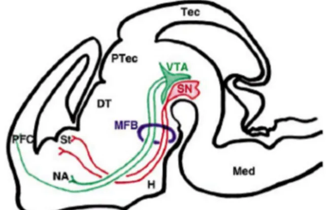
The dopaminergic system in brain comprises 3 distinct pathways, including the nigrostriatal, mesolimbic and mesocortical projections (FIGURE 1).
The nigrostriatal pathway runs from the SNc to the basal ganglia terminating specifically in the caudate putamen (CP). This pathway accounts for 75% of the DA neurons in the brain. The Striatum and the SNc are both situated in a part of the brain called the Basal Ganglia. The basal ganglia along with the cerebellum, is the key area assigned to motor coordination, movement and initiation of movement and therefore, responsible of initiates and maintains motor responses. The death of DA neurons in this pathway leads to PD. The mesolimbic pathway, beginning in the VTA in the midbrain, projects to parts of the limbic system including the nucleus accumbens, the amygdale, the septal area and the hippocampus. The nucleus accumbens is the site of rewarding effect. The mesocortical pathway is also located in the VTA, although this pathway terminates on the prefrontal cortex. Neurons of mesocortical pathway have an excitatory effect on the frontal cortex, where short term memories, planning, strategy and problem solving abilities take place. In fact, impairment in these pathways can lead to thinking disorder and logical thoughts deficits.
D2 receptors predominate in the nigrostriatal pathway, while in the mesolimbic and mesocortical pathway there are also D3 receptors. Furthermore, additional dopaminergic neurons are present in the hypothalamus. Therefore, the mainstay of symptomatic treatment for PD is dopamine replacement therapy (DRT). The criterion standard of symptomatic therapy is levodopa (L-dopa), the metabolic precursor of dopamine, in combination with carbidopa, a peripheral decarboxylase inhibitor. Motor, emotional and cognitive efficient presuppose an optimal level of dopamine, whereas dopamine levels increased or reduced lead motor and non-motor symptoms.
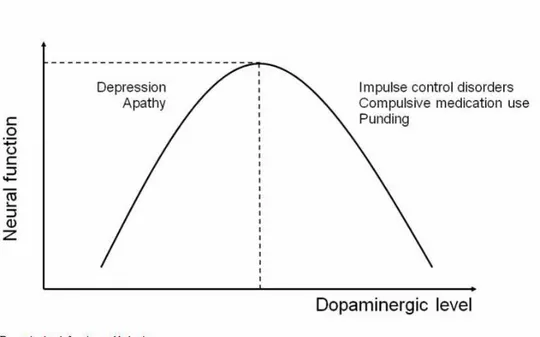
The motor and non-motor symptoms of PD can be classified as hypo-dopaminergic, directly related to the pathogenesis of degenerative disease, or hyper-dopaminergic, related to dopamine drug therapy. The dopaminergic activity can, therefore, be represented by a function as an inverted "U" (Figure 2).
Figure 1: Diagram of the dopaminergic projection in the embryonic mouse brain. VTA, Ventral Tegmental Area; SN, Substantia Nigra; MFB, Medial Forbrain Bundle; St, Striatum (Caudate Putamen); NA, Nucleus Accumbins; PFC, prefrontal cortex (Lin and Rosenthal 2003)
Figure 2: Dopamine level, function and behavior (Voon, Mehta et al. 2011)
The symptoms due to hypo-dopaminergic level includes the motor cardinal signs of the PD (akinesia, tremor, rigidity and flexed posture) and non motor symptoms as lethargy, apathy, anxiety, depression, and cognitive deficits (Menza 2000; Ishihara and Brayne 2006). More recently, decreased novelty seeking and increased tendency to harm avoidance have been associated to dopaminergic deficiency (Tomer and Aharon-Peretz 2004; Bodi, Keri et al. 2009).
The motor sign of the hyper-dopaminergic spectrum is characterized by dyskinesias while the non motor symptoms are recognized as disinhibitory psychopathologies. The disinhibitory psychopathologies include: disorders of Impulse Control (ICD), the pounding and the dopamine dysregulation syndrome (DDS). The DSM-IV-TR fourth edition of the American Psychiatric Association’s Diagnostic and Statistical Manual
(DSM-IV-TR) (APA 2000), defines an impulse control disorder as “a failure to resist an impulse, drive, or temptation to perform an act that is harmful to the person and others”; while the pounding is characterized by complex behaviours, stereotyped, repetitive and often aimless, how to handle continuous objects, sort, clean or accumulate things in an excessive way; and the dopamine dysregulation syndrome is characterized by addiction-like self-medication of high doses of levodopa and short-acting dopamine agonists .
DDS and pounding phenomena are more frequent during advanced stages of PD and are often accompanied by cognitive impairment or dementia, therefore are thought to fall outside of the ICD spectrum (Santangelo, Morgante et al. 2009).
It is documented that hyper-dopaminergic motor and non-motor symptoms often coexist and that is the effect of the sensitization to DRT. In fact, in a recent study, Solla et al. (2011), found a higher frequency of behavioral, neuropsychiatric and cognitive disorders in patients with motor complications, including dyskinesias and motor fluctuations. Moreover, a higher frequency of ICD was detected in patients with dyskinesias (22.2% - p < 0.001) and motor complications (12.2% - p < 0.05) (Solla, Cannas et al. 2011).
CHAPTER II
2.1 ICD- Definitions and phenomenology
According to the DSM-IV-TR, impulse control disorders (ICD) is a group of disorders characterized by a “failure to resist an impulse, drive, or temptation to perform an act that is harmful to the person or to others” (APA 2000). ICD described in PD include pathological gambling, hypersexuality, compulsive buying and compulsive eating. Occasional reports also mention pounding, dopamine dysregulation syndrome (DDS), kleptomania, impulsive aggressive disorder, trichotillomania, reckless driving and impulsive smoking. ICD aren’t a part of an obsessive-compulsive disorder (OCD), in which repetitive unwanted thoughts (obsessions) compel a person to perform ritualistic behaviors (compulsions) to reduce anxiety; in fact, impulsive behaviors are often motivated by pleasure, gratification or some other reward.
Although ICD are distinct from OCD, there are phenomenological overlaps, possibly indicating overlapping neurobiological mechanisms; in both disorders, the behaviors need to be “excessive” and result in “significant impairment” in major areas of life functioning to satisfy diagnostic criteria.
2.2.1 Pathological Gambling
Pathological gambling (PG) is defined according to DSM-IV TR criteria as a persistent and recurrent maladaptive gambling behavior as indicated by features such as: preoccupation about gambling, increasing amounts of money, unsuccessful attempts to control, restlessness or irritability when cutting down, lying to others about gambling, jeopardising relationships, work or education, and relying on others for money. For PG diagnosis, according to DSM-IV criteria (APA 2000) are required more or 5 criteria above mentioned.
PG is the most frequent ICD and therefore is the ICD more investigated. PG prevalence (either current or anytime during PD) in PD has been reported to vary from 2.2% to 7%; such percentages are higher than those reported in general population (Santangelo, Barone et al. 2013). This wide range of variability in prevalence percentages might depend on different diagnostic criteria or assessment methods, several setting of evaluation and various inclusion and exclusion criteria in experimental designs. However, the prevalence rates are higher in Caucasian samples than those reported in Asian countries; this divergence might depend on cultural and ethnic differences or genetic differences (Santangelo, Barone et al. 2013). Further studies are necessary to clarify this issue.
2.2.2 Hypersexuality
Hypersexuality is defined as being preoccupied with sexual feelings and thoughts. Sexual impulses become more intense, spontaneous and compulsive and can cause distress to both the patient and partner. Moreover, studies reported increased use of pornography, seeking out prostitutes, promiscuity engaging in exhibitionism and paraphilias. Validated criteria and validated scales for hypersexuality are lacking and the prevalence ranges from 2 to 10% (Weintraub, Koestler et al. 2010).
2.2.3 Compulsive buying
This is a preoccupation with repetitive impulsive and excessive buying or shopping whit experience of an irresistible urge to purchase more than patients need or can afford and may lead to financial stress. Patients with compulsive buying often describe an increasing urge or anxiety that can have a sense of completion only when a purchase is made. The prevalence estimates range from 3% to 6% of ICD and the younger people are more prone to develop it. Often, the maladaptive spending behavior is associated with serious psychological, social, occupational, and financial problems (Djamshidian, Averbeck et al. 2011).
2.2.4 Compulsive eating
The DSM-IV-TR (APA 2000) defines binge eating disorder as eating an amount of food that is definitely larger than most people would eat during the same period of time under similar circumstances coupled with a perceived lack of control over one’s eating. Binge eating has been defined as compulsive eating that occurs over a short period of time. Typically, patients binge eat in the evening or wake in the middle of the night to binge eat. Compulsive eating is usually associated with significant and undesired weight gain and the estimates prevalence is 4,3% (Weintraub, Koester et al. 2010).
2.3 Epidemiology of ICD
There is a great variability in the observed prevalence of ICD between different studies, however, the prevalence of ICD in PD patients treated with dopamine receptors agonists rages between 6.1 and 31.2% and is significantly higher than in the general population. The ICD is more frequent in male than female, especially hypersexuality, although some authors reported that compulsive eating and compulsive buying was considerably more common in women.
The occurrence of ICD in only a subset of patients, and not in all PD population, clearly suggests an underlying susceptibility that may be mediated by PD-related factors such as (a) the neurobiology of PD (e.g., pathology and compensatory mechanisms
that may also modulate underlying temperamental traits or cognitive processes), (b) PD-specific medication practices, and (c) individual factors underlying the vulnerability to ICD.
Young age or younger age at PD onset are risk factors to development ICD in PD patients. For instance, both PG and compulsive medication use are associated with younger PD onset (Voon, Thomsen et al. 2007), suggesting a potential role for selective vulnerability. However, this association may be confounded by the relatively greater use and higher doses of dopamine agonists used in young onset PD.
Familiar or personal history of substance abuse or bipolar disorder, personality traits variability labeled as “novelty seeking” and impulsiveness also appear to have a disadvantageous influences. More recently apathy, depressive mood and bipolar disorders have in addiction been reported as potential risk factors. Moreover, new reports found alexithymia an independent risk factor for ICD in PD patients. Alexithymia, a personality trait characterized by difficulties identifying and describing feelings and externally oriented thinking style, is associated with anxiety, depression, impulsivity and emotion suppression. (Poletti, Frosini et al. 2012; Goerlich-Dobre K.S., Probst C. et al. 2014).
Dopamine replacement therapy (DRT) of all type including levodopa, is a key precipitating factor in the emerge of the ICD in PD, and the use of dopamine agonists confers a two to three times greater risk of developing ICD (Weintraub, Koester et al. 2010). In a longitudinal study, Avilla et al. (Avila, Cardona et al.
2011), followed a series of 25 PD patients with ICD for a median of 12 months. They found that 15 of 25 patients (83%), who decreased their dopaminergic treatment, had a partial or full remission of ICD symptoms. The relevance of dopamine treatment for the manifestation of ICD is further supported by evidence that the ICD frequency in untreated PD patients, doesn’t differ from healthy control subjects (Antonini, Siri et al. 2011; Cilia and van Eimeren 2011).
The association between dopamine agonist therapy and ICD has been attributed to excessive or aberrant activation of the mesolimbic dopaminergic system, which under physiological conditions mediates the response to natural rewards. The expression of the D3 receptor is particularly rich within the limbic system and neuroanatomical, physiological, and behavioral data suggest the high D3 affinity of dopamine medications as the basis for these behavioral changes.
2.4 Pathophysiology of Addiction Processes
ICD are defined as “behavioral dependence”, characterized as mental imbalance status resulting from excessive motivational drive and impaired inhibition (Shaffer 1999; Holden 2001; Grant, Brewer et al. 2006; Potenza 2008). Thus, although exist differences in the phenomenology of ICD in PD, they are linked by their reward- or incentive - based and repetitive natures. The fifth edition of the DSM (APA 2000) underscores the ICD classification as “behavioral addiction”.
The main features of substance use disorders include: (a) enhanced salience attribution to drug-related stimuli in comparison to other natural rewards, (b) failure to inhibit the urge to obtain the drug, (c) withdrawal symptoms including cue-induced craving and (d) continued engagement in maladaptive risk- taking behaviors despite negative future consequences (Brewer and Potenza 2008). The neurobiological researches had investigated the circuitries of the development and maintenance of addictions, that include the ventral tegmental area; its ventral striatal, limbic, and prefrontal cortical dopaminergic projections; and the dorsal striatum and associated frontostriatal circuitry (Marechal, Denoiseux et al. 2014). Several of these common features are modulated by dopamine and are therefore of great interest, as they may constitute possible mediators of ICD in subjects chronically treated with dopaminergic drugs. There are several proposed theories explaining the higher risk in PD patients to develop ICD.
1. Downregulation of D2 receptors expression: PD patients exposed to long-term dopaminergic medication show a downregulation of D2 receptors expression (van Eimeren, Pellecchia et al. 2010) and as consequence, sensitivity for natural rewards, such as sex, food and money, decreased. ICD might be a compensatory effect in which the PD patients to obtain sufficient reward, needs higher than normal stimulation;
2. Tonic release of dopamine: phasic release of dopamine from the Ventral tegmental area (VTA) to the nucleus accumbens occurs at the time of exposure to
rewards, a process that is associated with anticipation of reward and receiving an unanticipated reward (i.e., reward prediction error) (Volkow, Wang et al. 2008). Conversely, phasic suppression occurs when a reward is expected but not received. The magnitude of dopamine release varies with the magnitude of the reward. In contrast to phasic release, tonic dopamine release occurs with anticipation of greatest reward uncertainty (i.e., when there is an equal probability or chance of either receiving or not receiving a reward). This observation has been interpreted to suggest that anticipation of conditions of high uncertainty -as, for example, gambling - may itself be rewarding (Fiorillo, Tobler et al. 2003). Repeated exposure to the reward–-drug or behavior–may result in over-activation of the reward and motivational circuits (including the medial orbitofrontal cortex, orbitofrontal cortex (OFC)) while decreasing the top–down control of inhibitory cortical areas (Potenza 2008). Pathologically increased saliency attribution to a particular reward produces an irresistible and increasingly compulsive drive toward that reward and away from other reinforcers, finally leading to withdrawal symptoms and craving, similar to substance use disorders and which has been hypothesized to be mediated by changes in dopamine and glutamate functioning (Kalivas and Volkow 2005). 3. The overdosing theory: in PD there is a dopamine
shortage in the nigrostriatal pathways, whereas the mesolimbic ad mesocortical pathways maintain relatively intact. SPECT and PET studies in PD patients report a resting overactivity in brain areas involved in reward and reward-based learning, motivation, impulse control, decision making and
memory processing (Cilia, Siri et al. 2008; Joutsa, Martikainen et al. 2012). Additionally, dopamine seems to have a binding preference to D3 receptors, rather than D2-receptors. The D3 receptor is expressed mainly in discrete brain areas belonging or related to the limbic system, especially in ventral striatum, whereas D1 and D2 receptors are widely expressed in all major dopaminoceptive areas.
4. Cognition: it has been propose that dopaminergic stimulation of dopamine- deficient dorsal striatal receptors is associated with cognitive enhancement on task requiring activation in this area, whereas dopaminergic activation of the relative intact ventral striatum is associated with impairment of cognitive functions in specific domains, e.g. the ability to modify behaviors by outcome (Cools, Barker et al. 2001). Some studies have reported higher cognitive performances in executive function in PG patients than PD patients without PG. Other observation didn’t confirm this results in which, less cognitive flexibility planning capability and poor feedback processes are found (Voon, Thomsen et al. 2007; Santangelo, Vitale et al. 2009).
5. Dopamine agonist withdrawal syndrome: it is defined by Raabinak as a drug –specific syndrome because of the observed lack of the symptomatic response when replaced by levodopa, antidepressant and anxiolytics. However, an improvement is observed with DA replacement. a severe cluster of physical or psychological symptoms that lead to significant distress or social/occupational dysfunction are observed (Rabinak and Nirenberg 2010). It is associated with higher greater cumulative DA
exposure, as can be observed in other drug withdrawal syndromes. The and long-term prognosis of Dopamine agonist withdrawal syndrome may result in severe cases in which DA can never be discontinued, leading to chronic ICD (Rabinak and Nirenberg 2010). High frequency of psychiatric symptoms (anxiety, panic attacks, depression, agitation, irritability, dysphoria, insomnia, fatigue, generalized pain) are been reported (Rabinak and Nirenberg 2010). The same authors, suggest that this patients belong to a “mesocorticolimbic variant” of PD, with disproportionate mesocorticolimbic (vs. relatively preserved nigrostriatal) dopaminergic dysfunction and therefore exhibit an increased vulnerability to Dopamine agonist withdrawal syndrome and ICD.
Although we focus on dopamine, other neurotransmitters (e.g., serotonin and opioids) may also be involved in the development of impulse control and addictive disorders. Serotoninergic neurons project form the raphe nucleus to mesocorticolimbic areas including prefrontal/orbitofrontal cortex, amygdala and hippocampus and are known to modulate delay discounting and reversal learning (Rogers 2011). Moreover, forebrain serotonin levels were found to be negatively correlated with measures of impulsivity (Brewer and Potenza 2008) and risk-taking behaviour (Moreno, Cardona et al. 2010). The opioid system is activated in response to gambling behavior (Shinohara, Mizushima et al. 2004) and is involved in the processing of hedonic responses and incentive motivation by modulating the nucleus accumbens and the ventral globus pallidum, either directly (Smith, Tindell et al.
2009) or indirectly, via gamma-aminobutyric acid input to the mesolimbic dopamine pathway (Grant, Brewer et al. 2006; Olmstead, Ouagazzal et al. 2009).
2.5 Psychiatric disorders in ICD patients
As explained previously, several studies have provided evidences for enhanced mesocorticolimbic dopamine neurotransmission associated with ICD in medicated PD patients. Alterations in mesolimbic dopamine function have also been linked to a number of psychiatric disorders (Di Giuda, Camardese et al. 2012). Thus, the negative sequelae of ICD and overlapping neurobiology with psychiatric disorders suggest that ICD are likely to be associated with significant psychiatric comorbidity.
Although psychiatric symptoms such as depression, anxiety, psychosis, anhedonia and apathy are relatively common in PD patients, studies investigating the comorbidity of psychiatric symptoms and ICD are scarce.
2.5.1 Depression
Depression affects approximately 35% of all PD patients during the progression of the disease (Aarsland, Pahlhagen et al. 2012). Core symptoms of depression in PD are similar to those observed in major depressive disorder: depressed mood and a loss of interest. Loss of interest, however, is also the cardinal symptom of apathy, a related disorder commonly defined as a reduction in goal directed
behavior (Marin 1991; Starkstein and Leentjens 2008) and may lead to considerable misdiagnosis (Aarsland, Pahlhagen et al. 2012). Other symptoms of depression, such as sleep disturbances, psychomotor retardation, fatigue and loss of expression, show strong clinical overlap with the motor and autonomic symptoms of PD (Aarsland, Pahlhagen et al. 2012), thereby hindering adequate recognition and management. Although the incidence of depression peaks around the time of PD diagnosis (Rickards 2005), there is considerable evidence that depression in PD is not simply an adjustment disorder but a highly prevalent symptom of the disease itself. First, epidemiological studies have shown that depressive symptoms are often evident before the onset of the typical motor symptoms (Shiba, Bower et al. 2000; Schuurman, van den Akker et al. 2002; Leentjens, Van den Akker et al. 2003) and a previous diagnosis of major depressive disorder is associated with an increased risk of subsequently developing PD (Schuurman, van den Akker et al. 2002; Leentjens, Van den Akker et al. 2003). Second, the incidence of depressive symptoms increases with progression of PD and the presumed spreading of the PD brain pathology (Schrag, Jahanshahi et al. 2001; Rickards 2005). Several studies have provided evidence for an association between ICD and depression (Pontone, Williams et al. 2006; Weintraub, Koester et al. 2010; Joutsa, Martikainen et al. 2012; Jaakkolaa, Kaasinena et al. 2014); in addition, symptoms of depression worsen in parallel with the development of novel ICD (Voon, Sohr et al. 2011; Joutsa,
Martikainen et al. 2012; Leroi, Andrews et al. 2012). A randomized double-blind placebo-controlled trial, showed that DRT through pramipexole exerts a direct antidepressant effect (Barone, Poewe et al. 2010) and PD patients report more depressive symptoms during “off” periods compared with “on” periods, which are unrelated to motor impairments (Maricle, Nutt et al. 1995; Storch, Schneider et al. 2013). Dopamine agonists also exert antidepressant effects in animal models of depression (Muscat, Papp et al. 1992; Breuer, Groenink et al. 2009). This suggests that depression can be conceptualized as a hypodopaminergic state, most notably of ventral striatal areas (Remy, Doder et al. 2005; Voon, Mehta et al. 2011). Low dopamine in the ventral striatum leads to a relative overactivity of the indirect pathway of the limbic cortico-striatal–thalamocortical circuit (Surmeier, Ding et al. 2007; Shen, Flajolet et al. 2008) and therefore a decreased stimulation of cortical areas involved in motivation and reward. A considerable number of studies have shown more severe dopaminergic deficits (i.e. neuronal cell loss and gliosis) in depressed PD patients compared to non-depressed PD patients (Spiegel, Hellwig et al. 2006; Frisina, Haroutunian et al. 2009). Furthermore, neurobiological studies have found that ventral striatal D3 receptor availability correlated negatively with severity of depressive symptoms in DRT-naïve early PD patients (Boileau, Guttman et al. 2009). Other reports found a reduced availability of the dopamine transporter (DaT) in presynaptic striatal dopamine neurons in depresssed PD patients (Remy, Doder et al. 2005; Hesse, Meyer et al. 2009;
Vriend, Raijmakers et al. 2013). The DaT eliminates dopamine from the synaptic cleft by reuptake and can serve as a marker for the integrity of the dopamine system (Scherfler, Schwarz et al. 2007). However, increased DaT availability in depressed PD patients (Felicio, Moriyama et al. 2010) and no differences between depressed and no depressed patients (Broussolle, Dentresangle et al. 1999) have also been reported. A DaT Single Photon Emission Computed Tomography (SPECT) study showed that severity of depressive symptoms in PD correlated negatively with DaT availability in the caudate nucleus, whereas the severity of motor symptoms showed a negative correlation with DaT availability in the putamen (Vriend, Raijmakers et al. 2013). This suggests that depressive symptoms are associated with degeneration of the dopaminergic projections to the caudate nucleus, that may partly originate from the VTA and is therefore, related to dysfunctional limbic and associative cortico-striatal–thalamocortical circuits. This result further underscores the contribution of the dopaminergic projection from the VTA to the pathophysiology of depression. Nonetheless, the pathophysiology of PD-related depression is highly complex and also involves deficits in the serotonergic and noradrenergic systems. In fact, it recognized the efficacy of serotonergic and noradrenergic antidepressants in PD-related depression (Menza, Dobkin et al. 2009; Richard, McDermott et al. 2012). Although neuroimaging studies on depression in PD are relatively scarce, data suggest differences in regional glucose metabolism between depressed and no depressed patients in the caudate nucleus and
inferior frontal prefrontal, orbitofrontal and anterior cingulate cortex (Mayberg, Starkstein et al. 1990; Ring, Bench et al. 1994; Mentis, McIntosh et al. 2002). Moreover, reduced activation in the medio-dorsal nucleus of the thalamus has been observed in PD patients in response to an emotional perception paradigm during functional (f)MRI scanning (Cardoso, Maia et al. 2009). The medio-dorsal nucleus of the thalamus is the output structure of the limbic cortico-striatal–thalamocortical circuit (Haber and Knutson 2010). Finally, severity of the depressive symptoms in PD is related to reduced volume of orbitofrontal cortex, the hippocampus and right inferior orbitofrontal region (Feldmann, Illes et al. 2008; Kostic, Agosta et al. 2010). The results of these neuroimaging studies are consistent with a dysfunction of reward and motivation related brain areas and are in line with studies on depression in non-PD samples (Carlson, Singh et al. 2006; Koolschijn, van Haren et al. 2009; Pizzagalli, Holmes et al. 2009; Price and Drevets 2012).
Depression often occurs together with anxiety (Kano, Ikeda et al. 2011; Weintraub and Burn 2011) and both symptoms are thought to be possible premotor symptoms of PD; thus, these symptoms are likely caused by the neurodegenerative disease itself (Kano, Ikeda et al. 2011). However, the results concerning anxiety in PD ICD remain inconsistent, despite the clear link between ICD and depression.
2.5.2 Apathy
Apathy is a state of decrement or lack of motivation or interest and loss of emotional reactivity, not attributable to loss of consciousness and\or cognitive or emotional disorder (Marin 1991). There are three dimensions of apathy: an emotional apathy, a cognitive apathy, and a behavioral apathy. The emotional apathy would be the consequence of the inability to associate affective and emotional signals to behavior that is achieving. Cognitive apathy denotes the reduction of behaviors aimed due to the alteration of cognitive functions necessary to draw up action plans. Behavioral apathy is the difficulty in activating thoughts or in starting a motor finalized program.
A significant percentage of patients with PD have experience of apathetic symptoms (20-50 %) (Isella, Melzi et al. 2002; Aarsland, Bronnick et al. 2007; Carriere, Besson et al. 2014).
As mentioned previously, apathy and depression show phenomenological overlap carrying a substantial risk of misdiagnosis, especially in PD patients in whom somatic symptoms, such as sleeping problems and psychomotor retardation, are mistakenly attributed to depression (Kirsch-Darrow, Zahodne et al. 2011; Aarsland, Pahlhagen et al. 2012). Apart from overlap in symptomatology, apathy and depression also show neurobiological similarities. Similar to depression, apathy in PD has been associated with dysfunction of the inferior frontal, anterior cingolate and orbitofrontal cortex; brain areas implicated in reward processing and
motivation (Reijnders, Scholtissen et al. 2010; Robert, Le Jeune et al. 2012; Skidmore, Yang et al. 2013). Furthermore, endogenous synaptic dopamine release appears diminished in the orbitofrontal cortex, amygdala and ventral and dorsal striatum in PD patients who developed apathy after deep brain stimulator implantation, compared with those did not develop apathy (Thobois, Ardouin et al. 2010). Lastly, there is evidence about a connection between severity of apathy and availability of dopamine and noradrenaline transporters: a negative correlation was found in the ventral striatum (Remy, Doder et al. 2005). A recent report have highlighted that apathy is associated with atrophy of the left nucleus accumbens and there is a positive correlation between the severity of behavioral disorder and atrophy (Carriere, Besson et al. 2014).
The overlap of symptoms of depression, apathy and the somatic symptoms of PD hampers the investigation of the underlying mechanisms of each specific symptom. Studies performed thus far on PD-related depression may partially reflect symptoms of apathy and viceversa. Further studies are therefore necessary to disentangle the neural basis related of each disorder (Kirsch-Darrow, Zahodne et al. 2011).
2.5.3 Anhedonia
Anhedonia refers to de reduced ability to experience pleasure from normal daily activities that where once enjoyable, such as going to the movies, working in the garden and doing sports. (APA 2000). It may be considered as a personality trait predisposing to de
development of schizophrenia and depression or core symptoms of these conditions.
Over the past three decades cognitive and behavioral neurosciences have expanded our understanding of anhedonia and other reward-related processes. Anhedonia is a core feature of reward deficits because the capacity to feel pleasure is a critical step during the normal processing of rewards. From a neurobiological perspective, a central dopaminergic dysfunction has been widely proposed as a neurobiological correlate of anhedonia. The dopaminergic mesolimbic and mesocortical circuits, which comprise the VTA, the ventral striatum, and part of the prefrontal cortex, are activated by rewarding events, behaviors, objects and physical or emotional states, with the function of ascribing them appositive value. In addiction to dopamine, other neurotransmitters mediate the hedonic experience, namely serotonin and endogenous opioids (Kranz, Kasper et al. 2010). Evidence including electrophysical and pharmacological as well as genetic and imaging studies, offer strong evidences for a serotonergic mediation as a fundamental mediator of emotional, motivational and cognitive aspects of reward representation, which makes it possibly as important as dopamine for reward processing (Kranz, Kasper et al. 2010). Moreover, Shinohara et al., demonstrated as the opioid system is activated in response to gambling behavior in the processing of hedonic responses (Shinohara, Mizushima et al. 2004). The opioid system is implicated, also, in incentive motivation, modulating the nucleus accumbens and the ventral globus
pallidum, either directly (Smith, Tindell et al. 2009) or indirectly, via gamma-aminobutyric acid input to the mesolimbic dopamine pathway (Grant, Brewer et al. 2006; Olmstead, Ouagazzal et al. 2009).
In literature, only Pettorruso et al., (Pettorruso, Martinotti et al. 2014) have studied anhedonia in PD patients with ICD. They found significantly higher incidence of anhedonia in pathological gamblers than PD patients without ICD.
CHAPTER III
1. Introduction
Impulse control disorders (ICD) are been progressively more recognized in patients affected by Parkinson’s disease (PD). There is increasing evidence that disorders in the impulsive-compulsive spectrum are related to the PD itself, to the pharmacological management of PD or to both. ICD are especially seen in PD patients with young age of onset, higher doses of antiparkinsonian drugs, pre-existent or current depression, pre-existing recreational drug or alcohol use, and high novelty seeking personality traits. Dopamine is not only implicated in voluntary movement control but also plays a significant role in the brain's reward system and in the modulation of behaviors. Although the pathophysiology of ICD in PD is still not clear, dopaminergic replacement therapies might play a critical role in the development of ICD, which may result from an overactivity of the mesolimbic dopaminergic system, due to its role in the modulation of reward circuits (Brewer and Potenza 2008). Therefore, the ICD might be best treated by reducing or replacing dopamine receptor agonists. Common ICD in PD include pathological gambling (PG), compulsive eating, compulsive shopping and hypersexuality. Majority of studies on ICD in PD have been on PG because the frequency is higher than
other compulsive behavior disorders and causes large financial losses and severe distress for patients and their family.
Studies of the neuro-cognitive features associated with ICD in PD have yielded mixed results. Executive dysfunction was shown in one study to be predictive of PG in a sample of PD patients without dementia (Santangelo, Vitale et al. 2009). Other report founded deficits in set shifting and spatial planning (Vitale, Santangelo et al. 2011),goal directed maintenance (Biundo, Formento-Dojot et al. 2011) and working memory (Djamshidian, Jha et al. 2010) abilities in PD patients with various ICD as compared with patients without ICD, consistently with studies of ICD in non-PD populations (Goudriaan, Oosterlaan et al. 2006). In contrast, others researchers have found a lack of difference in executive functioning (Voon, Thomsen et al. 2007) and equal or even higher performance (Siri, Cilia et al. 2010; Biundo, Formento-Dojot et al. 2011; Djamshidian, O'Sullivan et al. 2011; Shotbolt, Moriarty et al. 2012) in PD samples with ICD versus those without ICD.
Psychiatric disorders are another negative sequela of ICD. Although psychiatric symptoms such as depression, anxiety, psychosis, apathy and anhedonia are relatively common in PD patients (Coleman, Raymond et al. 2003; Achterberg, Ackermann et al. 2006; Cilia, Siri et al. 2008; Araujo, Santos et al. 2010; Lejoyeux and Weinstein 2010; Lorains, Cowlishaw et al. 2011; Voon, Sohr et al. 2011; Starkstein, Brockman et al. 2012; Jaakkolaa, Kaasinena et al. 2014); studies investigating
psychiatric symptoms comorbidity in ICD patients are scarce.
In recent years, neuro-anatomical and functional correlates of PG in PD patients have been explored by several studies (Cilia, Siri et al. 2008; Steeves, Miyasaki et al. 2009; Cilia, Cho et al. 2011; Joutsa, Martikainen et al. 2012; Ray, Miyasaki et al. 2012). Reported findings are not easy to summarize, due to different methodological approaches employed, therefore there is substantial convergence in highlighting that dysfunction of orbitofrontal cortex, anterior cingulate cortex, amygdala, insula and ventral striatum are often found in pathological gamblers. Cilia et. al., (Cilia, Cho et al. 2011) found a correlation between dysfunction of this network and gambling severity in PD patients. Furthermore, Biundo et al., (Biundo, Formento-Dojot et al. 2011) in a recent study using Voxel Based Morphometry (VBM) showed areas of significant brain atrophy in the middle and superior frontal gyrus in PD patients than healthy controls and no morphometric changes in ICD vs. PD without ICD. A recent VBM report, reveled that grey matter atrophy in the orbitolfrontal cortex differentiates PD patients with PG from those without PG, suggesting that this cortical area may play a critical role in the development of this drug-induced behavioral disorder (Cerasa, Salsone et al. 2014).
Although there are reports about neurocognitive, psychiatric and volumetric brain differences in PD patients with ICD than PD patients without ICD, studies using neuropsychological and psychiatric
extensive screening correlated with volume of brain region, are lacking.
Thus, our present study aimed to extensively investigate cognitive functioning, psychiatric symptomatology and functional imagin of brain in PD patients presenting ICD, as compared with PD patients without ICD and healthy controls who did not show clinically significant cognitive deficits. A second comparison between ICD patients with PG versus PD patients without ICD and healthy controls was performed, to examine the features in PG population with PD.
2. Patients and Methods
2.1 Participants
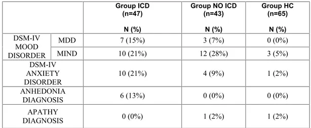
All PD patients, according to international guidelines (Hughes, Daniel et al. 2001), who warrant the DMS-IV-TR diagnostic criteria (APA 2000) for ICD were included in the study. A group of 47 PD patients with ICD (ICD) were recruited. Moreover, a group of 43 PD patients who did not present ICD (PD-No ICD) and 65 healthy controls (HC) were selected, which were matched to the ICD group for age and educational level. All patients were recruited at the outpatient services for movement disorders of two institutions (the I.R.C.C.S. Santa Lucia Foundation of Rome, and the Department of Neurological Sciences, University "Sapienza" of Rome).
For the experimental group, the inclusion criterion was PD - ICD diagnosis while the other inclusion and
exclusion criteria were the same for PD-No ICD group.
Inclusion criteria were: vision and hearing sufficient for compliance with testing procedures and compatibility to 3T MRI procedures.
Exclusion criteria were: 1) presence of major non stabilized medical illnesses (i.e. diabetes, obstructive pulmonary disease or asthma, hematologic/oncologic disorders, vitamin B12 or folate deficiency, pernicious anemia, clinically significant and unstable active gastrointestinal, renal, hepatic, endocrine or cardiovascular disorders); 2) known or suspected history of alcoholism, drug dependence and abuse, head trauma and mental disorders (apart from anxiety disorders) according to the DSM-IV-TR criteria (APA 2000); 3) history of neurological diseases other than Idiopathic PD; 4) history of clinically significant cognitive deficit or score ≤25 on the MMSE (Folstein, Folstein et al. 1975); and 5) unclear history of chronic dopaminergic treatment responsiveness.
The 65 HC were volunteer participants who didn’t suffer from PD or other major medical or neurological illnesses, without history of traumatic head injury with loss of consciousness, without present or past substance dependence or abuse; and without any other current or past DSM-IV-TR Axis I disorder as assessed by the SCID-P (First 2002).
The PD patients enrolled in the study have been under stable dopaminergic therapy for at least two months, when no booster doses of L-Dopa or dopamine agonists were required or pharmacological changes were needed.
The study was approved by the Ethical Committee of the I.R.C.C.S. Santa Lucia Foundation and, in accordance with the Helsinki Declaration, each subject signed an informed consent form prior to enrollment.
2.2 Neurological, psychopathological and
neuropsychological evaluation
Demographic data and clinical characteristics were collected by a neurologist during the clinical examination and the evaluation of motor symptoms was made using the Unified Parkinson’s Disease Rating Scale–Part III (UPDRS-III) (Fahn S. and R.L. 1987; Langston, Widner et al. 1992). All participants were subjected to a neuropsychological and psychopathological evaluation by an expert psychologist. All testing and clinical evaluations in PD patients were carried out during the “on” states, i.e. 2 hours after assumption of first daily dose of medication by trained personnel and blinded to the objectives of the study.
The neuropsychological evaluation included: 1) Mini Mental State Examination (MMSE) (Folstein, Folstein et al. 1975), a global index of cognitive impairment with the score ranging from 30 (no impairment) to 0 (maximum impairment); 2) tests taken from the Mental Deterioration Battery (Carlesimo, Caltagirone et al. 1996), a comprehensive neuropsychological battery that includes verbal and nonverbal tasks such as: a) Rey’s 15-word test – Immediate Recall (RIR) and Delayed Recall (RDR) to evaluate short- and long-term verbal memory, with total scores given by
the total number of words recalled in each test; b) Phonologic (PVF) and Semantic (SVF) Verbal Fluency tests to assess language abilities: in which the total score is the total number of words produced during each test; 3) Copy of the Rey-Osterrieth picture (CRO) and Delayed Recall of the Rey-Osterrieth picture (DRO) to evaluate complex constructional praxis and long-term visual memory (Osterrieth 1944), with scores ranging from 0 (maximum impairment) to 36 (no impairment) on both tests; 4) Wisconsin Card Sorting Test – short form (WCST-SF) (Greve 2001), to explore executive functions; and 5) Stroop Word-Color Test (SWCT) (Stroop 1935) to assess frontal abilities of simple attention, attention shifting and control, which consists of three subtest: “word reading”, “color naming” and “interference time”.
The psychopathological evaluation was conducted to assess the presence and severity of psychiatric disorders. The diagnosis of depression and anxiety were made on the basis of the Structured Clinical Interview for DSM- IV-TR– Patient Edition (SCID–P) (First, Spitzer et al. 2002) and depressive symptoms severity was evaluated by the 21-item Beck Depression Inventory (BDI) (Beck and Steer 1987) and by Hamilton Depression Rating Scale (Dmitrieva, Fyffe et al. 2014).
The diagnosis of apathy was made according to Starkstein’s criteria while the apathetic symptoms severity was assessed by the Apathy Rating Scale (Starkstein, Migliorelli et al. 1995). The diagnosis of anhedonia was made according to Snaith’s criteria and hedonic tone was assessed by the
Snaith-Hamilton Pleasure Scale (SHAPS) (Snaith, Snaith-Hamilton et al. 1995).
All testing and clinical evaluations were carried out from trained specialist who was blind to the aims of the study.
2.3 Image acquisition and processing
All participants underwent the same imaging protocol, which included standard clinical sequences (FLAIR, DP-T2-weighted), whole-brain 3D high-resolution T1-weighted and diffusion-weighted scanning using a 3T Allegra MR imager (Siemens, Erlangen, Germany). Volumetric whole-brain T1-weighted images were obtained using a modified driven equilibrium Fourier transform (MDEFT) sequence (TE/TR = 2.4/7.92 msec, flip angle 15º, voxel size 1 × 1 × 1 mm3).
All planar sequence acquisitions were obtained in the plane of the AC–PC line. Particular care was taken to center subjects' head in the head coil and to restrain their movements with cushions and adhesive medical tape.
High-resolution T1-weighted and DTI images were processed separately to obtain indices of brain macro and microstructural alteration.
First, T1-weighted images were processed and examined using the SPM8 software (Wellcome Department of Imaging Neuroscience Group, London, UK; http://www.fil.ion.ucl.ac.uk/spm), specifically the VBM8 toolbox
(http://dbm.neuro.uni-jena.de/vbm.html), running in Matlab 2007b (MathWorks, Natick, MA). The toolbox extends the unified segmentation model (Ashburner and Friston 2005) consisting of MRI field intensity in homogeneity correction, spatial normalization and tissue segmentation at several preprocessing steps in order to further improve the quality of data preprocessing. Initially, in order to increase the signal-to-noise ratio in the data, the optimized block wise non local-means filter proposed by Coupé et al. (2006) was applied to the MRI scans using the Rician noise adaption (Wiest-Daessle, Prima et al. 2008). Then, an adaptive maximum a posteriori segmentation approach extended by partial volume estimation (Manjon, Tohka et al. 2008) was employed to separate the MRI scans into gray matter (GM), white matter (WM) and cerebrospinal fluid (CSF). The segmentation step was finished by applying a spatial constraint to the segmented tissue probability maps based on a hidden Markow Random Field model (Cuadra, Cammoun et al. 2005) in order to remove isolated voxels which were unlikely to be a member of a certain tissue class and to close holes in clusters of connected voxels of a certain class, resulting in a higher signal-to-noise ratio of the final tissue probability maps. Then, the iterative high-dimensional normalization approach provided by the Diffeomorphic Anatomical Registration Through Exponentiated Lie Algebra (DARTEL) (Ashburner 2007; Bergouignan, Chupin et al. 2009; Klein, Andersson et al. 2009) toolbox was applied to the segmented tissue maps in order to register them to the stereotactic space of the Montreal Neurological Institute (MNI). The tissue deformations were used to
modulate the participants' GM and WM tissue maps in order to compare volumetric differences across groups. Voxel values of the resulting normalized and modulated GM and WM segments indicate the probability (between 0 and 1) that a specific voxel belongs to the relative tissue. Finally, the modulated and normalized GM and WM segments were written with an isotropic voxel resolution of 1.5 mm3 and smoothed with a 6 mm full with half maximum (FWHM) Gaussian kernel, thus obeying the ‘rule of thumb’ that the FWHM should be at least twice the voxel dimension in order to ensure a Gaussian distribution of the residuals of the General Linear Model(Moraschi, Hagberg et al. 2010). The segmented, normalized, modulated and smoothed GM and WM images were used for analyses.
3. Statistical analysis
3.1 Neurological, neuropsychological and
psychopathological data
Data processing and statistical analysis were performed using the software program “Stat View“. Comparisons between continuous variables in ICD and PG, PD-No ICD and HC groups, were performed using the Anova Test, while comparations between PG patients and ICD no PG groups were performed using Unpaired T Student Test. Chi Square test was used for compared categorical variables. The level of statistical significance was defined as p <0.05.
3.2 Neuroimaging data
In order to avoid possible edge effects between different tissue types, the VBM analyses of GM and WM volumes were carried out excluding all voxels with a probability of belonging to the relative tissue less than 20% (absolute threshold masking). Further, statistical analyses of MD maps were restricted to cortical and deep GM structure using an inclusive mask obtained by averaging subjects' GM segments and excluding all voxels with a probability of belonging to GM less than 30%. Finally, statistical analyses on FA maps were restricted to voxels in the WM skeleton.
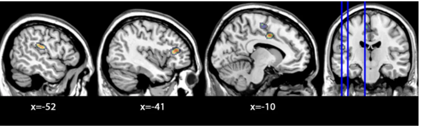
Differences in four neuroimaging parameters (i.e. GM and WM volume, GM MD and WM tract FA) between ICD PD or only PG, PD No-ICD and HC subjects were tested at the voxel level by means of ANOVA t-tests using SPM-8 within the framework of the General Linear Model.
Moreover, to identify the brain region in which patients showed GM or WM volumetric correlates of the psychopathological scores, was adopted a correlation model.
Statistical analyses were carried out at voxel level using SPM8. To avoid type I errors (i.e., accepting false positives) all these analyses were performed using the Random Fields Theory Family-wise error (FWE) correction (P < 0.05), which controls the possibility of any false positives across the entire volume (Ashburner and Friston 2005). Further, results were considered statistically significant if they
were part of a spatially contiguous cluster size of 50 voxels or greater.
To obtain fine anatomical localization of statistical results, two different brain atlases were used: (i) the automated anatomical labeling (Tzourio-Mazoyer, Landeau et al. 2002), which includes all main gyri and sulci of the cerebral cortex and the subcortical and deep GM structures for a total of 90 anatomical volumes of interest and (ii) the ICBMDTI-81 WM labels atlas (Mori, Wakana et al. 2005), which includes 50 WM tract labels created by manual segmentation of a standard-space average of diffusion MRI tensor maps from 81 subjects.
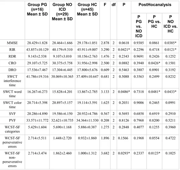
4. Results
Table 1 shows the sociodemographic and clinical characteristics of ICD, PD No-ICD and HC samples. As expected from the matching procedure, the patient groups did not significantly differences in age, gender, educational level, Hoehn and Yahr stage and UPDRS III score, L-dopa daily dose mg/die assumption, while, as expected, in ICD-PD patients, illness duration was significantly higher and age at onset PD was significantly lower than PD No-ICD group. Moreover, assumption of D-2 Agonists L-dopa equivalent daily dose and, consequently, L-dopa equivalent daily dose were significantly higher in ICD-PD than PD No-ICD patients.