UNIVERSITA’ DI PISA Facoltà di Medicina e Chirurgia
Scuola di Specializzazione in Oncologia Medica
TESI DI SPECIALIZZAZIONE
A Feasibility Study on
Cancer Stem Cells Sensitivity Assay
RELATORI
Chir.mo Prof. Alfredo Falcone Dr. Federico Cappuzzo
CANDIDATO
Dr. Manolo D’Arcangelo
INDEX
ABSTRACT ... 2
1.0 INTRODUCTION ... 5
1.1 STATE OF THE ART OF ADVANCED STAGE LUNG CANCER, COLORECTAL CANCER AND BREAST CANCER TREATMENT ... 5
1.2 CANCER STEM CELLS ... 9
1.3 CHEMOSENSITIVITY ASSAYS ... 13
2.0 STELLA: A FEASIBILITY STUDY ON STEM CELLS SENSITIVITY ASSAY ... 15
2.1 RATIONALE ... 15
2.2 STUDY END-POINTS ... 16
2.3 PATIENTS SELECTION CRITERIA ... 17
2.4 STUDY DESIGN ... 18 2.5 EXPERIMENTAL PROCEDURES ... 19 3.0 RESULTS ... 27 3.1 PATIENTS CHARACTERISTICS ... 27 3.2 CSCS ISOLATION ... 29 3.3 CHEMOSENSITIVITY ASSAY ... 31 3.4 PATIENTS TREATMENT ... 33 3.5 LABORATORY EXPERIMENTS ... 36 4.0 CONCLUSIONS ... 37 REFERENCES ... 41
ABSTRACT
Lung cancer (LC), colo-rectal cancer (CRC) and breast cancer (BC) are considered the biggest killers in oncology, accounting for about 40% of cancer deaths. During the last decade, improvement in cancer biology knowledge led to discovery and clinical use of new agents specifically targeting proteins critically involved in cancer growth. Although these new agents, including the Epidermal Growth Factor Receptor Tyrosine Kinase Inhibitors (EGFR-TKIs), the anti-EGFR antibodies (cetuximab, panitumumab), the anti-HER2 antibody (trastuzumab) and the anti vascular endothelial growth factor (VEGF) antibody (bevacizumab), are significantly contributing to increase duration of life, no patient with metastatic disease can obtain a definitive cure.
Available data suggest the hypothesis that cancer is driven by a small subpopulation of cells called “cancer stem cells” (CSCs) or “tumor initiating cells” with an unlimited proliferative potential and the ability to reproduce the original human tumor in experimental animal models. CSCs are responsible for tumor development, growth and progression. Current therapies are largely ineffective against the stem cell population, explaining the failure of standard treatments. In the present study we investigated whether CSCs isolation and in vitro
sensitivity assay are feasible, leading to identification of an effective treatment for chemorefractory NSCLC, CRC and BC.
Patients with heavily pretreated NSCLC, BC and CRC (median of 2 previous regimens) were included onto the study. CSCs were isolated from effusions or fresh cancer tissue from primary tumor or metastasis. Specific culture conditions select for CD133+ immature tumor cells. CSCs were propagated in vitro and further exposed to different chemotherapeutic and targeted agents. The agent or drugs combination inducing the highest CSCs mortality rate identified a possible tailored treatment. Moreover, by using cancer cell spheres, orthotopic xenograft models will be generated.
The study included 23 NSCLC and CRC patients with a median age of 66 years (range 42-85). The procedure for CSCs isolation was repeated in 1 patient. CSCs were obtained from liver metastases in 6 cases (25%), lung nodule excision in 2 cases (8%), lymph node excision in 3 cases (12,5%) and pleural, peritoneal and pericardial effusion in 13 cases (54%). CSCs were successfully isolated in 15 patients (63%). Failure in CSCs isolation was due to inadequate material (8 cases) or delivery accident (1 case). CSCs sensitivity assay was successfully performed in 7 patients (29%), with a median of 15 drugs or combinations tested (range 5-28) and a median time required for results of 51 days (range 37-95).
Preliminary data of our study indicate that CSCs isolation and in vitro sensitivity assay are feasible in metastatic NSCLC. Laboratory procedures for chemosensitivity assessment and for characterization of CSCs in vitro and in vivo are currently ongoing.
1.0 INTRODUCTION
1.1 State of the art of advanced stage lung cancer, colorectal cancer and breast cancer treatment
In 2011, Lung Cancer (LC), colorectal cancer (CRC) and breast cancer (BC) remained the leading causes of cancer-related death worldwide [1]. For patients with metastatic disease definitive cure is not achievable and median survival is approximately 1 year for non-small-cell lung cancer (NSCLC) and about 2 years for metastatic CRC and BC [2]. For NSCLC patients chemotherapy with third-generation platinum-based doublets represented the standard of care until recently, when major breakthroughs in the knowledge of cancer biology has granted the signaling out of numerous targeted therapies. Large phase III clinical trials demonstrated that a proper front-line therapy of a patient with metastatic NSCLC should de based on tumor histology and biology. Patients harboring activating Epidermal Growth Factor Receptor (EGFR) mutations benefit more from EGFR-Tyrosine Kinase Inhibitors (EGFR-TKIs) than from standard platinum-based chemotherapy at least in terms of response rate, progression-free survival (PFS), toxicity profile and quality of life [3-7]. Although no phase III data are currently available, patients with ALK translocation seem to derive a substantial and sustained benefit
when treated with crizotinib, an oral c-MET and ALK inhibitor [8]. In patients without any detectable specific target, histology is the major factor influencing therapy choice. Patients with non-squamous histology seem to benefit more from a pemetrexed-based chemotherapy [9], while in squamous histotype the classical combination of platin (cisplatin or carboplatin) together with gemcitabine, vinorelbine or a taxane remains the standard of care [2]. At the present time there are only three agents approved for second-line therapy, including pemetrexed, docetaxel and erlotinib that is also the only drug approved for third-line therapy. These three agents are considered equally effective in unselected patients, with a toxicity profile in favor of erlotinib and pemetrexed [10,11].
For CRC patients, over the past 2 decades the repertoire of chemotherapeutic agents has increased and extended median overall survival to more than 20 months. Today the active drugs for CRC include 5-Fluorouracyl, irinotecan, oxaliplatin and mytomicin C. An increasing body of evidence also supports the addition of targeted agents, those directed towards VEGF (bevacizumab) and EGFR (cetuximab, panitumumab), to expand treatment options for patients with metastatic disease. Bevacizumab, added to a 5-fluorouracil (5FU) ± irinotecan-based chemotherapy as first-line treatment, has been shown to improve response rates and survival of mCRC patients when
compared to chemotherapy alone [12-14]. An improvement of PFS was also shown in first-line with the addition of bevacizumab to oxaliplatin-based chemotherapy [15]. A randomized phase III study also reported a clinical efficacy of the association of bevacizumab and FOLFOX4 as second-line in metastatic CRC patients previously treated with a fluoropyrimidine and irinotecan, with a significant improvement in response rate, PFS and OS when compared to FOLFOX4 alone [16]. Moreover, the benefits of cetuximab in metastatic CRC are well documented in clinical trials. Cetuximab role is clear not only in irinotecan-refractory or heavily pretreated patients, but also in addition to FOLFIRI (irinotecan/5-fluorouracil/leucovorin) in first-line metastatic colorectal cancer, with an enhanced effect in patients with KRAS wild-type tumors [17]. In these patients, a recent meta-analysis of the pooled Cetuximab Combined with Irinotecan in First-Line Therapy for Metastatic Colorectal Cancer (CRYSTAL) and Oxaliplatin and Cetuximab in First-Line Treatment of CRC (OPUS) patients populations confirms that the addition of cetuximab to first-line chemotherapy achieves a statistically significant improvement in the best overall response, overall survival time, and progression-free survival (PSF) compared with chemotherapy alone [18].
In metastatic BC several options are available. In HER-2 positive patients anti-HER2 strategies, based on the use of
trastuzumab and lapatinib, can prolong patients life expectancy. Several phase III trials have demonstrated an improvement in terms of overall response rate, progression free survival and overall survival if these drugs are used in association to chemotherapy [19, 20]. In HER2 negative hormone-sensitive patients anti-hormonal drugs, both steroidal and non-steroidal, are available and their efficacy have been widely demonstrated [21]. In triple-negative patients recent trials have suggested a benefit when a PARP-inhibitor (olaparib) is associated to chemotherapy [22]. Several chemotherapeutic agents have demonstrated to be active in metastatic BC: antracyclines, fluoropyrimidines, taxanes and vinca alkaloids, even if none of them has been demonstrated to improve overall survival. On the other hand, the role of bevacizumab is not definitively clarified: discordant results on survival data from the phase III trials have brought recently to withdrawal of bevacizumab approval from the Food and Drug Administration [23].
Although many treatment options are available for LC, CRC and BC, none of the above mentioned drugs is able to cure any patient. Invariably, all patients relapse and die for their disease, clearly indicating that our therapies are able to eradicate only a part of the tumor, the sensitive phenotype.
1.2 Cancer stem cells
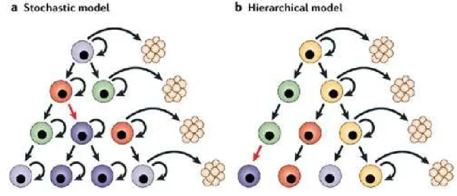
Cancer evolution has been historically meant as consequence of the competition between different cellular clones leading to the emergence of cell strains able to survive in particular microenvironments according to a Darwinian model (“clonal evolution model” or “stochastic model”).
A large number of recent studies underline the importance of a subpopulation of cancer cells with stem-cell like features, the so-called “cancer stem cells” (CSCs) or “tumor initiating cells”, which possess an unlimited proliferative potential and the ability to reproduce the original human tumor in experimental animal models. This small population is suggested to be responsible for tumor initiation, progression and spreading. The functional properties of CSCs make them able to give rise to the whole cancer population and explain the cellular heterogeneity of cancer [24]. It’s widely known that cells with different degrees of differentiation coexist within a tumor and this may be caused by CSCs existence [25]. In particular, this statement is explained by CSCs plastic behavior and self-renewal ability. They have, in fact, an asymmetric replicative modality: cellular division leads to the formation of two distinct cells, one retaining the parenteral phenotype and one destinated to differentiation. These aspects of functional biology led to postulate the
existence of a rigid hierarchy within the tumor, with a CSC placed at the top of the pyramid, operating as precursor of the entire cancer cell population (“hierarchical model”).
To date the two models mentioned above (schematically represented in figure 1) are not in contradiction as a certain grade of genetic heterogeneity has been described also at the top of the tumor pyramid, suggesting a clonal evolution also in the stem compartment [26].
Figure 1. Two alternative models explaining tumors initiation and development.
CSCs are thought to be the result of acquired epigenetic and genetic alterations that can forge signaling pathways controlling proliferation, differentiation and apoptosis. Such mutations would be
passed on to all of the stem cells’ progeny, allowing evolution towards malignancy. Evidence for the existence of CSCs was obtained first in the context of acute myeloid leukemia and thereafter in breast, colon, brain, prostate, ovarian tumors and melanoma [27-32]. The bio-pathology lab of our cooperative group characterized CSCs as CD133 positive cells and showed that these cultured cells retain the cancer-initiating potential upon injection into immune-deficient mice [33,34]. When injected in laboratory animals, CSCs exactly reproduce the parental tumour phenotype, not only histologically but also molecularly, and this feature makes them a good candidate for preclinical studies. In particular, tumor generated by CSCs in immuno-deficient mice replicate more faithfully the human origin tumor in terms of activation/deactivation of pro-tumorigenic processes pathways than the commercially available cell lines. This characterization has been performed with a high-throughput technology, called Reverse Phase Phosphoprotein Microarray (RPPM), that can estimate the state of activation of hundreds of molecular endpoints involved in key biological processes like uncontrolled proliferation, apoptosis, DNA repair mechanisms, self-renewal and epithelial to mesenchymal transition. It has been demonstrated that, if kept in culture for more than one year, the xenografts originated by CSCs maintain the same molecular signature
of the original tumor (figure 2) [35]. In conclusion, CSCs can be maintained in vitro indefinitely without losing their features and this charachteristic, in association with the possibility of propagating them in the experimental animal, make these cells a wonderful tool for a possible personalization of cancer treatment.
Figure 2. Venn diagram showing the percentage of molecular endpoints resulting by the comparison of patient’s tumor with xenografts obatained by CSCs (left) e by commercial cell lines (right).
Recent studies have revealed that CSCs produce high levels of anti-apoptotic proteins and growth factors making them refractory to antineoplastic treatments. Tumors have been widely described to evade death signals generated by therapeutic drugs through the development of anti-apoptotic mechanisms, but the molecular bases of chemotherapy failure has not yet been defined in the majority of tumors. One particularly intriguing property of CSCs is that they are highly resistant to drugs and toxins because of the expression of
several ABC transporters and anti-apoptotic factors and an active DNA-repair capacity [36]. Moreover, recently it has been shown that the apparent tumor debulking, obtained by chemotherapy, paradoxically causes the enrichment of the stem pool [37]. In addition, chemotherapeutic agent are directed at killing rapidly dividing cells, while CSCs are relatively slow cycling like normal stem cells. The inefficacy of conventional therapies towards the stem cell population might explain cancer chemoresistance and the high frequency of relapse shown by the majority of tumors. Therefore, the selective targeting of these cells appears necessary to eradicate tumors and prevent their recurrence.
1.3 Chemosensitivity assays
Although the great effort in the development of in vitro assays able to define sensibility/resistance to chemotherapeutic agents, conflicting results have been generated [38]. Kern and Weisenthal developed an assay to predict an extreme resistance (Extreme Drug Resistance, EDR) to chemotherapy in several solid tumors [39]. In this assay human cancer cells were cultivated in vitro and exposed to chemotherapeutic agents doses proportionally much superior than those used in clinical practice. Cancer cells surviving to this treatment show a condition of EDR. On the basis of these data it was suggested
that, in presence of a condition of EDR, this assay may have a negative predictive value. A great number of studies evaluated the technical aspects of the EDR assay and its correlation with response; anyway, only a little number of studies compared EDR-guided treatment to standard chemotherapy [38, 40, 41]. Due to conflicting results, lack of randomization and of long-term outcomes evaluation, in 2004 the American Society of Clinical Oncology (ASCO) did not recommend EDR as a possible tool guiding chemotherapy in clinical practice. In 2006 a multicenter randomized trial assessed the impact of EDR-guided chemotherapy (experimental arm) in confront of standard chemotherapy (control arm) in the first-line treatment of ovary cancer patients. An interim analysis of this study has not demonstrated a superiority for the experimental arm. Taken together, these data suggest that EDR test has a limited impact on predicting response to chemotherapy. In vitro drug sensitivity assays failure might be explained by the missing of the real target, that is the CSC. Only the elimination of these cells can theoretically bring to durable disease remissions.
2.0 STELLA: A FEASIBILITY STUDY ON STEM CELLS SENSITIVITY ASSAY
2.1 Rationale
1) LC, CRC and BC are major killers in oncology, accounting for about 40% of cancer deaths. Although progresses have been made in the last few years, unfortunately no patient with metastatic disease can obtain a definitive cure.
2) A recent hypothesis is that cancer is driven by a small subpopulation of cells called “cancer stem cells” (CSCs) with an unlimited proliferative potential and the ability to reproduce the original human tumor in experimental animal models. These cells are thought to be responsible for tumor development, representing the only cell population able to sustain tumor growth and progression. Current therapies are largely ineffective against the stem cell population, explaining the failure of standard treatments.
3) To date chemosensitivity assays studies failed in evidencing a predictive value. The failure may be explained by the missing of the real target, the CSCs.
4) Current technologies allow us to isolate and expand in vitro the CSCs from tumor specimens, testing in vitro their sensitivity to
different anticancer drugs. Therefore, there is the potential opportunity to identify and offer an individualized therapy to LC, CRC and BC patients.
5) Pathways responsible for CSCs homeostasis and global analysis can be analyzed (phosphoproteomic and signal transduction analysis, innovative drug testing, analysis of metastatization processes in vivo) in order to provide an overall picture of their activation state.
6) Orthotopic xenografts can be generated by CSCs modified in order to express a bioluminescent protein (luciferase). This may allow CSCs tracking in vivo. The local tumor and the invasiveness development will be monitored through whole-body imaging techniques. Non anesthetized and freely moving animals can be analyzed by this thecnique. Xenograft tumor can be removed for phosphoproteomic analysis. This system can provide information on specific molecular pathways involved in stem cells growth and spreading.
2.2 Study end-points Primary:
Secondary:
1. To identify LC, CRC and BC stem cells.
2. To investigate the sensitivity to anti-tumor agents in vitro. 3. To identify drugs potentially effective for a specific patient
2.3 Patients selection criteria Inclusion criteria
• Histologically/cytologically confirmed diagnosis of metastatic NSCLC, CRC and BC.
• Availability of tumor tissue suitable for CSCs extraction. • Performance status of 100% according to Karnofsky score
(appendix I).
• Failure of conventional therapies or no therapy of proven efficacy.
• Adequate hematological, renal and liver functions.
• No concomitant comorbidity potentially interfering with the study.
• Informed consent form signature.
• If female: childbearing potential either terminated by surgery, radiation, or menopause, or attenuated by use of approved contraceptive method (intrauterine contraceptive
device (IUD), birth control pills, or barrier device) during and for three months after trial.
Exlcusion criteria
• No possibility to obtain fresh tumor tissue.
• Performance status <100% according to Karnofsky score. • Patient suitable for standard therapies.
• Important comorbidity interfering with the study.
• Significant alteration of liver, hematological or renal function(s).
• No informed consent form signature.
2.4 Study design
The STELLA trial (Clinical Trials.gov: NCT01483001) was a prospective study assessing feasibility of individualized therapy in LC, CRC and BC patients. LC, CRC and BC patients with good performance status and tumor tissue collected before study enrollment, at failure of conventional therapies or without possibility to be treated with therapy of proven efficacy, were considered eligible for the study. Before study entry tumor tissue was collected, i.e tissue obtained during a diagnostic or therapeutic procedure, like
surgery or biopsies with other purposes than the protocol. In vitro tumor sensitivity to chemotherapy drugs was tested on tumor cell cultures per each patient. Drugs and their combinations were considered effective if they kill ≥ 60% of tumor stem cells in vitro test. During the period between collection of samples and assay results, the patient can be exposed to other therapies.
2.5 Experimental procedures CSCs identification
Isolation and characterization of CSCs was made starting from samples of tumor tissue obtained from patients with LC, CRC and BC before study inclusion. The surgical/bioptical samples collected were classified according to the specific histological and molecular characteristics of the tumor. From each sample, by means of enzymatic and mechanical procedures, the CSCs were obtained and then cultivated in adequate culture mediums to be subsequently used for biochemical and molecular studies. Each sample was associated with the patient history at the surgical time and an appropriate follow-up program consisting of periodic clinical and instrumental controls that in order to assign a prognostic value to the biological characteristics of the CSCs. Cells derived from the selected epithelial tumors then underwent analysis of surface and intracellular markers in
order to provide a definitive characterization of cellular phenotype. Stem cells derived from LC, CRC and BC were identified as a subset of tumor cells positive for the marker CD133.
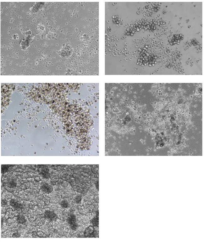
Tumor specimens were washed several times and left over night in DMEM:F12 medium supplemented with high doses of Penicillin/Streptomycin and Fungizone in order to avoid contamination. Tissue dissociation was carried out by enzymatic digestion and recovered cells cultured in serum-free medium containing 25 µg/ml insulin, 100 µg/ml apo-transferrin, 10 µg/ml putrescine, 0.03 mM sodium selenite, 20nM progesterone, 0.6% glucose, 5mM hepes, 0.1% sodium bicarbonate, 0.4% BSA, glutamine and antibiotics, dissolved in DMEM-F12 medium and supplemented with 20 ng/ml EGF and 10 ng/ml bFGF. Flasks non-treated for tissue culture were used in order to reduce cell adherence and favourite growth of undifferentiated tumour-spheres. These culture conditions select for immature tumor cells, while non malignant or differentiated cells are negatively selected as assessed for CSCs of different origin [30]. Surviving immature tumor cells slowly proliferate giving rise to tumour cell aggregates, “spheres”, within 1-2 months in these culture conditions. Sphere-forming cells can be expanded by mechanical dissociation of spheres, followed by re-plating of single cells and residual small cell aggregates in complete fresh medium (figure 3).
Differentiation of NSCLC, CRC and BC sphere-forming cells was obtained by cell culture in specific medium (Cambrex). Phenotype of NSCLC, CRC and BC spheres and their differentiated progeny will be analyzed by flow cytometric analysis or immunofluorescence. In particular stem cell markers such as CD133, CD34 and BCRP1 were analyzed.
In a second step of the trial, cancer spheres will be analyzed in order to define the status of pathways involved in the process of proliferation, self-renewal and survival. In particular, tumor-specific analysis will be carried out to investigate the activity and the possible alteration of pathways responsible for stem cell homeostasis and global analysis (phosphoproteomic and signal transduction analysis, innovative drug testing, analysis of processes metastatization in vivo) aimed to provide an overall picture of the activation state of the key cellular pathways.
Preclinical model
By using cancer spheres we will generate orthotopic xenograft models that recapitulate the parental tumor behaviour, including the aggressive features and the invasiveness potential. Orthotopic injection technique will be assessed in 5 weeks-old NOD/SCID mice. The injection procedure will be done with the support of a dissecting microscope. After anesthetization, 200 to 500 cancer sphere cells, modified in order to express a bioluminescent protein such as luciferase will be injected using a Hamilton syringe and 32-gauge needle. Metastatic and local tumors will be compared for their stem cell content through phenotypic analysis such as growth rate, or other stem cell properties including clonogenic capacity in soft agar or through limiting dilution assays. Infection of CSCs with lentiviral vector, coding for green fluorescent (GFP), as well as luciferase reporter proteins, will allow CSCs tracking in vivo. Particularly, the amphotropic packaging cell line 293T will be transfected by the calcium-phosphate/chloroquine method. Culture supernatants containing viral particles will be collected after 48 hours of transfection. Infection will be performed by culturing target cells in 0.45 µm filtered viral supernatant for 3 hours in a CO2 incubator. Two infection cycles will be performed to infect cells. Microscopic evaluation of GFP expression in viral packaging and target cells will
be performed by direct observation of cells using a reversed fluorescence microscope equipped with a FITC filter. After infection, cells transduced with luciferase will be sorted by flow cytometry to obtain a pure marked population. The local tumor and the invasiveness development will be monitored through whole-body imaging techniques, that will permit to detect, localize and quantify dynamically the optical signal - bioluminescence - in a non invasive localization of the marked cell population. This procedure will be performed using the Photon imager in vivo imaging system (Biospace Lab), assisted by the most recent software for acquisition and image analysis. Thanks to this system, characterized by a very high sensitivity and 20 ms temporal resolution, we will analyze non anesthetized and freely moving animals. The bioluminescence signal will be acquired simultaneously as a standard video of the animal. Once we are sure of the success of tumour growing, mice will be sacrificed. Tumor will be removed for morphological characterization and phosphoproteomic analysis. This latter will be performed through RPPM, which allows the achievement of a high degree of sensitivity, precision and linearity, making possible to quantify the phosphorylated status of signal proteins in immature and differentiated lung cancer cells. This system will provide information on specific molecular pathways involved in stem cells growth and spreading.
Chemosensitivity assay
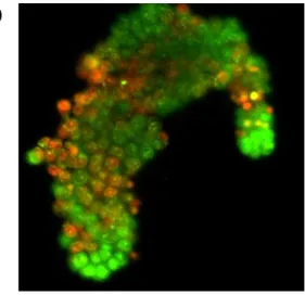
To selectively discriminate the effective therapeutic compounds against the putative tumor and initiating cells, we measured the viability of clonogenic LC, CRC and BC after the exposure to several anti-tumor drugs differentially combined at singular time point up to 96 hours. The measure is perfomed by staining with acridine orange: the green colour express cell vitality, while apoptotic cells appear orange/red (figure 4). The choose of drugs and combination groups to be tested i s defined by the clinician, according to histological and biological features of the primary tumor.
a) b)
c)
Figure 4. Staining with acridine orange after exposition to chemotherapeutic agents. a) Low sensitivity. b) Average sensitivity. c) High sensitivity.
3.0 RESULTS
3.1 Patients characteristics
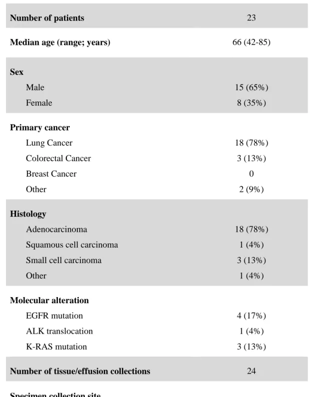
Twenty-three patients were enrolled onto the study. Median age was 66 years (range: 42-85), 15 patients were male (65%) and 8 female (35%). All subjects (100%) had a performance status of 100% according to Karnofsky score. Eighteen patients (78%) were affected by LC and their histotype was adenocarcinoma in 13 cases, squamous cell carcinoma in 1 case, undifferentiated NSCLC in 1 case and SCLC in 3 cases. Three patients (13%) had a CRC and 2 patients (9%) had another gastrointestinal cancer (1 small intestine adenocarcinoma and 1 pancreas adenocarcinoma). No patient enrolled into the study was affected with BC. Among LC patients, 4 subjects (17%) harboured an EGFR mutation, 1 (4%) an ALK translocation and 1 (4%) a KRAS mutation. Two (6%) of the three CRC patients harboured a KRAS mutation. Patients were heavily pretreated, with a median of previous treatment lines of 2 (range: 0-7). Collection of cancer tissue or effusion was performed 24 times (in one LC patient material collected by liver biopsy was not adequate and an additional thoracentesis was performed). Tumor sample for CSCs isolation was obtained from liver metastases in 6 cases (25%), lymph node biopsy in 3 cases (12,5%), lung nodule excision in 2 cases (8%) and by pleural/ peritoneal/pericardial effusion in 13 cases (54%).
Number of patients 23
Median age (range; years) 66 (42-85)
Sex Male Female 15 (65%) 8 (35%) Primary cancer Lung Cancer Colorectal Cancer Breast Cancer Other 18 (78%) 3 (13%) 0 2 (9%) Histology Adenocarcinoma
Squamous cell carcinoma Small cell carcinoma Other 18 (78%) 1 (4%) 3 (13%) 1 (4%) Molecular alteration EGFR mutation ALK translocation K-RAS mutation 4 (17%) 1 (4%) 3 (13%)
Number of tissue/effusion collections 24
Specimen collection site Liver biopsy
Lymph node biopsy Lung nodule excision
Ascitis/pleural/pericardial effusion
6 (25%) 3 (12,5%)
2 (8%) 13 (54%)
3.2 CSCs isolation
LC, CRC and BC patients with progressive disease after standard treatments and for whom the clinician requested a new tumor specimen collection for the evaluation of biological features, were identified at the Oncology Department of Livorno Civil Hospital, Italy. Due to the evidence of good results with cancer effusions, patients with ascitis, pleural and pericardial effusions were included into the study. In presence of effusions, it was given priority to the collection of these kind of samples. Specimens collection was executed preserving sterile conditions.
Effusions were centrifugated at 2500 rpm for 20 minutes and, after eliminating the supernatant, 3 ml of 9% saline solution was added. Liver, lymph node and lung biopsies were added with 3 ml of 9% saline solution. Samples were stored up at 4°C and then shipped at room temperature to the Cellular and Molecular Pathophysiology Laboratory of the University of Palermo, Italy. Delivery took up to 24 hours. Samples collected on Wednesday were stored at 4°C till the next Monday.
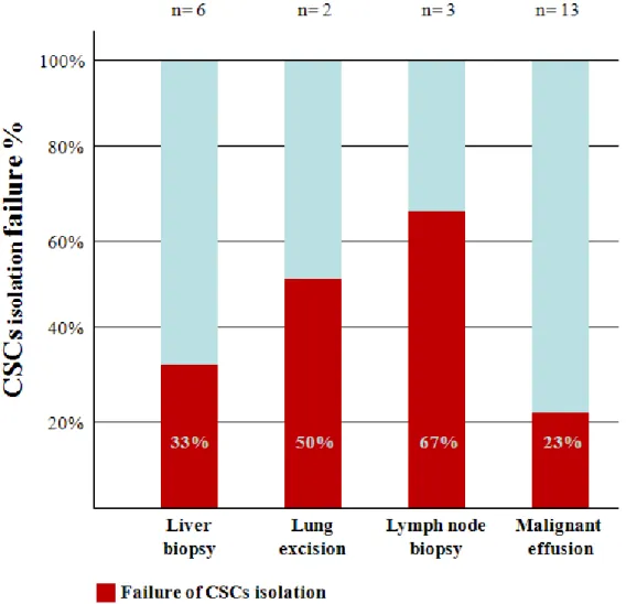
CSCs isolation was feasible in 15 cases (63%) of the 24 procedures. Main reasons for CSCs isolation failure (9 cases, 37%) included inadequate material (8 cases) and delivery accident (1 case). No sample was lost due to contamination. CSCs isolation failure was
more common with biopsies (5 failures on 11 cases, 45%) than with effusions (3 failures on 13 cases, 23%). In particular, lymph node biopsy seems to be the worse tissue for CSCs isolation (2 failures on 3 cases, 67%), while malignant effusions seems to be the samples with the best yield (3 failure on 13 cases, 23%) (figure 5).
Figure 5. CSCs isolation failure rate according to cancer specimens collection site. Failed procedures are represented in red, while successful isolation in blue.
According to the primary cancer, CSCs isolation failed in 5 of 15 cases (30%) of LC, in 2 of 3 cases (67%) of CRC and in 2 of 2 cases (100%) of other gastrointestinal malignancies.
3.3 Chemosensitivity assay
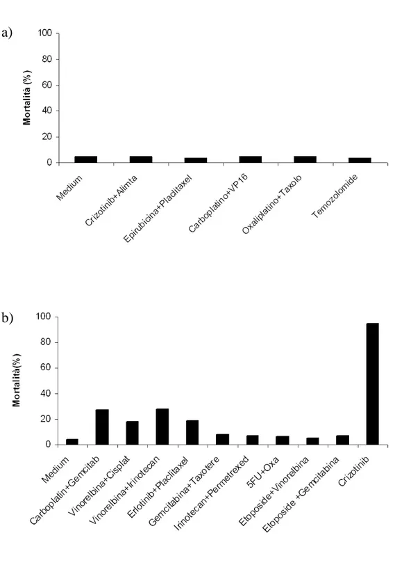
To date chemosensitivity assay was perfomed in 7 cases (29%). This data is not definitive, because other tests will be perfomed as soon as CSCs will be disposable. All tested patients had LC, specifically 5 patients had a lung adenocarcinoma, 1 patient an undifferentiated NSCLC and 1 patient a SCLC. According to site of specimens collection, the starting sample was a malignant effusion in 4 cases, a lung nodule in 1 case, a lymph node biopsy in 1 case and a liver biopsy in 1 case. The median time between sample collection and chemosensitivity assay results was 51 days (range: 37-95). The number of testable drugs and combinations depended on the number of available CSCs. In the study a median of 15 tested treatments (range: 5-28) was registered. In 6 of the 7 perfomed assays, no drug or combination showed a CSC mortality superior to 50%. In one case 4 regimens produced a CSC mortality superior to 50% and 1 combination gave a mortality of 80%. Figure 6 shows an example of the assay results received by the clinician. On the basis of these results the clinician might choose a personalized treatment for the patient.
a)
b)
Figure 6. Chemosensitivy assay results. a) Assay showing sensitivity to none of the tested drugs. b) Assay showing several grades of sensitivity to the tested agents.
3.4 Patients treatment
Our trial was a feasibility study and the correlation between sensitivity assay result and treatment outcome was not an end-point. No patient enrolled onto the study has been so far treated according to chemosensitivity assay results. In particular, one patient did not receive the tailored treatment because the chemosensitivity assay did not evidence any cell mortality when CSCs were exposed to drugs. One patient died before assay results, while the other patients are currently in treatment with chemotherapeutic/biological agents and are not yet in progression.
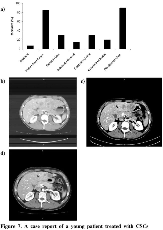
In a preliminary experience a young patient was treated with a sensitivity assay tailored treatment. He was a 26 years old man with a metastatic squamous cell lung carcinoma diagnosed two years before. After four months since surgery, the disease progressed and liver and lung metastases were detected. Molecular analysis revealed wild type EGFR, KRAS and HER-2 gene status and absence of EML4/ALK rearrangement. He had received three treatment lines (cisplatin/gemcitabine, carboplatin/paclitaxel and docetaxel), when he presented to our institution, asking for a valid treatment option. His clinical conditions were good (performance status: 0) and he complained of epigastric pain and moderate asthenia. CT scan showed bilateral lung metastases and an impressive liver involvement (figure
7a). We proposed the patient to undergo a liver biopsy, in order to collect fresh tumor tissue for CSCs isolation and in vitro chemosensitivity assay. After about 40 days since biopsy, the biologist tested six combinations of antiblastic agents and the combination of paclitaxel and oxaliplatin demonstrated to be the most active regimen, causing 95% of cells death in vitro. The patient received oxaliplatin 130 mg/mq day 1 and paclitaxel 175 mg/mq day 1 every three weeks for six cycles. After two cycles, a chest/abdomen CT scan surprisingly showed a notable reduction of liver involvement (figure 7b). Treatment was continued and the CT scans after the fourth cycle showed further improvement in liver disease (figure 7c). Unfortunately the disease progressed after the sixth cycle. Thanks to the tailored treatment, it was possible to reach a time to progression of 4 months and a considerable liver metastases debulk with a symptomatic benefit. This is a remarkable result, if we consider patients histology and previously received treatment lines. The sensitivity assay tailored treatment indicated a mortality percentage of 95%; it can be supposed that 5% of cells were resistant to the drugs indicated by the test and were responsible for disease progression.
a)
b) c)
d)
Figure 7. A case report of a young patient treated with CSCs sensitivity assay tailored therapy. a) Sensitivity assay results. b) Basal assessment. c) Response after two cycles. d) Response after four cycles.
3.5 Laboratory experiments
To date laboratory experiments have not yet started. Cancer spheres will be analyzed in order to define the status of pathways involved in the process of proliferation, self-renewal and survival. Moreover, we will generate orthotopic xenograft models for morphological characterization and phosphoproteomic analysis. The latter will be performed through RPPM, in order to quantify the phosphorylation status of signal proteins in immature and differentiated cancer cells. Through these experiments we will acquire a large number of info about CSCs features and the pathways they preferentially use to escape apoptosis signals and drugs cytotoxicity. The identification of a preferential pathway would be of great interest, because it would open the door to research on specific anti-CSCs drugs.
4.0 CONCLUSIONS
LC, CRC and BC are the most common cancer worldwide and account for about 40% of cancer-related deaths. Many progresses in the treatment of these diseases have been made in the last few years. The strict collaboration between basic researchers and clinicians has permitted the signaling out of fundamental pathways used by cancer cells. This led to design an impressive number of target agents and to large studies assessing their activity and efficacy. Although progresses have been made, no patient can be cured even by this new agents. A possible explanation of this statement probably stays in the missing of the real target. Till the present moment, researchers based their studies on the differentiated pool of cancer cells. These cells usually respond to treatments and in clinical practice complete and partial responses are commonly achievable, although no metastatic patient will reach a definitive cure. CSCs are resistant to many chemotherapeutic and biological agents and a complete elimination of these cells is difficult to reach. In our study we evaluated if it is possible to isolate CSCs and proceed to a chemosensitivity assay in vitro. The initial protocol contemplated the enrollment of LC, CRC and BC without standard treatment chances. No patient enrolled into the study had BC and this may be explained by the large number of drugs and hormones disposable for BC treatment. Among NSCLC patients enrolled into
the study, we had 4 patients with EGFR mutations (29%) and 1 patient harboring a KRAS mutation (7%), far from the percentages of mutations found in clinical practice (15% for EGFR mutations and 25% for KRAS mutation). The presence of EGFR mutation is an important prognostic factor and mutated subjects usually keep good clinical conditions for a long time. Conversely, KRAS mutated subjects have a more aggressive disease in confront of patients harboring EGFR mutations. We can postulate that the difference in genetic alterations percentage between our study and real world, is consequence of the strict selection criteria of the study. In fact, it was not possible to enroll patients with a Karnofsky performance status inferior to 100% and all patients must have already received all standard treatments. For CRC patients, two of the three patients enrolled harbored a KRAS mutation and this may depend on the reduced pool of drugs disposable for KRAS-mutated patients. The little number of CRC patients enrolled may depend on the lack of need for repeating biopsy in these subjects. To date, in fact, rebiopsy of CRC is of poor interest.
As for the biologic material used for CSCs isolation, we found that the best yield was obtained with malignant effusions. This is an important data, because thoracentesis and paracentesis are less
invasive than liver biopsies or lung nodule excision and are often necessary for symptoms relief.
In our study CSCs sensitivity assay has been performed in 7 cases, that is about 1 patient every 3. The last patient was enrolled onto the study at the end of January and the procedure for isolation and in vitro expansion are currently ongoing. Therefore, to date, the number of feasible sensitivity assays is underestimated. The median time between collection and results was 51 days. It’s possible to reduce this time if the oncology department and the laboratory are closest. In our study laboratory procedure were executed in Palermo and this may have altered results. If it was possible to eliminate shipment time and to facilitate the communication between clinician and biologist, time required to obtain a response may reduce. Moreover, in the study only in one case the test revealed a CSCs mortality superior to 80%. In all other patients the sensitivity assay results were disappointing. As seen above, chemotherapy exposure select for a bigger pool of resistant CSCs. The strong pre-treatment of our patients may be the cause of the unsuccessful test results. An earlier test may reduce the failure in identifying an active treatment. Another issue concerning the sensitivity assay is the choose of the drugs to be tested. In the STELLA trial the clinician empirically chose the drugs for the assay on the basis of tumor histology and molecular
status and, when a limited number of combinations were testable, the choose was difficult. A deeper knowledge of CSCs and of the pathways they use to overcome resistance and stay alive may help us in selecting the drugs to be tested. Predictive value of the test was not defined in this study. The trial was designed to evaluate the feasibility of the procedures of CSCs isolation and sensitivity assay in clinical practice. In our experience we assessed that these procedures are feasible and that a major skill in selecting patients, choosing tumor specimen collection sites, samples storing and selecting drugs to be tested may improve results. The STELLA trial permitted us to identify a percentage of successful CSCs isolation and sensitivity tests and to design a phase II trial. This study will recruit NSCLC patients in earlier treatment stages and its primary end-point will be the evaluation of the predictive value of CSCs sensitivity assay.
REFERENCES
1. American Cancer Society. Cancer facts & figures 2010. Atlanta: American cancer society. 2010
2. Schiller JH, Harrington D, Belani CP, Langer C, Sandler A, Krook J, Zhu J, Johnson DH; Eastern Cooperative Oncology Group. Comparison of four chemotherapy regimens for advanced non-small-cell lung cancer. N Engl J Med. 2002 Jan 10; 346(2): 92-8
3. Zhou C, Wu Y-L, Chen G et al. OPTIMAL (CTONG 0802) study comparing first-line erlotinib versus carboplatin (CBDCA) plus gemcitabine (GEM), in Chinese advanced non small-cell lung cancer (NSCLC) patients (pts) with EGFR activating mutations. Ann. Oncol. 2010; 21(Suppl. 8): Abstract LBA13.
4. Mok TS, Wu YL, Thongprasert S, Yang CH, Chu DT, Saijo N, Sunpaweravong P, Han B, Margono B, Ichinose Y, Nishiwaki Y, Ohe Y, Yang JJ, Chewaskulyong B, Jiang H, Duffield EL, Watkins CL, Armour AA, Fukuoka M. Gefitinib or carboplatin-paclitaxel in pulmonary adenocarcinoma. N Engl J Med. 2009 Sep 3; 361(10): 947-57.
5. Lee JS, Park K, Kim SW, et al. A randomized phase III study of gefitinib (IRESSATM) versus standard chemotherapy
(gemcitabine plus cisplatin) as a first-line treatment for never-smokers with advanced or metastatic adenocarcinoma of the lung. J Thor Oncol. 2009; 4(Suppl): PRS.4
6. Maemondo M, Inoue A, Kobayashi K, Sugawara S, Oizumi S, Isobe H, Gemma A, Harada M, Yoshizawa H, Kinoshita I, Fujita Y, Okinaga S, Hirano H, Yoshimori K, Harada T, Ogura T, Ando M, Miyazawa H, Tanaka T, Saijo Y, Hagiwara K, Morita S, Nukiwa T; North-East Japan Study Group. Gefitinib or chemotherapy for non-small-cell lung cancer with mutated EGFR. N Engl J Med. 2010 Jun 24; 362(25): 2380-8
7. Mitsudomi T, Morita S, Yatabe Y, Negoro S, Okamoto I, Tsurutani J, Seto T, Satouchi M, Tada H, Hirashima T, Asami K, Katakami N, Takada M, Yoshioka H, Shibata K, Kudoh S, Shimizu E, Saito H, Toyooka S, Nakagawa K, Fukuoka M; West Japan Oncology Group. Gefitinib versus cisplatin plus docetaxel in patients with non-small-cell lung cancer harbouring mutations of the epidermal growth factor receptor (WJTOG3405): an open label, randomised phase 3 trial. Lancet Oncol. 2010 Feb; 11(2): 121-8
8. Kwak EL, Bang YJ, Camidge DR, Shaw AT, Solomon B, Maki RG, Ou SH, Dezube BJ, Jänne PA, Costa DB, Varella-Garcia M, Kim WH, Lynch TJ, Fidias P, Stubbs H, Engelman JA,
Sequist LV, Tan W, Gandhi L, Mino-Kenudson M, Wei GC, Shreeve SM, Ratain MJ, Settleman J, Christensen JG, Haber DA, Wilner K, Salgia R, Shapiro GI, Clark JW, Iafrate AJ. Anaplastic lymphoma kinase inhibition in non-small-cell lung cancer. N Engl J Med. 2010 Oct 28; 363(18): 1693-70
9. Scagliotti GV, Parikh P, von Pawel J et al. Phase III study comparing cisplatin plus gemcitabine with cisplatin plus pemetrexed in chemotherapy-naïve patients with advanced-stage non-small-cell lung cancer. J Clin Oncol. 2008; 26(21): 3543-51.
10.Vamvakas L, Agelaki S, Kentepozidis NK et al. Pemetrexed (MTA) compared with erlotinib (ERL) in pretreated patients with advanced non-small cell lung cancer (NSCLC): Results of a randomized phase III Hellenic Oncology Research Group trial. ASCO Meeting Abstracts Jun 14, 2010: 7519.
11.Hanna N, Shepherd FA, Fossella FV et al. Randomized Phase III Trial of Pemetrexed Versus Docetaxel in Patients With Non– Small-Cell Lung Cancer Previously Treated With Chemotherapy. J Clin Oncol. 2004; 22(9): 1589-97.
12.Hurwitz H, Fehrenbacher L, Novotny W, Cartwright T, Hainsworth J, Heim W, Berlin J, Baron A, Griffing S, Holmgren E. Bevacizumab plus irinotecan, fluorouracil, and
leucovorin for metastatic colorectal cancer. N Engl J Med. 2004; 350(23): 2335–2342
13.Kabbinavar F, Hurwitz HI, Fehrenbacher L, Meropol NJ, Novotny WF, Lieberman G, Griffing S, Bergsland E. Phase II, randomized trial comparing bevacizumab plus fluorouracil (FU)/leucovorin (LV) with FU/LV alone in patients with metastatic colorectal cancer. J Clin Oncol. 2003; 21(1): 60–65 14.Kabbinavar FF, Schulz J, McCleod M, Patel T, Hamm JT,
Hecht JR, Mass R, Perrou B, Nelson B, Novotny WF. Addition of bevacizumab to bolus fluorouracil and leucovorin in first-line metastatic colorectal cancer: results of a randomized phase II trial. J Clin Oncol. 2005; 23(16): 3697–3705
15.Saltz LB, Clarke S, Diaz-Rubio E, Scheithauer W, Figer A, Wong R, Koski S, Lichinitser M, Yang TS, Rivera F. Bevacizumab in combination with oxaliplatin-based chemotherapy as first-line therapy in metastatic colorectal cancer: a randomized phase III study. J Clin Oncol. 2008; 26(12): 2013–2019
16.Giantonio BJ, Catalano PJ, Meropol NJ, O'Dwyer PJ, Mitchell EP, Alberts SR, Schwartz MA, Benson AB 3rd. Bevacizumab in combination with oxaliplatin, fluorouracil, and leucovorin (FOLFOX4) for previously treated metastatic colorectal cancer:
results from the Eastern Cooperative Oncology Group Study E3200. J Clin Oncol. 2007; 25(12): 1539–1544.
17.Van Cutsem E, Köhne CH, Láng I et al. Cetuximab Plus Irinotecan, Fluorouracil, and Leucovorin As First-Line Treatment for Metastatic Colorectal Cancer: Updated Analysis of Overall Survival According to Tumor KRAS and BRAF Mutation Status. J Clin Oncol. May 20, 2011: 2011-2019
18.Meads C, Round J, Tubeuf S et al. Cetuximab for the first-line treatment of metastatic colorectal cancer. Health Technol Assess. 2010 May;14 Suppl 1: 1-8.
19.Plosker GL, Keam SJ et al. Trastuzumab: a review of its use in the management of HER2-positive metastatic and early-stage breast cancer. Drugs. 2006; 66(4): 449-75.
20.Cameron D, Casey M, Oliva C et al. Lapatinib plus capecitabine in women with HER-2-positive advanced breast cancer: final survival analysis of a phase III randomized trial. Oncologist. 2010; 15(9): 924-34.
21.Goldhirsch A, Colleoni M, Gelber RD. Endocrine therapy of breast cancer. Ann Oncol. 2002; 13 Suppl 4: 61-8
22.Dent R.A., et al., Safety and efficacy of the oral PARP inhibitor olaparib (AZD2281) in combination with paclitaxel for the first- or second-line treatment of patients with metastatic
triple-negative breast cancer: Results from the safety cohort of a phase I/II multicenter trial. J Clin Oncol (Meeting Abstracts), 2010. 28(15_suppl): p. 1018
23.FDA begins process to remove breast cancer indication from Avastin label.
http://www.fda.gov/newsevents/newsroom/pressannouncements/ ucm237172.htm
24.Reya T, Morrison SJ, Clarke MF, Weissman IL. Stem cells, cancer, and cancer stem cells. Nature. 2001; 414: 105-11
25.Dieter SM, Ball CR, Hoffmann CM, Nowrouzi A, Herbst F, Zavidij O, et al. Distinct types of tumor-initiating cells form human colon cancer tumors and metastases. Cell Stem Cell. 2011; 9: 357-65
26.Pardal R, Clarke MF and Morrison SJ, Applying the principles of stem-cell biology to cancer. Nat Rev Cancer, 2003; 3(12): 895-902.
27.Bonnet D, Dick JE. Human acute myeloid leukemia is organized as a hierarchy that originates from a primitive hematopoietic cell. Nat Med. 1997; 3(7): 730-7
28.Dontu G, Jackson KW, McNicholas E, Kawamura MJ, Abdallah WM, Wicha MS. Role of Notch signaling in cell-fate
determination of human mammary stem/progenitor cells. Breast Cancer Res. 2004; 6(6): R605-15.
29.Lombardo Y, Scopelliti A, Cammareri P, Todaro M, Iovino F, Ricci-Vitiani L, Gulotta G, Dieli F, de Maria R, Stassi G. Bone morphogenetic protein 4 induces differentiation of colorectal cancer stem cells and increases their response to chemotherapy in mice. Gastroenterology. 2011; 140(1): 297-309.
30.Ricci-Vitiani L, Pallini R, Biffoni M, Todaro M, Invernici G, Cenci T, Maira G, Parati EA, Stassi G, Larocca LM, De Maria R. Tumour vascularization via endothelial differentiation of glioblastoma stem-like cells. Nature. 2010; 468(7325): 824-8. 31.Bapat SA, Mali AM, Koppikar CB, Kurrey NK. Stem and
progenitor-like cells contribute to the aggressive behavior of human epithelial ovarian cancer. Cancer Res. 2005 Apr 15; 65(8): 3025-9.
32.Fang D, Nguyen TK, Leishear K, Finko R, Kulp AN, Hotz S, Van Belle PA, Xu X, Elder DE, Herlyn M. A tumorigenic subpopulation with stem cell properties in melanomas. Cancer Res. 2005 Oct 15; 65(20): 9328-37.
33.Ricci-Vitiani L, Lombardi DG, Pilozzi E, Biffoni M, Todaro M, Peschle C, De Maria R. Identification and expansion of human
colon-cancer-initiating cells. Nature. 2007 Jan 4; 445(7123): 111-5.
34.Todaro M, Alea MP, Di Stefano AB, et al. Colon cancer stem cells dictate tumor growth and resist cell death by production of interleukin-4. Cell Stem Cell. 2007; 1(4): 389-402.
35.VanMeter A, Signore M, Pierobon M, Espina V, Liotta LA, Petricoin EF 3rd. Reverse-phase protein microarrays: application to biomarker discovery and translational medicine. Expert Rev Mol Diagn. 2007; 7: 625-33
36.Maugeri-Saccà M, Vigneri P, De Maria R. Cancer stem cells and chemosensitivity. Clin Cancer Res. 2011; 17: 4942-7
37.Li X, Lewis MT, Huang J, Gutierrez C, Osborne CK, Wu MF, et al. Intrinsic resistance of tumorigenic breast cancer cells to chemotherapy. J Natl Cancer Inst. 2008; 100: 672-9
38.Schrag D, Garewal HS, Burstein HJ, Samson DJ, Von Hoff DD, Somerfield MR. American Society of Clinical Oncology Technology Assessment: chemotherapy sensitivity and resistance assays. J Clin Oncol. 2004; 22: 3631-38
39.Kern DH, Weisenthal LM. Highly specific prediction of antineoplastic drug resistance with an in vitro assay using suprapharmacologic drug exposures. J Natl Cancer Inst. 1990; 82: 582-88
40.Markman M. Chemosensitivity and chemoresistance testing. J Clin Oncol. 2005; 23: 7363-7364
41.Samson DJ, Seidenfeld J, Ziegler K, Aronson N. Chemotherapy sensitivity and resistance assays: a systematic review. J Clin Oncol. 2004; 22: 3618-3630
APPENDIX I - Karnofsky performance status scale
GRADE (%)
STATUS
100 Normal, no complaints, no evidence of disease
90 Able to carry a normal activity: minor signs or symptoms of disease
80 Normal activity with effort: some signs or symptoms of disease
70 Cares for self: unable to do normal activity or to do active work
60 Requires occasional assistance but is able to care for most of his/her needs
50 Requires considerable assistance and frequent medical care
40 Disabled: requires special medical care and assistance 30 Severely disabled: hospitalization is indicated although
death is not imminent
20 Very sick: hospitalization necessary, active supportive treatment necessary
10 Moribund: fatal processed
0 Death
Per gentile concessione di Karnofsky et al: The use of the nitrogen mustards in the palliation treatment of carcinoma with particular reference to bronchogenic carcinoma, Cancer 1:634-656, 1948.