Alma Mater Studiorum – Università di Bologna
DOTTORATO DI RICERCA IN
SCIENZE DELLA TERRA, DELLA VITA E DELL’
AMBIENTE
Ciclo 29°
Settore Concorsuale di
afferenza Settore Scientifico disciplinare
05/A1 BOTANICA BIO/01BOTANICA GENERALE
FACTORS INVOLVED IN POLLEN
GERMINATION PLAYING A CRITICAL ROLE
ALSO IN
ALLERGIC SENSITIZATION
Dott.ssa IRIS ALOISI
Coordinatore Dottorato
Relatore
Prof.ssa Barbara
Mantovani
Prof. Stefano Del Duca
Index
Pg
Preface
1Introduction
3Microsporogenesis 3
Pollen adhesion to the stigma
Pollen rehydration 4
Pollen germination and tube elongation 6
PART I: Pollen as a model of study and polyamines as a tool
Chapter I Polyamines in pollen: from microsporogenesis to fertilization 21 Chapter II Natural polyamines and synthetic analogues modify the growth and the
morphology of Pyrus communis pollen tubes affecting ROS levels and causing cell death
35
Chapter III Spermine affects pollen tube growth by perturbing calcium concentration, actin organization and cell wall structure
69
PART II: Pollen as a source of allergens: air monitoring and in vitro
studies
Chapter IV Behaviour of profilins in atmosphere and in vitro, and their relationship with the performance of airborne pollen
113 Chapter V Differences in atmospheric concentration of airborne Poaceae pollen and
allergens
138 Chapter VI Purification and characterization of Amb a 1 and Art v 6, two pectate lyase
enzymes from weed pollen
152
Final Remarks
1691
Preface
Understanding the processes involved in pollen maturation and pollen tube growth is of great importance as these processes are involved in fertilization thus crop production.
However, besides the great agricultural interest behind pollen study, the ease of in
vitro growth of pollen, combined with the ease with which live imaging is
performed with pollen tube; make it one of the most exciting systems for the studies of polarity, tip growth and also cell wall deposition. Pollen has provided, over the last decades, important cues about the global rearrangements and the shaping of plant cells. For this reason, it was chosen in this study as a promising model for studying how external stimuli are integrated within the cell and how several signalling molecules and processes are interconnected. In particular, understanding how the overall shaping of the pollen tube tip and how several well-known factors are interconnected were the main goals of this study. In fact, while molecules and factors involved in the apical growth of the pollen tube are well-known individually, details on the reciprocal interactions between these factors are missing. The first part of the theses (“Pollen as a model of study and polyamines as a tool”) exactly deals with these doubts and goals and puts the basis for an integration of several factors of pollen tube growth. As the proper growth of the pollen tube depends on an elaborate mechanism that integrates several molecular and cytological sub-processes its growth mechanism is controlled by several signaling molecules such as polyamines (PAs), which control different aspects of pollen tube germination, e.g. by structuring pollen cell wall and by modulating protein assembly (a deepening of PAs involvement during the whole life span of pollen development can be found in Chapter 1). In pollen, the homeostasis of PAs is finely regulated and the perturbation of this balance has provided, over the years, interesting evidences about how PAs carry out some of their functions in the cell. For these reasons, PAs ware taken into consideration to be used as perturbing molecules, used to spread light in understanding not only the reciprocal interactions among factors, but also the timing of the events (Chapter 2 and 3).
Besides the great interest about pollen as a model of study, pollen is a relevant topic also because it is the main trigger of seasonal allergies, which are sometimes referred to as hay fever or allergic rhinitis. Seasonal allergies develop because the body’s immune system has become sensitized and overreacts to proteins present in the environment that are normally not harmful to other people, the so-called “allergens”. Thanks to a tight collaboration with Consiglio Nazionale delle Ricerche, CNR (Bologna) and Professor Delia Fernández-González (University of Léon, Spain) and a training period spent in the research group of Professor Fatima Ferreira (University of Salzburg) also the topic pollen as a source of allergens was investigated and the main findings can be found in the second part of the thesis (“Pollen as a source of allergens: air monitoring and in vitro studies”). It is well-known that many factors can influence the severity of allergy symptoms but also the abundance of airborne allergens. However, it often happens that the only “pollen count” do not completely mirror the potential allergenicity of the air. Here from, the necessity to integrate the forecasting of allergenic pollen in the atmosphere with the monitoring of airborne allergenic proteins, both if pan-allergens (Chapter 4) and specific pan-allergens (Chapter 5). In these studies, the amount of airborne allergens were put in correlation not only with pollen counts but also with meteorological factors in order to obtain a broad panel of evidences about factors that may affect pollen and allergen dispersion. Finally, in vitro studies also focused on the characterization of two allergenic enzymes, deeply involved in pollen cell wall organization and hugely secreted during pollen rehydration, i.e. pectate-lyases from Ambrosia artemisiifolia and Artemisia vulgaris. The physico-chemical characterization of these allergens is reported in Chapter 6.
Before starting, a brief introduction about what is pollen, how it develops and how pollen tube grows to achieve fertilization will be outlined.
3
Introduction
Microsporogenesis
Pollen, the mature male gametophyte (microgametophyte), is a highly specialized cell type that develops within the anthers of the flower through a complex series of processes.
During anther development, the reproductive or sporogenous cells, located centrally within the anther, give rise to the pollen mother cells; i.e. the microsporocytes, while the surrounding non-reproductive cells form sporophytic epidermal, cortical and tapetal cell layers. Pollen development from microsporocytes can be divided into microsporogenesis and microgametogenesis. During microsporogenesis, microsporocytes undergo a meiotic division, with the four haploid spores, initially staying together in the form of a tetrad. These tetrads are embedded in a thick wall, mainly consisting of callose, and surrounded by the locular fluid inside the anther locules. The innermost cell layer of the locule form the so-called tapetum, an essential tissue for microsporogenesis as it secrets nutrients, carbohydrates, cell wall components and enzymes needed for the proper maturation of the pollen grains and takes part also in the deposition of the pollen cell wall (Müller and Rieu, 2016).
Pollen grain cell wall is extremely unique and at maturity, the pollen surface can be divided into three principal layers, with the relative amount of each varying between species: an outer exine wall, itself multilayered, composed of the chemically resistant polymer sporopollenin and interrupted by openings called apertures; an inner intine, also sometimes multilayered, made primarily of cellulose; and a pollen coat, composed of lipids, proteins, pigments, and aromatic compounds, that fills the sculptured cavities of the pollen exine (Edlund et al., 2004).
Once pollen grains are completely formed, thanks to callases that digest the callose walls of the tetrads, they are released within the locules. During subsequent microgametogenesis, the microspores undergo vacuolization, expansion and a
mitotic, asymmetric division, resulting in the formation of binuclear pollen grains, harbouring a larger vegetative and smaller generative cell. At this stage, the tapetum undergoes programmed cell death. Pollen will then maturate and desiccate. In the case of tri-nucleate pollen, a second mitotic division of the generative cell into two sperm cells occurs before desiccation, while in binucleate pollen grains this happens after pollen germination (Müller and Rieu, 2016). In order to keep viable and metabolically quiescent, the extent of pollen desiccation is extremely high, ranging from 15 to 35% water content, when released from the anthers (Heslop-Harrison, 1979;Buitink et al., 2000;Edlund et al., 2004).
Pollen adhesion to the stigma
To capture pollen grains, stigmas engage biotic and abiotic pollinators (such as insects and wind) and use rapid and strong adhesive interactions to retain pollen grains. The pollen–stigma interface can differ from species to species as a result of the wide variability in the morphology and content of stigma exudates, exine layers, and pollen coats. After exine-mediated adhesion, mobilization of the pollen coat occurs, leading to mixing of lipids and proteins to form a first contact on the stigma surface. There is now extensive evidence that the proteins and lipids in the pollen coat, and proteins on the stigma surface, also contribute to adhesion, albeit most likely at a later stage than at the time of initial contact (Edlund et al., 2004).
Pollen re-hydration
As pollen lands on a stigma, water, nutrients, and other small molecules are transported rapidly into the grain from the stigma exudate or stigma papillae by mechanisms that remain unclear. The discovery of aquaporin expression in the stigma has prompted the exciting model that water channels are involved in the rapid and regulated water release from the stigma to the pollen (Dixit et al., 2001). Regardless of the mechanism of transfer, pollen hydration often is regulated, both temporally and spatially. Inappropriate hydration can have disastrous consequences, leading to premature germination within the anther (Johnson and McCormick, 2001) or germination on the wrong surface.
5 The genetic and molecular dissection of the lipid-rich matrices found in the pollen coat has progressed considerably in recent years. In Arabidopsis, the pollen coat contains long- and short-chain lipids along with a small set of proteins, including six lipases and eight Gly-rich oleosin proteins that contain a lipid binding domain. Disrupting pollen coat lipids or pollen coat proteins in Brassicaceae species can delay or block pollen hydration. Mutations that affect pollen coat proteins are less extreme, perhaps because of partial functional redundancy (Mayfield et al., 2001;Edlund et al., 2004;Fiebig et al., 2004).
The lipid-rich stigma exudate of plants with wet stigmas is thought to be functionally analogous, in part, to the pollen coat. Based on these findings, it is possible to propose a model in which the presence of lipids, whether provided by the male or the female surface, modulates water transfer to desiccated pollen, while highly diverse proteins and peptides mediate self and foreign pollen recognition(Dickinson, 1995;Edlund et al., 2004).
Independently of where and how the water required for pollen rehydration comes from, what is well known is that a huge amount of proteins is released in the hydration medium during this process. This phenomenon may, in part, be due to the bursting of some pollen grains, thus resulting in the release of proteins into the medium. However, it was demonstrated that several protein are over expressed and released into the rehydration medium (Sheoran et al., 2009). During in vitro germination of canola pollen in fact a huge number of proteins is released into the germination medium, including cruciferin, oleosin, cysteine-rich repeat secretory protein 26 precursor, enolase, pyrophosphatase, polygalacturonase and pectinesterase inhibitor (Sheoran et al., 2009), thus proteins involved in carbohydrate and energy metabolism, transport and wall remodeling. Besides proteins involved in those functions, also cell-wall remodeling protein, e.g. pectate lyases and expansins, and proteins involved in signalling are extruded from the pollen tube. What is worth of notice is that many proteins extruded actively or passively during pollen rehydration, is that they are often allergenic proteins (or allergen-related proteins), representing a threat to human health as pollen is an important source of seasonal allergens (Grote et al., 2000;Grote et al., 2003;Radauer and Breiteneder, 2006;Vega-Maray et al., 2006).
Pollen germination and tube elongation
Hydration transforms a pollen grain into a highly polarized cell as the grain organizes its cytoplasm and cytoskeleton to support the extension of the pollen tube. These changes occur within minutes after hydration and include the formation of a filamentous actin cytoskeleton highly polarized toward the site of tube emergence (Gossot and Geitmann, 2007;Cheung and Wu, 2008;Fu, 2015), reorientation of the vegetative nucleus so that it enters the extending tube before the generative cells (Åström et al., 1995;Laitiainen et al., 2002;Edlund et al., 2004), movement of mitochondria and polysaccharides at the site of tube emergence (Cresti et al., 1977), and delivering of secretory vesicle containing newly synthetized cell wall material at the site of tube emergence (Wang et al., 2005;Hepler et al., 2013).
It is not yet clear how the polarization signal is perceived and subsequently transduced to select a single point for tube emergence. Several candidate signals have been suggested, including water, lipids, and ions. Polarization signals ultimately trigger the recruitment of several signalling factors and the establishment of calcium gradients at tube tips (Edlund et al., 2004).
Once the cell has established its internal polarity relative to an external signal, the pollen tube must breach the exine wall to emerge from the grain and can enter the style, seeking to reach the ovary, after transiting the stigma barrier.
To efficiently reach the ovary, pollen tubes elongate at an astonishing rate (up to 1 cm/hr) to an extraordinary length by polarized tip growth, which is strictly dependent on polar exocytosis that delivers cell membrane and wall materials to the growing tip (Hepler et al., 2001;Qin and Yang, 2011). Pollen tube growth requires a highly polarized cytoplasmic organization and finely-tuned molecular machineries. The tip region displays an apical zone essentially packed with exocytic vesicles accumulated as a typical “V shape” to facilitate massive tip-targeted exocytosis. This zone is called “clear-zone” so named because the amyloplasts and vacuoles are prevented from moving into this apical part. In the subapical and shank regions there are organelles, nuclei, and vacuoles that may extend toward the grain (Cheung and Wu, 2008;Qin and Yang, 2011;Hepler et al., 2013). Cytoplasmic streaming drives organelles moving rapidly back and forth
7 along the main axis of the pollen tube in a reverse fountain pattern, which maintains the distribution of membranous structures and releases exocytic vesicles to the apical zone (Cai and Cresti, 2009). Several signaling networks control tip growth in pollen tubes via inter-connection with the cytoskeletal elements and the polarized exocytosis (Qin and Yang, 2011)
In the next part of the introduction, the main factors and processes involved in pollen tube growth will be discussed more in detail. As this process is extremely complicated and regulated and as the more that research investigates pollen tube growth, the more factors appear to be involved, a large number of molecules, although no less important, will not be discussed in detail.
The structural system The cytoskeleton
Pollen tubes contain both microtubules and actin microfilaments, which are highly organized and dynamic through their interaction with various actin-binding proteins (ABP) and microtubule-associated proteins (Staiger and Blanchoin, 2006;Fu, 2010;Staiger et al., 2010). While microtubules are thought to be mostly involved in organelle movement (Qin and Yang, 2011), recent discoveries show how microtubules are involved in exocytosis and endocytosis, besides the movement of organelles, suggesting a potential role for microtubules in the regulation of pollen tube tip growth (Idilli et al., 2013;Cai et al., 2015;Fu, 2015). Most assuredly is that actin microfilaments are essential for tip growth. They form long cables axially aligned in the shank, providing the main tracks for the movement of organelles and vesicles, regulating the cytoplasmic streaming (Vidali and Hepler, 2001;Bove et al., 2008;Cai et al., 2015). This cables-dependent cytoplasmic streaming rapidly brings exocytic vesicles to the subapical zone, where, a collection of shorter and thinner actin cables constitutes a ring-like actin structure. It was proposed that the subapical F-actin participates in vesicular
trafficking in the apical region (Gu et al., 2003). The dynamic microfilaments are regulated by Rho-related GTPase of plants (ROP) and many ABP. Pharmacological and genetic studies combined with live cell imaging have revealed multiple distinct populations of microfilaments that carry out specific functions in pollen tubes, respectively. Low concentrations of latrunculin B, which did not affect the longitudinal actin cables, inhibited pollen tube growth, suggesting an essential role for a dynamic form of microfilaments (Vidali et al., 2001).
Genetic studies of various ABPs more clearly define the role for each microfilament form. A series of ABPs that regulate the construction of longitudinal microfilament bundles have been characterized including actin-nucleating factor, actin-bundling proteins, fimbrin, villin, profilin and actin depolymerizing factor (ADF) that are important for the turnover of microfilaments in the shank region. The regulation of these ABP reflects the regulation of actin turnover and microfilaments organization, creating fine-tuned machinery, often difficult to be studied. For example, monomer-binding proteins, profilin and ADF, are thought to function synergistically to enhance turnover and the exchange of subunits between monomer and polymer pools. How individual actin filaments behave in living cells, however, remains largely unexplored (Staiger et al., 2010). Most assuredly is that the actin cables provide the tracks for the movement of large organelles and the cytoplasmic streaming during pollen tube tip growth. The subapical actin fringe is proposed to participate in vesicle trafficking in the apex as well as in control the apical clear-zone formation and maintenance (Qin and Yang, 2011;Fu, 2015). It maintains its length and apical location to keep in step with growth. How the subapical fringe modulates the directionality of pollen tube growth remains to be determined (Fu, 2015).
Cell wall composition and mechanics
The rapid and continuous tip growth has to rely on efficient and ample supply of membrane materials, enzymes, signaling molecules, and, mostly, of new cell wall components. Because of high turgor pressure in pollen tubes as in other plant cells, biosynthesis-based growth via exocytic delivery of cell wall materials has to be coordinated with the mechanics of the cell wall that facilitate localized cell expansion only at the tip. In fact, the pollen tube cell wall is not comparable to that
9 of other plant cells. The most striking difference is that deposition of new cell wall components occurs along the growth axis in an accurate temporal sequence. Methyl-esterified pectins are first secreted at the apex of pollen tubes (O’ Neill et al., 1990). After deposition, they are chemically converted into acid pectins at the apex/subapex edge (Rockel et al., 2008), where they bind calcium, thereby contributing to strengthen the cell wall (Palin and Geitmann, 2012;Wolf and Greiner, 2012). This prevents additional deformation of the cell wall and contributes to maintain the cylindrical shape of pollen tubes. The balance between pectin secretion and activity of pectin methyl-esterase (PME) thus yields a gradient at the pollen tube apex with highly methyl-esterified pectins at the extreme apex and a less esterified mix of polymers progressively away from the apex. As a consequence, the extreme apical wall is ‘softer’ than the cell wall in the pollen tube shank and, during oscillatory growth, episodes of rapid growth are preceded by local softening of the apical cell wall (Wolf and Greiner, 2012). In addition to pectins, the pollen tube cell wall contains other components such as callose, cellulose and arabinogalactan proteins. Callose is usually absent from the apex while both arabinogalactan proteins and cellulose could also be detected in the hemispherical apical dome (Mollet et al., 2013). In support of this, the cellulose synthase complex has been found close to the pollen tube apex in Nicotiana
tabacum (Cai et al., 2011) and Arabidopsis thaliana (Chebli et al., 2012). A temporal
analysis of secretion has been made using different methods and indicates that, as the pollen tube growth rate oscillates, so too does the exocytosis of pectin with the same period, but with a clearly different phase. An analysis of the phase relationship using cross-correlation reveals that pectin is deposited in advance of the increase in growth rate (McKenna et al., 2009;Hepler et al., 2013). While emphasis is usually focused on exocytosis, it is important to note that endocytosis is also a major contributor to pollen tube growth (Hepler et al., 2013). Based on the amount of material needed to support the ever-expanding cell wall, it becomes evident that a considerable excess of plasma membrane is delivered during the secretory process. This excess membrane is retrieved through endocytosis (Onelli and Moscatelli, 2013).
The regulatory system
Pollen grains generate pollen tubes in vitro in the absence of external signals, suggesting that their tip growth is controlled by a self-organizing system. Studies over the last decade have demonstrated the existence of a self-organizing signaling network, which is centered on a tip-localized Rho GTPase (ROP1) and tip-focused Ca2+ gradients and their interactions with the actin cytoskeleton and vesicular
trafficking (Qin and Yang, 2011). Also reactive oxygen species (ROS) are involved in keeping the polarity of the process, creating a cross-talk with the Ca2+ gradient
(Potocky et al., 2007;Lassig et al., 2014;Pottosin et al., 2014).
ROPs
ROP GTPases (also called RACs) belong to a subfamily of conserved Rho GTPases and have been established as one of the central regulators of tip growth in pollen tubes by virtue of their apical localization, their essential role for tip growth, and the spatiotemporal dynamics of their activities (Gu et al., 2003;Yan et al., 2009). These GTPases act as molecular switches by cycling between the biologically inactive GDP-bound and the active GTP-bound forms. In Arabidopsis pollen tubes, several ROPs are expressed, with the pollen-specific ROP1 playing a predominant role in determining polarity of the pollen tube at the apical plasma membrane (Li et al., 1998). Disrupting ROP1 signaling inhibits pollen tube tip growth, whereas expressing a constitutively active form of ROP1 induces a dramatically enlarged active ROP1 cap, leading to tip depolarization and balloon-like pollen tubes (Zheng and Yang, 2000;Guan et al., 2013).
ROP1 is activated at the apical region, where it activates multiple downstream pathways to determine the site and dynamics of exocytosis, which in turn leads to polarized growth. RIC3 and RIC4 interact with the active GTP-bound form of ROP1 at the apex of pollen tubes and regulate tip growth by regulating F-actin dynamics (Gu et al., 2003). The RIC4 pathway promotes F-actin assembly and induces the transport and accumulation of vesicles to the tip region. ROP1 also activates the RIC3 pathway, which promotes tip F-actin disassembly by regulating Ca2+
dynamics, thus facilitating the fusion of exocytic vesicles to the plasma membrane of tht tip of pollen tubes (Gu et al., 2005). As a key regulator of the self-organizing pollen tube system, the activity and distribution of ROP1 are fine-tuned by both
11 positive and negative feedback mechanisms (Hwang et al., 2010). The positive and negative ROP1 feedbacks might be coupled through F-actin-mediated exocytosis, continuously generating the oscillation of apical ROP1 activity to sustain tip growth. Apically localized active ROP1 promotes polarized exocytosis through RIC3- and RIC4-dependent F-actin dynamics, and exocytosis. F-actin-mediated exocytosis might facilitate the recruitment factors involved in ROP1 inactivation. As a result, localized ROP1 could be further activated and the distribution of active ROP1 would expand through the apex, leading to enhanced exocytosis and promoted pollen tube growth rate. The current understanding of RopGTPase signaling leaves many unanswered questions. The mechanisms underpinning the regulation of Ca2+ gradients through RIC3 are still unknown, and how Ca2+
mediates F-actin depolymerization remains to be elucidated (Guan et al., 2013).
Calcium gradients
Cytosolic free Ca2+ is an important secondary messenger in the signaling networks
regulating pollen tube elongation. In vivo pollen tube growth usually relies on external calcium stores in the pistil, and external Ca2+ enhances the elongation of
pollen tube in vitro (Franklin-Tong, 1999;Iwano et al., 2009). Application of either Ca2+ channel blockers or Ca2+ ionophores inhibits pollen tube growth, indicating
the requirement for fine-tuning of cytosolic Ca2+ concentration in the elongation of
pollen tubes (Guan et al., 2013). Growing pollen tubes display a tip-focused Ca2+
gradient, which oscillates in a correlated but lagged phase of growing pollen tube (Malho and Trewavas, 1996;Iwano et al., 2009;Hepler et al., 2012). The interplay between active ROP GTPases and a Ca2+ gradient is essential for the oscillating
patterns of both factors. Apical ROP1 activity oscillates ahead of both pollen tube growth and tip Ca2+ gradients. ROP1 activates its downstream effector RIC3 to
mediate the influx of Ca2+ across the plasma membrane. The increase of Ca2+ in the
tip causes F-actin disassembly, thus promoting exocytosis and regulating ROP1 activity. Elevated Ca2+ accumulation can then suppress ROP1 activity and balance
the F-actin-dependent apical ROP1 activation (Gu et al., 2005;Hwang et al., 2010;Guan et al., 2013).
In addition to the essential regulatory function in F-actin dynamics, Ca2+ signals
motif is found in several signaling components of pollen tubes (Guan et al., 2013). Ca2+ is perceived by the Ca2+ sensor proteins that mediate downstream signaling
responses or feedback regulation of Ca2+ gradient. Ca2+ sensor proteins include
calmodulin, calmodulin -like, calcium-dependent protein kinase, and calcineurin B-like protein. The requirement for calmodulin and calcium-dependent protein kinase in pollen tube growth has been elucidated in several studies. However, the detailed regulatory mechanisms of Ca2+ sensors and their potential targets in
pollen tube growth remain to be revealed (Guan et al., 2013).
The mechanisms that directly regulate tip-focused Ca2+ gradients are complex, and
involve multiple transport systems that increase or decrease the cytosolic Ca2+
concentration (Iwano et al., 2009). Plasma membrane-localized Ca2+ channels are
key regulators as they enable external Ca2+ to enter the cytosol. To date, the
characterized Ca2+ influx pollen tube plasma membrane channels include the cyclic
nucleotide-gated channel (CNGC) and glutamate receptor-like (GLR) channels (Frietsch et al., 2007;Michard et al., 2011). CNGCs are ion channels that function in response to the binding of cyclic nucleotides (cGMP and cAMP) and can be suppressed by elevated calcium levels together with calmodulin.
The negative regulation of tip Ca2+ gradient involves the efflux of Ca2+ on the
plasma membrane and sequestering Ca2+ within endomembrane system, both of
which may require
Ca2+ pumps. The plasma membrane -localized auto-inhibited Ca2+ ATPases (ACA) is
required for the efflux of Ca2+, whilst ER-localized Ca2+-ATPases may play an
important role in moving Ca2+ against its concentration gradient into the ER
(Steinhorst and Kudla, 2013). Calmodulins are activated by Ca2+ and are involved
in the regulation of Ca2+gradient. Active calmodulins can negatively regulate CNGCs
and positively regulate ACAs, resulting in the negative feedback regulation of cytosol Ca2+ gradient (Guan et al., 2013;Steinhorst and Kudla, 2013).
Reactive oxygen species
Reactive oxygen species (ROS) have long been known to play critical roles in processes involved in protecting the plant from biotic and abiotic stresses. However, ROS have also emerged as important regulators of plant development
13 where they play roles signal transduction and the modulation of cell wall polymer structure (Swanson and Gilroy, 2010).
ROS also play a key role during pollen tube emergence and elongation as they act as wall loosening or stiffening agents (Swanson and Gilroy, 2010;Speranza et al., 2012). In fact, hydroxyl radicals (OH•) produced in the cell wall induce cell-wall
loosening both in vitro and in vivo, modulating thus the elongation of pollen tube (Schopfer, 2001). Cytoplasmic ROS are mostly produced by mitochondria, chloroplasts and peroxisomes while plasma membrane NADPH-oxidases (NOX) are instead an important source for both cell wall and cytoplasmic ROS. In fact, they produce superoxide anions, O2•–, the precursor of both OH• and hydrogen
peroxide (H2O2), an important signalling molecule (Potocky et al., 2007). The
involvement of ROS during pollen tube growth was first speculated by evidences that showed how they localize in the apex of the tube and how the inhibition of NOX by Diphenilene Iodonium chloride (DPI) suddenly arrests tip growth and alters the morphology of the pollen tube (Potocky et al., 2007;Speranza et al., 2012). Moreover, there is a correlation between ROS and Ca2+ signaling, that is also
essential for plant development and tip-growth, since ROS induce Ca2+ influx
across cell membranes (Foreman et al., 2003), and cytosolic Ca2+ can activate NOX;
establishing a positive feedback between NOX activity and ROS-induced Ca2+ influx,
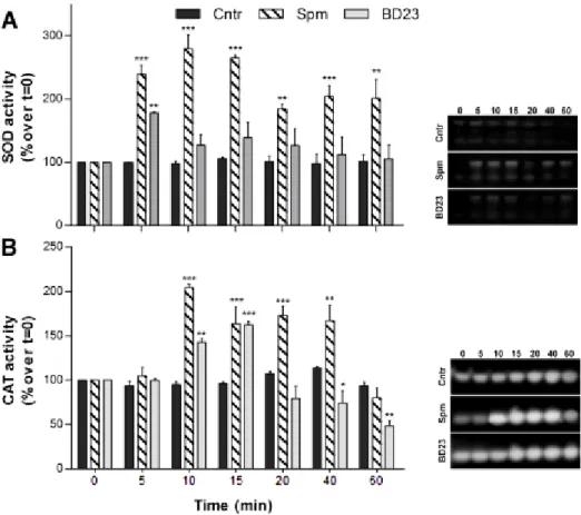
that supports polarized growth (Kaya et al., 2014;Lassig et al., 2014;Pottosin and Shabala, 2014). Moreover, to avoid an excess of ROS that could lead to oxidative damage, pollen has a powerful antioxidant machinery based on superoxide dismutase (SOD) that catalyzes the dismutation of O2.− into H2O2; the latter is
decomposed to water and oxygen by catalase (CAT).
References
Åström, H., Sorri, O., and Raudaskoski, M. (1995). Role of microtubules in the movement of the vegetative nucleus and generative cell in tobacco pollen tubes. Sex Plant Reprod 8, 61– 69.
Bove, J., Vaillancourt, B., Kroeger, J., Hepler, P.K., Wiseman, P.W., and Geitmann, A. (2008). Magnitude and direction of vesicle dynamics in growing pollen tubes using spatiotemporal image correlation spectroscopy and fluorescence recovery after photobleaching. Plant Physiol 147, 1646-1658.
Buitink, J., Leprince, O., Hemminga, M.A., and Hoekstra, F.A. (2000). The effects of moisture and temperature on the ageing kinetics of pollen: Interpretation based on cytoplasmic mobility. Plant Cell Environ 23, 967–974.
Cai, G., and Cresti, M. (2009). Organelle motility in the pollen tube: a tale of 20 years. J Exp Bot 60, 495-508.
Cai, G., Faleri, C., Del Casino, C., Emons, A.M., and Cresti, M. (2011). Distribution of callose synthase, cellulose synthase, and sucrose synthase in tobacco pollen tube is controlled in dissimilar ways by actin filaments and microtubules. Plant Physiol 155, 1169-1190. Cai, G., Parrotta, L., and Cresti, M. (2015). Organelle trafficking, the cytoskeleton, and pollen
tube growth. J Integr Plant Biol 57, 63-78.
Chebli, Y., Kaneda, M., Zerzour, R., and Geitmann, A. (2012). The cell wall of the Arabidopsis pollen tube-spatial distribution, recycling, and network formation of polysaccharides.
Plant Physiol 160, 1940-1955.
Cheung, A.Y., and Wu, H.M. (2008). Structural and signaling networks for the polar cell growth machinery in pollen tubes. Annu Rev Plant Biol 59, 547-572.
Cresti, M., Pacini, E., Ciampolini, F., and Sarfatti, G. (1977). Germination and early tube development in vitro of Lycopersicum peruvianum pollen: Ultrastructural features.
Planta 136, 239-247.
Dickinson, H. (1995). Dry stigmas, water and self-incompatibility in Brassica. Sex. Plant Reprod. 8, 1–10.
Dixit, R., Rizzo, C., Nasrallah, M., and Nasrallah, J. (2001). The brassica MIP-MOD gene encodes a functional water channel that is expressed in the stigma epidermis. Plant Mol Biol 45, 51-62.
Edlund, A.F., Swanson, R., and Preuss, D. (2004). Pollen and stigma structure and function: the role of diversity in pollination. Plant Cell 16 Suppl, S84-97.
Fiebig, A., Kimport, R., and Preuss, D. (2004). Comparisons of pollen coat genes across Brassicaceae species reveal rapid evolution by repeat expansion and diversification.
Proc Natl Acad Sci U S A 101, 3286-3291.
Foreman, J., Demidchik, V., Bothwell, J.H., Mylona, P., Miedema, H., Torres, M.A., Linstead, P., Costa, S., Brownlee, C., Jones, J.D., Davies, J.M., and Dolan, L. (2003). Reactive oxygen species produced by NADPH oxidase regulate plant cell growth. Nature 422, 442-446. Franklin-Tong, V.E. (1999). Signaling and the modulation of pollen tube growth. Plant Cell 11,
727-738.
Frietsch, S., Wang, Y.F., Sladek, C., Poulsen, L.R., Romanowsky, S.M., Schroeder, J.I., and Harper, J.F. (2007). A cyclic nucleotide-gated channel is essential for polarized tip growth of pollen. Proc Natl Acad Sci U S A 104, 14531-14536.
Fu, Y. (2010). The actin cytoskeleton and signaling network during pollen tube tip growth. J
Integr Plant Biol 52, 131-137.
Fu, Y. (2015). The cytoskeleton in the pollen tube. Curr Opin Plant Biol 28, 111-119.
Gossot, O., and Geitmann, A. (2007). Pollen tube growth: coping with mechanical obstacles involves the cytoskeleton. Planta 226, 405–416.
Grote, M., Valenta, R., and Reichelt, R. (2003). Abortive pollen germination: a mechanism of allergen release in birch, alder, and hazel revealed by immunogold electron microscopy. J Allergy Clin Immunol 111, 1017-1023.
15 Grote, M., Vrtala, S., Niederberger, V., Valenta, R., and Reichelt, R. (2000). Expulsion of allergen-containing materials from hydrated rye grass (Lolium perenne) pollen revealed by using immunogold field emission scanning and transmission electron microscopy. J Allergy Clin Immunol 105, 1140-1145.
Gu, Y., Fu, Y., Dowd, P., Li, S., Vernoud, V., Gilroy, S., and Yang, Z. (2005). A Rho family GTPase controls actin dynamics and tip growth via two counteracting downstream pathways in pollen tubes. J Cell Biol 169, 127-138.
Gu, Y., Vernoud, V., Fu, Y., and Yang, Z. (2003). ROP GTPase regulation of pollen tube growth through the dynamics of tip-localized F-actin. J Exp Bot 54, 93-101.
Guan, Y., Guo, J., Li, H., and Yang, Z. (2013). Signaling in pollen tube growth: crosstalk, feedback, and missing links. Mol Plant 6, 1053-1064.
Hepler, P.K., Kunkel, J.G., Rounds, C.M., and Winship, L.J. (2012). Calcium entry into pollen tubes. Trends Plant Sci 17, 32-38.
Hepler, P.K., Rounds, C.M., and Winship, L.J. (2013). Control of cell wall extensibility during pollen tube growth. Mol Plant 6, 998-1017.
Hepler, P.K., Vidali, L., and Cheung, A.Y. (2001). Polarized cell growth in higher plants. Annu Rev
Cell Dev Biol 17, 159-187.
Heslop-Harrison, J. (1979). An interpretation of the hydrodynamics of pollen. Am. J. Bot. 66, 737–743.
Hwang, J.U., Wu, G., Yan, A., Lee, Y.J., Grierson, C.S., and Yang, Z. (2010). Pollen-tube tip growth requires a balance of lateral propagation and global inhibition of Rho-family GTPase activity. J Cell Sci 123, 340-350.
Idilli, A.I., Morandini, P., Onelli, E., Rodighiero, S., Caccianiga, M., and Moscatelli, A. (2013). Microtubule depolymerization affects endocytosis and exocytosis in the tip and influences endosome movement in tobacco pollen tubes. Mol Plant 6, 1109-1130. Iwano, M., Entani, T., Shiba, H., Kakita, M., Nagai, T., Mizuno, H., Miyawaki, A., Shoji, T., Kubo,
K., Isogai, A., and Takayama, S. (2009). Fine-tuning of the cytoplasmic Ca2+ concentration is essential for pollen tube growth. Plant Physiol 150, 1322-1334.
Johnson, S.A., and Mccormick, S. (2001). Pollen germinates precociously in the anthers of raring-to-go, an Arabidopsis gametophytic mutant. Plant Physiol 126, 685-695.
Kaya, H., Nakajima, R., Iwano, M., Kanaoka, M.M., Kimura, S., Takeda, S., Kawarazaki, T., Senzaki, E., Hamamura, Y., Higashiyama, T., Takayama, S., Abe, M., and Kuchitsu, K. (2014). Ca2+-activated reactive oxygen species production by Arabidopsis RbohH and RbohJ is essential for proper pollen tube tip growth. Plant Cell 26, 1069-1080.
Laitiainen, E., Nieminen, K.M., Vihinen, H., and Raudaskoski, M. (2002). Movement of generative cell and vegetative nucleus in tobacco pollen tubes is dependent on microtubule cytoskeleton but independent of the synthesis of callose plugs. Sex Plant
Reprod 15, 195-204.
Lassig, R., Gutermuth, T., Bey, T.D., Konrad, K.R., and Romeis, T. (2014). Pollen tube NAD(P)H oxidases act as a speed control to dampen growth rate oscillations during polarized cell growth. Plant J 78, 94-106.
Li, H., Wu, G., Ware, D., Davis, K.R., and Yang, Z. (1998). Arabidopsis Rho-related GTPases: differential gene expression in pollen and polar localization in fission yeast. Plant
Malho, R., and Trewavas, A.J. (1996). Localized Apical Increases of Cytosolic Free Calcium Control Pollen Tube Orientation. Plant Cell 8, 1935-1949.
Mayfield, J.A., Fiebig, A., Johnstone, S.E., and Preuss, D. (2001). Gene families from the
Arabidopsis thaliana pollen coat proteome. Science 292, 2482-2485.
Mckenna, S.T., Kunkel, J.G., Bosch, M., Rounds, C.M., Vidali, L., Winship, L.J., and Hepler, P.K. (2009). Exocytosis precedes and predicts the increase in growth in oscillating pollen tubes. Plant Cell 21, 3026-3040.
Michard, E., Lima, P.T., Borges, F., Silva, A.C., Portes, M.T., Carvalho, J.E., Gilliham, M., Liu, L.H., Obermeyer, G., and Feijo, J.A. (2011). Glutamate receptor-like genes form Ca2+ channels in pollen tubes and are regulated by pistil D-serine. Science 332, 434-437. Mollet, J.C., Leroux, C., Dardelle, F., and Lehner, A. (2013). Cell Wall Composition, Biosynthesis
and Remodeling during Pollen Tube Growth. Plants (Basel) 2, 107-147.
Müller, F., and Rieu, I. (2016). Acclimation to high temperature during pollen development.
Plant Reprod 29, 107-118.
O’ Neill, M.A., Albersheim, P., and Darvill, A. (1990). The pectic polysaccharides of primary cell walls. . Methods in Plant Biochemistry, Carbohydrates, Dey P.M., Harborne J.B, editors.
, eds (London: Academic Press; ), , pp. 415–441.
Onelli, E., and Moscatelli, A. (2013). Endocytic Pathways and Recycling in Growing Pollen Tubes. Plants (Basel) 2, 211-229.
Palin, R., and Geitmann, A. (2012). The role of pectin in plant morphogenesis. Biosystems 109, 397-402.
Potocky, M., Jones, M.A., Bezvoda, R., Smirnoff, N., and Zarsky, V. (2007). Reactive oxygen species produced by NADPH oxidase are involved in pollen tube growth. New Phytol 174, 742-751.
Pottosin, I., and Shabala, S. (2014). Polyamines control of cation transport across plant membranes: implications for ion homeostasis and abiotic stress signaling. Front Plant
Sci 5, 154.
Pottosin, I., Velarde-Buendia, A.M., Bose, J., Zepeda-Jazo, I., Shabala, S., and Dobrovinskaya, O. (2014). Cross-talk between reactive oxygen species and polyamines in regulation of ion transport across the plasma membrane: implications for plant adaptive responses. J
Exp Bot 65, 1271-1283.
Qin, Y., and Yang, Z. (2011). Rapid tip growth: insights from pollen tubes. Semin Cell Dev Biol 22, 816-824.
Radauer, C., and Breiteneder, H. (2006). Pollen allergens are restricted to few protein families and show distinct patterns of species distribution. J Allergy Clin Immunol 117, 141-147. Rockel, N., Wolf, S., Kost, B., Rausch, T., and Greiner, S. (2008). Elaborate spatial patterning of cell-wall PME and PMEI at the pollen tube tip involves PMEI endocytosis, and reflects the distribution of esterified and de-esterified pectins. Plant J 53, 133-143.
Schopfer, P. (2001). Hydroxyl radical-induced cell-wall loosening in vitro and in vivo: implications for the control of elongation growth. . The Plant Journal 28, 679–688. Sheoran, I.S., Pedersen, E.J., Ross, A.R., and Sawhney, V.K. (2009). Dynamics of protein
expression during pollen germination in canola (Brassica napus). Planta 230, 779-793. Speranza, A., Crinelli, R., Scoccianti, V., and Geitmann, A. (2012). Reactive oxygen species are
17 Staiger, C.J., and Blanchoin, L. (2006). Actin dynamics: old friends with new stories. Curr Opin
Plant Biol 9, 554-562.
Staiger, C.J., Poulter, N.S., Henty, J.L., Franklin-Tong, V.E., and Blanchoin, L. (2010). Regulation of actin dynamics by actin-binding proteins in pollen. J Exp Bot 61, 1969-1986.
Steinhorst, L., and Kudla, J. (2013). Calcium - a central regulator of pollen germination and tube growth. Biochim Biophys Acta 1833, 1573-1581.
Swanson, S., and Gilroy, S. (2010). ROS in plant development. Physiol Plant 138, 384-392. Vega-Maray, A.M., Fernandez-Gonzalez, D., Valencia-Barrera, R., and Suarez-Cervera, M.
(2006). Detection and release of allergenic proteins in Parietaria judaica pollen grains.
Protoplasma 228, 115-120.
Vidali, L., and Hepler, P.K. (2001). Actin and pollen tube growth. Protoplasma 215, 64-76. Vidali, L., Mckenna, S.T., and Hepler, P.K. (2001). Actin polymerization is essential for pollen
tube growth. Mol Biol Cell 12, 2534-2545.
Wang, Q., Kong, L., Hao, H., Wang, X., Lin, J., Samaj, J., and Baluska, F. (2005). Effects of brefeldin A on pollen germination and tube growth. Antagonistic effects on endocytosis and secretion. Plant Physiol 139, 1692-1703.
Wolf, S., and Greiner, S. (2012). Growth control by cell wall pectins. Protoplasma 249 Suppl 2, S169-175.
Yan, A., Xu, G., and Yang, Z.B. (2009). Calcium participates in feedback regulation of the oscillating ROP1 Rho GTPase in pollen tubes. Proc Natl Acad Sci U S A 106, 22002-22007.
Zheng, Z.L., and Yang, Z. (2000). The Rop GTPase: an emerging signaling switch in plants. Plant
19
PART I
Pollen as a model of study and
21
Chapter 1
Polyamines in pollen: from
microsporogenesis to fertilization
This chapter is based on:I. Aloisi , G. Cai, D. Serafini-Fracassini and S. Del Duca, Front. Plant Sci. 7:155 (2016)
Abstract
The entire pollen life span is driven by polyamine (PA) homeostasis, achieved through fine regulation of their biosynthesis, oxidation, conjugation, compartmentalization, uptake, and release. The critical role of PAs, from microsporogenesis to pollen–pistil interaction during fertilization, is suggested by high and dynamic transcript levels of PA biosynthetic genes, as well as by the activities of the corresponding enzymes. Moreover, exogenous supply of PAs strongly affects pollen maturation and pollen tube elongation. A reduction of endogenous free PAs impacts pollen viability both in the early stages of pollen development and during fertilization. A number of studies have demonstrated that PAs largely function by modulating transcription, by structuring pollen cell wall, by modulating protein (mainly cytoskeletal) assembly as well as by modulating the level of reactive oxygen species. Both free low-molecular weight aliphatic PAs, and PAs conjugated to proteins and hydroxyl-cinnamic acids take part in these complex processes. Here, we review both historical and recent evidence regarding molecular events underlying the role of PAs during pollen development. In the concluding remarks, the outstanding issues and directions for future research that will further clarify our understanding of PA involvement during pollen life are outlined.
Forms, molecular partners, and tasks of
polyamines
In plant cells, metabolism of aliphatic PAs occurs in the cytosol and organelles (Figure 1A); Put has an aliphatic tetra methylene backbone deriving directly from ornithine or indirectly from arginine or citrulline via N-carbamoylputrescine. The biosynthesis of higher PAs occurs by the addition of one or two amino propyl groups to Put to form Spd and Spm, respectively. Whereas Put has positive charges on the primary amino groups, Spd and Spm also bear protonated internal iminic groups, at physiological pH. PAs are present in cells in both free and bound forms and their molecular mechanism of action is often associated with their polycationic groups able to establish hydrogen and ionic interactions with anionic groups of several biological molecules, among which proteins, nucleic acids, and membrane phospholipids. Moreover, they strongly bind in vitro to cell wall polysaccharides with a different binding capacity depending mainly upon the number of their positive charges. In addition, the covalent binding to some glutamyl residues of specific proteins, catalyzed by TGase, gives rise either to PA binding to proteins (mono- glutamyl-PAs) or to cross-links between proteins (bis-glutamyl-PAs) (Figure1B). These conjugates are components of the PCA-insoluble PA fraction (Del Duca et al., 2014). Covalent binding of PAs to phenylpropanoids, such as HCA, abundant in many plant families, give rise to hydroxyl-cinnamicacids amides (HCAAs) (Figure1C), components of the PCA-soluble fractions. These are involved in the organization of the cell wall and are associated to fertility (Martin-Tanguy, 2001; Grienenberger et al., 2009). In plant cells, PAs are mostly stored in the vacuole and in the cell wall, but Spm is present also in the nucleus (Belda-Palazon
et al., 2012). PAs play a molecular stabilizing role by crossing the DNA double helix
and covalently binding to histones, thus controlling transcription. Moreover, PAs are believed to act as radical scavengers thereby protecting DNA from ROS (Das and Misra, 2004). During catabolism, PAs and in particular Spm, are suggested as a source of free radicals (Takahashi and Kakehi, 2010). The role of PAs in plant cell life, therefore, appears multifaceted; in some instances, they act as pro-survival molecules, whereas in others they accelerate PCD (Cai et al., 2015a). Indeed, it is
23 not astonishing that the perturbation of PA homeostasis influences many fundamental cell processes (Tiburcio et al., 2014), such as organogenesis, cell proliferation, differentiation, senescence/PCD, and stress- and external stimuli-induced homeostatic adjustments. Special issues on PAs have been reported (http://www.sciencedirect.com/science/journal/09819428/48/7). Polyamines also control many aspects of pollen development, both under normal and stress conditions. Here, we summarize the involvement of PAs during the entire developmental program and functioning of pollen.
FIGURE 1. PAs metabolism and their conjugating pathways to proteins and to hydroxyl-cinnamic acids (HCA). Free PA biosynthetic and catabolic pathways are highlighted in the yellow rectangle (A). The covalent binding to glutamyl residues of proteins gives rise to mono- glutamyl-PAs or to cross-links between proteins (bis- glutamyl-PAs) (B). The biosynthetic pathway of hydroxyl-cinnamicacids amides (HCAAs) in Arabidopsis thaliana stamens is reported according to Fellenberg et al. (2012) (C). ADC, arginine decarboxylase; ARG, arginase; AIH, agmatine imino-hydrolase; CDC, citrulline decarboxylase; NCPAH, N-carbamoylputrescine amidoimino-hydrolase; ODC, ornithine decarboxylase; SAMDC, S-adenosylmethionine decarboxylase; SPDS, spermidine synthase; SPMS, spermine synthase; PAO, polyamine oxidase; SSAT, spermidine/spermine N1
-acetyltransferase; DAO, diamine oxidase; TGase, transglutaminase; SHT, Spd hydroxycinnamoyl transferase; CYP98A8/CYP98A9,P450cytochromes; AtTMS1, Arabidopsis thaliana tapetum-specific methyl transferase, SDT, spermidine disinapoyl transferase.
Polyamines in pollen
Microsporogenesis
Transcripts for enzymes involved in PA biosynthetic and oxidative metabolisms are present starting from the early pollen stages as observed during Nicotiana
tabacum pollen formation inside the anthers (Figure2A). At the stage of
uninucleate microspore, transcripts for enzymes involved in the biosynthesis of PAs, mostly Put, have been found, namely transcripts for ADC and ODC (Bokvaj et
al., 2015) (Figure2B). At the bicellular pollen stages, other transcripts are present
for the oxidative metabolism of Put (e.g., DAO) (Figures1A and 2C); additional transcripts for enzymes that participate in the urea cycle and metabolism of amino groups (e.g., N-carbamoylputrescine amidase) are also present (Figure1A). Both the sporophytic tapetal layer of the anther and the gametophyte contribute to the formation of the pollen grain cell wall, consisting of the inner intine and the outer exine layers. This process is not only strictly related to the deposition of cell wall components necessary for fertilization and protection against biotic and abiotic stresses, but is also essential for enzymatic reactions. When present, tryphine, the soluble part of the pollen exine, is the preferential accumulation site of soluble HCAAs. Recent studies in Arabidopsis thaliana demonstrated that HCAAs are exported from the tapetum prior to dehiscence of the anthers, which occurs by PCD (Quilichini et al., 2014). HCAAs form a highly variable mixture, made of at least 30different (HCA)-Spd conjugates (Handrick et al., 2010) (Figure1C). These compounds were shown to crosslink different cell wall polymers via ester and ether linkages, suggesting a role in modulating the rigidity of the cell wall (Moschou et al., 2012). The enzyme SHT (Figure1C), catalyzing the conjugation of hydroxycinnamoyl CoA to Spd in anthers, was recently shown to take part in the organization of the cell wall. The sht mutant displayed irregularities, depressions and decreased auto-fluorescence of the pollen grain (Grienenberger et al., 2009). It also displayed disappearance of tris-HCAAs from Spd conjugates, whereas the qualitative and quantitative pattern of bis-HCAAs was much less affected (Handrick et al., 2010). These conjugates have been found sporadically in other species but their role remains to be established (Fellenberg and Vogt, 2015). Elejalde-Palmett et al. (2015) showed that an acyltransferase of Malus domestica
25 was able to complement the sht mutant of Arabidopsis thaliana. Based on bioinformatic analyses of putative SHT orthologs, authors showed a genetic linkage among SHT sequences and argued for a common ancestral origin of the SHT gene in a common core Eudicotyledon ancestor (Elejalde-Palmett et al., 2015). Recently, a second transferase, Spd disinapoyl transferase (SDT), was shown to be considerably expressed in stamens and involved in the formation of HCAAs (Fellenberg et al., 2012). In addition to the reaction catalyzed by SHT/SDT, at least two subsequent reactions that add phenolic rings were shown to be catalyzed by tapetum-specificCYP98A8/CYP98A9 (Matsuno et al., 2009) and anAtTMS1 (Fellenberg et al., 2008) (Figure1C). Recently, the biosynthetic pathway of (HCA)-Spd based on the analysis of several Arabidopsis knock-out mutants was proposed (Fellenberg et al., 2009). PAs were thus shown to contribute directly to wall architecture. It was, however, proposed that they also control wall stiffening indirectly by regulating PME (Figure2G) (Charnay et al., 1992). When oxidized by PAO, PAs may play an additional role during pollen development in so far as the reaction product H2O2 is involved in cell wall stiffening. Pollen PAOs (Wu et al.,
2010; Fincato et al., 2012), but also apoplastic PAOs secreted from the anther, appear to be involved (Figure2C). In Oryza sativa seven PAO isoforms have been identified, and one of these, OsPAO7, is specifically expressed in anthers, with an expression peak at the bicellular pollen stage (Figure2C); OsPAO7 produces H2O2
about 100 times more efficiently than other PAO isoforms (Cona et al., 2006; Liu et
al., 2014). In the dioecious kiwifruit, Put and Spd represent biochemical markers
for male sterility in female plants by being involved in female pollen degeneration. During microgametogenesis, ADC, ODC, and SAMDC, the latter involved in Spd/Spm biosynthesis (Figure1A) are active. The aborted pollen grains showed high SAMDC activity in wall residues, while functional pollen (from the male-fertile anthers) showed low SAMDC activity, suggesting a possible regulatory role of Spd in the functionality of kiwifruit pollen (Falasca et al., 2010). The involvement of tapetal SAMDC in pollen development and male fertility was also demonstrated in tomato by RNAi techniques. Down-regulation of several tapetal SAMDC homologs not only led to reduction in cellular PA levels, particularly in the bound and conjugated forms, but also caused partial or complete male sterility in transgenic plants. RNAi- mediated down-regulated SAMDC lines showed morphological
abnormalities only in the pollen grains, which were shrunken and distorted (Sinha and Rajam, 2013).
Quiescence and viability
Pollen can be stored for extended periods without loss of viability under dry and low-temperature conditions leading to reduced metabolism. PAs may contribute to maintaining viability during natural quiescence and/or storage (Figure2D), when the main PA biosynthetic enzymes (i.e., ADC, ODC and SAMDC) were present and active in vitro (Falasca et al., 2010). Two different SAMDC gene transcripts were highly expressed together with weak ADC transcription. The combined application in planta of competitive inhibitors of SAMD (methylglyoxal-bis guanylhydrazone) and Spd synthase (SPDS) (cyclohexylamine), or D-arginine (inhibitor of Put synthesis) led to abnormal pollen grains in male-fertile plants with reduced viability and germination (Falasca et al., 2010). Reduced pollen viability was associated to a lower activity of the PA biosynthetic enzymes upon rehydration; in fact, exogenous PAs applied to germination medium were able to restore germination and fertilization of aged pollen grains (Song and Tachibana, 2007) (Figure2D).
Pollen rehydration and pollen tube emergence
Different RNAs and proteins are synthesized at the onset of pollen germination (Linskens et al., 1968; Bagni et al., 1981). Spd was shown to play a role in male gametophyte development of Marsilea vestita, a heterosporous fern, by unmasking the translationally inhibited stored mRNAs (Deeb et al., 2010; Boothby et al., 2013). Spd was hypothesized, but not demonstrated, to play a similar role in pollen of flowering plants. It is noteworthy that inhibition of pollen germination by the transcriptional inhibitor actinomycin D (Speranza et al., 1986) or by the protein synthesis inhibitor cycloheximide could be overcome by treatment with exogenous Spd and Spm (Song and Tachibana, 2007). High activities of PA biosynthetic enzymes, in particular during the very early stages of germination, were detected in different pollens (Bagni et al., 1981; Falasca et al., 2010) (Figure2E). Moreover, the inhibition of PA biosynthetic enzymes by bis (guanylhydrazone) strongly affected pollen germination (Antognoni and Bagni, 2008). Despite high
27 biosynthetic enzyme activities, the amount of both free and bound Spd was shown to decrease concomitantly. The PA was released into the germination medium together with RNAs, neo-synthesized proteins (Bagni et al., 1986), and TGase, suggesting their possible involvement in pollen tube/style adhesion (Di Sandro et
al., 2010). In general, profiles of PAs, RNAs, and proteins during germination seem
to be finely co-regulated. As PA homeostasis must be finely tuned, exogenous application of PAs has dramatic effects on pollen germination. Low concentrations of exogenous PAs were often shown to stimulate pollen tube emergence while high concentrations drastically altered tube growth and morphology (Antognoni and Bagni, 2008; Wu et al., 2010; Rodriguez-Enriquez et al., 2013; Aloisi et al., 2015). It was suggested that Spd could increase in vitro pollen germination by reducing local effects of pollen density, which negatively affects this process (Rodriguez- Enriquez et al., 2013). Interestingly, both RNA and protein biosynthesis (Bagni et
al., 1981) were shown to be stimulated by addition of Spd, but were inhibited by
an excess of Spm, as first observed in Petunia (Linskens et al., 1968). Because PAs (which can also be RNA bound) promote both transcription and translation, a positive feedback could be hypothesized (Bagni et al., 1973, 1986). It has been proposed that Spd and Put may play a role in the developmental change from monosomes to polysomes, the process needed for active protein synthesis during pollen tube germination (Falasca et al., 2010).
Pollen tube growth
A strict regulation of the influx/efflux of inorganic ions (mostly Ca2+ and K+) across
the plasma membranes, the apical pool of ROS (Potocky et al., 2007) and a highly dynamic and polarized cytoskeleton ensure polarized growth at the pollen tube apex. In Rosaceae, the effect of exogenous PAs during pollen tube growth seems multifactorial and was shown to involve the organization and assembly of the cytoskeleton (Del Duca et al., 2009) and cell wall deposition (Di Sandro et al., 2010). The action of PAs is at least in part mediated by TGase that is present in distinct cell sites, including cytosol, organelles, membranes and cell walls, all involved in PA metabolism. TGase was reported to mediate pollen germination and pollen–style interactions (Del Duca et al., 2013) (Figures2F, G). In fact, during
pollen tube growth, the activity of cytoplasmic TGase was mainly detected in the tube apex and in the region closest to the grain. PA conjugation to actin and tubulin, catalyzed by TGase, affected their ability to assemble and their interaction with motor proteins both in vivo and in vitro (Del Duca et al., 2009). TGase, co-localizing with pectins and arabinogalactan-proteins in the cell wall, was released during tube elongation (Del Duca et al., 2013). This extra-cellular TGase and its products localized as aggregates at the surface of Malus domestica pollen tubes. As specific TGase inhibitors blocked tube growth, a role for TGase in tip growth and in the reinforcement of the cell wall, supporting the migration of pollen tubes through the style, was proposed (Del Duca et al., 2013) (Figures2F,G). Moreover, pollen TGase secreted into the medium catalyzed the covalent linkage of PAs to released proteins and their cross- linking in vitro. This feature may contribute to regulating the pollen tube-style interaction (Di Sandro et al., 2010). In addition, PAs might also control the assembly and properties of cell wall polysaccharides, such as pectins, which bind to PAs by ionic linkages (D’Orazi and Bagni, 1987). In cell walls of soybean, positively charged PAs competed with acidic pectins in binding calcium ions; moreover, PAs were reported to regulate the activity of PME, thereby leading to decreased levels of acidic pectins and, therefore, to softer cell walls (Charnay et al., 1992) (Figure2G). In Arabidopsisthaliana pollen tubes, exogenously supplied Spd increased the concentration of cytosolic Ca2+; Spd
oxidation by PAOgeneratedH2O2, which activated Ca2+ channels, thus inducing
Ca2+ influx beyond optimal levels and causing the inhibition of tube growth.
Activation of Ca2+ currents by Spd was significantly disrupted in pao knock-out
mutants, but Ca2+ channelscouldstillbeactivatedfollowingapplicationofH2O2 (Wu
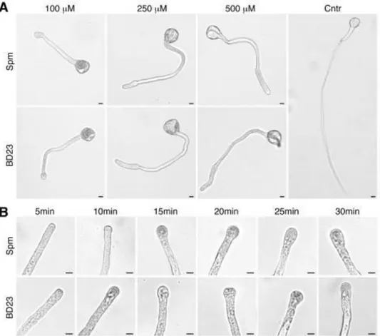
et al., 2010). Spm was the most effective PA in inhibiting pear pollen tube
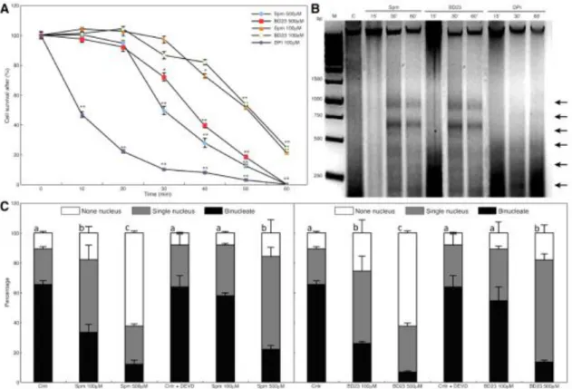
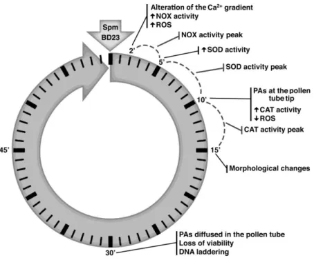
elongation (Aloisi et al., 2015). Spm rapidly entered the pollen tube tip and caused swelling of the apex, suggesting cell wall relaxation. Spm rapidly induced ROS formation (Pottosin et al., 2014; Aloisi et al., 2015), causing the reduction of pollen viability, followed by activation of the antioxidant machinery. The final event after Spm supply was the degradation of nuclear DNA leading to cell death; this process was proposed to be induced either by Ca2+-activated signaling or by the altered
29 Pollen–pistil interaction during fertilization and self-Incompatibility
When pollens land on an incompatible stigma they may undergo the Self Incompatibility (SI) response. This is the most important evolutionary system of the Angiosperms to prevent inbreeding and requires a species-specific cell–cell recognition system. The female determinants can be either a cell membrane receptor as in Papaver rhoeas or a released molecule, such as stigma/style ribonucleases (termed S-RNases) in Solanaceae, Rosaceae and Plantaginaceae; they enter the pollen and are degraded in compatible pollen while they are active in incompatible ones causing the degradation of pollen RNA (Dresselhaus and Franklin-Tong, 2013). The involvement of PAs in the SI response has been reported both in Pyrus communis and in Citrus grandis. In Pyrus communis the content of free PAs (Put and Spm) was lower during incompatible as compared to compatible pollination (Figures2F, G). This could be related to the inhibitory effect of PAs on RNases; in fact, Put and Spd, and, even more, Spm, have been shown to halve the activity of RNase in Malus domestica pollen (Speranza et al., 1984), as also observed in Solanum tuberosum (Altman, 1982). The accumulation of PCA-soluble PAs in reproductive organs, and particularly in pollen, has been associated with fertility. Triferuloyl-Spd, a HCAA of tryphine, is involved in pollination and in pollen–stigma interaction. Moreover, the amount of PCA-soluble PAs was lower in SI-pollinated styles compared to compatible pollinated ones. In the SI-pollination styles, an increase of PCA-insoluble PAs and a higher TGase activity were also observed, concomitantly with the arrest of tube growth and the appearance of a TGase plug at the tip (Del Duca et al., 2010). In contrast to compatible pollination, SI pollination in Citrus grandis was characterized by higher amounts of PCA-insoluble PAs, enhanced TGase activity, and increased production of glutamyl-PAs, together with arrested pollen tube growth (Gentile et al., 2012). The direct involvement of the cytoskeleton in SI was so far solely reported in incompatible Papaver tubes, where a high Ca2+ influx took place after pollen–stigma interaction.
Subsequently, F-actin foci were formed by a still uncharacterized cross-linking mechanism, leading to the arrest of tube elongation and to pollen PCD (McClure and Franklin-Tong, 2006). Since enhancedCa2+ influx is a general feature of the SI
dependent enzyme) was stimulated in Pyrus communis and Citrus grandis. This could have led to cross-links among cytoskeleton proteins, generating high-mass aggregates, similar to the actin foci observed in Papaver, and forming the tube tip plug (Del Duca et al., 2014; Cai et al., 2015b).
FIGURE 2. Polyamine involvement during pollen development. PA biosynthetic and oxidative metabolisms occur from the early stage of pollen formation inside the anthers (A), when both microspores and the tapetal cell layer of the anther contribute to microspore cell wall architecture (B). Pollen accumulates high levels of free PAs and HCAAs, mainly localized in the cell wall. PA catabolism by PAO and DAO modulates the rigidity of the cell wall (C). Once dehydrated, pollen grains are released and PAs contribute to maintain pollen viability (D). During germination on a stigma (E), PAs promote the translation of transcripts and they are also released in the extracellular space, together with TGase (F). During pollen tube growth in a compatible style, PAs take part in the cytoskeleton organization, in cell wall deposition and remodeling by the PME enzyme as well as in the regulation of ion transport through the plasma membrane. PAs also exert an inhibitory effect on RNase enzymes (G).