DOTT.SSA NICOLETTA BERNARDINI
Medico ChirurgoSpecialista in Dermatologia e Venereologia Matricola 696083
Vincitrice del Concorso di Dottorato di ricerca in Anatomia, Dermatologia e Chirurgia Plastica, 31° ciclo “Tecnologie innovative nelle malattie dello scheletro, della cute e del distretto oro-cranio-facciale”, della durata di 3 anni, Università gli Studi di Roma “La Sapienza”
TUTOR: PROF. DIEGO RIBUFFO, PROF.SSA CONCETTA POTENZA
TITOLO: “Nuovi meccanismi infiammatori nella patogenesi della psoriasi: effetti dell’IL-17A nel rilascio e cargo delle vescicole extracellulari in cheratinociti umani”
TITLE: “New pathogenetic inflammatory mechanisms in psoriasis: Interleukin-17A affects extracellular vesicles release and cargo in human keratinocytes”
RAZIONALE: La psoriasi è riconosciuta come uno dei più comuni disordini immuno-mediati, dove sia l’immunità acquisita che quella innata giocano un ruolo fondamentale nello sviluppo e nel mantenimento della patologia. Per tale ragione la psoriasi viene considerata un vero e proprio modello di studio dei meccanismi di infiammazione cronica
Nella psoriasi si assiste alla disregolazione dell’asse citochinico IL-23/IL-17 da cui ne consegue da una parte l’attivazione del subset linfocitario Th-17 e dall’altra una risposta proliferative cheratinocitaria.
Questo induce la secrezione di differenti citochine, chemochine e peptidi antimicrobici da parte dei cheratinociti stessi. L’induzione di specifici fattori chemiotattici richiama, nel sito dell’infiammazione, cellule dendritiche mieloidi, linfociti Th17 e neutrofili.
Il processo infiammatorio psoriasico, oltre alle interazioni cellulari, coinvolge anche le strutture vascolari con il rilascio di citochine, ormoni e secondi messaggeri. È stato, inoltre, dimostrato che le cellule comunicano anche attraverso il rilascio di vescicole contenenti DNA, RNA, microRNA e proteine. Sebbene siano state riportate alterazioni del traffico micro-vescicolare in disturbi ematologici ed infettivi, vi sono poche evidenze di tali alterazioni nella patogenesi della psoriasi.
I risultati preliminari ottenuti nei nostri laboratori indicano come l’IL-17 sia in grado di modificare, sia qualitativamente che quantitativamente, il carico e il rilascio extracellulare di vescicole. L’obiettivo del nostro studio, basato su un gruppo di ricerca multidisciplinare, è identificare nuovi biomarkers della psoriasi.
Background: psoriasis is a chronic inflammatory skin disease caused by the excessive
secretion of inflammatory cytokines. Deregulation of the interleukin-23/-17 axis allows the activation of Th17 lymphocytes and the reprogramming of keratinocytes proliferative response, thereby inducing the secretion of cyto-/chemokines and antimicrobial peptides. Currently, one anti-IL-17 biological agent is approved for the treatment.
Psoriasis-associated inflammation also affect systemic functions associated with extracutaneous manifestations. Beside cell-to-cell contacts and release of cytokines, hormones and second messengers, cells communicate each other through the release of extracellular vesicles containing DNA, RNA, microRNAs and proteins. It has been reported the
alteration of extracellular vesicles trafficking in several diseases, but there is scarce evidence of the involvement of extracellular vesicles trafficking in the pathogenesis of psoriasis.
Objective: the main goal of the study was to characterize the release, the cargo content and
the capacity to transfer bioactive molecules of extracellular vesicles produced by keratinocytes following recombinant IL-17A treatment if compared to untreated keratinocytes.
Methods: a combined approach of standard ultracentrifugation, RNA isolation and Real Time
RT-PCR techniques was used to characterize extracellular vesicles cargo. Flow cytometry was used to quantitatively and qualitatively analyze extracellular vesicles and, in association with
structured illumination microscopy, to evaluate cell-to-cell extracellular vesicles transfer.
Results: we report that the treatment of human keratinocytes with IL-17A significantly modify
the extracellular vesicles cargo and release. Vesicles from IL-17A-treated cells display a specific pattern of mRNA which is abrogated by Secukinumab neutralization. Extracellular vesicles are taken up by acceptor cells irrespective of their content but only those derived from IL-17A-treated cells enable receiving cells to express psoriasis-associated mRNA.
Conclusion: the obtained results imply a role of extracellular vesicles in amplifying the
pro-inflammatory cascade induced in keratinocyte by pro-psoriatic cytokines.
This work was supported by Ateneo Research Projects, Sapienza University of Rome 2016 (C26H153BSX to CP), and 2017 (RM11715C81B4052B to GR).
Introduction
Psoriasis is an inflammatory skin disease, with chronic relapsing course. The classical clinical presentation consists of erythematous-scaly lesions, even if other variants have been described. Patients with psoriasis typically have sharply demarcated chronic erythematous plaques covered by silvery white scales, and the typical erythematous plaque contains histopathological hallmarks that include hyper-proliferation of epidermal keratinocytes and hyperkeratosis, as well as infiltration of immunocytes along with angiogenesis (for review see Mahil et al.).1 Psoriasis affects about 2% of the caucasian population and it is associated wide
range of systemic comorbidities ranging from rheumatologic to cardiovascular disease.2, 3
To date, the classical view of a primary keratinocytes’ dysregulation has been revised, being psoriasis considered as a multifactorial disease, where both innate and adaptive immunity have pivotal roles. Keratinocytes play as the trigger of the inflammatory response, culminating into the production of a pro-inflammatory cytokine network responsible for the typical dermal and epidermal histopathological psoriatic alterations.4 The role of Th1 and Th17 subsets in the
pathogenesis is well characterized with a common pattern of Th1 and Th17 cytokines upregulation, T cell activation and local and systemic expression of endothelins and adhesion molecules. Surrounding the area of inflammation, activated Th1 cells produce interferon (IFN)-γ, interleukin (IL)-2, tumor necrosis factor (TNF)-α, whereas IL-17A, IL-17F and IL-22 are released by Th17 cells. Binding of IL-17A with its receptor activates the transcription of nuclear factor-κB leading to pro-inflammatory gene expression and cytokines production, as IL-6, TNF-α, and IL-1.5 IL-17 acts with IFN-γ by increasing the production and release of IL-6, IL-8 and
other cytokines by keratinocytes.6 IL-8 production by keratinocytes is also induced by TNF-α,
of ICAM-1 by keratinocytes, thus allowing the interaction between lymphocytes and endothelial cells, as well as the expression of chemokines as CXCL9, CXCL10, CXCL11 and receptors thereof as CXCR3.6,8 Nevertheless, some aspects in the settlement of the pro-inflammatory
milieu remain cytokine-specific. In primary human keratinocytes IFN-α, IL-17 and IL-22 modulate distinct and specific inflammatory response pathways.9 IL-17A treatment uniquely
induce the expression of β-defensin 2, CXCL-1, -3, -5, -6, -8 and CCL20, thereby generating the influx of neutrophils, dendritic cells and memory T cells in lesional skin. Instead, IL-22 downregulates genes involved into the differentiation of keratinocytes, thereby determining epidermal alterations in an organotypic skin model. Therefore, IL-17 and IL-22 mediate different pathways associated with clinical aspects of psoriasis: IL-17 is more proinflammatory, while IL-22 retards keratinocyte differentiation.9 On the other hand, the expression of Th2
cytokines IL-4 and-13 is reduced in psoriatic lesions and they have proved to inhibit keratinocytes activation through STAT6, SOCS1 and SOCS3.7,10-13 Even Treg subset is
dysregulated in psoriasis, getting the basis for chronicity.14
Besides the release of cytokines, chemokines, growth factors and soluble messengers, cells communicate each other via the delivery of extracellular vesicles (EVs). Depending of their biogenesis and size, EVs are classified as ectosomes or Microvesicles (MVs) and exosomes.15
All the different EVs subsets contain DNA, RNA, microRNAs and proteins able to modulate the functions of receiving cells. Emerging evidence demonstrates a role of EVs in a variety of fundamental physio- and pathological processes ranging from T cell activation to Alzheimer's disease also correlating their levels to poor prognosis in hematological malignancies.16-18
Conversely, only few data correlate EVs release to inflammatory skin diseases as psoriasis. Seminal observations reported an increase of both endothelial- and platelet-derived circulating
EVs in psoriatic patients.19,20 Conflicting results are obtained in anti-IL-12/23 or anti-TNF-α
treated patients compared to untreated ones.21,22 It has been recently reported the ability of T
cell-derived EVs to induce mast cell production of IL-24 23 as well as the ability of mast
cell-derived exosomes to present neolipid antigens to psoriatic T cells.24 No data are, instead,
available on the ability of IL-17 to modulate the release of EVs by keratinocytes and/or their content in term of bioactive products.
To test the hypothesis of an involvement of EVs in psoriasis pathogenesis, we resorted to an
in vitro model using a spontaneously transformed keratinocyte cell line HaCaT derived from
adult human skin. We treated these cells with recombinant human IL-17A testing, both quantitatively and qualitatively, the EVs release. EVs cargo was also evaluated in the presence of a clinical used approved neutralizing anti-IL-17A antibody (i.e. Secukinumab).
Materials & Methods
Cells and Reagents. The spontaneously transformed aneuploid immortal keratinocyte HaCaT
cell line, derived from adult human skin, was purchased from Cell Line Service GmBH (Eppelheim, Germany) and was cultured in Dulbecco’s Modified Essential Medium (DMEM), 4.5 g/L glucose, supplemented with 10% heat-inactivated Fetal Bovine Serum (FBS), 2 mM L-glutamine, 100 UI/mL Penicillin, 100 µg/mL Streptomicin (Mediatech/Corning, Manassas, VA). Cells were routinely maintained in culture in a humidified atmosphere of 5.5% CO2 at 37°C. Recombinant human IL-17A (rIL-17A) was purchased from Peprotech (London, UK) and used at 100 ng/mL. 5(6)-CFDA-SE (5-(and-6)-Carboxyfluorescein Diacetate, Succinimidyl Ester, CFSE) was purchased from Invitrogen/Thermo Fischer Scientific (Waltham, MA). PKH67 Green Fluorescent Cell Linker Midi Kit for General Cell Membrane Labeling and Actynomicin D were both purchased from Sigma-Aldrich/Merck (Saint Louis, MO). Secukinumab (Cosentyx) was a kind gift of Novartis Pharmaceuticals (Camberley, UK). All the experiments were performed using Exosome-depleted FBS Media Supplement (System Biosciences, Palo Alto, CA).
EVs isolation and staining. Cells were seeded at 1.5x10⁶ cells/mL in 100 mm diameter Petri
dishes (Falcon/Corning, Duhram, NC). After 24 h, cells were washed in complete medium and stimulated for 48 hours with rIL-17A. At the end of stimulation, supernatants were collected and EVs isolated by serial centrifugations. Supernatants were spun 500g for 10’ and 2000g for 10’ to remove detached cells and cellular debris, then ultracentrifugated 100,000g for 60’. Pellet containing EVs was either lysed to isolate total RNA or resuspended in complete medium or phosphate buffered saline (PBS) depending on the subsequent use.
PHK67 fluorophore staining was performed on pelleted EVs resuspended in 0.5 µl of PHK67 in 250 µl of Diluent C buffer, for 5’ at RT. At the end of incubation staining was stopped by adding 250 µl of 1% Bovine Serum Albumin, 1X PBS for 1’ at RT. Exceeding dye was removed by ultracentrifugation.
EVs Cytometric analysis. Cells were seeded at 1.5x10⁶ cells/mL in a 10 cm diameter Petri
dishes. Cells were washed twice in PBS and stained for 10’ at 37°C in serum free medium plus 5 µM CFSE according to the manufacturer’s instruction. Cells were, then, washed twice in complete medium and stimulated with rIL-17A. After 48 h, supernatants were collected, EVs isolated as previously described and resuspended in PBS. Samples were acquired on a FACs ARIA II Cell Sorter equipped with FACs DiVa software v6.1.1 (Becton Dickinson, San Jose, CA) set with a minimal threshold (i.e. 200) on FITC channel and no thresholds on forward and side scatter. Acquisition field was calibrated using Megamix-Plus SSC beads (Biocytex, Marseille, France). Samples were acquired for 30” at minimal flow rate. To avoid carry over, fluidic was extensively washed with 0.1 µm filtered, distilled water before each sample acquisition. Results were analyzed using Flowing software v2.5.1 (Turku Centre for Biotechnology, University of Turku, Finland).
EVs internalization assay and structured illumination microscopy. Cells were seeded at
1.5x10⁶ cells/mL in a 10 cm diameter Petri dishes and stimulated for 48 h with rIL-17A. EVs
were collected and stained with PHK67 as described in the previous section and resuspended in PBS. Unstained cells were incubated at 1:1 v/v ratio with PHK67-labelled EVs at both 37°C and 4°C for different time then cells were analyzed by flow cytometry. For microscopic observations cells (2x10⁴ per well) were seeded on 8 well Culture Slide (Corning Falcon,
EVs was added and incubated for 30’ at both 4°C and 37°C. Cells were, then, washed twice with PBS, counterstained with 4’,6-diamino-2-phenylindole (DAPI, Sigma Aldrich, Saint Louis, MO) and mounted with Mowiol 4-88 Reagent (Calbiochem, Merck Millipore, Billerica, MA). Images were acquired on a Nikon Eclipse Ni-E microscope and processed with NIS-Elements AR software v4.40 (Nikon Instruments, Melville, NY).
RNA isolation and Real Time RT-PCR on cells and EVs. Cells were seeded at 1.5x10⁶
cells/mL in a 10 cm diameter Petri dishes and stimulated as described in the previous section. Supernatants were collected and EVs were isolated as previously described, whereas cells were washed twice in PBS. Total RNA was extracted using the Total RNA purification Kit (Norgen Biotech Corp.). 500 ng of total RNA was retro-transcribed using the Tetro cDNA synthesis kit (Bioline) and cDNA products were analyzed by Real Time PCR using the SensiMix SYBR Hi-ROX Kit (Bioline). Data were normalized using as endogenous control HPRT-1, or 18S rRNA for EVs analysis, and expressed using the 2-ΔΔCT method.25 Primers
used to quantify cytokines, chemokines and β-Defensin 2 were reported in Supplementary
Table S1.
Statistical analysis. Two-tail P value Mann-Whitney t-test was performed to compare two
sample groups. To compare three or more groups, non-parametric ANOVA with post-hoc Kruskal-Wallis test was performed. *: p<0.05; **: p<0.01; ***: p<0.001.
Results
IL-17A reduces the release of EVs. First, we tested the hypothesis that the treatment of
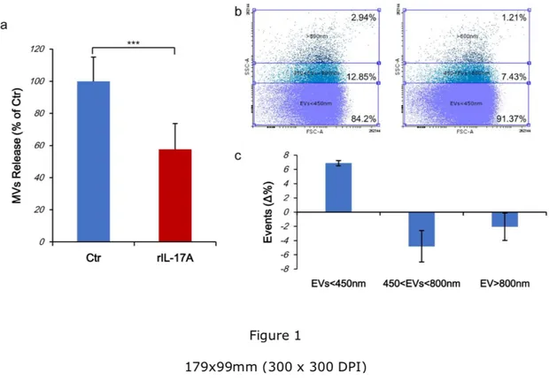
keratinocytes with rIL-17A could affect both quantitatively and/or qualitatively the synthesis and release of MVs. For this purpose, cells were stained with CFSE, treated for 48 h with rIL-17A and released EVs quantified by flow cytometry. rIL-17A treatment decreased the release of EVs in a statistically significant manner (42.512% reduction, p<0.0001, Fig. 1a). Otherwise, the scatter analysis revealed a significant change in the size of EVs released by rIL-17A-treated cells compared to unrIL-17A-treated cultures. A reproducible increase in the percentage of EVs<450 nm isolated from rIL-17A treated HaCaT cells compared to untreated cells was observed (6.87±0.36%, Fig. 1b, 1c and Supplementary Fig. S2). Accordingly, the release of both EVs>800 nm and included in the range of 450 to 800 nm were reduced in IL-17A treated cells compared to control (2.05±1.91% and 4.81±2.21% respectively, Fig. 1b, 1c and
Supplementary Fig. S2). As the CFSE dye does not leak out from undamaged particles, we
also conclude that EVs were intact.
EVs from IL-17A treated cells contain specific mRNAs. In human primary keratinocytes the
treatment with IL-17A induces a specific molecular signature characterized by the upregulation of 28 genes.9 Between these, the most expressed are b-Defensin 2 and the neutrophil
chemoattractants CXCL1, -3, -5, -6 and -8. IL-17A also induces the expression of CCL20 which regulate memory T and dendritic cells traffic. We, then, tested the presence of these mRNAs in EVs isolated from rIL-17A-treated cells. HaCaT cells were treated from 1 to 72 hours with rIL-17A and the expression of several pro-inflammatory cytokines and chemokines mRNA was tested by RT Real Time PCR. In our experimental system all the chemokines previously reported as upregulated by IL-17A were induced at different degrees and with
different kinetics (Supplementary Fig. S3). We also detected the upregulation of IL-6 mRNA as already observed in IL-17 treated murine keratinocyte.30 All these effects were
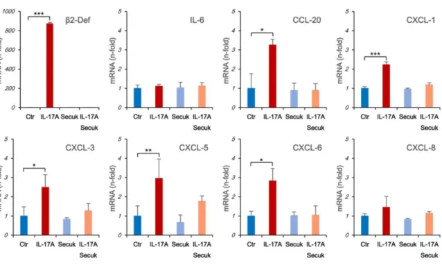
IL-17A-dependent because Secukinumab pre-treatment abrogate these inductions. Then, we analyzed the mRNA content in EVs isolated from both untreated keratinocytes or treated for 72 hours with rIL-17A. EVs isolated from rIL-17A treated cells, embedded mRNA for all chemokines and b-Defensin 2 but not IL-6 and CXCL-8. Again, this feature was completely abrogated by Secukinumab pre-treatment (Fig. 2).
Chronic exposure to IL-17A induces higher b-Defensin 2 mRNA expression in EVs. In vivo psoriatic keratinocytes were continuously exposed to IL-17A produced by chronically
activated Th17 lymphocytes.31 We simulated chronic exposure by stimulating HaCaT cells for
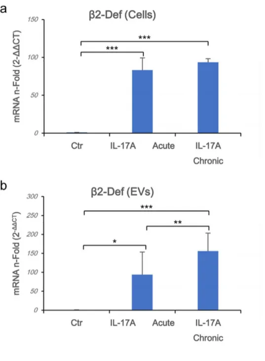
5 days with IL-17A either in acute mode (i.e. one stimulation at the beginning of the experiment) or in chronic stimulation (i.e. daily stimulation with the cytokine), to test if there are any differences between these conditions. Between the previously tested cytokines/chemokines, we measured higher levels of β-Defensin 2 mRNA in samples exposed to IL-17A in chronic rather than in acute manner in both cells and EVs (Fig. 3a and 3b, respectively). According to the kinetic of induction in cells, a shorter (i.e. 72 hours) chronic rIL-17A treatment up-regulated the expression of mRNAs of CCL20 and CXCL3 to a greater extent than an acute stimulation in EVs (data not shown). These results suggest that chronic exposure to IL-17A quantitatively improves the cargo content of EVs.
EVs are internalized by acceptor cells irrespectively of IL-17A. Once we recorded the
ability of HaCaT cells to release EVs, we tested the hypothesis that these vesicles could be transferred to acceptor cells. Then, we isolated EVs from untreated (Ctr-EVs) or IL-17A-treated (IL-17A-EVs) HaCaT cells, staining them with PHK67, a green fluorescent dye able to label
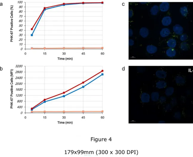
membranes. In both cases, acceptor unstained HaCaT cells were incubated for different time points with stained EVs. Incubation was performed at both 37°C or 4°C to discriminate between EVs endocytosis and surface adsorption, respectively. EVs were rapidly internalized by acceptor cells as 30 to 40 % of acceptor cells were positive to PHK67 when incubated at 37°C for 5’ but not at 4°C, thereby excluding any adsorption to acceptor cell surface (Fig. 4a). Internalization curves of HaCaT treated with Ctr-EVs and IL-17A-EVs were superimposable. Nevertheless, Mean Fluorescence Intensity (MFI) of acceptor cells treated with IL-17A-EVs are slightly higher than Ctr-EVs (Fig. 4b). We also perform immunofluorescence analysis on EVs treated cells using structured illumination microscopy. Again, both Ctr-EVs (Fig. 4c) and IL-17A-EVs (Fig. 4d) were internalized at 37°C with the same efficiency and no major differences in the intracellular localization was detected whereas no fluorescence was detected at 4°C (data not shown).
b-Defensin 2 mRNA is over-represented in acceptor cells. In view of the previously
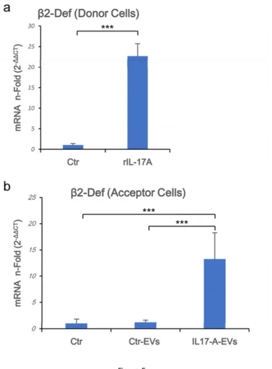
reported results, we wondered if cargo mRNAs could be transferred to acceptor cells. Therefore, HaCaT cells were treated with both Ctr-EVs and IL-17A-EVs. Acceptor cells were daily stimulated with EVs for 5 days to simulate chronic exposure to EVs (Fig. 5a) and, to monitor the cargo transfer, we measured the b-Defensin 2 mRNA as readout. As depicted in
Fig. 5b b-Defensin 2 mRNA was specifically upregulated in cells treated with IL-17A-EVs.
IL-17A-EVs specifically induces endogenous b-Defensin 2 mRNA in acceptor keratinocytes. Based on the previous result, two possible scenarios are conceivable: i)
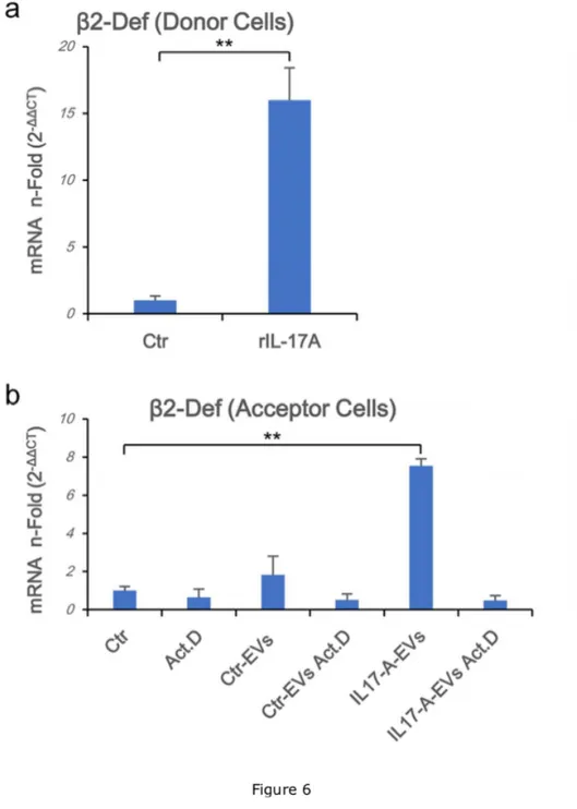
IL-17A-EVs transfer their cargo to acceptor cells enabling them to express “exogenous” mRNAs or ii) IL-17A-EVs allow acceptor cells to express endogenous b-Defensin 2 mRNA. To discriminate between these two hypotheses, acceptor cells were pre-treated with Actinomycin
D thereby interfering with cellular mRNA synthesis, then incubated with EVs collected from both donor untreated or IL-17A-treated cells, containing b-Defensin 2 mRNA (Fig. 6a). Cells pre-treated with Actinomycin D failed to upregulate b-Defensin 2 mRNA following IL-17A-EVs challenge (Fig. 6b). As Actinomycin D was unable to inhibit the expression in acceptor cells of a viral derived mRNA (i.e. HPV-16 E6 mRNA shipped by CaSki-derived EVs, Supplementary
Fig. S3), these results suggest that EVs derived from IL-17A treated keratinocytes, but not
from untreated cells, are able to induce the expression of specific mRNAs (i.e. b-Defensin 2) in acceptor cells in the absence of the primitive stimulus (i.e. IL-17A).
Discussion
The increased knowledge of EVs biology and their ability to enrich tissue-specific biomarkers in normal and pathological conditions, suggest that they may provide a valuable tool to identify diagnostic signatures, develop new diagnostic assays and help guide therapeutic approaches. In the case of psoriasis, the comprehension of the role of EVs relased from different cell type (i.e. Th17 cells, mieloid dendritic cells, keratinocytes, mast cells) is just at an early stage. Reports have demonstrated an increase in the number of circulating EVs derived form platelets and endotelial cells in patients compared to healthy subjects19,20 but none addressed
the issue of keratinocytes-derived EVs. Our in vitro model suggests that IL-17A, a driver cytokine in the pathogenesis of psoriasis, reduces the delivery of EVs (Fig. 1a). Differences between our data and those reported by others are due by either the different system used (i.e. plasma form patients versus cell culture supernantants) and by the different nature of producing cells (i.e. platelet/endothelial cells versus keratinocytes). We also observed an effect of IL-17A in the size of the EVs population released by keratinocytes. Indeed, EVs produced from IL-17A-treated cells are smaller in size compared to those released from untreated cells (Fig. 1b) suggesting the presence in the former sample of a heterogeneous population of EVs mostly composed from exosomes and/or small MVs (in the range of 100 nm). The exposure of HaCaT cells to IL-17A not only have a sizeable impact on the EVs release, but also affect their cargo in terms of mRNA. Results obtained in EVs isolated from IL-17A-treated HaCaT cells (Fig. 2) demonstrate the presence of mRNAs for chemokines/alarmins representing an IL-17A-specific signature overlapping the intracellular expression pattern in keratinocytes.9 According
to Nograles et al., the most induced mRNA in both cells and EVs is b-Defensin 2 also in our experimental system. b-Defensin 2 is a psoriasis-associated marker together with CCL20,
another mRNA we founded up-regulated in IL-17A-EVs.32,33 Another streaking feature is the
ability of Secukinumab to inhibit the expression of IL-17A-related mRNAs in EVs and in keratinocytes. Nevertheless, Secukinumab does not reduce the release of EVs (Data not shown). This suggest that EVs release takes place through an IL-17A-independent pathway, whereas EVs cargo is Secukinumab-sensitive.
Beside the prognostic/diagnostic use, EVs represent an almost unexplored way of cell-to-cell and cell-to-extracellular matrix communication. From this point of view one of the crtitcal step is the interaction between EVs and target/acceptor cells. Our results (Fig. 4) demonstrate that not only EVs derived by keratinocytes homotypically interact with acceptor cells but also that these EVs are internalized. Fluorescent-labelled EVs stain at 37°C, but not at 4°C, acceptor HaCaT cells regardless of rIL-17A treatment of donor cells. This suggest that the interaction between EVs and acceptor cells arise trough a molecule constitutively expressed on EVs surface. Nevertheless, only EVs derived from cells treated with IL-17A induce the expression of b-Defensin 2 mRNA in acceptor cells. Results obtained pre-treating acceptor cells with Actinomycin D indicate that the b-Defensin 2 mRNA expression is due to de novo synthesis of endogenous mRNA rather than the transfer from donor to acceptor cells. Indeed, when EVs expressing a heterologous viral mRNA (i.e. HPV-16 E6 mRNA) were used, Actinomycin D is unable to inhibit its expression in acceptor cells (Supplementary Fig. S3). Since it is known that, Depending on concentration, Actinomycin D blocks pinocytosis and endocytosis,34,35
using fluorescent-labelled EVs we verified that the concentration used was able to inhibit RNA neo-synthesis but not pino- and endocytosis (data not shown).
Our results indicate that IL-17A affects the production of EVs and their size in keratinocytes also reshaping the cargo content through the overexpression of psoriasis-associated mRNAs
like b-Defensin 2. These EVs interact and are internalized by acceptor cells which, in turn, express b-Defensin 2 in the absence of primitive stimulus (i.e. IL-17A). This mechanism could play a role in the amplification of the chronic inflammatory state in the surroundings of the psoriatic lesions and, if EVs reach the blood stream, a role in the dissemination of the disease in other anatomical sites as heart, eye or joints in the case of psoriatic arthritis could be also hypothesized.
References
1. Mahil SK, Capon F, Barker JN. Update on psoriasis immunopathogenesis and targeted immunotherapy. Semin Immunopathol 2016;38(1):11-27.
2. Ni C, Chiu MW. Psoriasis and comorbidities: links and risks. Clin Cosmet Investig
Dermatol 2014;7:119-32.
3. Ahlehoff O, Gislason G, Hansen PR. Cardiovascular aspects of psoriasis: an updated review. Int J Dermatol 2014;53(6):e337.
4. Nograles KE, Davidovici B, Krueger JG. New insights in the immunologic basis of psoriasis. Semin Cutan Med Surg 2010;29(1):3-9.
5. Tan KW, Griffiths CE. Novel systemic therapies for the treatment of psoriasis. Expert
Opin Pharmacother 2016;17(1):79-92.
6. Boca AN, Talamonti M, Galluzzo M, Botti E, Vesa SC, Chimenti S, et al. Genetic variations in IL6 and IL12B decreasing the risk for psoriasis. Immunol Lett 2013;156(1-2):127-31.
7. Szondy Z, Pallai A. Transmembrane TNF-alpha reverse signaling leading to TGF-beta production is selectively activated by TNF targeting molecules: Therapeutic implications. Pharmacol Res 2017;115:124-32.
8. Chiricozzi A, Saraceno R, Chimenti MS, Guttman-Yassky E, Krueger JG. Role of IL-23 in the pathogenesis of psoriasis: a novel potential therapeutic target? Expert Opin Ther
Targets 2014;18(5):513-25.
9. Nograles KE, Zaba LC, Guttman-Yassky E, Fuentes-Duculan J, Suárez-Fariñas M, Cardinale I, et al. Th17 cytokines interleukin (IL)-17 and IL-22 modulate distinct inflammatory and keratinocyte-response pathways. Br J Dermatol 2008;159(5):1092-102.
10. Duffin KC, Woodcock J, Krueger GG. Genetic variations associated with psoriasis and psoriatic arthritis found by genome-wide association. Dermatol Ther 2010;23(2):101-13.
11. Heidenreich R, Röcken M, Ghoreschi K. Angiogenesis drives psoriasis pathogenesis.
Int J Exp Pathol 2009;90(3):232-48.
12. Gelfand JM, Yeung H. Metabolic syndrome in patients with psoriatic disease. J
Rheumatol Suppl. 2012;89:24-8.
13. Wilson NJ, Boniface K, Chan JR, McKenzie BS, Blumenschein WM, Mattson JD, et al. Development, cytokine profile and function of human interleukin 17-producing helper T cells.
Nat Immunol 2007;8(9):950-7.
14. Richetta AG, Mattozzi C, Salvi M, Giancristoforo S, D'epiro S, Milana B, et al. CD4+ CD25+ T-regulatory cells in psoriasis. Correlation between their numbers and biologics-induced clinical improvement. Eur J Dermatol 2011;21(3):344-8.
15. Raposo G, Stoorvogel W. Extracellular vesicles: exosomes, microvesicles, and friends.
J Cell Biol 2013;200(4):373-83.
16. Raposo G, Nijman HW, Stoorvogel W, Liejendekker R, Harding CV, Melief CJ, et al. B lymphocytes secrete antigen-presenting vesicles. J Exp Med 1996;183(3):1161-72.
17. Valadi H, Ekström K, Bossios A, Sjöstrand M, Lee JJ, Lötvall JO. Exosome-mediated transfer of mRNAs and microRNAs is a novel mechanism of genetic exchange between cells.
Nat Cell Biol 2007;9(6):654-9.
18. Kim HK, Song KS, Park YS, Kang YH, Lee YJ, Lee KR, et al. Elevated levels of circulating platelet microparticles, VEGF, IL-6 and RANTES in patients with gastric cancer: possible role of a metastasis predictor. Eur J Cancer 2003;39(2):184-91.
19. Tamagawa-Mineoka R, Katoh N, Kishimoto S. Platelet activation in patients with psoriasis: increased plasma levels of platelet-derived microparticles and soluble P-selectin. J
Am Acad Dermatol 2010;62(4):621-6.
20. Pelletier F, Garnache-Ottou F, Angelot F, Biichlé S, Vidal C, Humbert P, et al. Increased levels of circulating endothelial-derived microparticles and small-size platelet-derived microparticles in psoriasis. J Invest Dermatol 2011;131(7):1573-6.
21. Ho JC, Lee CH, Lin SH. No Significant Reduction of Circulating Endothelial-Derived and Platelet-Derived Microparticles in Patients with Psoriasis Successfully Treated with Anti-IL12/23. Biomed Res Int 2016;2016:3242143.
22. Pelletier F, Garnache-Ottou F, Biichlé S, Vivot A, Humbert P, Saas P, et al. Effects of anti-TNF-α agents on circulating endothelial-derived and platelet-derived microparticles in psoriasis. Exp Dermatol 2014;23(12):924-5.
23. Shefler I, Pasmanik-Chor M, Kidron D, Mekori YA, Hershko AY. T cell-derived microvesicles induce mast cell production of IL-24: relevance to inflammatory skin diseases. J
Allergy Clin Immunol 2014;133(1):217-24.e1-3.
24. Cheung KL, Jarrett R, Subramaniam S, Salimi M, Gutowska-Owsiak D, Chen YL, et al. Psoriatic T cells recognize neolipid antigens generated by mast cell phospholipase delivered by exosomes and presented by CD1a. J Exp Med 2016; 213(11):2399-412.
25. Livak KJ, Schmittgen TD. Analysis of relative gene expression data using real-time quantitative PCR and the 2(-Delta Delta C(T)) Method. Methods 2001;25(4):402-8.
26. Keller C, Keller P, Marshal S, Pedersen B. IL-6 gene expression in human adipose tissue in response to exercise--effect of carbohydrate ingestion. J Physiol 2003;550(Pt 3):927-31.
27. Zeeuwen PL, de Jongh GJ, Rodijk-Olthuis D, Kamsteeg M, Verhoosel RM, van Rossum MM, et al. Genetically programmed differences in epidermal host defense between psoriasis and atopic dermatitis patients. PLoS One 2008;3(6):e2301.
28. Iuliano M, Mangino G, Chiantore MV, Zangrillo MS, Accardi R, Tommasino M, et al. Human Papillomavirus E6 and E7 oncoproteins affect the cell microenvironment by classical secretion and extracellular vesicles delivery of inflammatory mediators. Cytokine 2018;106:182-9.
29. Palanichamy JK, Mehndiratta M, Bhagat M, Ramalingam P, Das B, Das P, et al. Silencing of integrated human papillomavirus-16 oncogenes by small interfering RNA-mediated heterochromatization. Mol Cancer Ther 2010;9(7):2114-22.
30. Wu L, Chen X, Zhao J, Martin B, Zepp JA, Ko JS, et al. A novel IL-17 signaling pathway controlling keratinocyte proliferation and tumorigenesis via the TRAF4-ERK5 axis. J Exp Med 2015;212(10):1571-87.
31. Miossec P, Kolls JK. Targeting IL-17 and TH17 cells in chronic inflammation. Nat Rev
Drug Discov 2012;11(10):763-76.
32. Kolbinger F, Loesche C, Valentin MA, Jiang X, Cheng Y, Jarvis P, et al. β-Defensin 2 is a responsive biomarker of IL-17A-driven skin pathology in patients with psoriasis. J Allergy
Clin Immunol 2017;139(3):923-32.e8.
33. Liu Y, Lagowski JP, Gao S, Raymond JH, White CR, Kulesz-Martin MF. Regulation of the psoriatic chemokine CCL20 by E3 ligases Trim32 and Piasy in keratinocytes. J Invest
34. Alsharif NH, Berger CE, Varanasi SS, Chao Y, Horrocks BR, Datta HK. Alkyl-capped silicon nanocrystals lack cytotoxicity and have enhanced intracellular accumulation in malignant cells via cholesterol-dependent endocytosis. Small 2009;5(2):221-8.
35. Kuhn DA, Vanhecke D, Michen B, Blank F, Gehr P, Petri-Fink A, et al. Different endocytotic uptake mechanisms for nanoparticles in epithelial cells and macrophages. Beilstein J Nanotechnol 2014;5:1625-36.
Figure Legends
Figure 1. IL-17A reduces the release of EVs. Cells (5x10⁵) were seeded in 60 mm diameter
Petri dishes. After 24 h, cells were stained with CFSE and stimulated for 72 h with rIL-17A (100 ng/mL). Supernatants were collected, EVs isolated and acquired on a FACs ARIA II cytometer. (a) CFSE positive EVs were enumerated, normalized to cell counts and results are expressed as percentage compared to untreated cells. Blue bar: EVs from untreated cells (Ctr); red bar: EVs from IL-17A treated cells. (b) A representative dot plot of CFSE stained EVs derived from untreated (left panel) and rIL-17A treated cells (100 ng/mL, right panel). EVs sizes were set according to the calibration beads size. (c) The variation in the EVs sub-populations in response to rIL-17A treatment was expressed as the difference between the
percentage of each EVs sub-population measured in IL-17A treated cells and the percentage in untreated controls (Δ%).
Figure 2. EVs from IL-17A treated cells contain specific mRNAs. Cells (1.5x10⁶ in 100 mm
diameter Petri dishes) were stimulated for 72 h with rIL-17A (100 ng/mL) pre-incubated for 1 h at 37°C with or without Secukinumab, with Secukinumab alone or left untreated. Supernatants were collected, EVs were isolated, total RNA was purified and Real Time RT-PCR was performed as described in materials and methods section. Results were expressed as fold of induction (n-Fold) according to the 2-ΔΔCT method, using the value of control as reference and
the value of RNA 18S as an internal loading control. Blue bar: EVs from untreated cells (Ctr); red bar: EVs from rIL-17A treated cells; light blue bar: EVs from Secukinumab (Secuk) treated cells; light red bar: EVs from Secukinumab plus IL-17A treated cells.
Figure 3. Chronic exposure to IL-17A induces higher b-Defensin 2 mRNA expression in EVs. Cells (1.5x10⁶ in 100 mm diameter Petri dishes) were stimulated for 72 h with rIL-17A
(100 ng/mL) or left untreated. Supernatants were collected and EVs were isolated whereas cells were washed twice in PBS and lysed. Total RNA was purified, and β-Defensin 2 Real Time RT-PCR was performed on both cells (a) or EVs (b) as described in materials and methods section. Results were expressed as fold of induction (n-Fold) according to the 2-ΔΔCT
method, using the value of control as reference and the value of RNA 18S (EVs) and HPRT-1 (Cells) as internal loading controls. Ctr: untreated cells.
Figure 4. EVs is internalized by acceptor cells irrespectively of IL-17A. (a, b) Cells were
seeded at 1.5x10⁶ in 100 mm diameter Petri dishes. After 24 h, cells were stimulated for 72 h
with rIL-17A (100 ng/mL). At the end of the stimulation, supernatants were collected and EVs were isolated and stained with PHK67 as described in materials and methods. HaCat cells (105 cells/100 µl in PBS) were incubated with PHK67-labeled EVs in a 1:1 v/v ratio for 5’, 15’,
30’, 45’ and 60’ at both 37°C or 4°C. Incubation was stopped by adding 900 µl of cold PBS and cells were analyzed by flow cytometry. Results were expressed as percentage of PHK67 positive cells (a) or as Mean Fluorescent Intensity (MFI, b). Blue line: untreated cells, 37°C;
red line: rIL-17A, 37°C; light blue line: untreated cells, 4°C; light red line: rIL-17A, 4°C. (c, d) Cells (5x10⁵) were seeded in 60 mm diameter Petri dishes. After 24 h, cells were stimulated
for 72 h with rIL-17A (100 ng/mL). At the end of the stimulation, supernatants were collected and EVs were isolated and stained with PHK67 as described in materials and methods. HaCat cells (2x10⁴ cells) were seeded on culture slide and incubated for 30’ at 37°C with
PHK67-labeled EVs. (c) HaCaT cells treated with EVs from unstimulated cells; (d) HaCaT cells treated with EVs from rIL-17A stimulated cells.
Figure 5. b-Defensin 2 mRNA is overrepresented in acceptor cells. Donor cells (1.5x10⁶ in
left untreated. Supernatants were collected, EVs from rIL-17A-treated cells (IL-17A-EVs) and from unstimulated cells (Ctr-EVs) were isolated and then added to acceptor cells seeded at 1.5x10⁶ in 100 mm diameter Petri dish. Acceptor cells were incubated with or without EVs for
24 h then washed twice in PBS and lysed. Total RNA was purified, and β-Defensin 2 Real Time RT-PCR was performed as described in materials and methods section on both donor
(a) and acceptor cells (b). Results were expressed as fold of induction (n-Fold) according to
the 2-ΔΔCT method, using the value of control as reference and the value of HPRT-1 as an
Figure 6. IL-17A-EVs specifically induce endogenous b-Defensin 2 mRNA in acceptor keratinocytes. Donor cells (1.5x10⁶ in 100 mm diameter Petri dishes) were daily stimulated
for 5 days with rIL-17A (100 ng/mL) or left untreated. Supernatants were collected, EVs from rIL-17A-treated cells (IL-17A-EVs) and from unstimulated cells (Ctr-EVs) were isolated. Acceptor cells were seeded at 1.5x10⁶ in 100 mm diameter Petri dish and pre-treated with
Actinomycin D (500 ng/mL) for 30’ at 37°C or left untreated. Cells were, then, washed twice with medium and stimulated with or without EVs for 24 h at 37°C. At the end of the stimulation cells were washed twice in PBS and lysed. Total RNA was purified, and β-Defensin 2 Real Time RT-PCR was performed as described in materials and methods section on both donor
(a) and acceptor cells (b). Results were expressed as fold of induction (n-Fold) according to
the 2-ΔΔCT method, using the value of control as reference and the value of HPRT-1 as an
Supplementary Materials
Supplementary Figure S2. IL-17A alters the relative expression of EVs sub-populations.
Cells (5x10⁵) were seeded in 60 mm diameter Petri dishes. After 24 h, cells were stained with
CFSE and stimulated for 72 h with rIL-17A (100 ng/mL). Supernatants were collected, EVs isolated and acquired on a FACs ARIA II cytometer as described in materials and methods. CFSE positive EVs were enumerated and the results were expressed as the percentage of positive cells belonging to each subpopulation. Blue bar: EVs from untreated cells (Ctr); red bar: EVs from IL-17A treated cells.
Supplementary Figure S3. Cells treated with IL-17A contain specific mRNAs. Cells
(1.5x10⁶ in 100 mm diameter Petri dishes) were stimulated for 1, 6, 24, 48 and 72 h with
rIL-17A (100 ng/mL) pre-incubated for 1 h at 37°C with or without Secukinumab, with Secukinumab alone or left untreated. Cells were washes twice with PBS and lysed. Total RNA was purified, and Real Time RT-PCR was performed as described in materials and methods
section. Results were expressed as fold of induction (n-Fold) according to the 2-ΔΔCT method,
using the value of control as reference and the value of RNA 18S as an internal loading control. Blue line: untreated cells (Ctr); red line: rIL-17A; light blue line: Secukinumab (Secuk); light red line: Secukinumab plus rIL-17A.
Supplementary Figure S4. Actinomycin D is unable to inhibit HPV16 E6 mRNA expression in acceptor keratinocytes. CaSki cells (1.5x10⁶ in 100 mm diameter Petri
dishes) were cultured for 5 days at 37°C. Supernatants were collected and EVs were isolated. Acceptor HaCaT cells were seeded at 1.5x10⁶ in 100 mm diameter Petri dish and pre-treated
twice with medium and stimulated with or without EVs for 24 h at 37°C. At the end of the stimulation cells were washed twice in PBS and lysed. Total RNA was purified, and HPV16 E6 Real Time RT-PCR was performed as described in materials and methods section on acceptor cells. Results were expressed as fold of induction (n-Fold) according to the 2-ΔΔCT method,
using the value of control as reference and the value of HPRT-1 as an internal loading control. Ctr: untreated cells; Act D: Actinomycin D.
