Alma Mater Studiorum
Alma Mater Studiorum –
– Università di Bologna
Università di Bologna
DOTTORATO DI RICERCA IN
MORFOFISIOLOGIA E PATOLOGIA VETERINARIA CON
APPLICAZIONI BIOTECNOLOGICHE
Ciclo
xxv
Settore Concorsuale di afferenza: 07/H1 Settore Scientifico disciplinare: VET/01
TITOLO TESI
TASTE RECEPTORS IN THE GUT: A CHEMOSENSITIVE
MECHANISM FROM FISH TO HUMAN
Presentata da: ROCCO LATORRE
Coordinatore Dottorato: Chiar.mo Prof. Eraldo Seren
Relatore: Chiar.mo Prof. Paolo Clavenzani
i
Riassunto
L’ingestione di un pasto evoca una serie di processi digestivi che consistono nelle funzioni essenziali dell’apparato digerente, trasporto degli alimenti, attività secretiva, assorbimento dei nutrienti digeriti e l’espulsione dei residui non assorbiti. La gastrointestinal chemosensitivity è caratterizzata da elementi cellulari endocrini della mucosa gastroenterica e da fibre nervose, soprattutto di natura vagale. Una ampia gamma di mediatori endocrini e/o paracrini possono essere rilasciati da varie cellule endocrine in risposta a nutrienti introdotti con la dieta. Tali ormoni, oltre alla loro attività diretta, agiscono attraverso recettori specifici attivando azioni di assoluta importanza nel controllo di varie funzioni tra cui l’introito calorico e l’omeostasi energetica dell’organismo. Ad integrazione di questo complesso sistema di controllo della chemosensitività gastrointestinale, recenti evidenze dimostrano la presenza di recettori del gusto (o taste receptors, TR) appartenenti alla famiglia dei recettori correlati alle proteine G espressi a livello della mucosa del tratto gastrointestinale di diversi mammiferi e dell’uomo.
La presente ricerca, suddivisa in diversi progetti di ricerca, è stata concepita al fine di chiarire il rapporto tra TR e nutrienti. Per definire questo rapporto sono stati usati diversi approcci scientifici, che sono andati a valutare le variazioni delle molecole segnale dei TR in particolare dell’α-transducina in condizioni di digiuno e a seguito di rialimentazione standard nel tratto gastrointestinale di suino, la mappatura della stessa molecola segnale nel tratto gastrointestinale di pesce (Dicentrarchus Labrax), il signaling pathway dei bitter TR in colture cellulari endocrine STC-1 ed infine il coinvolgimento dei bitter TR, in particolare del T2R38 in pazienti con un eccessivo introito calorico. I risultati hanno evidenziato come ci sia una stretta correlazione tra nutrienti, TR e rilascio ormonale e come questi siano coinvolti non solo nella percezione del gusto propriamente detto ma probabilmente anche in patologie croniche come l’obesità.
ii
Abstract
The ingestion of a meal evokes a series of digestive processes, which consist of the essential functions of the digestive system: food transport, secretory activity, absorption of nutrients and the expulsion of undigested residues do not absorbed. The gastrointestinal chemosensitivity is characterized by cellular elements of the endocrine gastrointestinal mucosa and nerve fibers, in particular of vagal nature. A wide range of mediators endocrine and/or paracrine can be released from various endocrine cells in response to nutrients in the diet. These hormones, in addition to their direct activity, act through specific receptors activating some of the most important functions in the control of energy intake and energy homeostasis in the body. For integration of this complex system of control of gastrointestinal chemosensitivity, recent evidence demonstrates the presence of taste receptors (TR) belonging to the family of G proteins coupled receptor expressed in the mucosa of the gastrointestinal tract of different mammals and human. This thesis is divided into several research projects that have been conceived in order to clarify the relationship between TR and nutrients. To define this relationship I have used various scientific approaches, which have gone on to evaluate changes in signal molecules of TR, in particular of the α-transducin in the fasting state and after refeeding with standard diet in the gastrointestinal tract of the pig, the mapping of the same molecule signal in the gastrointestinal tract of fish (Dicentrarchus labrax), the signaling pathway of bitter TR in the STC-1 endocrine cell line and finally the involvement of bitter TR in particular of T2R38 in patients with an excessive caloric intake. The results showed how there is a close correlation between nutrients, TR and hormonal release and how they are useful both in taste perception but also likely to be involved in chronic diseases such as obesity.
iii
INDEX
RIASSUNTO/ABSTRACT...
I
LIST OF TABLES...
Vii
LIST OF FIGURES...
Vii
AKNOWLEDGEMENTS...
INTRODUCTION...
1
THE GASTROINTESTINAL TRACT IN MAMMALS...
1
I)
THE STOMACH...
1
i) Structure...
2
ii) Fundic Mucosa...
4
iii) Cardial Mucosa...
6
iv) Pyloric Mucosa...
6
II) INTESTINE...
7
i) The Small Intestine...
7
a. Duodenum...
8
b. Jejunum...
8
c. Ileum...
9
d. Structure...
9
ii) The Large Intestine...
11
a. Cecum...
11
b. Colon...
12
c. Rectum...
13
d. Structure...
13
iv
THE GASTROINTESTINAL TRACT IN FISH...
14
I)
THE HEADGUT...
15
II) THE FOREGUT...
15
i) The esophagus...
15
ii) The stomach...
15
III) THE MIDGUT AND HINDGUT...
17
THE SENSE OF TASTE……….
18
TASTE BUDS………
19
BITTER TASTE RECEPTORS……….………
22
α GUSTDUCIN AND TRANSDUCIN SUBUNIT……….
24
CELLULAR SIGNAL OF TASTE TRANSDUCTION……….
26
I)
BITTER………..….
26
II) SWEET………...
28
III) SALTY………..
29
IV) SOUR………..
29
V) UMAMI……….
30
TASTE RECEPTORS IN THE GI TRACT………..
31
GUT CHEMOSENSING AND ENTEROENDOCRINE SYSTEM……..
33
FOOD INTAKE MECHANISMS……….…….
37
I)
GASTRIN………
38
II) SOMATOSTATIN……….……...
39
III) CHOLECYSTOKININ………
40
IV) GHRELIN……….…..
41
V) OBESTATIN………..……
43
v
VI) GLUCAGONE LIKE PEPTIDE‐1………..
43
VII) SEROTONIN……….
45
CONCLUSION………...
46
REFERENCES………... 47
CHAPTER 1...
Enteroendocrine profile of α‐transducin immunoreactive cells in the
gastrointestinal tract of the European sea bass (Dicentrarchus labrax)
66
CHAPTER 2...
Expression and regulation of α‐transducin in the pig gastrointestinal
tract
92
CHAPTER 3...
Activation of Enteroendocrine STC‐1 Cell Signaling By Bitter
Compounds and Bacteria Quorum Sensing Molecules (N‐Acyl
Homoserine Lactone)
119
CHAPTER 4...
Modulation of the T2R38 (Bitter Taste Receptors) in Healthy Human
Subjects by Diet
144
vi
LIST OF TABLES
Table Page
INTRODUCTION
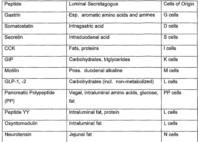
Tab I Gastrointestinal peptide, function and localization in endocrine cells in the GI tract
38
CHAPTER I
Tab I List of antibodies used in this study 85
Tab II Peptides used for absorption test 86
CHAPTER II
Tab I List and diluition of primary and secondary antibodies 96 Tab IIa Mean number of Gαtran/CgA-IR cells in the pig GI tract 100 Tab IIb Percentage of gastrin/total CgA-IR cells in the pig GI tract 100 °Tab III Mean number and percentage of the co-localized Gαtran/total GHR-IR
cells in the cardiac and pyloric mucosa
103
Tab IV Mean number and percentage of the co-localized Gαtran/total CCK-IR cells in the jejunum
104
Tab I
CHAPTER III Molecular composition of HBSS solution
CHAPTER IV
128
Tab I List of primary and secondary antibodies used in this study 148
LIST OF FIGURES
Figure INTRODUCTION Page
Fig 1 A. External and internal anatomy of the stomach B. Region of the pig stomach
2 Fig 2 General cell composition and location in a gastric glands 5 Fig 3 Schematic representation of human and pig GI tract 7 Fig 4 Different types of epithelial cells present in the intestine 11 Fig 5 Overview of gastrointestinal system in the bone fish 14
Fig 6 Different shapes of fish stomachs 16
Fig 7 Taste bud organization 19
vii
Fig 9 Bitter taste signalling pathways 23
Fig 10 General transduction mechanisms in taste 26
Fig 11 Sweet and bitter taste transduction 28
Fig 12 Salt and sour taste transduction 30
Fig 13 Possible mechanisms involved in GI chemosensing 34 Fig 14 Different GI hormones regulate food intake through bloodstream and
vagal afferents 37
Fig 15 Biological action of gastrin hormone 39
Fig 16 Nutritional stimulation of CCK 41
Fig 17 Biological action of ghrelin hormone 42
Fig 18 Biological action of GLP-1 44
CHAPTER I
Fig 1 Western blot analysis 87
Fig 2 Representative images of seabass gastric mucosa 89 Fig 3 Representative images illustrating different subpopulation of ECC
cells in seabass gastric and intestinal mucosa 91 CHAPTER II
Fig 1 Localization of Gαtran-IR in the pig GI tract 106 Fig 2 Graphs, mean number of Gαtran-IR in the different segment of the pig
GI tract
107 Fig 3 Colocalization of Gαtran-IR with ghrelin in the pyloric mucosa and
CCK in the jejunum
119 Fig 4 Enteroendocrine cells of the duodenum co-expressing Gαtran and
SOM-IR 111
Supplementary data CHAPTER II
Fig 1 Western blot of Gαtran antibody 114
Fig 2 Western blot of Gαgust antibody 114
Fig 3 Western blot of GAS/CCK antibody 114
CHAPTER III
Fig 1 Different MapK pathways 124
Fig 2 Mechanisms mediating AHL effect of mammal cells 126 Fig 3a WB results showing the % of phosphorilation for MapKp44/42 on
STC-1 after 3’ incubation with increasing PTC concentration
129 Fig 3b Wb results showing the % of phosphorilation for MapK on STC-1
after 3’ incubation with increasing DB concentration
130 Fig 4a PTC induces MAPk phosphorilation, is blocked by GF-1 in a dose
dependent manner in STC-1 cells
viii
Fig 4b DB induces MAPk phosphorilation, it is not blocked by GF-1 in STC-1 cells
131 Fig 4c DB induces MAPk phosphorilation, is blocked by nitrendipine in a
dose dependent manner in STC-1 cells 132
Fig 4d PTC induces MAPk phosphorilation, it is not blocked by nitrendipine in STC-1 cells
132 Fig 5a WB results showing the % of phosphorilation for MapKp44/42 on
STC-1 after STC-10’ incubation with increasing AHL concentration
133 Fig 5b AHL induces MAPk phosphorilation, is blocked by GF-1 in a dose
dependent manner in STC-1 cells
134 Fig 5c AHL induces MAPk phosphorilation, it is not blocked by
nitrendipine in STC-1 cells
134 Fig 6a PTC induces MAPk phosphorilation, is blocked by Probenecid in a
dose dependent manner in STC-1 cells
135 Fig 6b AHL induces MAPk phosphorilation, is blocked by Probenecid in a
dose dependent manner in STC-1 cells
135 Fig7 The bitter stimuli PTC and the QS molecule AHL rapdly increase
intracellular [Ca2+]i in STC-1
CHAPTER IV
136
Fig 1 Effect of a different BMI on mT2R38 expression 149 Fig 2 Representative image of human colonic mucosa. hT2R38
immunoreactivity colocalised with ChrA in normal weight and overweight/obese group.
150
Fig 3 Representative image of human colonic mucosa. hT2R38 immunoreactivity colocalised with GLP-1.
150
Fig 4 Representative image of human colonic mucosa. hT2R38 immunoreactivity colocalised with CCK.
1
INTRODUCTION
THE GASTROINTESTINAL TRACT IN MAMMALS
The alimentary canal consists of the esophagus, stomach, small intestine, large intestine and the anal canal. Associated to it there are two large glands that release their secretions into the intestinal lumen: liver and pancreas.
I will consider only the gastrointestinal tract, and so I will not treat the esophagus and the associated glands. I will try to describe the generality of the gastrointestinal tract, highlighting the key parts in pigs and humans
.
I) THE STOMACH
The stomach is a dilated portion of the digestive tract, which follows the esophagus at the level of cardia and it is continuous through the small intestine, in correspondence of the pylorus [1] [2] [3].
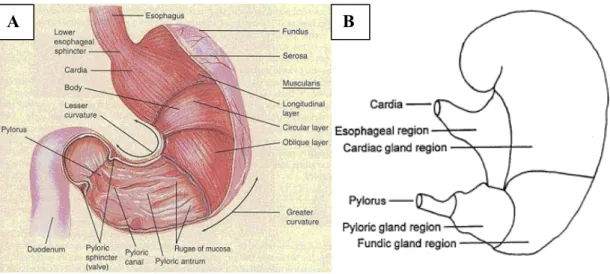
The stomach receives the insalivated boluses of food from the oral cavity through the esophagus. The bolus is temporarily stored in the stomach and soaked by gastric juice which is secreted by the gastric glands [1] [2] [3] [4]. This solution is mainly composed by pepsin, rennin and hydrochloric acid, which act on proteic substances [1]. The food, under the combined action of gastric juice and peristaltic movements is transformed into a fluid mass called chyme and is moved into the duodenum [1] [2] [3] [4]. The size and the structure of the stomach depends on the habits and on the food behavior of the singular species. Moreover the structure of the stomach depends on different feeding and lifestyle of the various species. pigs and humans are monogastric animals, but they show some differences: the human stomach (Fig 1) (glandular stomach) has a glandular mucosa covered with simple columnar epithelium and a capacity of 1,3 liters, while the stomach of the pig has a proventricular portion (Fig 1) (nonglandular mucosa; characterized by stratified squamous epithelium) and the presence of an esophageal-like mucosa, more or less extended from the cardia. The capacity of the pig’s stomach is 4 liters [2] [3].
2
Fig 1: A. External and internal anatomy of the stomach of human (Tortora and Grabowski, 1996), B. Region of the pig stomach (http://www.thepigsite.com/articles/2749/digestive-system-anatomy-and-function)
The stomach is characterized by two curvatures: the greater curvature is convex and directed ventrally toward the left, while the lesser curvature is concave and direcetd dorsally to the right [2]. The stomach can be divided into three anatomical regions: the fundus, or blind sac, overlooking the cardia, the body, located ventrally to the fundus, which continues on to the pyloric region. The latter, which corresponds to the flexed lower portion of the stomach, consists of the pyloric antrum and pyloric canal [2].
i) Structure
The wall of the stomach consists of a mucous membrane, a muscular, and serous coat. The tunica mucosa (Mucous membrane) can be divided into layers: surface epithelium, lamina propria mucosae, lamina muscolaris mucosae, and tela submucosa [4].
Tunica Serosa
The serosa is constituted by the visceral peritoneum; it is formed by two sheets, one anterior and one posterior. In proximity of the small curvature the serosa is coated with a small amount of elastic fibers, which seem to have the task of maintaining the two ends of the stomach close together.
The tunica serosa is continuous at the level of the greater curvature with the greater omentum, near the diaphragm with the gastrophrenic ligament, and at the level of small curvature with the lesser omentum [2] [3].
3 Tunica Muscolaris
The musculature of the stomach is constituted by two fundamental layers of smooth muscle cells, one on the surface (longitudinal layer) and one in deep (circular layer). Depending on the considered levels we can observe a dissociation, a thickening or a change of direction of the muscle planes, in relation to the conformation and function of each region of the organ. In the fundus and in the body of the stomach, two oblique layers are added to the two fundamental layers, one of which is internal and the other external. [2] [3].
-Longitudinal muscle layer
The longitudinal muscle layer is incomplete and is placed immediately after the subserosa. It is reduced to two straps, one of which runs along the small curvature and the other runs along the greater curvature [2] [3].
The first one is continuous on the surface of the esophagus and opens like a fan on the faces of the stomach, reaching the gastric incisure. The other one is relatively thin and extends from the left edge of the fundus to the pyloric area, where it is reinforced by elastic fibers. In the proximity of the pylorus the longitudinal layer is complete and thickened and then continues with the longitudinal layer of the intestine [2] [3].
-External oblique fibers
This layer is a continuation of the longitudinal layer, which is located only in the vicinity of the fundus and the part next to it, i.e. the body. This is well developed in the pig, where it forms a relatively superficial and flat thickened layer. These external oblique fibers are poorly developed in the human stomach.
-Circular muscle layer
The circular layer is not present at the levelof the fundus and is thin in the adjacent part of the body of the stomach. The circular layer occupies an intermediate position between the layers descibed above and those of the internal oblique fibers; at the level of the pylorus this layer is in direct relation to the submucosa. Precisely in the pyloric part (pyloric canal), the circular layer becomes stronger and forms the pyloric sphincter, which through its contraction, completely closes the communication with the intestine [2] [3].
-Internal oblique fibers
This layer is present only at the level of the fundus and in the body of the stomach. It is a thickened layer that is continuous with the circular bundles of the left face of the esophagus and which is made up of two parts: cardiac loop and the oblique layer.
4 Tunica Submucosa
This layer is immediately beneath the mucosa; it is a layer of loose to dense connective tissue containing blood and lymphatic vessels. The submucosa also contains the submucous plexus, a critical component of the digestive tract’s nervous system which provides nervous control to the mucosa [4].
Tunica Mucosa
When proventricular mucosa is present it is an esophageal-like mucosa. The rest of the stomach has a glandular mucosa, which has structural changes that allow us to divide it into three zones: fundic mucosa, cardiac mucosa and pyloric mucosa [2] [3].
-Proventricular mucosa
The proventricular mucosa (non-glandular mucosa) of the stomach is often slightly folded, it is white and dry and very similar to esophageal mucosa; in the pig the proventricular mucosa extends for 3-4 cm on the right of the small curvature, while it reaches 7-8 cm in width to the left where it leads up to the margin of the gastric diverticulum [2] [3].
The muscularis mucosa is relatively thick and has two floors of irregular bundles, which are often dissociated. The tunica propria mucosa is thick and is formed by dense connective tissue which is rich in elastic fibers [2] [3].
Like in the esophagus, the epithelium is stratified squamous non-keratinized, thick and cornified. There are no glands in the proventriculum [2] [3].
ii) Fundic mucosa
It is considered that the cardial mucosa and the pyloric mucosa derive from the fundic mucosa. The fundic mucosa occupies the fundus in humans and carnivores, whilst in pigs it is positionated fully into the body of the stomach and therefore it is not found in the fundus. The fundic mucosa is thick, soft, red to brown [2] [3].
Its overall organization is characterized by the provision of the muscularis mucosa, which is well developed and is provided with two floors of bundles, of which the exterior is longitudinal and the inner one is transverse. The transverse layer projects numerous bundles of cellular fibers between glands. They separate groups of glands forming lobules, which leads to the formation of small areas, called gastric areas, on the surface of the mucosa.
5
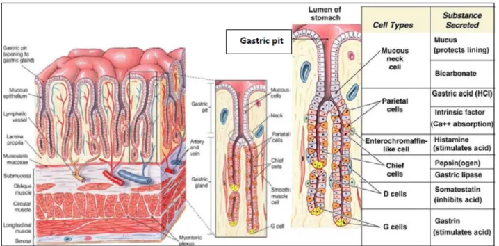
Each lobule is divided into small groups of glands, whose excretory ducts open into deep and narrow depressions called: gastric pits or crypts (Fig 2).
Fig 2.General cell composition and location in a gastric gland. G and D cells are mainly in pyloric glands, while paretial and chief cells in oxyntic (fundic) mucosa. (Basic and Clinical Pharmacology. Mc Graw-Hill, 2012)
-Lamina propria mucosa is the delicate connective tissue interposed between glands. It is rich in vessels with a thick subglandular layer particularly developed in carnivores. Numerous lymphocytes infiltrate the lamina propria mucosa in particular in the subglandular area, and in some cases they form lymphatic nodules, which are especially developed and evident in the pig. The epithelium is simple, formed by a single row of cells supported by a basement membrane. Many tubular glands are present in the thickness of the tunica propria, these glands occupy almost the entire thickness of the mucosa. In the adjacent part of the crypts and on the surface of the mucosa, the epithelium is formed by high and clear prismatic cells, which become lower and cubic towards the bottom of the crypts, in correspondence of the glandular orifices [2] [3].
-Region of proper gastric glands, the fundic glands or proper gastric glands are closely packed tubular glands with a rectilinear course and perpendicular to the surface they become sinuous or convoluted in the vicinity of the muscularis mucosae. Each gland is composed of a narrow and cylindrical neck, a large cylindrical and slightly flexuous body, and finally a base convoluted with trend and terminating in a blind end [2] [3].
The epithelium has 4 types of cells: 1) the mucous neck cells have a cubical shape, are positionated in the neck where they form the coating, 2) the gastric chief cells, which are
6
positioned in the body and at the base of the glands they are clear and their volume changes in response to their function, as well as the position of the core which can be central or baseline, 3) the parietal cells or oxyntic cells, which are localized primarily in the body of the glands, while they are very rare at the base of the glands. In the glands they are in an eccentric position, between the chief cells and the basement membrane, their cytoplasm is acidophilus. The chief and the parietal cells secrete different products; the first process the pepsinogen and chymosin or rennin; the latter are involved in the secretion of hydrochloric acid 4) the enterochromaffin cells are located between the chief cells and the basement membrane; they are equivalent to the endocrine cells of the intestine, and are so called because their cytoplasm contains granules that stain with the Ag or Cr salts. They definitely have an endocrine function [2] [3].
iii) Cardial Mucosa
The cardial mucosa gets its name because it is interposed between the esophageal mucosa and the fundic mucosa. In the majority of species including man, this takes place in the vicinity of the cardia, however in the pig it is positioned in the fundus and is well developed. Its organization is similar to that of the mucosa of the fundus, but the cardial glands are tubular branched and very tortuous. Their epithelium is composed of a single row of light-colored, cube-shaped mucous cells with basal nucleus [2] [3].
iv) Pyloric Mucosa
The pyloric mucosa is located in the pyloric part of the stomach and is also located in part of the body of the stomach. It is thinner and lighter than the fundic mucosa and is also less crinkled, with the exception that it is in the immediate vicinity of the pylorus. It has the same overall organization, but the grouping in lobules of the glands is more evident than in other parts of the stomach and the crypts are much deeper and narrower. The pyloric glands are more branched and tortuous than the proper gastric glands. It lines most of the pyloric part in the pig's stomach, except for an area near the greater curvature. Apart from some exceptions, the pyloric glands are devoid of parietal cells and their epithelium is composed only of a layer of clear cells [2] [3].
7
II) INTESTINE
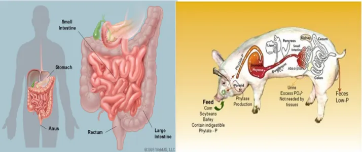
The intestine extends from the pylorus of the stomach to the anus, with the exception of some rare cases. It is easy to recognize two major parts in the gut: the first part is narrow and relatively long and is called small intestine, the second is more voluminous and variable and is called large intestine [2] [3] (Fig 3).
Fig 3. Schematic representation of Human and Pig gastrointestinal tract (www.webmed.com, www.vetmed.vt.edu).
i) Small Intestine
The small intestine is a long cylindrical tube of nearly uniform caliber, where the most important stages of digestion occur. The small intestine is divided into three successive and unequal segments: duodenum, jejunum and ileum. Among these, only the duodenum is clearly demarcated, whilst the boundary between the other two segments is barely visible [2] [3].
The duodenum receives the secretion from the liver and pancreas, while in the jejunum and ileum there is only the secretion from their own mucosa. These secretions continue and complete the action started by gastric juices in the stomach due to the fact that certain substances such as carbohydrates are not modified in the stomach [2] [3]. In order to perform this function the small intestine needs many specilaized structures, a large amount of digestive enzimes and a large amount of mucus, which is indispensible to preserve the mucosa from mechanical insults and irritating compounds.
8
The size of the small bowel depends on the habits and on the food behavior of the singular species. It is 10 times the length of the body in humans and 13 times in pigs, but may vary also from animal to animal of the same species [2] [3] [4].
a. Duodenum
Duodenum is the first part of the small intestine and extends from the pylorus to the jejunum. Its mesentery, the mesoduodenum, is relatively short, with the exception of carnivores. Two flexures divide the duodenum in 4 parts: 1) cranial part, which passes to the right along the visceral surface of the liver and ends at the cranial flexure; 2) descending part, which runs caudally from the cranial flexure towards the right kidney; this part of the duodenum is shorter in both humans and pigs; 3) transversal part, which runs towards the left and differs from animal to animal; in humans it is elongated, whilst it is shorter in carnivors and significantly shorter in ruminants; 4) ascending part, which is found in the vicinity of the left kidney; it passes cranially and as its mesoduodenum becomes longer it turns ventrally at the duodenojejunal flexure and continues into the jejunum [2] [3] [4]. The descending and ascending parts of the duodenum form a U-shaped loop around the caudal aspect of the root of the mesentery and the cranial mesentery artery [4]. The cranial part of the duodenum is closely related to the liver and pancreas; the duodenum receives the bile duct from the liver and the pancreatic duct from the pancreas [4].
b. Jejunum
The jejunum begins at the duodenojejunal flexure at the cranial end of duodenocolic fold[4]. The jejunum is a long cylindrical tube covered by peritoneum, and continues from the duodenum. In the pig it is found mainly in the ventral part of the right half of the abdominal cavity, but it extend along the floor into the left half and lie ventrally to the coiled ascending colon and cecum [4]. The internal structure of the jejunum consists of a soft epithelium with many villosities; in some areas the mucosa has a particular appearance due to the partial lack of villi and accumulation of lymphnodes. This accumulation of limphonodes forms the peyer’s patch [2] [3]. Contractions called peristalsis occur-in this structure, but they never at the same time as in the ileum. Peristalsis is the contraction of
9
the muscle layer that helps the chyle to continue into the other parts of the gut [2] [3]. Peristalsis occur in each segmant of the gut.
c. Ileum
Ileum is the short terminal part of the small intestine and forms the link between the small and large intestine[4]. It terminates at the cecocolic junction of the large intestine forming the ileal orifice [4]. The anatomy of the terminal part of the ileum suggests that the junction of the ileum and large intestine is not only an anatomical division, but also an important functional division of the alimentary canal [4]. Similarly to the jejunum the ileum also has a cylindric shape and a soft epithelium, with many villosities and in some areas peyer’s patches.
d. Structure
The duodenum, the jejunum and the ileum have a very similar structure; they only differ in some aspects. As in the rest of the digestive tract, there are four different layers: tunica serosa, tunica muscolaris, tunica submucosa, tunica mucosa [2] [3].
Tunica Serosa
The whole small intestine is covered with a thin tunica serosa, which derives from the peritoneum [2] [3]. The serosa adheres closely to the tunica muscolaris and near the mesentery it thickens and forms elastic connective tissue to facilitate the changes of caliber that the body undergoes during its functions [2] [3] [4].
Tunica Muscolaris
As in all species, the tunica muscolaris is composed of two layers: a thick circular inner layer and a longitudinal and thinner outer layer. Both are relatively thin at the level of the duodenum and thicken towards the ileum [2] [3]. Between these two layers there is a thin layer of connective tissue which welcomes a network of nerve fibers with ganglion cells, which belong to the myenteric plexus [5].
10 Tunica Submucosa
The submucosa can be thinner or thicker depending on the segment considered. It is formed by a layer of connective fibers and contains some elastic fibers, which allow it to form folds. Moreover, a submucosal plexus is present at this level with an extensive neuronal network mixed with ganglion cells. Furthermore, deep in the submucosa there are submucosal glands (Brünner glands). In the pig the secretion of these glands lubrificates the epithelial surface and protects it from the acidity of gastric chyme [2] [3].
Tunica Mucosa
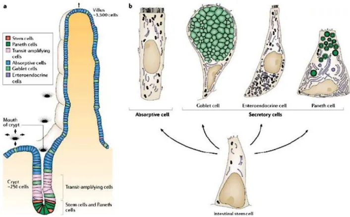
The tunica mucosa is the most characteristic part of the bowel; it has a myriad of tiny finger-like processes called villi, which take on various shapes and positions and are specific organs that are very important for absorption [2] [3]. Each villus is coated by epithelium and presents connective tissue. Inside it is possible to note blind end lymphatic vessels surronded by an extensive network of capillaries formed by a small artery and drained by a small venule. Moreover, the villus axis presents smooth muscle cells from the muscolaris mucosae. Between the villi there are depression zones or crypts, which are considered intestinal glands [5]. The epithelium lining of the intestinal mucosa is a simple columnar epithelium, and it is formed by different types of cells (Fig 4).
-Enterocytes, these cells are prismatic or pyramidal, with the restricted part facing towards the basement membrane. In an optic microscope the apical part of these cells presents a thickened and finely striated area, with microvilli which are very important for absorption [2] [3].
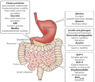
-Goblet cells, these cells are interspersed among the enterocytes; there are fewer than enterocytes. They are glandular simple columnar epithelial cells and secrete mucin, which in the end becomes mucus; they use both apocrine and merocrine methods for secretion. Mucus mainly consists of glycoproteins and glycosaminoglycans (PAS positive) [2] [3]. -Enteroendocrine cells, these cells are specilized endocrine cells, which produce hormones such as serotonin, somatostatin, colechistokinin (CCK), ghrelin, glucagone peptide-1 (GLP-1), polypeptide YY (PYY) and regulate the digestive cycles. The enterochromaffin cells present in the stomach are also considered enteroendocrine cells [2] [3].
11
Fig 4. Different types of epithelial cells present in the intestine (Nature Reviews 2006).
ii) Large intestine
The large intestine is the part of the digestive tract that follows the small intestine and ends with the anus. It is divided into the following three segments: the cecum, the colon and the rectum plus a short anal canal. The large intestine has an almost uniform structure and retains, in all its segments, which are variable in shape and size, an anatomical and functional unit. The last part of digestion takes place in this portion of the digestive tract, and in particular the absorption of liquids, which is very high despite the lack of villi [2] [3].
a. Cecum
The cecum is the initial part of the large intestine and appears as a blind end, which is more o less voluminous and present in the gut between the ileum and colon. Depending on the species in question the cecum can be very small or well-developed, in most cases it presents sacculations and teniae [3]. In herbivores and omnivores it is quite developed, whilst it is
12
short and poorly developed in carnivores. It reaches maximum development in horses and rabbits. In the pig the cecum is 30-40cm long, it is positioned on the left side, and has very pronounced sacculations interrupted by longitudinal folds (tenie) [4]. Between adjacent sacculations, semilunar folds project into the interior of the gut and increase the initial surface area (the same process occurs in the colon) [4]. In humans the cecum, even though short and not voluminous, consists of two unequal segments, one proximal and one distal. The vermiform appendix is part of the distal segment [3].
b. Colon
The colon is the largest part of the large intestine, followed by the cecum, when present, and terminates with the rectum. The size and shape of the colon is related to the diet. The description of the colon in comparative anatomy is based on human nomenclature. The simple arrangement in man gave rise to the division into an ascending colon, which passes cranially on the right, a transverse colon, which passes from right to left in front of the mesenteric artery, and a descending colon, which passes caudally on the left [4].
The ascending colon in the pig has a spiral-shaped cone arrangement with apex on the left side and transverse axis disposed vertically [6], whilst the transverse colon is short and the descending colon presents a smooth appearance and is generally smaller than the ascending colon [3].
-Ascending Colon, this part of the colon, is very developed in ungulates and rabbits. In the pig it is from 2 to 4 meters long, wheras it is 5-10 meters long in the cow, 12-15 meters in the camel, 30-35 cm in the rabbit and 15-25 cm in humans. Due to these sizes the ascending colon is forced to bend and roll up to find a place in the abdomen [3]. In the pig it is coiled on itself and forms the spiral loop of the colon, which is between the cecum and the transverse colon and its bends are piled up to form a thick cone [4].
-Transverse Colon is relatively long in humans, from 50 to 60 cm, and is delimited by two angles, between which it forms a curve. The transverse colon is smooth in pigs, whereas it is bumpy in humans [3]. It passes from right to left in front of cranial mesenteric artery in the abdomen [4].
-Discending Colon is very long and suspended by the long descending mesocolon. The large coils of the descending colon are found in the left dorsal quadrant of the abdominal cavity [4]. It has a simple arrangement and often extends in a straight line. It is smaller than
13
the ascending colon and has sacculations in humans, while it is smooth in pigs [3]. As in the cecum, the colon is also provided with semilunar folds which project into the lumen of the gut and increase the initial surface area.
c. Rectum
The rectum, so called because it does not describe any convolutions, is smooth and caudal and becomes enlarged, forming the ampulla recti before ending at the short anal canal [4] [6]. Infact the rectum is a straight piece of gut which continues from the descending colon into the pelvic cavity.
d. Structure
The large intestine consists of four concentrically arranged layers proceeding from outside to the lumen: tunica serosa, tunica muscularis, tunica submucosa, tunica mucosa.
Tunica Serosa
The tunica serosa is very thin and it is derived from the peritoneum. The serosa does not cover the entire large intestine but finishes at the rectum. The rectum is covered and surronded by dense connective tissue [3].
Tunica Muscolaris
The muscular coat is composed of two layers, one circular and internal, and the other longitudinal and external. However the longitudinal layer in some places is considerably thickened and forms longitudinal bands called teniae in latin; in this part the elastic fibers are abundant (cecum and colon segments) [3] [5].
Tunica Submucosa
14 Tunica Mucosa
The mucosa lacks villi and the epithelium is reminiscent of the small intestine. It forms the Liberkuhn glands, which extend perpendicularly from the surface to the proximity of the muscolaris mucosae. These glands are more abundant and bigger than in the small intestine and are rich in mucous cells. These cells seem to be less in number in the cecum, but their number increases in proximity of the rectum [3].
THE GASTROINTESTINAL TRACT IN FISH
The gut is a tubular structure beginning at the mouth and ending at the anus (Fig 5). The digestive system of the fish is divided into 4 parts[7]:
• The head gut is generally divided into the oral (buccal) and gill (branchial, pharyngeal) cavities. As it is not part of the gastrointestinal tract it will not be treated here
• The foregut begins at edge of the gills and includes the esophagus, the stomach, and the pylorus.
• The midgut includes the intestine posterior to the pylorus,.and often includes a variable number of pyloric caecae (pyloric appendages) near the pylorus.
• The hindgut is marked by an increase in diameter of the gut and it ends is the anus. Some species of fish, such as the cyprinus, lack both a stomach and pylorus, in this case the foregut consists of the esophagus and an intestine anterior to the opening of the bile duct.
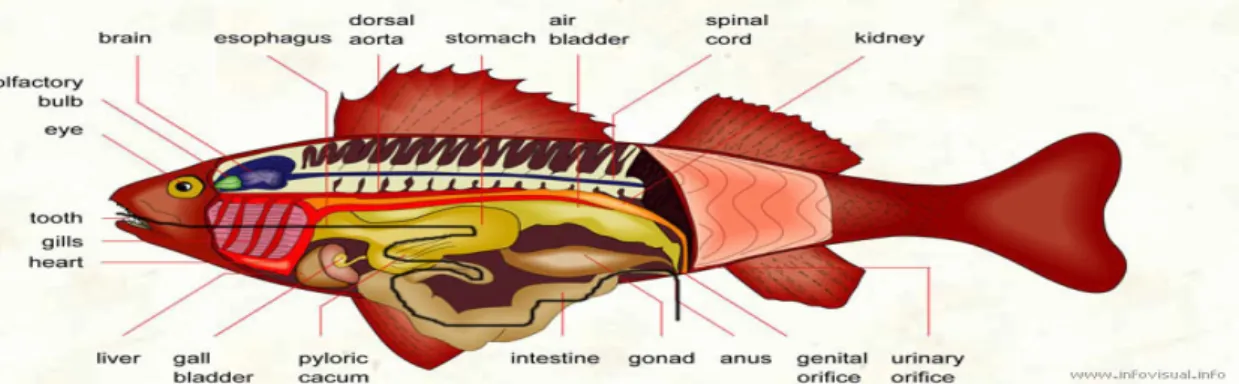
Fig 5. Overview of gastrointestinal system in the bone fish (black line is the the black line is the path of food from ingestion to expulsion) (http://www.infovisual.info/02/033_en.html).
15
I) THE HEAD GUT
As previously mentioned the head gut is not part of the gastrointestinal tract, but is a fundamental part of the digestive tract of the fish.
II) THE FOREGUT
The foregut includes the esophagus and the stomach. the anterior limit is given by the gills, while the caudal limit is given by the pylorus[8].
i) The esophagus
I will only briefly introduce the esophagus, because it is only the anterior limit of the stomach, thus it is not part of the gastrointestinal tract that begins with the stomach.
The esophagus is a large, short and straight tube, constituted by outside to the lumen of: tunica serosa, tunica muscolaris, tunica submucosa e tunica mucosa. There are many mucous cells that secerne mucus-like substances, which make the esophagus viscous [7].
ii) The stomach
The stomach of teleosts, when it is present, presents a variety of different shapes, and in any case represents the caudal part of the foregut [7].
The stomach can be straight, like a tube of uniform diameter with no marked anatomical differences between the esophagus and stomach as in the Northern pike (Esox Lucius), U-shaped or in the form of a round and muscular structure situated at the end of the esophagus and with a cardiac and pyloric region as in most teleosts, or Y-shaped with a blind sac of variable size and a cardiac and pyloric region as some teleosts and sharks [7] [8] (Fig 6).
16
Fig 6. Different shapes of fish stomachs (Dicentrarchus labrax have a Y-shaped stomach) (http://diversityofanimalsystem.wikispaces.com/digestive+system)
The stomach is absent in different fishes, when present it has numerous gastric pits (like crypts) immersed in the mucosa and at the bottom there is the opening of the gastric glands [8]. The epithelium of the stomach and of the lining of the crypts consists of a single layer of high columnar prismatic cells; these cells in the apical part are positive to PAS reaction and so they secrete a protective mucus [8]. There are two different types of glands in the mucosa of the stomach: fundic and pyloric; the fundic glands cover most of the mucosa of the body of the stomach, while the pyloric glands are only present in the pyloric part of the stomach [8]. The fundic branched tubular glands possess more than one type of cell, this gland cell (oxyntopeptidic cell) contains acidophilic granules and produces pepsin and hydrochloric acid. The pyloric glands are less closely associated than the fundic glands; they are shorter, less frequently branched tubules [8]. Their epithelium is similar to that of the stomach, the stroma of the mucosa of the stomach contains many lymphocytes and eosinophilic granular cells, the muscularis mucosae is present and consists almost entirely of smooth, longitudinally-disposed muscle cells [7] [8]. The submucosa contains
Y shape stomach NO STOMACH
17
eosinophilic granulocytes and is rich in networks of nerves, arteries and veins, while the muscolar coat is formed by a circular, longitudinal and additional inner oblique layer [8].
III) THE MIDEGUT AND THE HINDGUT
The segment of the intestine that follows the stomach is called midgut, while the terminal segment of the intestine is the hindgut. Unlike mammals, in fish there is no distinction between the small intestine and large intestine. The intestines of fish are mostly a tubular structure that can vary in size depending on their eating habits; in fact, carnivorous species often have a shorter intestine compared to herbivorous fish [7] [8]. Some species of bony fishes have an intestine with a smooth surface, others have longitudinal folds or folds which form a rather complex pattern or network. Moreover, some fish, such as higher vertebrates, have villi as their intestinal wall lining, which can be different sizes and shapes depending on the species. A villus is a finger-like process of the mucosa which is composed of an epithelial covering and a core of connective tissue containing blood and lymph capillaries. Many species have a number of protrusions extending from the midgut close to the pylorus [7] [8]. These blind-ending structures are the pyloric cecae, they possess a multi folded intestinal epithelium and their role seems to be to increase the area for the absorptive process and the duration of food retention in the intestine. The intestinal epithelium can be of a simple or pseudostratified columnar type; it is composed of cells that possess a well marked striated border called microvilli and goblet cells, which are mucus secreting cells [8]. These cells have different functions, such as absorption and secretion. In some fishes ciliated cells have been described among the ordinary prismatic cells of the intestine [8]. In the intestine of fish are present some glands similar to liberkhum glands [8]. The lamina propria and submucosa of some species contain large numbers of eosinophilic granular cells and lymphoid tissue [8]. The eosinophilic granular cells are similar to mastcells; they contain antimicrobal peptides and their release can increase the vascular permeability and promote neutrophil adhesion (innate immunity and inflamation) [8]. The muscularis mucosa is composed of a thin layer of smooth muscle, and the submucosa is generally composed of a loose connective tissue with blood vessels. In most fishes, as in mammals, the muscular coat of the intestine is very developed to ensure peristaltic contraction [8]. The rectum is the terminal part of the fish intestine [7] [8].
18
THE SENSE OF TASTE
Taste is the ability to respond to dissolved molecules and ions called tastants. The chemical senses (taste and smell) are the most ancient of the sensory modalities in any living species. There is no doubt that the gustatory system is essential for nutrition and survival. In fact, the discrimination between nutrient and/or potentially harmful compounds has effect on animal and human behaviour and, therefore, on their major organic and biological functions [9].
It is difficult to imagine how the many taste sensations that we perceive can be only related to four and more recently five types of taste: sweet, bitter, salty, sour and umami. These five types of taste, however, can be mixed together to produce many shades and hues of flavour.
Taste at a molecular level is very similar to the other senses. Tastants are recognized and bound by taste receptor signaling to sensory neurons in order to convey the chemosensory information to the central nervous system (CNS). However the relationship between tastants and taste is not linear connection. It would be easy to think that the five main tastants can be recognized via respective receptors, one for each type of taste. In contrast, the complex web of information generated by various tastants results from a polymodal function of taste receptors, i.e. each receptor recognizes different stimuli. The same stimuli, can be evoked by many different tastants, and each taste modality may use more than one processing mechanism. Moreover each tastant can be recognized by taste receptors only if it reaches the right threshold. For example, some compounds such as sucrose and lactose, which elicit a sweet taste in humans, activate taste receptors only at high concentration [9], while bitter substances have a nanomolar concentration threshold.
19
TASTE BUDS
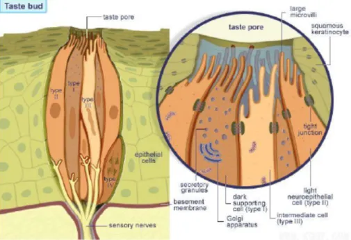
The perception of different gustatory stimuli originates from the interaction of the molecules present in oral fluid as saliva. Found primarily on the tongue’s surface, taste cells are organized in specialized structures (specialized sensory cells) referred to as “taste buds” [10] (Fig 7).
In mammals, the taste buds are located on the tongue, epiglottis, pharynx and in the upper part of esophagus [11]. In fish the distribution of taste cells is on the whole body surface, lips, gills, skin and barbells as well as in the mouth, pharynx and esophagus [12] [13] [14]. The distribution of taste buds in fish reflect the eating habits, hunting strategy and the different fish habitats [14] [15] [16].
Taste buds are approximately 50µm in diameter and appear to be composed of 50-150 taste cells that detect sugar (sweet taste), aminoacids (umami taste), poisons (bitter taste), acids (sour taste) and minerals (salty taste). Taste receptor cells are long and spindle shaped, with microvilli at their tips.
Fig 7. Taste bud organizations
The beginning of taste recognition occurs at the pore, an opening in taste buds where the microvilli of receptor cells contact the outside environment. Tastants penetrate into the pore and make contact with receptor molecules and channel within the microvillar membrane of the taste receptor cells [10][17]. Microscopic studies of taste buds, highlight four morphologically different cell types (Fig 8): light cells (type I cells), dark cells (type II cells), intermediate cells (type III cells) and basal cells [18].
20
The basal cells, small and rounded, are located at the base of taste buds and are considered to be stem cells, because it is believed that all of the other cell types derive from the basal cells. The lifespan of an individual taste cell is only from 10 days to 2 weeks, thus cells within the taste buds are continually being replaced [11] [19].
The light cells are the mature taste cells and their primary function is to support dark and intermediate cells [20], while the dark and intermediate cells are different stages of differentiations of immature taste cells [11]. Functionally all four cell types are referred to as taste cells, they are elongated cells and extend from the bottom of taste buds up to their taste pore.
The most studied cell are certainly the type II cell that expresses G-protein coupled receptors (GPCRs) for the detection of sweet, umami and bitter compounds [21]. Type III cells are thought to express sour taste receptors and detect acid taste [22]. This cell type also expresses the pan neuronal marker protein gene product 9.5 (PGP 9.5) and contains 5-hydroxytryptamine (5-HT) [23]. Type I cells express nucleoside triphosphate diphosphohydrolase-2 (NTDPase2) and the oxytocin receptor [24] [25].
Fig 8. Cells types in taste bud: light cells (type I cells) are supporting cells, dark cells (type II cells) contain taste receptor, intermediate cells (type III cells) form synapses with afferent nerves, basal cells (type IV cells) are progenitor cells.
Mammalian taste buds are localized in structures called papillae, varying in number depending on the species considered. In humans, three types of papillae are present on the
21
tongue of domestic animals, each with morphological differences and with a different localization.
The fungiform papillae are numerous (in terms of hundreds) on the tip of the tongue of humans and pigs, in addition in pigs other fungiform papillae can be found on the anterior two-thirds of the tongue. This type of papillae has a simple structure generally containing a single taste bud. On the back of the tongue there are the circumvallate papillae, with a more complex structure, V-shape and many taste buds (in the pig there are only two papillae - one at each side of the back of the tongue, in humans they range from 8 to 12). Taste buds in circumvallate papillae line the side of the grooves with their pore facing the cleft [2] [3]. The foliate papillae are positioned on the lateral margin towards the posterior part of the tongue, they have a similar structure to the circumvallate papillae and also taste buds line the cleft of the papillae, their shape being like a leaf [2] [3]. These papillae are well developed and numerous in pigs [3].
Taste cells detect stimuli, but taste receptor cells have to convey taste information to the CNS. Nerve fibers representare the link between the taste receptor cells and the brain. Taste cells secrete neurotransmitters into the synapse, passing information from the taste receptor cells to neurons. The latter spike action potentials that signal to the brain [10] [26].
22
BITTER TASTE RECEPTORS
Bitter stimuli is perceived as dangerous and harmful and therefore the gustatory system induces an adverse reaction [27]. In fact the bitter stimuli has evolved as a central warning system against the ingestion of potentially toxic substances, including the alkaloid and other environmental toxins [28].
The recognition of these potentially dangerous signals by the gustatory system is associated with the development of T2R family in the oral cavity. This is known thanks to the discovery of T2Rs genes in several animal species. In the human genome about 25 T2Rs have been identified [29] [30], in birds for example only three genes, whilst in amphibians about 50 T2Rs genes. Generally in mammals the expression of T2Rs genes ranges between 15 and 36 [11] [29] [31] [32]. In general T2Rs belong to the guanine nucleotides, which are bound to the GPCRs superfamily, with a short NH2 terminal segment and seven transmembrane α-helics, three extracellular loops, three cytoplasmatic loops and a COOH-terminal segment [10]. Specific G alpha subunits are common to all taste GPCRs called: gustducin and transducin (Gi/Go proteins) [33] (Fig 9).
The T2R family is composed of many receptors, only some of them, to date, are associated with a specific ligand (6% of total receptors), this is the case of phenylthiocarbamide (PTC) and denatonium benzoate (DB), which bind T2R138 (T2R38 in humans) and T2R108 (T2R4 in humans) respectively in mice.
Most T2Rs are generally known as “orphan receptors” (about 80% of total T2Rs receptors) because it is not known exactly which substance can be used as an agonist [34] [35]. It should be remembered that a single receptor can also bind different substances, this further complicates the task of researchers who seek to shed light into the great family of T2Rs. A recent study[35] compared 25 hT2Rs with more than 100 natural and synthetic compounds, going on to establish a ranking of the most “broadly tuned” T2Rs. So today it is known that some T2Rs can respond to a wide range of bitter substances and others can have intermediate characteristics to recognize only a few bitter substances. In one of our studies, we treated the enteroendocrine STC1 culture cells with increasing doses of PTC and DB.
This was done to evaluate the activation of phosphorylation MAPkp44/42, because PTC and DB bind T2R138 and T2R108 respectively, leading to a dose response activation of MAPkp44/42, which confirms the involvement of these two substances with T2Rs.
23
Fig 9. Bitter taste signalling pathways
24
α (gustducin and transducin)-SUBUNIT OF G PROTEIN
COUPLED RECEPTOR SUPERFAMILY
Gustducin plays an important role in transducing bitter and sweet gustatory signals in the taste buds of the lingual epithelium. Outside of the oral cavity α-gustducin (an alpha subunit of GPCR) has been localized in the gastrointestinal tract of rats and mice [36] [37] [38] [39] and in the pancreas [40], suggesting a role for taste sensing mechanisms in the gastrointestinal tract [37] [41]. Alpha transducin was originally described in the photoreceptor cells of the retina, but it is now well established that this subunit of the G protein is present in the taste cells of the lingual epithelium and thus is implicated in taste signal transduction [42].
The presence of taste receptors in the oral cavity was confirmed through the use of immunohistochemistry, which identified the expression of gustducin (Gi) and α-transducin (Go) cells in the stomach and intestine mucosa of rats [43], mice [39] [44] [45], pigs [46] and man [47] [48].
In particular, both α-gustducin and α-transducin are stained in different subpopulations of enteroendocrine cells (98% of them). This has been established using immunohistochemestry to localize such as chromogranin A (an estabilished marker of endocrine cells in the GI tract), PYY, GLP-1, ghrelin, CCK, serotonin and somatostatin [46] [47] [48] [49] [50] [51].
The presence of the alpha subunit of G protein coupled receptors has been found not only in the oral cavity and gastrointestinal tract but also in the respiratory system [52]. In these systems, the alpha subunit of G protein coupled receptors has been localized in specific cells called brush cells distributed to the pancreas, stomach and intestine [53]. As described by Rozengurt and Sternini, these cells are morphologically different compared to the endocrine cells which have a “bottleneck” or “pear” shape or have an elongated pyramidal shape and the ability to secrete CCK, PYY, GLP-1 upon activation induced by taste stimuli [54] [55] [56].
Brush cells have an elongated soma with a basolateral rootlet and an apical tuft of microvilli that extends into the lumen [57], they do not contain granules and secrete neurotransmitters.
Using immunohistochemistry, α-gustducin [39] [43] and α-transducin [46] are expressed throughout the epithelium surface in the brush cells in the rat and pig gut.
25
In conclusion, much remains to be understood about the distribution and function of the alpha subunit of the G protein coupled receptors involved in taste perception. The α-gustducin seems to be more involved in the perception of the bitter and sweet stimuli, whilst α-transducin seems to have a secondary for scientific community. However, the study of Clavenzani et al [46] and the most complete study of Mazzoni et al [58]showed that α-transducin is localized throughout the gastrointestinal tract of pigs with exception of the esophagus. In that study, the α-transducin co-localizes with several neurotransmitters and fasting and refeeding evoke a modification in the expression of this protein in the entire gastrointestinal tract. This evidence highlights the role of α-transducin and α-gustducin in taste transduction [58].
26
CELLULAR SIGNAL OF TASTE TRANSDUCTION
Food intake causes a series of processes that lead the gustatory system and therefore our body to distinguish between different molecules, nutrients but also potentially hazardous or toxic substances.
In fact taste cells are able to discriminate between various substances such as ions and complex compounds as sweet and bitter. Depending on their chemical nature, the ingested substances are reception and transduction with different kinds of processes (Fig 10). It is also true that different molecules may also be perceived as one [11].
Once received and recognized, taste stimuli are transduced with different cellular mechanisms (e.g., membrane potential or change in the concentration of free Ca2+), which lead to the release of neurotransmitters carryng information to CNS.
Fig 10. General transduction mechanisms in taste (publishing as Cummings B. 2006, in Cur Opin in Neurobiol)
I) BITTER
The bitter taste is often associated with toxic or harmful substances, in fact for an organism sensitivity to bitter stimuli is a protective mechanism for poison avoidance [59]. There is a
27
wide range of compounds that are very different chemically, but that induce the sensation of bitter.
It is well known that different substances such as caffeine, nicotine, strychnine, drugs and plants alkaloid evoke the taste of bitter [10]. For this reason the perception of bitter taste is of fundamental importance for the survival of the animal kingdom.
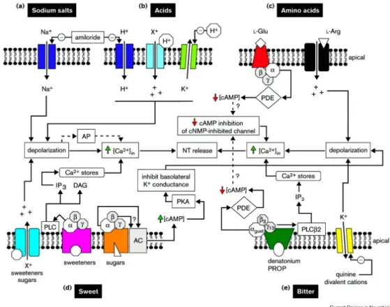
Bitter compounds are divided in lipophilic (kinin), which penetrate the membrane directly, hydrophilic (DB), which instead must use a mechanism as a receptor to enter in the cell. This suggests how there can be more than one intracellular signal pathway involved in bitter transduction (fig 11). The transduction of bitter stimuli is primarily mediated by the T2Rs, a G protein coupled receptor superfamily (a family of about 30 receptors). To date, we know two mechanisms of signal transduction for bitter taste: the activation of cell surface receptors and the following activation cascade of secondary messengers which involves phospholipase C that in turn activates the inositol tri-phosphate (IP3), which is well known to stimulate the release of calcium ions from intracellular stores [17] [60]. The Ca2+ thus liberated causes the hyperpolarization of the cell via K+ channel, but we have to remember that Ca2+ can also directly activate the release of neurotransmitters. The second way is identical to the first until the activation of G protein, which involves the activation of phosphodiesterase, which reduces the intracellular levels of cAMP or cGMP (cyclic nucleotides) [10] [17]. The decrease of the cAMP level activates protein kinase A, which regulates the passage of Ca2+ through the ion channel.
The α-gustducin subunit is definitely involved in the bitter signal transduction. This is confirmed by several in vitro and in vivo studies on laboratory animals. One study in particular confirmed the α-gustducin as an important mediator of bitter stimuli. In this study the author used KO mice for α-gustducin gene, in which the responce of Ca2+ to bitter compounds was measured through calcium imaging technique [61]. The result shows how the bitter stimuli are transduced mainly by gustducin, because in KO mice for α-gustducin gene, the response to bitter compounds was low but not zero. This evidence is very important because this means that there is a different subunit of GPCR to transduce the same bitter stimuli, maybe the α-transducin.
As proof of this, the Clavenzani et al. study on the pig GI tract shows how the same enteroendocrine cells can co-express both α-gustducin and α-transducin [46].
It should be remembered that the mechanism which implicates GPCRs is valid only for hydrophilic molecules, because lyphophilic molecules use the ions channels directly (eg. K+ channel) to penetrate into the cell.
28 II) SWEET
The transduction of sweet stimuli is similar to bitter stimuli. In fact, when sweet substances bind the taste receptor the stimulatory G protein is activated inside the taste cell. The activation of the taste receptors by sweet substances such as sugar, saccharin, aspartame, and alcohol, causes a depolarization of the taste cell due to the action of cyclic nucleotides cAMP and cGMP [62] (Fig 11).
Avenet et al showed that the addition of cAMP in the taste cells of the frog, causes the activation of protein kinase, an enzyme that induces the closure of K+ channels [63] on basolateral side of the plasma membrane blocks this in turn, the exit of the K+ ions, causing the depolarization of taste cells and the release of neurotransmitters [19] [62].
The T1R receptors are responsible for transducing the sweet stimuli, this is a small family of receptors (T1R1, T1R2, T1R3) linked to G-protein that perform their function alone or in combinations (eg. T1R1 and T1R3 for umami taste) [64]. Likewise bitter, even the sweet taste is composed of very heterogeneous substances, in fact the gustatory signal transduction requires many processes and it seems unlikely that a single receptor can incorporate all sweet stimuli.
29 III) SALT
Salt stimuli are transduced by simple ion channels. Sodium is the most popular salty substance, it represents 90% of the inorganic ions in the extracellular fluid. About 30 yars ago, it was thought that taste cells were impermeable to ions, but different studies showed that Na+ could be transported across the tongue ephitelial membranes in dogs [65]. In addition, it was shown that the drug amiloride inhibits the passage of Na+ from outside to inside the taste cells, leading to a decreased perception of salt stimuli. This occurs because the amiloride blocks the Na+ channel, as demonstrated in humans and rats [66]. The salty taste is transduced through ionotrophic mechanisms and mediated by a particular receptor, an ion channel for the amiloride-sensitive sodium, known as ENaC [10] [17]. The entry of Na+ into the cells, depolarizes cells, leading to the release of neurotransmitters and, as a result, nerves convey sensory information to the brain (fig 12).
IV) SOUR
Sour stimuli are perceived when in the oral cavity there are compounds that increase the H+ ions. Indeed sour tasting acids and substances may be inorganic, such as hydrochloric acid, or organic, such as lactic acid, both evoking H+ release. The signal transduction occurs through the modulation of the potassium channel. The increase of H+ block K+ channels and this effect prevents the release of K+ from the cells and depolarizes the cell membrane, thereby leading to neurotransmitters release [67] (fig 12). Other mechanisms may operate in sour stimuli transduction, such as the activation of Na+ channel due to elevated H+ concentrations [68].
30
Fig 12. Salt and Sour taste transduction (Callmethedoctor.co.uk)
V) UMAMI
Umami is a recently discovered taste. It is given from substances such as L-glutamate, and 5’-ribonucleotides, including guanosine monophosphate (GMP) and inosine monophosphate (IMP) [69]. This taste was first discovered in 1908 by K. Ikeda, who coined the term “umami”. Most dietary proteins contain high amounts of glutamate (e.g. protein of meat, poultry, seafood and vegetables).
Transduction for umami taste is still unclear. Biochemical studies showed that taste receptors are responsible for the sense of umami, and some receptors are taken into consideration for umami transductions, e.g. T1R1-T1R3 dimer and some modified glutamate receptors such as mGLuR4, mGLuR1. All of these receptors are found in the taste buds on the tongue [70] [71] [72]. However, to date, the specific role of these receptors in taste buds remains unclear.