UNIVERSITÀ DEGLI STUDI DI SALERNO
Dipartimento di Farmacia
Dottorato di ricerca
in Scienze Farmaceutiche
Ciclo XIV — Anno di discussione 2016
Coordinatore: Chiar.mo Prof. Gianluca Sbardella
Biomolecular and Biophysical approaches to
interrogate epigenetic targets: a platform for
drug discovery
Settore scientifico disciplinare di afferenza: CHIM/08
Dottorando Tutore
Dott. Alessandra Feoli Chiar.mo Prof.
Gianluca Sbardella
Co-tutore Chiar.mo Prof.
INDEX
ABSTRACT ... VII
1. INTRODUCTION ... 1
1.1 Epigenetics ... 1
1.2 Epigenetic Writers ... 2
1.2.1 Lysine Acetyltransferases (KATs) ... 3
1.2.1.1 p300/CBP ... 3 1.2.2 Protein Methyltransferases (PMTs) ... 6 1.2.2.1 Arginine Methyltransferases (PRMTs) ... 7 1.2.2.2 Lysine Methyltransferases (PKMTs) ... 7 1.3 Epigenetic Erasers ... 9 1.3.1 Histone Demethylase (HDMs) ... 9
1.3.2 Histone Deacetylase (HDACs) ... 9
1.4 Epigenetic Readers ... 10
1.4.1 Bromodomains ... 11
1.4.2 The Royal Superfamily ... 11
1.4.2.1 Tudor Domains ... 12
1.5 Epigenetics and Drug Discovery ... 13
1.6 Biophysical Methods ... 15
1.6.1 Surface Plasmon Resonance (SPR) ... 16
1.6.2 Differential Scanning Fluorimetry (DSF) ... 17
1.6.3 Microscale Thermophoresis (MST) ... 20
1.7 Biochemical Methods ... 21
1.7.1 Radioactivity Based Assays ... 22
1.7.2 Fluorescence Based Assays ... 23
1.7.2.1 AlphaScreen Technology ... 24
2. AIM OF THE WORK ... 27
3.1 p300 ... 29
3.1.1 SPV106 analogues as modulators of acetyltransferase activity.. 29
3.1.1.1 Chemistry ... 31
3.1.1.2 Biophysical Screening: Surface Plasmon Resonance Assay .. 33
3.1.1.3 Biochemical Screening: Radiometric Assay ... 35
3.1.1.4 Evaluation of Cellular Activity ... 37
3.1.2 From Garcinol to 5Benzylidenebarbituric Acid Derivatives ... 41
3.1.2.1 Biochemical Screening: Radiometric Assay ... 43
3.1.2.2 Biophysical Screening: Surface Plasmon Resonance Assay .. 46
3.1.2.3 Stability assays ... 49
3.1.2.4 Chemical Stabilization of 15b Derivative ... 50
-3.1.2.5 Biochemical and Biophysical Characterization of 15h Derivative- 51 3.1.2.6 Inhibition Mechanism of 15h Derivative ... 53
3.1.2.7 Evaluation of Cellular Activity ... 55
3.2 SETD8 ... 58
3.2.1 HisSETD8FLAG expression and purification ... 59
3.2.1.1 Microscale Thermophoresis (MST) experiments at LMU ... 60
3.2.2 Cloning, Expression and Purification of GSTSETD8 ... 62
3.2.2.1 Cleavage of GST tag ... 64
3.2.3 Gene Reporter System ... 65
3.3 TUDOR DOMAINS OF PHF20 ... 68
3.3.1 UNC1215 analogues as Tudor modulators ... 68
3.3.1.1 Protein expression and purification ... 68
3.3.1.2 Biophysical Screening: nanoDSF ... 70
-3.3.1.3 AlphaScreen assay: design, optimization and evaluation of UNC1215 analogues activity ... 71
4. CONCLUSIONS ... 77
5.1 Chemistry: Synthesis of SPV106 Analogues ... 81
5.1.1 General procedure for the synthesis of derivatives 2a–d: ... 82
5.1.2 General procedure for the synthesis of derivatives 3a–d: ... 83
5.1.3 General procedure for the synthesis of derivatives 4a–b: ... 85
5.1.4 General procedure for the synthesis of derivatives 5a–b: ... 85
5.1.5 General procedure for the synthesis of derivatives 6a,b8a–f: .. 86
5.2 Surface Plasmon Resonance ... 89
-5.3 Histone acetyl transferase IC50 profiling ... 90
5.4 Cell viability assay ... 91
5.5 Cellcycle analysis ... 91
5.6 Western blot analysis of acetyllysines ... 91
5.7 Garcinol Derivatives Stability Assays ... 92
5.8 Parallel Artificial Membrane Permeability Assay (PAMPA) ... 92
5.9 Kinetic Characterization of 15h Inhibitor ... 93
5.10 HisFLAGSETD8 expression and purification ... 94
5.11 SETD8 Labelling and Microscale Thermophoresis experiments .... 95
5.12 SETD8 cloning in pGEX4T1 ... 96
5.13 GSTSETD8 Expression and Purification ... 98
5.14 GSTSETD8 Thrombin Cleavage ... 98
5.15 AlphaLISA SETD8 assay ... 99
5.16 Optimized Conditions for the Gene Reporter System ... 99
5.17 Tudor domains of PHF20 expression, purification and cleavage. . 100
5.18 nanoDSF ... 101
-5.19 Tudor Alphascreenassay optimization ... 102
ACKNOWLEDGMENTS ... 103
-ABSTRACT
The term epigenetics refers to heritable changes in gene expression that do not involve changes in the DNA sequence. A large number of enzymes, which act mainly on histone tails and DNA, carries out epigenetic modifications, influencing several biological mechanisms.
The interplay between epigenetic enzymes and chromatin is highly complex, and despite great progress has been made in understanding the role of these proteins in biological contexts, much remains still unknown. On the other hand, it is widely reported that specific epigenetic modifications are associated with disease states, therefore epigenetic enzymes represent potential therapeutic targets. However, the lack of specific and robust screening methods to evaluate epigenetic enzyme activity limits the identification and development of epigenetic modulators.
In this scenery, this thesis is focused on the development of a robust and widely usable combined screening platform to identify small-molecule modulators of epigenetic proteins. Different biochemical and biophysical techniques were used in order to evaluate potency, selectivity, binding and mechanism of action of the modulators synthesized in the Epigenetic Medicinal Chemistry Laboratory (EMCL).
As model systems, among all the epigenetic enzymes, the attention was focused on the acetyltransferase p300, the methyltransferase SETD8 and the readers Tudor domains of PHF20. By the use of a combined approach, a set of small-molecule modulators was identified. These compounds could be used as chemical probes to further investigate the biological role of these enzymes and their implications in physiological and/or pathological processes.
1. INTRODUCTION
1.1 Epigenetics
The concept of Epigenetics was pioneered by Conrad H. Waddington in 1942 as “the branch of biology which studies the causal interactions between genes and their products, which bring the phenotype into being.” Epi comes from the Greek word over, and thereby epigenetics is the study of the molecular, cellular, and environmental aspects of heredity, which are not explained by simple changes in the underlying DNA sequence. In fact, epigenetic processes influence the chromatin structure but not the gene sequence. Unlike somatic mutations and other genetic alterations which irreversibly alter the genes, epigenetic processes are reversible and thus provide environmental plasticity and cell adaptation to new microenvironments. Moreover, the chromatin structure has an intrinsic memory and it is transmitted over cell generations.1
Chromatin is made up of building blocks called nucleosomes. Each nucleosomal unit contains an octamer of four histone proteins (H2A, H2B, H3 and H4) around which genomic DNA is wound almost twice. The nucleosomes undergo recurrent structural rearrangements through DNA unwrapping, rewrapping, and histone core disassembly and assembly, and they are subject to covalent modifications. These modifications, called epigenetic marks, have been identified on both DNA and histones. Whereas DNA can primarily be methylated, histones are capable of carrying a wide array of Post-Translational modifications (PTMs). A large number of PTMs have been discovered on the histone tails that protrude from the nucleosomal core and are freely accessible to epigenetic enzymes.2
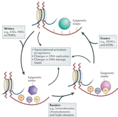
Histone modifying proteins have been categorized as “writers”, “erasers” or “readers” (Figure 1.1). Writers are those epigenetic enzymes that lay down
the removal of these marks. Readers are those proteins that contain domains that recognize these histone marks and bind to them.
Figure 1.1: Schematic representation of the main mechanisms for epigenetic
modifications.
1.2 Epigenetic Writers
Epigenetic writers are chromatin-associated proteins that catalyse the deposition of the PTMs mainly on histone tails and introduce dynamic modifications that respond rapidly to environmental changes.3
Among all the possible chemical modifications that can be introduced (methylation, acetylation, phosphorylation, ubiquitination, sumoylation, propionylation, butyrylation, crotonylation, ADP-ribosylation, citrullination), histone acetylation and methylation are the most abundant and widely studied. These modifications are carried out by Acetyltransferases and
Methyltransferases respectively, which are able to modify both histone and non-histone substrates.
1.2.1 Lysine Acetyltransferases (KATs)
Lysine Acetyltransferases (KATs) mediate the transfer of an acetyl group from acetyl coenzyme A (acetyl-CoA) to the ε-amino groups of lysine residues in histones and other proteins. After the acetylation, the positive charge of lysine is neutralized and, in the case of histones, the interaction with the negatively charged DNA backbone is diminished, producing an open chromatin status that is responsible for the promotion of gene expression.3
KATs have been classified into type A, which are nuclear proteins that acetylate chromatin-associated proteins and histones, and type B, which are located both in the nucleus and in the cytoplasm and acetylate newly synthesized cytoplasmic histones to promote their nuclear localization and deposition onto nascent DNA chains. While KAT1 is the only KAT in the type B group,3 type A KATs can be classified into different families by structural homology and biochemical mechanism of action. Despite several KAT families have been identified, only four have been extensively studied: the Gcn5-related N-acetyltransferase (GNAT) family (KAT2),4 the E1A-associated 300 kDa protein (p300)/CREB-binding protein (CBP) family (KAT3), the MYST family (KAT6)5 and the regulation of Ty1 transposition gene product 109 (Rtt109) family (KAT11). Among all the above-mentioned acetyltransferases, one of the most interesting protein is p300, which is a key enzyme in higher eukaryotes where it acts as an effector in several signalling pathways. Because of its fundamental role in different biological functions, this protein is the first target on which this thesis has been focused.
1.2.1.1 p300/CBP
is about 300 kDa in size (with 2414 amino acids). p300 was firstly reported in 1985 and 1989 in studies looking for proteins that bind E1A, an adenoviral oncogenic transcription factor. Meanwhile, CBP (also called CREBBP or KAT3A) is composed of 2441 amino acids and was reported for the first time in 1993 in a study of proteins that bind CREB, a transcription factor that binds cAMP response elements (CREs). Because of the high sequence homology between CBP and p300, together with the little sequence homology between them and other acetyltransferases in the human genome,6 the two proteins are now collectively referred to as p300/CBP and classified as a separate family class of KATs.
Despite they were first identified for their binding to E1A (p300) and CREB (CBP), it has been later demonstrated that these two proteins contribute to transcriptional regulation through their histone acetyltransferase activity. p300 and CBP contain several protein interaction domains (Figure 1.2); in particular, the structure of their HAT domain suggests a “hit and-run” (Theorell-Chance) catalytic mechanism in which, after binding of acetyl-CoA, the lysyl residue of the substrate peptide snakes through the p300 tunnel and reacts with the acetyl group. Interacting with a large number of transcription factors, CBP and p300 are involved in different cellular processes and misregulation of their activity is frequently implicated in many human diseases.
Figure 1.2: Domain architecture of p300. NRID, nuclear receptor interaction domain;
TAZ1, transcriptional adaptor zinc-finger domain 1; KIX, kinase-inducible domain of CREB-interacting domain; Bd, bromodomain; RING, really interesting new gene; PHD, plant homeodomain; HAT, histone acetyltransferase domain; ZZ, ZZ-type zinc-finger domain; TAZ2, transcriptional-adaptor zinc-zinc-finger domain 2; IBiD, IRF3-binding domain.
Modifications to histone lysines associated to p300/CBP have been studied as a major factor in cancer pathogenesis, but they also play a role in neurodevelopmental disorders, neurodegenerative, autoimmune and cardiovascular disease, metabolic and psychological disorders. 7, 8
For instance, mutations in the CBP (rarely p300) gene causes Rubinstein-Taybi syndrome, characterized by a short stature, moderate to severe intellectual disability, distinctive facial features, and broad thumbs and first toes. Moreover, CBP and p300 were demonstrated to be involved in hematopoietic homeostasis, such that mutations in the CBP/p300 interaction domain of different transcription factors were found in hematologic malignancies and chromosomal translocations involving CBP or p300 genes are associated with leukemia and lymphomas. CBP and p300 promote prostate cancer progression by activating androgen receptor-regulated transcription and colon cancer progression by microsatellite instability and they are involved in the development of drug resistance. 8
The inhibitors of CBP/ p300 described so far are of two types: the compounds that inhibit the binding of other proteins targeting the interacting domains and the derivatives that directly affect the acetyltransferase activity. To the first class belong the bromodomain-interacting molecules, such as the 5-isoxazolylbenzimidazoles recently reported as potent and selective ligands, those targeting the TAZ1 domain, such as chetomin, and those (namely sekikaic acid and lobaric acid) targeting the KIX domain. The second class inhibits the enzymatic activity of the HAT domain; in addition to many non-selective inhibitors (e.g., the natural products anacardic acid,9 garcinol,10 curcumin,11
plumbagin,12 and their analogues or semisynthetic derivatives), the only selective p300 HAT inhibitors described to date are the bisubstrate inhibitor Lys−CoA conjugate,13 which is not cell permeable, the pyrazolone C646,14
histone substrate, and the isogarcinol derivative LTK14,15, 16 again not very cell permeable and hardly optimizable due to its structural complexity.
For all the above considerations, there is still need for selective modulators of CBP/p300 activity not only as useful tools to dissect the role of their physiological and pathological role but also as potential leads for the development of drug candidates for specific diseases.17-19
1.2.2 Protein Methyltransferases (PMTs)
The protein methyltransferases (PMTs) catalyse methyl transfer from their universal methyl donor, S-adenosyl-L-methionine (SAM), to a nitrogen atom of lysine or arginine side chains forming S-adenosyl-L-homocysteine (SAH) as a byproduct of their mechanism.
In contrast to acetylation, histone methylation does not affect chromatin structure directly because this chemical modification does not change the charged state of an amino acidic residue. Depending on each specific residue, methylation is associated with activated euchromatic genes or with silenced heterochromatic genes. Moreover, each type of methyl mark represents a specific modification that is recognized as a docking site for chromatin-associated proteins that maintain chromatin architecture or regulate gene expression.3
The PMT enzyme class is composed of two distinct families of enzymes, based on their active site structure and on the amino acid to which they transfer methyl groups: the protein arginine methyltransferases (PRMTs) and the protein lysine methyltransferases (PKMTs). There is one exception to this general structural bifurcation of the PMT class: the enzyme DOT1L acts as a lysine methyltransferase, but its active site structure is most closely aligned with that of the protein arginine methyltransferases.20
1.2.2.1 Arginine Methyltransferases (PRMTs)
Arginine methylation is a common post-translational modification that has been implicated in signal transduction, gene transcription, DNA repair and mRNA splicing, among others.
Three types of methyl arginine species exist: ω-NG-monomethyl-arginine (MMA), ω-NG,NG-asymmetric dimethyl-arginine (ADMA) and ω-NG,N’G-symmetric dimethyl-arginine (SDMA).
Protein arginine methyltransferases are classified into type I or type II, according to modification types: although all PRMTs catalyse the formation of a monomethyl-arginine intermediate, type I PRMTs (PRMT1, 2, 3, 4, 5, and 8) can catalyse the production of asymmetric dimethyl-arginine, and type II PRMTs (PRMT5 and 7) are able to catalyse the production of symmetric dimethyl-arginine.21
1.2.2.2 Lysine Methyltransferases (PKMTs)
Histone lysine methylation is a marker of both transcriptionally active and inactive chromatin, depending on the residue that is methylated, its degree of methylation (mono-, di-, or trimethylation), and its position within the gene and in the genome. Except for DOT1L and WRAD complex,22 all known PKMTs contain a conserved SET (Su(var)3-9, Enhancer of Zeste, Trithorax) domain harbouring the enzymatic activity.23 To date, more than 50 PKMTs have been identified and characterized. On the basis of sequence homology, SET-containing KMTs can be divided into different subfamilies: the SUV39 family, the EZH family, the SET2 family, the PRDM family and the SMYD family.24 Several studies demonstrated that many SET domain PKMTs catalyse site-specific methylation of lysine residues in non-histone proteins, including transcription factors and other chromatin modifying enzymes, illustrating that lysine methylation is a widespread post-translational modification in signal
Among the several PKMTs identified so far, SETD8 is the second enzyme on which this thesis is focused. In the following paragraph there will be a brief description of its mechanism of action and its implication in biological processes.
1.2.2.2.1 SETD8
SETD8 (also known as PR-Set7, SET8, or KMT5A) is the sole mammalian enzyme known to catalyze the monomethylation of histone H4 Lys20 (H4K20me1). SETD8 protein expression is tightly regulated during the cell cycle, being highest during G2/M and early G1 and nearly absent during S phase.
SETD8 activity is essential in cell cycle progression and in the DNA damage response and it has been associated with mitotic chromosomes during cell division. SETD8 promotes transcriptional repression and mediate transcriptional activation. Recently, a direct involvement of H4K20me1 modification in the assembling of the pre-replication complex (pre-RC) on the replication origins of metazoans was demonstrated, highlighting the important role of SETD8 in this process. Besides H4K20, SETD8 methylates p53 at lysine residue 382, preventing p53 promoter binding and thereby inhibiting apoptosis. In regard to its function and role in human diseases, SETD8 is overexpressed in different types of cancer tissues and cancer cell lines including bladder cancer, non-small cell and small cell lung carcinoma, chronic myelogenous leukemia, hepatocellular carcinoma and pancreatic cancer. Furthermore, SETD8 is implicated in cancer invasiveness and metastasis through its interaction with TWIST, a master regulator in epithelial-mesenchymal transition (EMT). 26-28 Selective SETD8 inhibitors would serve as useful chemical probes to further investigate the cellular effects of SETD8 inhibition in both normal and diseased cells and as lead structures for the development of anticancer therapeutics. However, only few inhibitors have been reported so far for this enzyme.29-33
Very recently, three quinone-containing inhibitors endowed with cellular activity were identified from a high-throughput screening campaign, yet they irreversibly inhibit the enzyme, probably by a covalent bond with a Cys residue.29 These consideration prompted the research to the development of new small-molecule scaffolds for SETD8 inhibition.
1.3 Epigenetic Erasers
The erasers are enzymes responsible for the specific removal of epigenetic marks deposited by writers. Epigenetic erasers are classified in several groups of enzymes and the most studied ones are histone demethylases (HDMs) and histone deacetilases (HDACs).
1.3.1 Histone Demethylase (HDMs)
Until a decade ago, histone methylation, together with DNA methylation, was considered a stable chemical modification. This view changed with the discovery of lysine-specific demethylase 1 (LSD1) and the identification of the JMJC domain-containing lysine demethylase family. Several members of the histone demethylase family appear to be genetically amplified and overexpressed in some human tumours and these findings make the histone demethylases very interesting targets for drug discovery.
1.3.2 Histone Deacetylase (HDACs)
HDACs are enzymes responsible for the removal of the acetyl group of lysine residues in histones. After histone deacetylation, the positive charge of lysine is restored, promoting the condensation of chromatin and consequently transcriptional repression. HDACs are divided into five classes based on their phylogenetic comparison with yeast enzymes. Class I comprises HDAC1, HDAC2, HDAC3 and HDAC8; class IIa consists of HDAC4, HDAC5, HDAC7
sirtuins from SIRT1 to SIRT7; and class IV contains only HDAC11. Enzymes from classes I, II and IV require a zinc ion for catalysis, whereas sirtuins are NAD+ dependent enzymes with protein deacetylase and ADP ribosylase
activity.3
More than 10 years ago, it was discovered that deregulation of HDAC activity in association with chromosomal translocation was involved in the stimulation of leukemogenesis. To date, several studies have provided evidence of aberrant acetylation and altered expression of HDACs in cancer cells and tumour tissues. Therefore, using HDAC inhibition to reverse epigenetic aberrancies in cancer cells is a powerful approach for the treatment of several tumour types and four HDAC inhibitors are now approved: vorinostat34 is used for cutaneous T-cell lymphoma and is being explored for treatment of other cancers, romidepsin35 is used for peripheral and cutaneous T-cell lymphoma, belinostat36 is used for the same lymphomas but can be used in combination with other drugs to treat ovarian cancer and panobinostat37 is used for the treatment of multiple myeloma.
1.4 Epigenetic Readers
Epigenetic readers are specialized domains able to recognize and bind to specific epigenetic marks produced by the writers and erasers. Chromatin readers are able to identify not only different modified amino acids, but also different modification states of the same amino acid.
The most known reader domains of histone PTMs can be divided in different families able to recognize acetyl (Bromodomains) and methyl marks (Tudor, Chromo, MBT, PWWD and plant agenet domains, which belong to the “Royal Superfamily”).
1.4.1 Bromodomains
Bromodomains bind to acetylated lysine and they are probably the best-characterized epigenetic readers. The structure of these readers is highly conserved. More than 50 bromodomain proteins are encoded by the human genome and they can be clustered in nine subfamilies according to sequence homology.3
A well-known example of a bromodomain family is the BET (Bromodomain and extraterminal domain family) that includes four protein members (BRD2, BRD3, BRD4 and BRDT), which contain a tandem bromodomain at the N-terminal.38 These proteins play a decisive role in the regulation of transcription and cell growth; in addition, BET proteins are usually part of large nuclear complexes that are involved not only in transcription processes, but also in chromatin remodelling, replication and DNA damage. Hence, dysregulation of BET proteins has been reported in several diseases.39
1.4.2 The Royal Superfamily
The royal superfamily includes Tudor, Chromo, MBT, PWWD and plant
Agenet domains; all the members of this superfamily possess a structurally
related barrel-like protein fold, which is composed of 3 to 5 antiparallel β-sheets and forms the core structure. This conserved structure probably originated from a common ancestor that specifically recognized protein methylation. In distinct royal subfamilies, additional structural elements flanking the core structure contribute to the binding properties of its members. Members from each subfamily recognize either methylated lysine or methylated arginine residues in target proteins using a common binding mode, in which an aromatic binding pocket in the barrel accepts the methylated side chain. The pocket is usually composed of 2 to 4 aromatic residues, which provide electrostatic and
1.4.2.1 Tudor Domains
Among all the methyl readers of the Royal Superfamily, particularly interesting are the Tudor domains. Mammalian Tudor proteins can contain a single Tudor domain alone, multiple tandem Tudor domain repeats or one or more Tudor domains in conjunction with other types of domain. The Tudor protein family can be largely divided into two groups, one containing methyl-arginine binding Tudor domains and the other containing methyl-lysine binding Tudor domains. Most methyl arginine binding Tudor proteins are predominantly involved in RNA-related processes while methyl-lysine binding Tudor proteins are often implicated in chromatin biology, a general feature of the other royal superfamily members.41
Specifically, this thesis is focused on the recently identified Tudor domains of PHF20, which have a crucial role in the stabilization and activation of the tumour suppressor protein p53.42
1.4.2.1.1 Tudor Domains of PHF20
The PHD finger protein 20 (PHF20/GLEA2/HCA58) was initially described as an immunogenic antigen that elicits a strong antibody response in glioblastoma and adenocarcinoma patients. Later, it was shown that PHF20 can transcriptionally activate p53 and be downregulated through phosphorylation by the Akt kinase. PHF20 was also identified as a component of the “male absent on the first” (MOF) lysine acetyltransferase, which together with O-linked β-N-acetylglucosamine transferase, isoform 1 (OGT1) form the nonspecific lethal (NSL) complex.42
This protein is a component of some mixed-lineage leukemia (MLL) methyltransferase complexes with the core components MLL, ASH2L, WDR5 and RBBP5. PHF20 is a multidomain protein and it comprises two N-terminal Tudor domains, a central C2H2-link finger domain and a C-terminal zinc-binding PHD domain (Figure 1.4).
Figure 1.4: Domain architecture of PHF20.
Although little is known about its cellular role, the domain organization of PHF20 and its association with MLL core complexes suggest it to function as a transcription factor. Previous studies have indicated that the second Tudor domain is capable of binding methylated residues (preferentially dimethylated ones, one of the most common is H4K20me2) on histone tails, while no such function has been ascribed to the first Tudor domain.43 Moreover, the second Tudor domain of PHF20 interacts specifically with p53 peptides monomethylated or dimethylated at Lys370 or Lys382.
Whether by mutation or overexpression, Tudor domains of PHF20 were found to be implicated in cancer and other diseases through aberrant binding of chromatin, which results in aberrant activation or repression of genes.
1.5 Epigenetics and Drug Discovery
Over the last decade, the cellular machinery that creates the epigenetic modifications has been the subject of intense scientific investigation. As previously discussed, epigenetics play a fundamental role in all the biological processes and modulating epigenetic mechanisms is highly relevant for many diseases. Because of the importance of epigenetic proteins in physiological and pathological processes, great efforts have been done in the discovery and development of small molecule modulators of chromatin modifying proteins. Despite all these efforts, to date only six epi-drugs are available in therapy (four inhibitors of HDACs and two of DNA methyltransferase) and few other
enzymes for which no modulators have been reported. Therefore, despite recent advances in epigenetic modulators development, the challenge of identifying potent and selective molecules remains still open.
One of the main reasons that led to the lack of modulators is that not all the epigenetic enzymes are druggable, because of unfavourable pocket features as well as the competition with cofactors for targeting the active site.44
Proteins belonging to the families of HDACs, HDMs, HMTs and bromodomains are druggable, indeed robust and selective chemical probes are available to study these enzymes. On the other hand, for example, it has been very difficult to find potent, selective, and drug-like small molecule inhibitors of HATs. Beside the druggability, the identification and development of epigenetic modulators is also affected by the lack of specific and robust screening methods to evaluate their activity.
From a practical standpoint, developing assays to screen compound libraries for the identification of small-molecule modulators of epigenetic enzymes is not straightforward. In most cases, the primary screening is based on biochemical assays, which allow to evaluate the effect of the compounds on the activity of the enzyme. An ideal biochemical assay should reproduce the activity of the protein in the cellular context. However, many epigenetic targets exist as large complexes of proteins and, in some cases, these complexes are necessary for their activity, so the use of isolate proteins may be misleading. In addition, the substrates used are not always canonical histones (often peptides are preferred) and this choice could affect the results.
Cellular assays for epigenetic proteins have also proven to be somewhat complex. For many epigenetic targets, the effects of inhibitors in cell culture often take several days to see histone mark changes or effects on target genes, while phenotypic responses may require up to 7 to 10 days to be observed. In
addition, there is a lack of antibodies to detect effects on target proteins and even fewer for specific sites.45
In the following paragraphs, there will be an overview of the most widely used biophysical and biochemical methods in the drug discovery process for identification of epigenetic modulators.
1.6 Biophysical Methods
Biophysical methods are well-established in many areas of drug discovery. Application of these methods was once restricted to a relatively small number of scientists using specialized, low throughput technologies and methods. Now, automated high-throughput instruments are available in a growing number of laboratories.
Many biophysical methods are capable of measuring the equilibrium binding constants between pairs of interaction partners including protein-protein, protein-small molecule, and protein-nucleic acid interactions, and they can be used to measure the kinetic and/or thermodynamic components controlling biological processes in which they are involved. For a full characterization of a binding process, the determination of stoichiometry, the binding mode and any conformational change associated with such interactions are issues that should be addressed. The biophysical methods that are now available represent a powerful toolbox of techniques that can effectively deliver this full characterization. Biophysical methods are increasingly considered as a primary approach for hit finding. Among all, thermal-shift assay (mainly Differential Scanning Fluorimetry), calorimetry assays, optical biosensors and thermophoresis are techniques that have been recently successfully employed at this stage.46
1.6.1 Surface Plasmon Resonance (SPR)
Surface plasmon resonance (SPR) is a label-free detection method that has emerged during the last two decades as a suitable and reliable platform for biomolecular interactions. The technique allows the measurement of interactions in real-time with high sensitivity and without the need of labels, immobilizing target biomolecules on the sensor surface while a solution of ligand is flowed over the surface. Since it was first introduced in the early 1990s, SPR has been proven to be one of the most powerful technologies to determine specificity, affinity and kinetic parameters of a protein-ligand interaction. Surface plasmon resonance occurs when a photon of incident light hits a metal surface (typically a gold surface). At the incidence angle of total internal reflection, a portion of the light energy couples with the electrons of the metal surface layer, which then oscillate with the light wave. The electrons moving are called plasmons, and they propagate parallel to the metal surface. The plasmon oscillation in turn generates an electric field whose range is around 300 nm from the boundary between the metal surface and sample solution. The defined SPR angle, at which resonance occurs, is dependent on the refractive index of the material near the metal surface. Consequently, when there is an interaction between the target biomolecules immobilized onto the surface and the analyte solution, a small change in the reflective index occurs and it is detected (Figure 1.6.1).47
Figure 1.6.1: Typical set-up for a Surface Plasmon Resonance biosensor48.
The main advantages of this technique are: Real time analysis.
Kinetic information derived.
Label free technique: no need for radioactive, fluorescent or any other labelling.
On the other side, operator have to face with several problems:49 Mass transport can affect kinetic analysis.
Any artefactual refraction index change (other than from the interaction) can also give signal.
One of the interacting molecules must be immobilized on the surface. Lack of sensitivity when monitoring low molecular weight ligands.
1.6.2 Differential Scanning Fluorimetry (DSF)
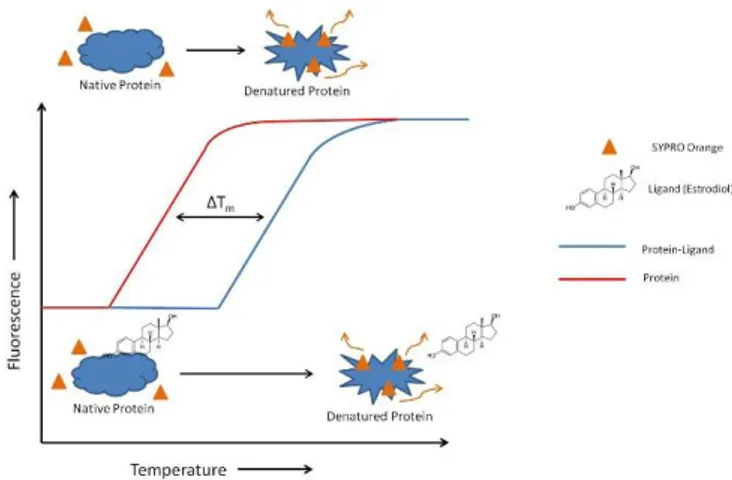
Differential Scanning Fluorimetry (or Thermofluor assay) belongs to a class of technologies that investigate the interactions between a biological target and its binding ligand. This assay use the well-established thermodynamic principle that the thermal stability of a protein, often quantified as the midpoint of thermal denaturation or melting point (Tm, the temperature at which both native and
concentration and potency-dependent manner. In a Thermofluor assay, a compound with a low fluorescence signal in a polar environment (such as in aqueous solution) but with high fluorescence in a non-polar environment is added to a protein solution. The fluorescence of the solution is monitored while the solution is heated. When the protein chain begins to unfold, the hydrophobic core becomes exposed and the signal increases until the protein is completely denatured. Thus, the temperature of hydrophobic exposure at which half of the protein population is unfolded, is determined (Figure 1.6.2).50, 51
Figure 1.6.2: Typical recording of fluorescence intensity versus temperature for the
unfolding of protein with and without the ligand in presence of SYPRO orange.
The commonly used fluorescent reporter dyes in thermal shift assays include Sypro Orange, Nano Orange and Sypro Red. Sypro Orange is usually the preferred choice because of its large intensity change upon binding to unfolded proteins. Intrinsic fluorescence from buried aromatic residues can also report protein unfolding as in the case of the nanoDSF. The basis of label-free fluorimetric analysis of protein unfolding lies in the properties of the fluorescent amino acid tryptophan. Since tryptophan is a hydrophobic amino acid, it is mostly located in the hydrophobic core of proteins where it is shielded from the surrounding aqueous solvent. Upon unfolding, tryptophan is exposed, and its
photophysical properties are altered. By detecting changes in tryptophan fluorescence intensity and its emission peak shift, the transition of a protein from the folded to the unfolded state can be precisely determined (Figure 1.6.3).52
Figure 1.6.3: nanoDSF principle.
DSF is a useful screening method for the following reasons: Use of small amount of protein.
Label-free and no-immobilization required. Easy optimization of assay conditions. Fast assay preparation and data analysis.
Likewise any other type of assay, this method has also intrinsic limitations: Not all biomolecules are responsive to fluorescence based thermal shift
assays. In general, proteins with large hydrophobic surfaces, such as membrane-embedded, lipophilic, complex-forming or misfolded proteins, produce high background of fluorescence as a result of dye binding before melting.
The assay is adversely affected by false positive or negative results: a weak binder that associates promiscuously to multiple sites on the protein surface may appear as a more potent hit.
Interference by many detergents in the assay buffer. Possible competition of the reporter dye with the ligand.
1.6.3 Microscale Thermophoresis (MST)
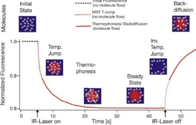
MicroScale Thermophoresis (MST) is a powerful technique to quantify biomolecular interactions. It is based on thermophoresis, the directed movement of molecules in a temperature gradient, which strongly depends on a variety of molecular properties such as size, charge, hydration shell or conformation. Thus, this technique is highly sensitive to virtually any change in molecular properties, allowing for a precise quantification of molecular events independent of the size or nature of the investigated sample. During a MST experiment, a microscopic temperature gradient is induced by an infrared laser: the directed movement of molecules through this microscopic gradient is detected and quantified using either covalently attached or intrinsic fluorophores (Figure 1.6.4).53, 54
Figure 1.6.4: Typical signal of a MST experiment. Initially, the molecules are
homogeneously distributed and a constant ‘‘initial fluorescence’’ is detected. Within the first second after activation of the IR laser, the ‘‘T-Jump’’ is observed, which corresponds to a rapid change in fluorophore properties due to the fast temperature change. Subsequently, thermophoretic movement of the fluorescently labelled molecules out of the heated sample volume can be detected.
MST not only allows for a precise determination of binding constants, but can also be used to derive additional information about the molecular mechanism of the investigated interaction.
From different points of view, MST technology is superior to other methods in determining the parameters of molecular interactions. The advantages of MST technology are:
Low sample consumption.
Robust and sensitive detection (broad concentration and size range). Rapid analysis.
No surface immobilization required.
Close to native protein conditions (possibility to work without labelling).
Live detection of aggregation, sticking and precipitation effects. On the other side, the technique suffers of the following drawbacks:
For a label-free analysis, at least one Tryptophan in the protein sequence is needed.
Absorption or Emission phenomena of small molecule compounds can interfere with protein signal.
Target and substrate must be soluble in the same buffer conditions.
1.7 Biochemical Methods
Biochemical methods are the most common techniques used for the study and characterization of small molecule compounds activity in drug discovery. While biophysical methods allow interaction studies between the compound and the target, biochemical assays give a quantitative measure of the ability of the
with a purified enzyme preparation that demonstrates catalytic activity on a specific substrate in a cell-free context. Usually, procedures for enzyme assays are well documented or cited in literature, but even accurate observance gives no guarantee of an unequivocal outcome: the same assays performed independently may yield quite different results. In fact, the enzyme activity depends on multiple factors and the understanding of particular features of each enzymes is required. The enzyme activity depends decisively on defined conditions with respect to temperature, pH, nature and strength of ions and enzyme assays can only be reliably compared, if such conditions are strictly observed.55
Many different approaches are available to measure enzymatic activity and can be differently classified. The most commonly used assays are based on the measurement of radioactivity and fluorescence.
1.7.1 Radioactivity Based Assays
Traditionally, assays based on radioactive probes (usually tritiated or iodinated ligands) have been successfully used to determine enzymatic activity because of the high sensitivity of the technique and the possibility to perform experiments on unmodified receptors expressed in native tissues and even in transfected cells. However, the use of radioactive ligands as tracers in these assays presents several drawbacks, both technical and financial. Technically, a classic radioactivity assay cannot be performed in a homogeneous format. Consequently the assay requires multiple washing steps before reading the radioactivity. This need adds complexity to the procedure and makes these assays more difficult to perform in high-throughput screening leading to extra cost. Additionally, the washing steps prevent any possibility of carrying out kinetics experiments on a single sample. Over the past decade, strategies such as scintillation proximity assays (SPA), which can be performed in homogeneous conditions, have been developed. SPA utilize microscopic beads containing scintillant: the interaction of these beads with β-particles, generated
by the radioactive decay of tritium or iodine, releases photons that may be measured with scintillation counters or charge-coupled device (CCD) imagers. A variety of SPA bead formats allows utilization of various substrates, but these assays are still expensive due to the production cost of beads.
A second technical drawback rely on the nature of the radioactive probe: it is difficult, in part for health reasons, to perform saturation assays that necessitate high concentrations of radioactive ligand. Furthermore, due to the hazardous nature of the compounds, the use of radioactive probes has some restrictions in term of radioactive waste disposal, delimitation of working area and staff medical follow-up. As a result, other techniques have been introduced to replace the use of radioactive assays, such as fluorescence techniques, without completely supplanting them.56
1.7.2 Fluorescence Based Assays
Among the most popular enzyme assays, the fluorescence-based ones are largely used in screening campaigns. These assays are commonly based on synthetic substrates that incorporate a chromophore, whose fluorescence properties change as a result of the enzyme reaction. The key advantage of these substrates is that the assay is simple and the signal produced is directly related to the enzyme-catalysed reaction. If the fluorescent product is soluble, the assay is well-suited for microtiter plate based assays. To date, almost all examples of fluorescent substrates focus on a small family of fluorophores and chromophores, in particular umbelliferones, nitrophenols, fluoresceins, rhodamines and BODIPY dyes, all of which are relatively large aromatic groups which tend to influence both substrate binding, catalytic turnover, and solubility.57
With the aim to overcome the limitations of traditional fluorescence-based assays, in the last few decade there was the introduction of other new
luminescence based techniques. Among all of them, one of the most widely used is the Alphascreen technology.
1.7.2.1 AlphaScreen Technology
An AlphaScreen assay utilizes proximity-based fluorescence detection through the tethering of donor and acceptor beads by a protein-ligand interaction (Figure 1.7.1).
Figure 1.4.1: Principle of the Alphascreen assay technology.
Initially developed underneath the name LOCI® (Luminescent Oxygen Channeling Immunoassay), the reagents and bead technologies for drug discovery are currently exclusively commercially available under the name AlphaScreen by Perkin Elmer. In this assay, the photosensitizer phthalocyanine is dissolved on a polystyrene donor bead. Excitation with 680 nm light induces phthalocyanine to convert ambient oxygen to singlet oxygen molecules with a 4 μs half-life. These molecules can diffuse 200 nm freely through solution. If a polystyrene acceptor bead is within the lifetime of the singlet oxygen species, the singlet oxygen will react with thioxene derivatives on the bead, resulting in a dioxetane product followed by a diester fluorescent product. If the emission is at 615 nM, the assay is called AlphaLISA: both assays rely on the same Donor beads but use different Acceptor beads. AlphaScreen Acceptor beads are embedded with three dyes: thioxene, anthracene, and rubrene. Rubrene, the final fluorophore, emits light detectable between 520-620 nm. In the AlphaLISA Acceptor beads, anthracene, and rubrene are substituted with an Europium
chelate. The Europium (Eu) chelate is directly excited by the 340 nm light resulting from the conversion of thioxene to a di-ketone derivative following its reaction with singlet oxygen. The excited Europium chelate generates an intense light detectable within a much narrower wavelength bandwidth centred around 615 nm. In contrast to the AlphaScreen, the AlphaLISA emission is therefore less susceptible to interference by either artificial or natural compounds that absorb light between 500-600 nm.
In both cases, the detection of the chemiluminescent readout depends on binding of the protein and its related ligand. Typically, the donor bead captures substrates (by the biotin-streptavidin interaction) while the acceptor bead interacts with the modifications on the substrate, after the enzymatic reaction. The interaction of protein and substrate results in chemical energy transfer of acceptor and donor beads, culminating in a luminescent signal. Lack of binding fails to bring acceptor and donor beads into sufficiently close proximity and the singlet oxygen decays without the production of light. Since the beads are coated with hydrogel, non-specific interactions are minimized, providing a large signal-to-background assay window.58, 59
However, Alphascreen may be sensitive to different types of interferences. For example, antioxidants or other quenchers of reactive oxygen species like metal ions can strongly affect the emitted signal, as well as biotin-like compounds can compete for the interaction of biotinylated substrate with Donor beads. Moreover, since the Alphascreen detection is only based on a fluorescence intensity measurement, coloured compounds absorbing in the 500-600 nm wavelength range can artificially decrease the signal and therefore may be detected as false positives in HTS.60
2. AIM OF THE WORK
Epigenetic enzymes are involved in the development of several diseases and, as previously discussed, they are considered very interesting targets for drug discovery. The complexity of these proteins, together with the lack of a complete understanding of their biological role, are responsible for the limited number of currently identified modulators.
Different methodologies are available for the identification of epigenetic modulators (Chapters 1.6 and 1.7), nevertheless to date there is not a gold standard approach for the evaluation of their activity. It is worth mentioning that in a drug discovery programme it is necessary to use different technologies. The reasons of this need are different. First, each technique suffers of intrinsic limitations that could affect the outputs, as previously reported, so it is necessary to validate data. In addition, a single method is not suitable to deeply characterize the modulators activity (for example binding, mechanism of action, potency and so on). Furthermore, the use of different methodologies allow the identification of pan-assay interference compounds (PAINS), which encompass some 400 structural classes (for example enones, catechols, isothiazolones and so on). Usually, during the screening of compounds libraries, the activity of 5-12% of compounds does not depend on a specific, drug-like interaction between molecule and protein. PAINS can give false readouts in a variety of ways. Some are fluorescent or strongly coloured. In certain assays, they give a positive signal even when no protein is present. Other compounds can trap the toxic or reactive metals used to synthesize molecules in a screening library or used as reagents in assays. These metals then give rise to signals that have nothing to do with a compound’s interaction with a protein. Other PAINS coat a protein or sequester metal ions that are essential to a protein’s function, or they may alter proteins chemically without fitting specifically into a binding site.
All of these mechanisms prevent further attempts to improve the biological activity of a molecule by modifying its structure. The only method to discover PAINS is to use orthogonal assays that can give different readouts, so it is possible to define “true hits”.
In this scenery, the aim of my phD project was to develop a robust and widely usable combined screening platform to identify small-molecule modulators of epigenetic enzymes (Figure 2.1). To overcome the limitations of each technique and with the aim to identify “true hits”, different biophysical and biomolecular techniques were exploited to interrogate three epigenetic proteins (p300, SETD8 and Tudor domains of PHF20) and we validated the outputs. It is important to highlight that the use of multiple technologies enabled also a deep characterization of modulators activity, allowing the determination of binding, mechanism of action and potency.
3. RESULTS AND DISCUSSION
3.1 p300
3.1.1 SPV106 analogues as modulators of acetyltransferase activity
Different approaches have been used to identify p300 modulators, but only a limited number of small molecule inhibitors have been described, with various degrees of selectivity and cell permeability. Ethnomedicine has inspired the identification of a few natural products, including anacardic acid,9 garcinol,10 curcumin,11 plumbagin,12 and guttiferone A61 as inhibitors of different classes of KATs. Among them, in the Epigenetic Medicinal Chemistry Laboratory (EMCL) we focused our attention on anacardic acid. Anacardic acid, a small molecule compound extracted from cashew nut shell liquid which is known to have antitumor activity, inhibits acetyltransferase activity of p300, but it’s not a selective inhibitor.9
In 2008, EMCL reported on the activity of a series of long-chain alkylidenemalonates (LoCAMs) as protein acetyltransferases modulators. The design of LoCAMs was inspired by the structural simplification of anacardic acid (Figure 3.1.1).62, 63
Figure 3.1.1: From anacardic acid to LoCAMs.
One of these compounds, the diethyl pentadecylidenemalonate 1 (SPV106), has a unique activity profile. This compound exhibits inhibitory properties against p300 with a potency comparable to that of anacardic acid, and it simultaneously enhances the acetylating activity of PCAF. Therefore, it is the first mixed
As a result of its peculiar activity profile, derivative 1 was successfully used as a chemical probe in a study that correlated Duchenne cardiomyopathy with PCAF-mediated lysine acetylation levels of connexin 43.64 It was also examined
during investigations of the role of KAT enzymes in the regulation of the extinction of conditioned fear and neuronal plasticity.65 More recently,
treatment with compound 1 has been shown to reverse alterations in human cardiac mesenchymal cells obtained from diabetic patients and restore cellular function.66
The uncommon biological properties of derivative 1 prompted us to explore the structure-activity relationships of LoCAMs. First, we focused on the alkyl chain and the flexibility of the scaffold:62 we found that variations in the alkyl chain length influenced the activity profile of KAT modulators, whereas all other modifications, such as variations in flexibility/rigidity of the core structure and the introduction of substituents, were detrimental.62 Indeed, not only the selectivity toward different KAT enzymes, but also the inhibitory and activating properties, varied depending on the substitution pattern.62
As a further exploration of the structure-activity relationships of this class of KAT modulators, and with the aim of identifying potential structural features that differentially affect the activity of acetyltransferases, my project started with the synthesis of a number of 1,3-dicarbonyl derivatives (Figure 3.1.2). The synthesized compounds formally derived by the replacement of one or both ester functions with keto- or carboxylic acid groups while the alkyl chain length was kept constant. In addition, because it was previously found that the diketo-analogue of compound 1 (derivative 2d, Figure 3.1.2) retained inhibitory activity against p300,62 a few shorter and longer-chain homologues of 2d were also prepared.
Figure 3.1.2: 1,3-Dicarbonyl derivatives 2a-e, 3a-d, 4a,b, 5a,b, 6a,b, 7a,b, and 8a-f.
3.1.1.1 Chemistry
Novel SPV106 derivatives 2-5 were prepared by Knoevenagel condensation of a 1,3-dicarbonyl derivative with the appropriate alkyl aldehydes. The aldehyde used, if not commercially available, were prepared from the corresponding alcohol by oxidation with 2,2,6,6-tetramethylpiperidine 1-oxyl (TEMPO),67 (Scheme 3.1.1). Derivatives 2a-e, 3a-d, and 4a,b were prepared by reacting, respectively, pentane-2,4-dione, ethyl 3-oxobutanoate, tert-butyl 3-oxobutanoate, or malonic acid with the appropriate aldehyde in dichloromethane using piperidine and acetic acid as the catalysts (Scheme 3.1.1).
Scheme 3.1.1: Reagents and conditions: a) alkyl aldehyde, AcOH, piperidine, CH2Cl2,
RT, 2–12 h; b) TFA/CH2Cl2 (5:95), RT, 30 min.
The hydrolysis of tert-butyl esters 3c,d was performed with trifluoroacetic acid (TFA), yielding the corresponding 2-alkylidene-3-oxobutanoic acids 5a,b. By Knoevenagel condensation of ethyl oxobutanoate and tert-butyl 3-oxobutanoate with alkyl aldehydes, derivatives 3a,b and 3c,d were obtained, as mixtures of E and Z isomers. After the separation of the two isomers by silica gel chromatography, it was observed a spontaneous interconversion of the compounds. For this reason, compounds 3a-d and the corresponding acids 5a,b were tested as the E/Z mixtures. Alkylthiomethylidenepentane-2,4-diones 6a,b and alkylaminomethylidenepentane-2,4-diones 8a-f were obtained in high yield (Scheme 3.1.2) by reacting, respectively, the appropriate alkyl amine or alkyl thiol with 3-(ethoxymethylene)-2,4-pentanedione 9 in THF at reflux.68
Scheme 3.1.2: Reagents and conditions: a) alkyl amine, thiol, or alcohol (n=11, 12),
THF, reflux, 2 h.
Alkyloxymethylene-2,4-pentanediones 7a,b were obtained in good yield under the same conditions from the corresponding alkyl alcohols. Unfortunately, compounds 7a,b were unstable even in presence of low amount of water. Therefore, any attempt to purify these derivatives by chromatography was unsuccessful. Moreover, because an aqueous medium is required for biological screening, any further purification efforts was abandoned.
This pool of SPV106 analogues synthesized was used to develop a screening strategy, using biophysical and biochemical techniques.
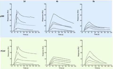
3.1.1.2 Biophysical Screening: Surface Plasmon Resonance Assay
To avoid time-consuming, expensive assays, all compounds were preliminarily screened using a Surface Plasmon Resonance (SPR)-based binding assay. As discussed in the introduction section, this biophysical technique is well suited for a primary screening.69 Recently, at EMCL, an SPR-based binding assay was established and successfully employed to study the real-time interactions between small-molecule derivatives and p30062, 63 as well as other epigenetic targets.70
human recombinant p300 (aa 1284-1673) and PCAF (aa 492-658) catalytic domains were immobilized (up to ̴10 000 response units (RU)) on the flow cells of the biosensor chip, and 1,3-dicarbonyl derivatives 2-8 were injected at various concentrations (from 25 to 100 µM) over the protein surface. To decrease false positives related to detergent-sensitive and aspecific aggregation-based binding, 0.005% of Surfactant P20 was added to the running buffer in all experiments. In addition, to evaluate potential non-specific binding, all compounds were injected on an immobilized myoglobin. The binding of each compound was read in real time as the change in mass at the sensor surface. After injection, running buffer was allowed to flow over the surface, and the dissociation of compounds from the surface was observed.
These experiments showed that tetradecylidene- and pentadecylidene-substituted pentane-2,4-diones, malonic acids, and 3-oxobutanoic acids (2c,d,
4a,b, and 5a,b, respectively) efficiently interact with the immobilized proteins,
as demonstrated by the concentration-dependent responses and the clearly evident exponential curves during both the association and dissociation phases (the sensorgrams for compounds 2d, 4b, and 5b are displayed in Figure 3.1.2). For the compounds 2d, 4b and 5b equilibrium dissociation constant (KD) values were derived from the ratio between kinetic dissociation (kd) and association (ka) constants, obtained by fitting data from all injections at different concentrations of each compound, using the simple 1:1 Langmuir binding fit model of the BIAevaluation software. The compound 2d showed a KD of 5.85 x 10⁻⁵ against p300 and of 2.74 x 10⁻⁵ against pCAF, while for the compound
4b the KD obtained was of 4.32 x 10⁻⁷ against p300 and of 1.53 x 10⁻⁵ against
pCAF. Regarding the compound 5b, the KD against p300 was of 3.05 x 10⁻⁶ while the one reported against pCAF was of 1.52 x 10⁻⁴.
On the other hand, both 3-oxobutanoates 3a-d and derivatives 6-8, which are characterized by the presence of a heteroatom in their aliphatic chains, showed negligible interaction. In addition, in agreement with what was previously
reported for LoCAMs,62 it was shown that variations in the alkyl chain length influenced the binding profile of the tested pentane-2,4-diones 2a-e. In fact, their superior (2e) and inferior (2a and 2b) homologues also produced good sensorgrams but showed low and/or concentration-independent responses.
Figure 3.1.3: Sensorgrams obtained from the SPR interaction analysis of compounds 2d, 4b, and 5b binding to immobilized p300 (catalytic domain, aa 1284-1673, top row)
and PCAF (catalytic domain, aa 492-658, bottom row). Each compound was injected at four different concentrations (25, 50, 75, and 100 μM).
3.1.1.3 Biochemical Screening: Radiometric Assay
SPR, used as a filtering method, gives information about the interaction of the molecules with targets and explain the modulatory activity, but cannot be used to define the potency of molecules. For this reason, the effect of the selected derivatives (SPR+) 2c,d, 4a,b, and 5a,b on the catalytic activity of the human recombinant acetyltransferase enzymes p300 and PCAF was determined in radiometric activity assays, which were performed by Reaction Biology Corporation (Malvern, PA, USA), using curcumin and anacardic acid as reference compounds. Two negative controls (SPR-, 6b and 8b) were also tested
As shown in Table 3.1.1, all the SPR+ derivatives substantially affected the activity of one or both the HAT enzymes.
Table 3.1.1: Effects of compounds 2c,d, 4a,b, 5a,b, 6b, and 8b on the activity of p300
and PCAF. Compounds were tested in 10-dose IC50 mode with threefold serial dilutions
starting at 100 µM. Data were analyzed with GraphPad Prism software (version 6.0) for IC50 curve fits. Enzyme activity percentage was determined at 100 µM with respect
to DMSO. Histone H3 was used as substrate (5 µM), and [acetyl-3H]acetyl coenzyme
A (3.08 µM) was used as an acetyl donor.
In particular, pentane-2,4-diones 2c,d induced a marked dose-dependent increase in the enzymatic activity of PCAF (HAT activity at 100 µM: 229% and 389%, respectively; Table 3.1.1), while they did not affect the activity of p300. In contrast, malonic acids 4a,b strongly inhibited both enzymes, with IC50
values in the low micromolar range (1.3 and 1.1 µM, respectively, for p300, and 50.3 and 21.1 µM, respectively, for PCAF; see Table 3.1.1), which are similar to or more potent than the reference compounds (IC50: 6.5 µM and 33.9 µM for
curcumin and anacardic acid, respectively; Table 3.1.1). Notably, 3-oxobutanoic acids 5a,b efficiently inhibited p300 (IC50 values of 2.4 µM and
4.7 µM, respectively) but also caused strong amplification of PCAF enzymatic activity in a dose-dependent manner (HAT activity at 100 µM: 346 and 497%, respectively; Table 3.1.1). This effect was even more significant than that
observed for pentane-2,4-diones 2c,d. In accordance with the SPR experiments, the negative controls negligibly affected the enzymatic activity of p300 and PCAF (see 6b and 8b in Table 3.1.1).
3.1.1.4 Evaluation of Cellular Activity
After the characterization of the enzymatic activity of compounds 2c, 2d, 4a,
4b, 5a and 5b using a biophysical and a biochemical method, the screening
approach continued with the aim to evaluate their effect on cells. Cellular assays were carried out on three different cell lines, namely human leukemic monocyte lymphoma U937 cells and human cervical carcinoma C33A and HeLa cells. First, an MTT assay was performed after the treatment with selected derivatives to assess the maximum concentration of compounds that could be used without significantly affecting cell viability. For solid C33A and HeLa tumor cell lines, it was observed no significant decrease in the number of metabolically active cells after 24 h of treatment with concentrations up to 200 µM for derivatives
2c,d and 4a,b and up to 50 µM for compounds 5a,b.
On the other hand, in the case of the more sensitive U937 cell line (Figure 3.1.4a), it was registered a significant decrease in cell viability starting from lower concentrations of tested compounds (100 µM for 2c,d and 4a,b and 10 µM for 5a,b, respectively). Therefore, the derivatives were evaluated on U937 cell line for their effects on the cell cycle. After 24 h of treatment, compounds
4b, 5a, and 5b were able to arrest the cell cycle in the G1 phase (Figure 3.1.4b).
In the case of the latter two compounds, this occurred at concentrations as low as 10 µM. This result is consistent with the importance of acetylation for control of the G1/S transition.71-74 Under the same conditions, the other derivatives had
Figure 3.1.4: Cell viability and cell-cycle analysis in U937 cells by
fluorescence-activated cell sorting (FACS). a) Cell viability was assessed by measuring the mitochondrial-dependent reduction of MTT to formazan. b) U937 cells were treated with compounds 2c,d, 4a,b, and 5a,b at the indicated concentrations for 24 h, stained with propidium iodide, and subjected to flow cytometric analysis to determine the distribution of cells in each phase of the cell cycle. Data are reported as the mean ±SD of at least three independent experiments.
Finally, the effects of compounds 2d, 4b, and 5b were determined on the acetylation levels of specific lysine residues of core histones H3 (K9) and H4 (K5) in the three cell lines. Cells were incubated for 24 h with vehicle and tested with compounds at the indicated concentrations, using as reference compound the suberoylanilide hydroxamic acid (SAHA; 5 µM).75 The histone extracts were then immunoblotted with antibodies to specific histone acetylation sites
(Figure 3.1.5). Consistently with the different patterns of acetylation recently described by Garcia and co-workers,76 it was observed that the tested compounds differentially affected the H4K5ac and H3K9ac levels in the three cell lines. This observation is not surprising, considering that in the cellular context, acetyltransferases are similar to other enzymes by not being isolated; they participate in complex pathways and actively crosstalk with each other and with other proteins.
Specifically, the effect of pentane-2,4-dione 2d on both markers was negligible in U937 cells (Figure 3.1.5, left and middle panels), whereas a decrease was observed in both cervical carcinoma cell lines, which was more evident for H4K5ac (Figure 3.1.5). Both malonic acid 4b and 3-oxobutanoic acid 5b induced a marked decrease in the acetylation of lysine H4K5 in C33A cells (Figure 3.1.5, left panel), whereas no appreciable effect on the same marker was detected in HeLa cells, and an increase was observed in U937 cells (Figure 3.1.5).
In contrast, derivative 4b induced a moderate decrease in the acetylation of H3K9 in C33A and HeLa cells (Figure 3.1.5, middle panels), and there was no appreciable variation in U937 cells (Figure 3.1.5, middle panel). The compound
5b induced a decrease in the acetylation of H3K9 in C33A and HeLa cells
(Figure 3.1.5, middle panels) and a marked increase in the acetylation level in U937 cells (Figure 3.1.5, middle panel). As expected, treatment with the reference compound SAHA reliably showed a significant increase in the lysine acetylation level (Figure 3.1.5).
Figure 3.1.5: Western blot analyses performed with compounds 2d, 4b, and 5b at the
indicated concentrations for 24 h on the acetylation of the specific lysine residues H4K5 (left column) and H3K9 (middle column) in histone extracts from a) C33A and b) HeLa carcinoma cells, and c) U937 leukemic monocyte lymphoma cells. Acetylation was detected by immunoblotting with antibodies specific for histone acetylation sites as indicated. Total histone H3 was used to check for equal loading. SAHA (5 µM) was used as a reference compound. Signals were detected with the ImageQuant LAS 4000 digital imaging system and quantified by ImageQuantTL software (version 8.1); total histone H3 levels were used for normalization. The results are reported as the mean ±SD of three independent experiments.
The differences observed in the activity profile of the structurally related compounds 2c,d, 4a,b, and 5a,b against the two acetyltransferases could have a number of explanations, and the mechanisms underlying their biological effects remain unclear. However, it has been previously reported that small modifications in structurally related compounds lead to dramatic differences in terms of selectivity between PCAF and p300.15 Moreover, is worth mentioning that the catalytic mechanisms of the two enzymes are different, with PCAF