Full Terms & Conditions of access and use can be found at
http://www.tandfonline.com/action/journalInformation?journalCode=tizo21
The European Zoological Journal
ISSN: (Print) 2475-0263 (Online) Journal homepage: http://www.tandfonline.com/loi/tizo21
Reproductive and tissue plasticity in Arca noae
(Bivalvia: Arcidae)
F. Ghribi, G. Bello, R. Zupa, L. Passantino, N. Santamaria, M. El Cafsi & A.
Corriero
To cite this article: F. Ghribi, G. Bello, R. Zupa, L. Passantino, N. Santamaria, M. El Cafsi & A. Corriero (2017) Reproductive and tissue plasticity in Arca noae (Bivalvia: Arcidae), The European Zoological Journal, 84:1, 473-487, DOI: 10.1080/24750263.2017.1368725
To link to this article: https://doi.org/10.1080/24750263.2017.1368725
© 2017 The Author(s). Published by Informa UK Limited, trading as Taylor & Francis Group.
Published online: 01 Sep 2017.
Submit your article to this journal
Article views: 121
View related articles
Reproductive and tissue plasticity in
Arca noae (Bivalvia: Arcidae)
F. GHRIBI
1, G. BELLO
2, R. ZUPA
2, L. PASSANTINO
2, N. SANTAMARIA
2,
M. EL CAFSI
1, & A. CORRIERO
2*
1
Unit of Physiology and Aquatic Environment, Faculty of Mathematical, Physical and Natural Sciences of Tunis, University of Tunis El Manar, Tunis, Tunisia, and 2Veterinary Clinics and Animal Production Unit, Department of Emergency and Organ Transplantation, University of Bari Aldo Moro, Valenzano, Italy
(Received 8 June 2017; accepted 11 August 2017)
Abstract
The reproductive strategy of an unexploited population of Arca noae from the salt-water Bizerte Lagoon (Tunisia, western Mediterranean), including its tissue plasticity, was studied. In total 309 individuals, collected monthly from October 2013 to September 2014, were examined; 142 were females, 42 were males and five were hermaphrodites. They were used in histological and immunohistochemical (stem marker: Pou5f1; proliferation marker: proliferating cell nuclear antigen (PCNA)) analyses of gonads and adjacent tissues (N = 189) as well as to compute the monthly condition index (N = 120). Water salinity, temperature and chlorophyll a concentration were recorded. Ripe ovaries were observed in two discrete periods, October–November and April–August. Both gonad ripenings were followed by spawning periods, November–April and July–September. The mature oocyte density showed that the first spawning period was less vigorous than the second one. These data also indicated that A. noae is a multiple spawner. Five cases of protandric hermaphroditism occurred from December to April. Gonad tissue was strictly associated and intermingled with the digestive gland and mantle muscle fibres. Seasonal variations were observed in the relative proportions of digestive gland and gonads: the former predominated when the latter regressed (March) and vice versa (peak in June). Seasonal transitions from germinal to somatic tissue and vice versa were hypothesised to occur through transdifferentiation mechanisms based on the activity of stem and proliferating cells. The condition index roughly increased along with gonad ripening and decreased during the spawning periods, although it did not run parallel to gonad evolution, because it also depended on chlorophyll a concentration, a proxy for phytoplankton density. The condition index was significantly correlated, by multiple regression, to both mature oocyte density and chlorophyll a concentration. Arca noae appears to have evolved aflexible reproductive strategy that makes it capable of exploiting diverse environmental conditions, which also involves tissue transdifferentiation. Keywords:Mollusca, reproduction, body condition, stem cells, Mediterranean Sea
Introduction
The Noah’s ark, Arca noae Linnaeus, 1758 (Bivalvia: Arcidae), is a commercial, edible bivalve widespread in the whole Mediterranean Sea and the eastern Atlantic Ocean from Portugal to Angola (Gofas2008). Hence, it is a warm-affinity mollusc. It lives attached by a solid byssus on rocky grounds or other solid substrate (e.g. dead mollusc shells), from the low-tide level to about 120 m depth (Hrs-Brenko & Legac 1996). In the Mediterranean, it becomes sexually mature at an early age, i.e. 2 years, when slightly larger than 15 mm and 20 mm in males and females, respectively (compare
data in Bello & Paparella2001; Peharda et al.2006), and reaches 12 cm in length and 25 years of age (Puljas et al.2015). It takes 3 to 7 years to grow to about 50 mm length (Peharda et al.2002,2003), which corresponds to the minimum commercial size. It is the most impor-tant edible arcid species in the Adriatic Sea and is commercially exploited in Croatia, Italy and Slovenia (Valli & Parovel 1981; Peharda et al. 2006) where it attains high market quotations (Poutiers1987).
The reproductive biology of A. noae has been investigated only in the Adriatic Sea. Studies on its life traits have been carried out by Bello and Paparella (2001), Peharda et al. (2002, 2003, 2006,
*Correspondence: A. Corriero, Veterinary Clinics and Animal Production Unit, Department of Emergency and Organ Transplantation, University of Bari Aldo Moro, S.P. per Casamassima km. 3, Valenzano, BA 70100, Italy. Tel: +39 5443907. Fax: +39 5443907. Email:[email protected]
Vol. 84, No. 1, https://doi.org/10.1080/24750263.2017.1368725
© 2017 The Author(s). Published by Informa UK Limited, trading as Taylor & Francis Group.
This is an Open Access article distributed under the terms of the Creative Commons Attribution License (http://creativecommons.org/licenses/by/4.0/), which permits unrestricted use, distribution, and reproduction in any medium, provided the original work is properly cited.
2009), Bello et al. (2013), Župan et al. (2014) and Puljas et al. (2015).
Nofishery management has ever been implemented for A. noae, although several papers have suggested that its Adriatic populations are overfished and require a suitable conservation policy (Bello & Paparella2001; Peharda et al.2002, 2003, 2006,2009; Župan et al.
2012; Bello et al. 2013). The overexploitation of A. noae populations alters their structure both size- and sex-wise, since females, which are larger than males, are proportionally harvested more than males. This hinders the ascertainment of actual natural biological parameters, including sex ratio by size/age class and size/age at sex change (Bello et al.2013).
The presence of A. noae has been occasionally recorded in Tunisia: in the Gulf of Gabes (Risso1978; Darmoul et al. 1980; Ben Mustapha et al. 1999; Enzenross & Enzenross 2001), the North lagoon of Tunis (Saubade & Risso 1983), the Bizerte Lagoon, Tabarka (Enzenross & Enzenross2001) and the Gulf of Tunis (Zouari1985; Ayari & Afli2008). Due to their negligible commercial exploitation, none of these (sub) populations has ever been studied and no information is available on their demographic structure and reproduc-tive biology.
Since the reproductive cycle of A. noae was already satisfactorily described by Valli and Parovel (1981) and Peharda et al. (2006), the present study aimed mainly at knowing the reproductive strategy of this species in the Bizerte Lagoon, including periods and pattern of reproduction. This information is necessary for establishing a knowledge base (Gosling 2003) for
an unexploited (sub)population in view of the man-agement of stocks that will be likely exploited in the near future. Moreover, the information obtained from the study of unexploited stocks provides an insight into the natural biological parameters of a species, which in turn are an essential reference tool for the assessment and management of overexploited stocks from other geographical areas.
In addition to gaining this set of information, the histological analyses of A. noae gonads provided insights into the structural plasticity of this marine invertebrate, which displayed an unreported capability of modifying the structure and volume of visceral organs on a seaso-nal basis, most likely through a transdifferentiation mechanism also observed in other marine and terrestrial invertebrates but never reported in any bivalve.
Materials and methods
Specimens of Arca noae were collected in the Bizerte Lagoon, Tunisia, western Mediterranean Sea, close to the Sicilian–Tunisian sill. This is a salt-water coastal lagoon 150 km2 wide, with a
maximum depth of 12 m (Souissi 1981). It is
connected to the Mediterranean Sea by a large, 8-km-long channel, which allows a considerable water exchange with the sea. Freshwater inputs to the lagoon derive from the Ichkeul Lake, through a 5-km-long channel, and from several wadis (seaso-nal streams) as well as from rainfall. Although the Bizerte Lagoon is affected by agricultural runoff, the sampling station for A. noae, in its southern
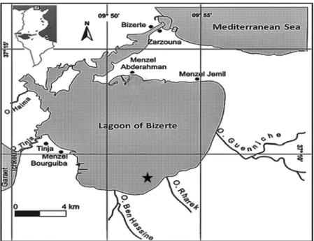
part (coordinates: 37°08’36”N, 9°52’20”E), was far away from urban and industrial sources of pol-lution (Figure 1).
A total of 309 specimens were hand-collected by scuba diving at about 3 m depth from October 2013 to September 2014, on the 15th± 1 day of each month (Table I), and carried to the laboratory in a cool box. In the laboratory, the shell length (SL) of all specimens was measured with a digital calliper and rounded down to the nearest 0.1 mm. The gonads from 10 to 31 sexually mature individuals per month were used for histological and immunohistochemical analyses (in total 189 specimens; SL range = 16.4 to 75.5 mm, all of them > 12 mm, size atfirst maturity according to Peharda et al. (2006)). The gonads werefixed in 10%
buffered formalin, dehydrated in ethanol and
embedded in paraffin wax; 5-µm-thick sections were obtained by means of a microtome and stained with haematoxylin–eosin. In order to detect the spawning period of the species in the sampling area, the occur-rence of mature specimens– i.e. specimens with ripe or partially spawned gonads (cf. Walker & Power2004; Peharda et al.2006)– was recorded.
The identification of stem cells was carried out through the immunohistochemical identification of Pou5f1, a transcription factor involved in the main-tenance and self-renewal of undifferentiated and pluripotent cells (Sánchez-Sánchez et al. 2010; Schulz et al. 2010; Lacerda et al.2014).
The identification of proliferating germ cells was performed through the immunohistochemical localisa-tion of proliferating cell nuclear antigen (PCNA), a polymerase delta accessory protein that is synthesised in late G1 and S phases of the cell cycle and is, there-fore, used as a nuclear marker of proliferation.
The immunohistochemical detection of Pou5f1 and PCNA was performed using the same protocol, with the exception of an antigen retrieval procedure that was
applied only to Pou5f1 immunostaining. This proce-dure was performed by boiling testis sections in citrate buffer (0.01 M, pH 6.0; 4 × 5 min cycles) in a micro-wave oven on high power (750 watts). Endogenous peroxidase was inhibited by treating sections for 10 min with 3% hydrogen peroxide (H2O2) and then rinsing them with distilled water and phosphate-buf-fered saline (PBS, 0.01 M, pH 7.4, containing 0.15 M sodium chloride [NaCl]). Subsequently, sections were incubated for 30 min in normal horse serum (NHS; Vector, Burlingame, CA), to block non-specific bind-ing sites for immunoglobulins, and then incubated overnight in a moist chamber at 4°C with rabbit poly-clonal antibodies raised against synthetic peptide of Pou5f1 (Abnova, Taipei, Taiwan) and monoclonal antibodies to PCNA (Santa Cruz Biotechnology Inc., Dallas, Texas). Anti-Pou5f1 and anti-PCNA antibo-dies were diluted 1:500 and 1:100, respectively, in PBS containing 0.1% bovine serum albumin (BSA; Sigma– Aldrich, Milan, Italy). After rinsing for 10 min in PBS, immunohistochemical visualisation was obtained using the Vectastain Universal Elite Kit (Vector, Burlingame, CA). This method uses the avidin –bio-tin–peroxidase complex (ABC) procedure. Peroxidase activity was visualised by incubating for 10 min with a Vector DAB Peroxidase Substrate Kit (Vector, Burlingame, CA), which produces a brown precipitate. To confirm the specificity of the immunoreaction, a control-staining procedure was carried out by replace-ment of the primary antibody with NHS and PBS.
To estimate the mean monthly density of mature oocytes (MOD), a representative histological slide per each female specimen was examined and all of the oocytes larger than 50 µm occurring infive dif-ferent microscopefields (field surface = 0.244 mm2) were counted. The mean density (oocytes/mm2) of each month was compared with that of the contig-uous months by the Student’s t-test.
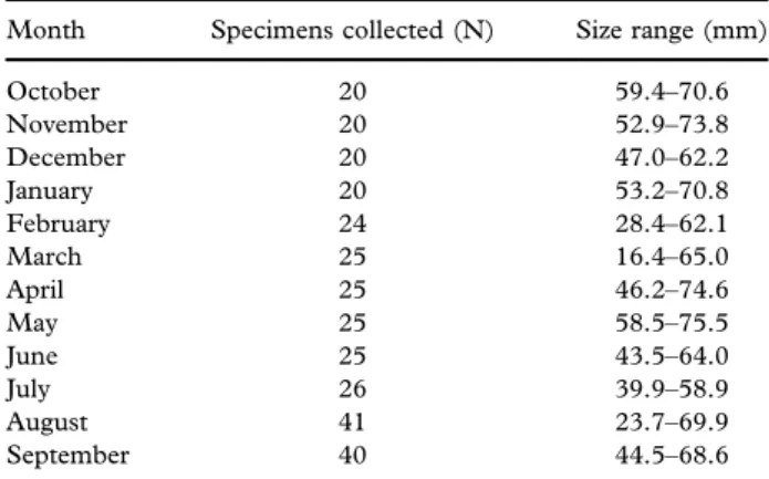
In addition to the microscopic examination of gonads, the analysis of“body condition” – an indi-cator of the individual overall body mass, which depends on both trophic state of soma and gonad state – was carried out. Ten specimens per month were chosen at random and sexed macroscopically by the colour of their gonads: whitish in males, orange to purple-red in females (Figure 2).
Their soft tissues were carefully removed from the shell and dried in the oven at 60°C for 48 h; the shells were scraped to remove all epibiotic material and dried in an oven at 60°C for 48 h. Soon after, the mass of both dried soft tissues (BW) and dried shells (SW) were measured with a digital scale to the nearest 0.01 g. Incidentally, in this comparatively irregularly shaped mollusc, shell mass was found to
Table I. Number (N) and size range of Arca noae specimens from the Bizerte Lagoon.
Month Specimens collected (N) Size range (mm) October 20 59.4–70.6 November 20 52.9–73.8 December 20 47.0–62.2 January 20 53.2–70.8 February 24 28.4–62.1 March 25 16.4–65.0 April 25 46.2–74.6 May 25 58.5–75.5 June 25 43.5–64.0 July 26 39.9–58.9 August 41 23.7–69.9 September 40 44.5–68.6
be a better descriptor of the animal’s overall size than shell length. To evaluate the year-round evolution of “body condition”, the monthly relationship between BW and SW was analysed; since no statistically
sig-nificant differences were found in any month
between the male and female BW/SW ratio, the raw data for the two sexes were pooled. The monthly individual BW and SW values were log-transformed (natural logarithms) and fitted to predictive linear regression equations (model I of Sokal & Rohlf
2012) of the type ln BW = a + b ln SW. The set of 12 regression equations so obtained was tested by analysis of covariance (ANCOVA). In order to avoid the effects of body size (see Results), the monthly adjusted means of log-transformed soft tissue dry masses, ln BWadj, were computed by ANCOVA as suggested by Trippel and Hubert (1990). Pairs of ln BWadj values of consecutive months were compared
by the Tukey–Kramer multiple-comparison test
(Sokal & Rohlf2012).
The size class frequency distributions of males and females were compared by the non-parametric Mann–Whitney U-test (Sokal & Rohlf2012).
The mollusc sampling was complemented by temperature (°C) and salinity (S‰) records at 1 m depth using a WTW-197i multimeter, as well as by surface water collection for the determination in the laboratory of chlorophyll a concentration (Chl a [mg l−1]). Chlorophyll a was extracted using
Whattman GF/F filters with 90% methanol, and
its concentrations were determined
spectrophoto-metrically at 665 and 750 nm (Aminot &
Chaussepied 1983). The seasonal variation of Chl a was monitored during this study as a proxy for the primary production, and hence food availabil-ity, for bivalves.
Correlations among these parameters, as well as between them and body condition and between the latter and mature oocyte density, were examined by both single and multiple regressions, in order to understand how the environmental situation affects oocyte maturation and animal condition.
Results Gonad structure
The Arca noae gonad tissue was found to be strictly associated and mixed with the digestive gland and with mantle muscle fibres (Figure 3(a,b)) (see the section Gonad/digestive gland tissue plasticity).
Both ovaries and testes were arranged in acini (follicles), in which germ cells at different stages of development were observed (Figure 3(a,b)). Gonad acini conveyed their products in small gonadal ducts
(250–370 µm in diameter) lined with a simple
columnar ciliated epithelium with scattered secretory cells containing acidophilic granules (Figure 3(c)). Small gonadal ducts merged into a large main gona-dal duct (700–800 µm in diameter) lined with a simple columnar ciliated epithelium containing both cells with acidophilic secretory granules and cells with large vacuoles (Figure 3(d), inset). Gonadal ducts containing gametes were observed in few specimens (Figure 3(c,d)).
Reproduction seasonality
Out of the 189 histologically analysed gonads, 142 were ovaries, 42 testes and five hermaphroditic. Incidentally, the Mann–Whitney U-test showed that the females in the sample were significantly larger than the males (P < 0.05).
FEMALES (N = 142; SL range: 23.7–75.5 mm;
mean SL = 56.6):
Specimens with mature oocytes, inside either ripe or partially spent ovaries, were found throughout the year (Figure 4).
Ripe ovaries showed densely packed mature oocytes, polygonal in shape, which enclosed a large euchromatic nucleus with a single eccentric nucleo-lus (Figure 5(a)). Oocytes at this stage incorporated plenty of acidophilic yolk granules, mainly accumu-lated at their lumen-facing pole (inset of Figure 5 (a)). Females with ripe ovaries were collected from October to November and from April to August.
Partially spawned ovaries (Figure 5(b)) showed mature oocytes with the same morphological aspect as those observed in ripe females; however, these ovaries had released part of their oocytes and hence their acini were partially empty. This condition was
Figure 2. Fully ripe specimens of Arca noae collected in late July from the Bizerte Lagoon. (a) Male. (b) Female. Asterisk, right gonad. Scale bar = 1 cm.
considered evidence of an effective spawning activ-ity. Females in this condition were found in two discrete periods: from November to April and from July to September.
MALES (N = 42; SL range: 16.4–72.0 mm; mean SL = 53.0):
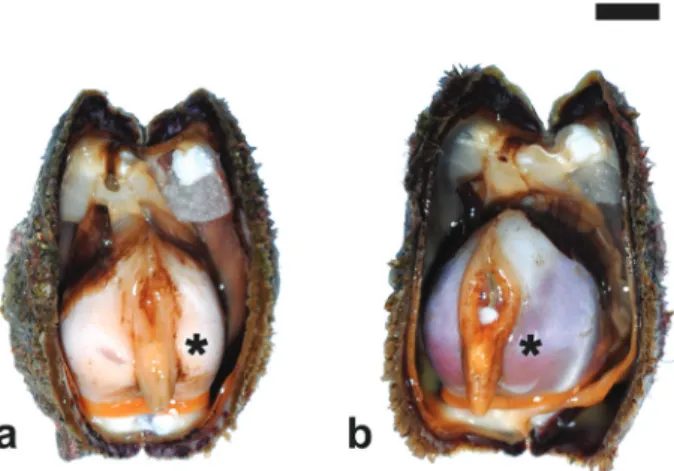
The histological examination of testes showed that all males collected in March (N = 4) had polygonal-shaped ripe acini and seemingly had not started to spawn yet (Figure 6(a)). Spermatogonia in active
proliferation, as well as spermatocytes and
spermatids, were assembled in different layers at the periphery of the acini, the lumina of which were packed with flagellated spermatozoa. The nuclei of both spermatogonia and spermatocytes immuno-reacted with anti-PCNA antibodies (Figure 6(b)). Testes from all other months (N = 38) showed ripe as well as partially emptied acini, the latter state
being indicative of ongoing spawning activity
(Figure 6(c)).
HERMAPHRODITES (N = 5; SL range: 47.2–
56.4 mm; mean SL = 52.0):
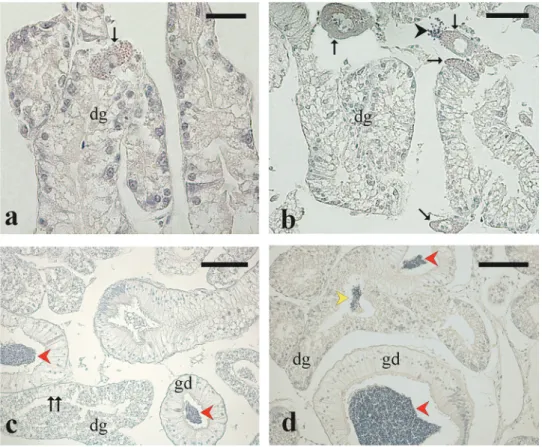
Figure 3. Microphotographs of Arca noae visceral mass sections showing gonad tissue associated and mixed with digestive gland and with muscle fibers. (a) Arca noae specimen collected in August showing partially spawned ovarian acini and digestive gland. (b) Arca noae specimen showing partially spawned testicular acini and digestive gland. (c) Small sperm ducts containing spermatozoa along with digestive gland in an Arca noae specimen collected in June. Note the degeneration of the digestive gland adenomeres concomitant with the proliferation of gonad duct epithelial cells. (d) Main sperm duct containing large amount of sperm between testis (left) and digestive gland (right) tissues. The inset shows the ciliated columnar epithelium of the sperm duct. Haematoxylin–eosin staining. Arrowhead, transition between digestive gland and gonadal duct; double arrowhead, secretory granules; arrow, muscle tissue; asterisk, proliferating gonadal duct epithelial cells, dg, digestive gland; sp, spermatozoa. Scale bars: a, b = 200μm; c = 50 μm; d = 600 μm; inset of d = 50 μm.
The co-occurrence of female and male germ cells in one gonad, i.e. hermaphroditism, was observed infive specimens (Figure 7). They were collected in December (SL = 47.5 mm), February (SL = 55.3 mm), March (SL = 47.2 mm) and April (SL = 53.6 and 56.4 mm). All the hermaphroditic gonads were purple red-coloured and could be set apart from normal ovaries only through histological examination.
The February specimen’s acini contained previtello-genic oocytes, still attached to the acini wall, along with few luminal ripe oocytes. Clusters of spermatozoa were visible within the acini lumen intermixed with mature oocytes (Figure 7). The hermaphroditic gonads from the following months were at a further advanced maturation phase, showing acini either full of ripe oocytes or partially empty (i.e. with a lower oocyte density). As for the male component, there were only
Figure 5. Microphotographs showing ripe and partially spawned Arca noae ovaries. (a) Ripe ovary showing large vitellogenic oocytes. (b) Partially spawned ovary showing mature oocytes and empty spaces, signs of oocyte release. Haematoxylin–eosin staining. Arrowhead, nucleolus; mo, mature oocyte; n, nucleus; pea, partially empty acini; y, yolk granules. Scale bars: a = 100 μm; inset of a = 20 μm; b = 100 μm.
Figure 6. Microphotographs showing ripe and partially spawned Arca noae testes. (a) Ripe testis showing large acini filled with spermatozoa. Haematoxylin–eosin staining. (b) Section from a ripe testis immunostained with anti-PCNA antibodies showing dividing cells confined to the periphery of the acini (arrowheads). Inset: particulars of two acini from (b) showing the nucleus of dividing cells stained in brown. (c) Partially spawned testis show-ing partially empty acini. Haematoxylin–eosin staining. pea, par-tially empty acini; ra, ripe acini; sp, spermatozoa. Scale bars: a, b, c = 200μm; inset of b = 20 μm.
residual spermatozoa grouped in comparatively small clusters either inside the acini or among them.
This February specimen was at an earlier phase of the protandric sex-change than the March and April hermaphrodites, because its gonad male component was more conspicuous and the female component was at an earlier maturation stage than the others. In particular, the March specimen had seemingly already spawned a fraction of its oocytes and some of the remaining ones were degenerating (see below).
As for the scantiness of hermaphroditic indivi-duals, the low overall percentage of hermaphrodites in the Bizerte Lagoon (2.1%) masks a much higher percentage. In fact, when only the male fraction of the only period when hermaphrodites occurred (December to April) is taken into consideration, the percentage of hermaphroditic specimens reaches 33.3 (calculation according to Bello et al. 2013).
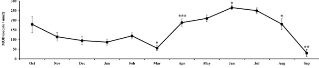
Mature oocyte density monthly progression
From October to March, the MOD decreased from 178.9 to 55.7 oocytes/mm2(Figure 8), thus indicat-ing that the ovary was steadily releasindicat-ing its products. This is in agreement with the monthly progress of
mature and partially spawned gonads: we found only mature individuals in October, mature and partially spawned individuals in November, and only partially spawned individuals from December to March (Figure 4), which trend is a robust indication that spawning occurred gradually, in fractionated batches from October to March. Hence, the oocyte density increased progressively through June (MOD = 267.4 oocytes/mm2) because of the progressive ripening of the ovaries and the accumulation of mature oocytes. Afterwards, a second oocyte release phase ensued through September, which was faster and more mas-sive than the previous one (MOD = 267.4 to 30.4 oocytes/mm2; note the rapid drop in MOD from July to September inFigure 8).
This overall picture is corroborated by the histolo-gical examination of the ovaries. They were for the most part partially spawned from November to March, packed with mature oocytes from April to June, and partially spawned or spent in July– September after the massive summer spawning.
Gonad/digestive gland tissue plasticity
In the examined histological sections, the portions occupied by digestive and gonadal components showed seasonal variations; that is, digestive gland tissues predominated when the gonad regressed and vice versa. In March, when oocyte density was at a minimum and gonad acini were almost completely empty, the gonad tissue appeared to be degenerating with the disintegration of the gonad acini structure and the degeneration and reabsorption of residual oocytes and spermatozoa (Figure 9(a,b)). In June, when gonads were fully ripe and ready to spawn, and mature oocyte density peaked, a massive develop-ment of the gonadal duct system was observed con-comitantly with the digestive tissue regression. This was shown, in histological sections, by the degenera-tion of adenomere cells, which were replaced by gonadal duct cells. In particular, in some males, clutches of spermatozoa were visible inside the lumen of well-structured gonadal ducts, as well as within degenerating digestive glands, which tissue
Figure 7. Microphotograph from an Arca noae hermaphrodite specimen sampled in February (shell length (SL) = 55.3 mm). Spermatozoa are visible within ovarian acini containing previtello-genic oocytes, still attached to the acini walls, along with few ripe oocytes in the lumina. Haematoxylin–eosin staining. sp, sperma-tozoa. Scale bar = 100μm.
Figure 8. Monthly oocyte density (MOD) progression for female Arca noae from the Bizerte Lagoon. Significance levels: *, P < 0.05; **, P < 0.01; ***, P < 0.001.
was seemingly being remoulded into gonadal ducts (Figure 9(c,d)).
The immunohistochemical staining with antibo-dies against the stemness marker Pou5f1 labelled single small cells (diameter 6.1 ± 1.4 μm) scattered in the interstitial tissue as well as at the periphery of ovarian acini and around adenomeres of digestive glands (Figure 10(a)). The development of the gona-dal duct system was supported by cells arranged within the glandular tissue that actively proliferated and gave rise to the epithelium lining the gonadal ducts (Figure 10(b,c)).
Environmental parameters
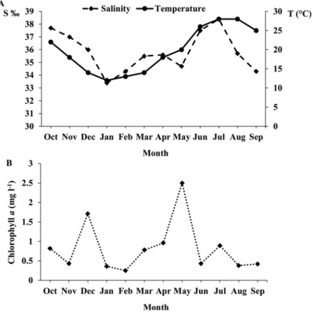
Monthly variations in water temperature (°C), salinity (S‰) and chlorophyll a concentration are shown in Figure 11. The highest water temperature (28°C) was recorded in July and August and the lowest (12°C) in
January. Salinity varied from 33.4‰ in the winter (January), which is marked by rainfall and freshwater input from the Ichkeul Lake and several wadis, to 38.5‰ in the summer (July), when rainfall is fairly rare and water evaporation is very high (Figure 11 (a)). The first notable Chl a peak was recorded in December (1.7 mg l−1) and was likely due to the input of nutrient-rich fresh water after heavy rains. A second, much more marked peak occurred in May (2.5 mg l−1) depending on spring phytoplankton blooms (Figure 11(b)). No statistically significant cor-relation was found between Chl a and either °C or S‰ (P = 0.76 and 0.99, respectively).
“Body condition”
The examination of monthly regressions of ln BW on ln SW showed that the correlation coefficient b was significantly different from 1 in most months, which
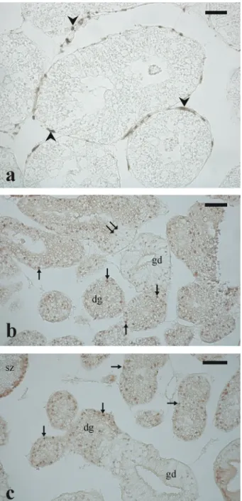
Figure 9. Micrographs from Arca noae gonad/digestive gland sections. (a) and (b) show the development of the digestive gland concomitant with the degeneration of the gonad in a female and a hermaphrodite specimen, respectively. In (a) note the presence of a reabsorbing vitellogenic oocyte inside a digestive gland adenomere. In (b) a few degenerating vitellogenic oocytes (arrows) and a cluster of spermatozoa (black arrowhead) are visible within the digestive gland tissue. (c) and (d) show degenerating digestive gland adenomeres along with developing gonadal ducts. Spermatozoa are visible within gonad ducts and in the lumen of a digestive gland adenomere. Haematoxylin– eosin staining. Arrow: degenerating oocyte; red arrowhead, spermatozoa within gonadal duct; yellow arrowhead, spermatozoa within the lumen of a digestive gland adenomere (please, refer to the online version for the explanation of coloured symbols). Double arrow indicates a digestive gland adenomere in which lining epithelial cells, morphologically similar to gonadal duct cells, are developing. dg, digestive gland, gd, gonadal duct. Scale bars: a = 25μm; b, d = 50 μm; c = 100 μm.
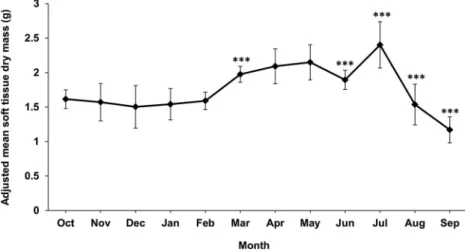
was evidence of a significant size effect; that is, soft tissue mass was not proportional to shell mass throughout the A. noae size range. Hence, in order to avoid size bias, the computation of monthly adjusted means of log-transformed soft tissue dry masses, BWadj, was carried out. Monthly BWadj values generally increased as gonads ripened, and decreased during the spawning periods (Figure 12). However, such a relationship appeared to be modu-lated by other factors (see the following section). In particular, BWadj remained fairly stable from October to February, despite the progressive reduc-tion of mature oocyte density, and hence loss of gonadal mass. This seemingly depended on the fact that while the gonad tissue was being reduced, somatic mass was accreting anew. BWadj started to increase in March, when Chl a increased thanks to spring phytoplankton blooms, and peaked in May, when oocyte density went on increasing and all spe-cimens became mature, concomitantly with the highest Chl a (compareFigures 8 and 11(b)). The significant June drop depended as well on the con-comitant considerable drop of phytoplankton den-sity, in spite of the fact that oocyte density had reached its annual maximum. The subsequent decrease of BWadj, from July to September, is related with the release of gametes.
Correlations between different parameters
The monthly mean mature oocyte density was found to be positively correlated to temperature and to sali-nity. Their regression equations are, respectively,
MOD = −923.832 + 29.898 S‰ (r = 0.674;
P < 0.05) and MOD = 1.962 + 7.357 °C (r = 0.577; P < 0.05). Moreover, the multiple regression of MOD on °C and S‰ for all months was not statistically significant (P = 0.064) but, when excluding the out-lying September data from computation, the correla-tion of MOD to the two environmental factors became highly significant: MOD = 67.40 + 10.44 °C − 3.12 S ‰ (R2 = 0.713; P < 0.01). Together °C and S‰ explained 71.3% of the MOD variance.
“Body condition”, BWadj, was found to be signi fi-cantly correlated with the mature oocyte density (regression equation: BWadj = 1.292 + 3.120x10⁻³ MOD; r = 0.675; P < 0.05) and, to some degree although not significantly, with the chlorophyll a concentration (P = 0.07). The multiple regression examination showed that BWadjis best correlated to both mature oocyte density and Chl a, a proxy for
phytoplankton density (regression equation:
BWadj= 1.146 + 2.781x10⁻³MOD + 0.221 Chl a;
Figure 10. Micrographs from Arca noae testis/digestive gland sections immunostained with antibodies against the stemness marker Pou5f1 and with proliferating cell nuclear antigen (PCNA). In (a) the stem cells, scattered around digestive gland adenomeres, have brown-labelled nuclei. (b) and (c) show anti-PCNA positive (i.e. dividing) cells within digestive gland adenomeres, testicular acini and gonadal ducts. The immunostaining is stronger in the nucleus of digestive gland cells than in the epithelial cells lining the gonadal ducts. Arrowhead, stem cell; arrow, dividing cell. Double arrow indi-cates a digestive gland adenomere in which lining epithelial cells, morphologically similar to gonadal duct cells, are devel-oping. dg, digestive gland, gd, gonadal duct. Scale bars: a = 25 μm; b, c = 50 μm.
R2= 0.615; P < 0.05). Together, the two regressors explained 62% of the BWadjvariance.
Discussion
The histological analyses of gonads coupled with the monthly succession of mature oocyte density sug-gested that, in the examined year, Arca noae from the Bizerte Lagoon underwent two reproductive phases, differing from each other in the pattern of gonad maturation as well as in the ensuing spawning. The October maturation was followed by a long spawning period, extending from November to March. A second ovarian maturation phase encom-passed a three-month period, from April to June, and was followed by a comparatively rapid and intense spawning period from July to September.
The two spawning phases differed substantially from each other. Thefirst one, extending throughout the autumn, winter and early spring, was charac-terised by protracted partial releases of gametes (i.e. multiple spawning). The second maturity/spawning phase, which started in April–May, involved all examined females. During this phase, the density of mature oocytes increased steadily, peaked in June
and decreased gradually from July through
September (Figure 8), thus showing that the animals were actively spawning from July to mid-September. Temperature appeared to modulate the two gonad
maturation/spawning phases. Comparatively low
temperatures (12–14°C) were most probably respon-sible for a less intense release of gametes, in the winter spawning period, with respect to the summer, when temperatures were as high as 25–28°C (Figures 8and11(a)).
The fact that individuals of A. noae from the Bizerte Lagoon reproduced by multiple spawning, i.e. partial progressive release of gametes, is clearly shown by the progressive decrease of mature oocyte density during both spawning periods (Figure 8) as well as by the histological analysis of the gonads. Similarly, Peharda et al. (2006) reported the occur-rence of partially spawned specimens in the two months following the period of gonad ripening in A. noae from the Adriatic Sea.
The spawned oocytes appeared to derive from one single cohort, contrary to what happens in other bivalves, e.g. Venus nux, in which several cohorts of oocytes were detected throughout the year (Tirado et al.2011a). In this respect, Holopainen and Hanski (1986) suggested that populations of the same spe-cies might be either semeleparous or iteroparous
Figure 11. Monthly variation of (a) physico-chemical parameters, (b) chlorophyll a concentration (Chl a, µg l−1) in the surface water of the Bizerte Lagoon.
depending on the environmental circumstances that affect growth. These authors demonstrated in their work on Pisidium species that individuals living under favourable conditions are likely to be semel-parous, while populations living under unfavourable conditions are likely to be iteroparous. A similar shift in reproductive modes was reported in Musculium species by Mackie et al. (1976a,b)) and Way et al. (1980). Therefore, the long, low-intensity late autumn to early spring multiple spawning period in A. noae from the Bizerte Lagoon may be related to the comparatively unfavourable conditions of that season (mainly low temperatures).
In the Bizerte Lagoon, the A. noae reproductive pattern showed peculiar features with respect to the other known Mediterranean areas. Indeed, the few existing studies on this topic were carried out on the Adriatic (sub)population (Valli & Parovel 1981; Peharda et al. 2006); several other papers have focused on accessory topics (Bello & Paparella
2001; Peharda et al. 2002, 2003, 2009; Bello et al.
2013; Župan et al. 2014; Puljas et al. 2015). Different reproductive patterns were described for the A. noae (sub)population of the Adriatic Sea, a comparatively small and closed sea. Peharda et al. (2006) showed that in Mali Ston bay (mid-eastern Adriatic Sea) this species reproduces by a single annual gametogenic cycle extending from October– November to April–May, and one spawning peak in the summer (July and August), with slight timing fluctuations from one year to another. A quite simi-lar cycle was observed along the coast of Bari (south-western Adriatic Sea; P. Paparella and G. Bello, unpublished personal observations; see also Bello & Paparella (2001)). In the Gulf of Trieste (northern Adriatic Sea), according to Valli and Parovel (1981), A. noae reproduces throughout an extended season spanning from the spring to the autumn, with two spawning peaks in March and September, respec-tively. Two spawning peaks per year were also reported for the Gulf of Manfredonia (south-western Adriatic Sea) stock (Bello & Paparella 2001).
Contrary to the Adriatic reproductive patterns of A. noae with a single, more or less prolonged game-togenic cycle, either single- or two-peaked spawning, this study reports for thefirst time a rather different pattern of two discrete maturing/spawning phases differing in terms of both length and intensity. Fretter (1984), quoted by Baqueiro and Aranda (2000) in a detailed review of reproductive patterns of Mexican bivalves, classified them into two cate-gories according to their reproductive strategies: tachitictic, with short and limited reproductive peri-ods, and braditichtic, with an extended reproductive activity. Furthermore, the same population of a
bivalve species can display differences in its repro-ductive activity from one year to another, i.e. either a short synchronic spawning or a long, partially asyn-chronous spawning or several partial spawnings (Bricelj & Malouf 1980; Baqueiro 1981; Jaramillo et al.1993). Conversely, different spawning timings can be observed in the same bivalve from two geo-graphically separate sites, depending on water tem-perature and pabulum concentration (Tirado et al.
2011a,b). The possibility of shifting bewteen bradi-tichtic and tachitictic reproductive modes appears to pertain to A. noae, strategies which are sequentially exploited by this bivalve according to the season. Similar findings were reported about the oyster Crassostrea (= Magallana) gigas by Enríquez-Díaz et al. (2009), who observed different reproductive patterns in oysters cultured at different latitudes in the Atlantic Ocean. Those from the Baie des Veys (English Channel) showed a single partial spawning period in August, whereas those from Marennes-Oléron (Gulf of Biscay) displayed a partial spawning in July and a massive release of gametes during August. Enríquez-Díaz et al. (2009) assumed that the oyster spawning intensity was related to food availability. Interestingly enough, in the case of the Bizerte Lagoon, three Chl a peaks were observed, one between November and January; another one, the highest, between April and June; and the third one, lower, between June and August. The highest, spring peak corresponded with gonad ripening rather than spawning, whereas the other two peaks coin-cided with the two spawning periods, the slow winter one coinciding with the second highest Chl a peak.
The environmental peculiarities of the Bizerte Lagoon, chiefly the overall comparatively high chlor-ophyll a concentrations, were also related to the longer reproductive period in the oyster Magallana gigas cultured there with respect to an oyster stock cultured in an adjacent open sea site (Dridi et al.
2014).
Arca noae from the Bizerte Lagoon showed the presence of mature gonads during most of the year, which makes the reproductive pattern of this (sub) population more similar to that of tropical bivalves (e.g. the tropical oyster, Crassostrea corteziensis; Rodríguez-Jaramillo et al. 2008) than to the other known Mediterranean A. noae (sub)populations. This largeflexibility confirms that bivalve reproduc-tive strategies are not strictly species-specific since they are driven by complex interactions of environ-mental and genetic factors (Jovanovich & Marion
1989; Baqueiro & Aranda2000).
In addition to Chl a, the second most important environmental factor to which mature oocyte density is significantly correlated, namely temperature,
appears to modulate the gametogenic cycles.
Comparatively low temperatures (12–14°C) are
most probably the cause for a slower release of gametes in the winter spawning period with respect to the summer, when temperatures are as high as 25–28°C (Figures 8and11(a)). Mature oocyte den-sity is also multiple correlated to temperature and salinity except in the month of September, when oocyte density reached its lowest value following total gamete release in most specimens.
“Body condition” – in any of its many expressions found in the literature– is generally deemed a good descriptor of bivalve reproductive cycles (e.g. Peharda et al. 2006; Tirado et al. 2011a,b). In the present study the customary “soft tissue mass/shell mass” ratio (or similar expressions of “body mass/ body size”), without any further transformation, was deemed inappropriate to describe body condition because it does not compensate for the effects of body size (Trippel & Hubert 1990; Labocha et al.
2014). In fact, in our case, (a) specimens of a com-paratively large size range were used; (b) more importantly, body mass (BW) was not isometric to shell mass (SW) in most months (the slope of monthly ln BW/ln SW regression equations signi fi-cantly differed from 1); and (c) the monthly ln BW/ ln SW regression lines significantly differed from each other either in slope and/or position. That is
why “body condition” was expressed as mean
adjusted soft tissue dry mass BWadj.
The monthly progression of “body condition” for A. noae from the Bizerte Lagoon is in agreement with
the seasonal trend of mature oocyte density.
However, the June “body condition” relative nega-tive peak conflicts with the peak of mature oocyte density (Figures 8and12). This may be explained by
the concomitant massive reduction of phytoplankton in the lagoon, as shown by the sudden drop of chlor-ophyll a concentration (Figure 11(b)), which in turn caused a dramatic decrease in weight of the soma, and hence of the June BWadj. This supposition is markedly supported by the statistically significant multiple correlation of BWadj with both mature oocyte density and chlorophyll a concentration. It is also corroborated by the fact that the June BWadj variance was the lowest in the whole year; that is, all June specimens employed in the “body condition” analysis had rather similarly reduced soft tissue mass, hence they similarly suffered a shortage of food resources. Whatever the cause, the June decrease of BWadj seemingly did not affect the reproductive cycle. In addition, the release of oocytes from November to March is not paralleled by the decrease of BWadj likely because the release of gametes was compensated by the somatic mass growth; this is particularly evident in March, when BWadj showed a significant increase concomitantly with phyto-plankton early spring blooms.
To conclude, caution should be exerted when using “body condition” seasonal progression as a proxy for the reproductive cycle (see also Matozzo et al. 2005; Boussoufa et al. 2015) since this index changes according to both gonad and somatic mass variations.
With regard to the occurrence of hermaphroditic A. noae individuals, the protandric sex change was gen-erally deemed occasional (Valli & Parovel 1981; Peharda et al.2006) or obligate in this bivalve (Bello et al. 2013). In the present case as well, the male fraction in the examined samples was found to be smaller in size (SL) than the female fraction, which
corroborates the obligate hermaphroditism
Figure 12. Monthly progression of adjusted mean soft tissue dry weight, BWadj, for Arca noae from the Bizerte Lagoon. Significance levels:
hypothesis. However, data about A. noae hermaphro-ditism are too scanty to positively delineate a precise model of sex change, whether occasional or obligate and whether depending, to some extent at least, on the socio-demographic structure of the population, as has been clearly shown in other molluscs, e.g. Patella ferruginea (Rivera-Ingraham et al.2011). Most likely, hermaphroditism is a potential feature in A. noae, which is expressed when and in the proportion needed to maintain population homeostasis, under the influence of either biotic or abiotic environmental factors (cf. review by Breton et al.2017). Incidentally, in this case as well, the sex change occurred in a restricted period of time, shortly before the late spring massive gamete maturation. Moreover, the corrected percentage of hermaphroditic individuals in the Bizerte Lagoon sample (33.3%) is very close to that computed for the Croatian (sub)population (35%) and higher than the Apulian one (20%) (Bello et al.
2013).
The reproductive flexibility observed herein is not the only trait of A. noae’s extreme adaptability. The gonads and the gonad duct system are strictly asso-ciated and combined with other visceral organs as well as with mantle musclefibres, which is a feature com-mon to many bivalve species (Baccetti et al. 1991). During the reproductive cycle, gonads and the gonadal duct system undergo remarkable seasonal modi fica-tions in volume and structure, which are connected with changes in the volume and structure of visceral organs. In particular, the relative surfaces of the histo-logical sections covered by the digestive and gonadal components vary on a seasonal basis: spent gonads are associated with a well-developed digestive gland sys-tem, and conversely gonad ripening is associated with a reduction of the digestive gland volume. Furthermore, the gonadal duct system develops during the spawning season at the expense of the digestive gland. The latter displays adenomere cell degeneration and proliferation of new lining epithelial cells that assemble the gonadal duct system. The immunohistochemical analysis showed anti-PCNA positive, i.e. proliferating, cells within digestive gland adenomeres when gonad matur-ity reached its annual peak. This proliferating activmatur-ity is most likely associated with the development of gonadal ducts.
To the best of our knowledge, no data are avail-able in the bivalve literature on transdifferentiation – that is, the process of transformation of differen-tiated somatic cells into different cell types. Indeed, several recent studies demonstrate that aquatic invertebrate taxa, including cnidarians, platyhel-minthes, echinoderms and urochordate ascidians, exhibit widespread multiple cell types with stem
cell attributes; hence, transdifferentiation is ubiqui-tous in both anatomically simple and highly evolved invertebrates (Rinkevich et al.2009). In the present study anti-Pou5f1 immunopositive cells – that is, cells sharing stemness characteristics– were distrib-uted around and within digestive gland adeno-meres. These cells may retain the potentiality to differentiate and proliferate, giving rise to diverse cell lineages, so their occurrence can be related to the hypothesised, albeit highly probable, tissue transdifferentiation in A. noae. Therefore, the observed modifications in the A. noae reproductive and digestive gland systems should be interpreted as a mosaic piece of a wider and astonishing anato-mical and physiological plasticity widespread in invertebrate organisms.
Conclusions
Arca noae has evolved aflexible reproductive strategy that makes it capable of exploiting diverse environ-mental situations in consonance with exogenous
fac-tors, mainly pabulum availability and water
temperature. The present study, carried out on an unexploited (sub)population, widened the frame of the A. noae reproductive strategies and further cor-roborated it. Present results represent a landmark for a future management of the Bizerte Lagoon A. noae stock and, for thefirst time, offer interesting insights into mechanisms underlying seasonal changes in gonad and visceral organs of a bivalve species.
Acknowledgements
We are grateful to Prof. Carmen Salas Casanova (University of Malaga, Spain) for providing useful
suggestions, and to Mr Michele Carrassi
(University of Bari, Italy) for his technical
support.
ORCID
G. Bello http://orcid.org/0000-0002-0902-0556
A. Corriero http://orcid.org/0000-0001-7068-9438
References
Aminot A, Chaussepied C.1983. Manuel des Analyses Chimiques en Milieu Marin. Brest: Centre National d’Exploitation des Océans. 395 pp.
Ayari R, Afli A.2008. Functional groups to establish the ecologi-cal of soft benthic fauna within Tunis bay (western Mediterranean). Vie et Milieu 58:67–75.
Baccetti B, Baldaccini NE, Bedini C, Brandmayr P, Capanna E, Chieffi G, Cobolli M, Ferraguti M, Ghirardelli E, Ghiretti F,
Giusti F, Grigolo A, Mainardi D, Minelli A, Papi F, Parrinello N, Ricci N, Ruffo S, Sarà M, Scali V, Zullini A.
1991. Zoologia. Trattato Italiano. Bologna: Editoriale Grasso. 389 pp.
Baqueiro CE. 1981. Reproductive cycle of six species of bivalve molluscs with aquaculture potential from the Panamic province. World Conference on Aquaculture, Venice, Italy, 1981 – Abstracts Book. Venice: European Mariculture Society. p. 367.
Baqueiro CE, Aranda DA. 2000. A review of reproductive pat-terns of bivalve mollusks from Mexico. Bulletin of Marine Science 66:13–27.
Bello G, Paparella P. 2001. Struttura di popolazioni di Arca noae (Bivalvia, Arcidae) insediate su substrati diversi nell’Adriatico meridionale. Atti della Società Italiana di Scienze Naturali e del Museo Civico di Storia Naturale di Milano 141:175–185.
Bello G, Paparella P, Corriero A, Santamaria N.2013. Protandric hermaphroditism in the bivalve Arca noae (Mollusca: Arcidae). Mediterranean Marine Science 14:86–91. DOI: 10.12681/ mms.326.
Ben Mustapha K, Hattour A, Mhelti M, El Abed A, Tritar B.
1999. Bionomie des étages infra- et circa- littorales du golfe de Gabès. Bulletin de l’Institut National des Sciences et Technologie de la Mer de Salammbô 26:1–55.
Boussoufa D, Ghazali N, Rabeh I, Soudan N, Navarro JC, El Cafsi M. 2015. Reproductive cycle and gonad development of the commercial clam Donax trunculus from the Bay of Tunis (Northern Tunisia, South Mediterranean coast): Effects of environmental variability. Cahiers de Biologie Marine 56:369–380.
Breton S, Capt C, Guerra D, Stewart D.2017. Sex determining mechanisms in Bivalves. Preprints 2017:2017060127. DOI:
10.20944/preprints201706.0127.v1.
Bricelj MV, Malouf RE. 1980. Aspects of reproduction of hard clams (Mercenaria mercenaria) in Great South Bay, New York. Proceedings of the National Shellfisheries Association 70:216– 229.
Darmoul B, Hadj Ali S, Vitello P.1980. Effets des rejets indus-triels de la région de Gabès (Tunisie) sur le milieu marin récepteur. Bulletin de l’Institut National des Sciences et Technologie de la Mer de Salammbô 7:5–61.
Dridi S, Romdhane MS, El Cafsi M.2014. Gametogenic cycle of Crassostrea gigas in contrasting Mediterranean habitats: Marine (Gulf of Tunis) and continental (Bizert lagoon) culture sites. Journal of Biological Research-Thessaloniki 21:13. DOI:
10.1186/2241-5793-21-13.
Enríquez-Díaz M, Pouvreau S, Chávez-Villalba J, Le Pennec M.
2009. Gametogenesis, reproductive investment, and spawning behavior of the Pacific giant oyster Crassostrea gigas: Evidence of an environment-dependent strategy. Aquaculture International 17:491–506. DOI:10.1007/s10499-008-9219-1. Enzenross L, Enzenross R. 2001. Untersuchungen über das Vorkommen mariner Mollusken in tunesischen Gewassern. Schriften für Malakozoologie 17:45–62.
Fretter V.1984. Prosobranchs. In: Tompa AS, Verdonk NH, Den Biggelaar JAM, editors. The Mollusca. Volume 7: Reproduction. Orlando, Florida: Academic Press. pp. 1–45. Gofas S.2008. Arca noae. In: MolluscaBase (2017). Accessed
through: World Register of Marine Species. Available:
http://www.marinespecies.org/aphia.php?p=taxdetails&id= 138788. Accessed May 2017 30.
Gosling E.2003. Bivalve Molluscs - Biology, Ecology and Culture. Oxford: Fishing News Books, Blackwell Publishing. 443 pp. Holopainen IJ, Hanski I.1986. Life history variation in Pisidium
(Bivalva: Pisidiidae). Ecography 9:85–98. DOI: 10.1111/ j.1600-0587.1986.tb01195.x.
Hrs-Brenko M, Legac M.1996. A review of bivalve species in the eastern Adriatic Sea: II. Pteromorphia (Arcidae and Noetidae). Natura Croatica 5:221–247.
Jaramillo R, Winter J, Valencia J, Rivera A.1993. Gametogenic cycle of the Chiloé scallop (Chlamys amandi). Journal of Shellfish Research 12:59–70.
Jovanovich MC, Marion KR.1989. Gametogenic cycle of Rangia cuneata (Mactridae, Mollusca) in Mobile Bay, Alabama, with comments on geographic variation. Bulletin of Marine Science 45:130–138.
Labocha MK, Schutz H, Hayes JP.2014. Which body condition index is best? Oikos 123:111–119. DOI: 10.1111/j.1600-0706.2013.00755.x.
Lacerda SMDSN, Costa GMJ, De França LR.2014. Biology and identity of fish spermatogonial stem cell. General and Comparative Endocrinology 207:56–65. DOI: 10.1016/j. ygcen.2014.06.018.
Mackie GL, Qadri SU, Clarke AH.1976a. Intraspecific variations in growth, birth periods and longevity of Musculium securis (Bivalvia: Sphaeriidae) near Ottawa, Canada. Malacologia 15:433–446. Mackie GL, Qadri SU, Clarke AH. 1976b. Reproductive habits of
four populations of Musculium securis (Bivalvia: Sphaeriidae) near Ottawa, Canada. Nautilus 90:6–86.
Matozzo V, Tomei A, Marin MG.2005. Acetylcholinesterase as a biomarker of exposure to neurotoxic compounds in the clam Tapes philippinarum from the Lagoon of Venice. Marine Pollution Bulletin 50:1686–1693. DOI: 10.1016/j. marpolbul.2005.07.011.
Peharda M, Bolotin J, Vrgoč N, Jasprica N, Bratoš A, Skaramuca B.2003. A study of Noah’s Ark shell (Arca noae Linnaeus, 1758) in Mali Ston Bay, Adriatic Sea. Journal of Shellfish Research 22:705–709.
Peharda M, Mladineo I, Bolotin J, Kekez L, Skaramuca B.2006. The reproductive cycle and potential protoandric development of the Noah’s Ark shell, Arca noae L.: Implications for aqua-culture. Aquaculture 252:317–327. DOI: 10.1016/j. aquaculture.2005.07.007.
Peharda M, Richardson CA, Onofri V, Bratoš A, Crnčević M.
2002. Age and growth of the bivalve Arca noae L. in the Croatian Adriatic Sea. Journal of Molluscan Studies 68:307– 310. DOI:10.1093/mollus/68.4.307.
Peharda M, Stagličić N, Ezgeta D, Vrgoč N, Isajlović I, Krstulović-Šifner S.2009. Distribution and population struc-ture of Arca noae in Pašman channel. Ribarstvo 67:3–10. Poutiers JM. 1987. Bivalves. In: Fischer W, Bauchot ML,
Schneider M, editors. Fiches FAO d’identification des espèces pour les besoins de la pêche. (Révision 1). Méditerranée et Mer Noire. Rome: Food and Agriculture Organization of the United Nations, FAO. pp. 369–512. Puljas S, Peharda M, Župan I, Bukša F. 2015. Maximum
recorded life span of Arca noae Linnaeus, 1758 in the marine protected area Telaščica, Adriatic Sea. Cahiers de Biologie Marine 56:163–168.
Rinkevich Y, Matranga V, Rinkevich B.2009. Stem cells in aqua-tic invertebrates: Common premises and emerging unique themes. In: Rinkevich B, Matranga V, editors. Stem cells in marine organisms. Berlin: Springer. pp. 61–103.
Risso JC. 1978. Faune malacologique de la plateforme Tunisienne. Etude de quelques dragages et carottages effectués à l’intérieur et au large du golfe de Gabès. Bulletin de l’Institut National Scientifique et Technique d’Océanographie et de Pêche Salammbô 5:17–41.
Rivera–Ingraham GA, Espinosa F, García–Gómez JC. 2011. Conservation status and updated census of Patella ferruginea (Gastropoda, Patellidae) in Ceuta: Distribution patterns and new evidence of the effects of environmental parameters on population structure. Animal Biodiversity and Conservation 34:83–99.
Rodríguez-Jaramillo C, Hurtado MA, Romero-Vivas E, Ramírez JL, Manzano M, Palacios E.2008. Gonadal development and histochemistry of the tropical oyster, Crassostrea corteziensis (Hertlein, 1951) during an annual reproductive cycle. Journal of Shellfish Research 27:1129–1141. DOI: 10.2983/0730-8000-27.5.1129.
Sánchez-Sánchez AV, Camp E, García-España A, Leal-Tassias A, Mullor JL. 2010. Medaka Oct4 is expressed during early embryo development, and in primordial germ cells and adult gonads. Developmental Dynamics 239:672–679. DOI:
10.1002/dvdy.22198.
Saubade AM, Risso JC. 1983. Quelques dragages et carottages récents dans le lac de Tunis: La malacofaune, témoin de l’histoire de la lagune. Bulletin de l’Institut National des Sciences et Technologie de la Mer de Salammbô 3:1–72. Schulz RW, de França LR, Lareyre -J-J, Le Gac F,
Chiarini-Garcia H, Nóbrega RH, Miura T.2010. Spermatogenesis in fish. General and Comparative Endocrinology 165:390–411. DOI:10.1016/j.ygcen.2009.02.013.
Sokal RR, Rohlf FJ.2012. Biometry: The principles and practice of statistics in biological research. 4th ed. New York: Freeman WH & Co. 859 pp.
Souissi N. 1981. Mécanismes de la sédimentation et évolution paléogéographique de la lagune de Bizerte (Tunisie) durant le quaternaire récent. Thèse. Université de Toulouse. 229 pp.
Tirado C, Rueda JL, Salas C. 2011a. Reproductive cycles in Atlantic and Mediterranean populations of Venus nux Gmelin, 1791 (Bivalvia: Veneridae), from southern Spain. Journal of Shellfish Research 30:813–820. DOI: 10.2983/ 035.030.0322.
Tirado C, Rueda JL, Salas C. 2011b. Reproduction of Donax trunculus in the littoral of Huelva (southern Atlantic Spain): Is there any difference with the Mediterranean population from the Andalusian coast? Iberus 29:47–57.
Trippel EA, Hubert JJ.1990. Common statistical errors infishery research. In: Hunter J, editor. Writing for fishery journals. Bethesda: American Fisheries Society. pp. 93–102.
Valli G, Parovel C.1981. Aspects de la reproduction et de la biométrie chez Arca noae L. (Mollusca: Bivalvia). Rapports et procès-verbaux des réunions. Commission Internationale pour l’Exploration Scientifique de la Mer Méditerranée 27:135–136.
Walker RL, Power AJ.2004. Growth and gametogenic cycle of the transverse ark, Anadara transversa (Say, 1822), in coastal Georgia. American Malacological Bulletin 18:55–60. Way CM, Hornbach DJ, Burky AJ.1980. Comparative life history
tactics of the sphaeriid clam, Musculium partumeium (Say), from a permanent and a temporary pond. American Midland Naturalist 104:319–327. DOI:10.2307/2424872.
Zouari S. 1985. Contribution à l’étude systématique des Lamellibranches des côtes tunisiennes. Tunis: D.E.A. Université de Tunis. 245 pp.
Župan I, Peharda M, Dolenec T, Dolenec M, Žvab Rožić P, Lojen S, Ezgeta-Balić D, Arapov J.2014. Aquaculture assess-ment of Noah’s ark (Arca noae Linnaeus, 1758) in the Central Adriatic Sea (Croatia). Journal of Shellfish Research 33:433– 441. DOI:10.2983/035.033.0212.
Župan I, Peharda M, Ezgeta-Balić D, Šarić T.2012. Noah’s ark shell (Arca noae Linnaeus, 1758) - What do we need to know for starting up its aquaculture? Croatian Journal of Fisheries 70:71–81.