Alma Mater Studiorum – Università di Bologna
University College London
DOTTORATO DI RICERCA IN Cognitive Neuroscience
Ciclo XXV
Settore Concorsuale di afferenza: AREA 11 Settore Scientifico disciplinare: M-PSI/02
Plasticity in body and peripersonal space representations
Presentata da: Dott.ssa Elisa Canzoneri
Coordinatore Dottorato Relatore
Prof.ssa Elisabetta Làdavas Prof.ssa Elisabetta Làdavas
ABSTRACT ... 6
OVERVIEW OF THE THESIS ... 7
CHAPTER ONE: A MULTISENSORY BODY ... 9
1.1 The body in its space ... 10
1.1.1 Neurophysiological evidence ... 11
1.1.2 Sensory-to-motor function ... 18
1.1.3 Neuropsychological evidence ... 19
1.1.4 Behavioural evidence ... 23
1.1.5 Neural basis of PPS in humans ... 29
1.2 From the body to its representation(s) ... 31
1.2.1 Unimodal low-level body representations ... 33
1.2.2 Neuropsychological evidence ... 36
1.2.3 Behavioural evidence ... 39
1.2.4 Taxonomies of BR ... 44
1.2.5 Neural basis ... 46
CHAPTER TWO: PLASTIC PROPERTIES OF PERIPERSONAL SPACE REPRESENTATION AND BODY REPRESENTATIONS ... 51
2.1 Plastic properties in Peripersonal Space representation ... 51
2.1.2 Neurophysiological evidence ... 51
2.1.2 Neuropsychological evidence ... 52
2.1.3 Behavioural evidence ... 55
2.1 Plastic properties in Body Representations ... 61
2.1.1 Plastic changes in unimodal Body Representations ... 62
2.1.2 Plastic changes in multisensory Body Representations ... 65
Summary ... 68
CHAPTER THREE: NEURAL BASIS OF PERIPERSONAL SPACE REPRESENTATION ... 70 3.1 Introduction ... 70 3.2 Experiment 3.1A ... 71 3.2.1 Methods ... 72 3.2.2 Results ... 76 3.3 Experiment 3.1B ... 77 3.3.1 Methods ... 77 3.3.2 Results ... 79 3.3.3 Discussion ... 79 3.4 Experiment 3.2 ... 80 3.4.1 Methods ... 82 3.4.2 Results ... 85 3.5 Discussion ... 87
CHAPTER FOUR: DYNAMIC SOUNDS CAPTURE THE BOUNDARIES OF PERIPERSONAL SPACE REPRESENTATION ... 92
4.1 Introduction ... 92
4.2 Experiment 4.1 ... 94
4.2.1 Methods ... 94
4.2.2 Results ... 99
4.3 Discussion ... 105
CHAPTER FIVE: PLASTIC MODIFICATION OF BODY AND PERIPERSONAL SPACE REPRESENTATION AFTER TOOL USE ... 109
5.1 Introduction ... 109
5.2 Experiment 5.1 ... 114
5.2.2 Results ... 119 5.3 Experiment 5.2 ... 131 5.3.1 Methods ... 131 5.3.2 Results ... 133 5.4 Experiment 5.3 ... 137 5.4.1 Methods ... 138 5.4.2 Results ... 139 5.5 General Discussion ... 141
CHAPTER SIX: A NEURAL NETWORK UNDERLYING EXTENSION OF PERIPERSONAL SPACE ... 147 6.1 Introduction ... 147 6.2 Experiment 6.1 ... 151 6.2.1 Methods ... 151 6.2.2 Results ... 154 6.3 Discussion ... 160
CHAPTER SEVEN: PLASTIC MODIFICATION OF BODY AND PERIPERSONAL SPACE REPRESENTATION AFTER AMPUTATION ... 165
7.1 Introduction ... 165
7.2 Methods ... 167
7.3 Results ... 173
Experiment 7.1: tactile distance perception task ... 173
Experiment 7.2A: audio-tactile interaction task ... 176
Experiment 7.2B ... 179
7.4 Discussion ... 182
CHAPTER EIGHT: SOCIAL MODULATION OF PERIPERSONAL SPACE BOUNDARIES ... 188 8.1 Introduction ... 188 8.2 Experiment 8.1 ... 191 8.2.1 Methods ... 191 8.2.2 Results ... 197 8.3 Experiment 8.2 ... 205 8.3.1 Methods ... 205 8.3.2 Results ... 209 8.4 Experiment 8.3 ... 212 8.4.1 Methods ... 212 8.4.2 Results ... 214 8.5 General Discussion ... 216
CHAPTER NINE: GENERAL DISCUSSION ... 219
9.1 Studying multisensory PPS in humans through audio-tactile interaction. ... 220
9.2 Neural correlates of PPS representation: a (multi) sensory-motor system to represent the space around the body. ... 223
9.3 A dynamic representation of the space around the body ... 227
9.4 Plasticity in PPS and BR: tool-use ... 230
9.5 Mechanism of plasticity in PPS representation ... 231
9.6 Plasticity in PPS and BR: amputation ... 234
9.7 PPS and BR: a representation of the body in space ... 236
9.8 Extension or incorporation effects? ... 237
9.9 PPS as a social interface ... 240
ABSTRACT
A successful interaction with objects in the environment requires integrating information concerning object-location with the shape, dimension and position of body parts in space. The former information is coded in a multisensory representation of the space around the body, i.e. peripersonal space (PPS), whereas the latter is enabled by an online, constantly updated, action-orientated multisensory representation of the body (BR) that is critical for action. One of the critical features of these representations is that both PPS and BR are not fixed, but they dynamically change depending on different types of experience. In a series of experiment, I studied plastic properties of PPS and BR in humans. I have developed a series of methods to measure the boundaries of PPS representation (Chapter 4), to study its neural correlates (Chapter 3) and to assess BRs. These tasks have been used to study changes in PPS and BR following tool-use (Chapter 5), multisensory stimulation (Chapter 6), amputation and prosthesis implantation (Chapter 7) or social interaction (Chapter 8). I found that changes in the function (tool-use) and the structure (amputation and prosthesis implantation) of the physical body elongate or shrink both PPS and BR. Social context and social interaction also shape PPS representation. Such high degree of plasticity suggests that our sense of body in space is not given at once, but it is constantly constructed and adapted through experience.
OVERVIEW OF THE THESIS
The aim of this dissertation is to investigate functional and plastic properties of body and space representation after different types of experience.
In order to successfully interact with objects in the external world, the brain needs to integrate information concerning the object location with the shape, dimension and position of body parts in space. Two different representations are thought to support this function. On one side, the notion of Peripersonal Space (PPS) captures the idea of a specific portion of space where body-objects interactions take place: tactile stimuli applied on a part of the body are integrated with visual and acoustic stimuli delivered on or near the same body part, taking into account proprioceptive information about the position of body parts in space. On the other side, information relative to dimension and position of the different body parts is processed by an online, constantly updated, action-orientated multisensory representation of the body (BR) and its parts. Properties and features of these two representations have been reviewed in Chapter 1 and 2. One of the critical features of these representations is that both PPS and BR are not fixed, but they can be dynamically modulated by different types of experience. In this dissertation, PPS and BR properties in humans, with a particular interest on plastic properties of these two representations, have been experimentally tested in a series of studies.
In the first part of this dissertation, properties of a multisensory representation of space in humans have been investigated using an audio-tactile interaction task. In particular, in Chapter 3 the neural basis of this representation have been studied, while in Chapter 4 I presented a new audio-tactile paradigm specifically developed to
measure the extent of PPS representation. This task has been used across the thesis to measure PPS representation in different contexts and after several kinds of experience. In the second part of this work we focused on changes in PPS and BR as a function of different types of experiences. Particularly, in Chapter 5 we investigated plastic extension effects on PPS and BR after a change in body function, such as after a brief training with a tool. I then focused on a possible mechanism explaining plasticity in PPS representation after tool-use. As suggested by a neural network model, the extension of PPS could depend not on the physical presence of a tool, but it raises because of pairing of tactile stimuli at the hand with synchronized multisensory stimuli presented in the far space where the tool is used. In Chapter 6 I presented an experiment run to test this hypothesis.
In Chapter 7 we investigated whether PPS and BR change after a sudden change in the structure of the physical body, such as after amputation and prosthesis implantation. Finally, in Chapter 8, we investigated how our perception of space is shaped by social experience. Particularly, we studied whether PPS is shaped both by the presence of an unknown individual and by social interactions with other people.
“The body is our general medium for having a world.” Maurice Merleau-Ponty, Phenomenology of Perception, p169
“Visible and mobile, my body is a thing among things; it's caught in the fabric of the world, and its cohesion is that of a thing. But, because it moves itself and sees, it holds things in a circle around itself.” Maurice Merleau-Ponty, The Visible and the Invisible, 1964, p163
CHAPTER ONE: A MULTISENSORY BODY
Perception has been traditionally described as a modular function, with the different sensory modalities operating as independent and separated processes. Accordingly, distinct cognitive, sensory and motor functions can be localized in distinct areas of the brain. Although different sensory modalities have often been studied in isolation, in order to perceive the external and internal environment, our brain uses multiple sources of sensory information obtained from several sensory modalities. The coexistence of different sensory channels can potentially enhance the detection and identification of external stimuli. This property has obviously a high adaptive value, since external stimuli could be either potentially dangerous or particularly interesting, so they would need to be detected rapidly (Ernst & Bulthoff, 2004; Stein & Meredith, 1993; Stein, 1998). Since interactions with external stimuli have such an important value, it is not difficult to think that stimuli presented in the space close to the body – where preferentially any physical interaction with the environment takes place - should be specially treated by the brain in comparison to stimuli far from the body.
Despite the apparent unitary character of space representation, indeed, evidence from neurophysiology, neuropsychology and experimental psychology demonstrated the existence of different neuronal representations of space, each built in relation to the behavior we can perform. The notion of Peripersonal Space captures the idea of a multisensory representation of the space immediately surrounding the body, coding for position of external stimuli with respect to the body itself.
In every successful interaction however the human brain needs to concurrently represent not only the position and movements of external stimuli in the near space, but also the position and shape of body parts used to perform a successful interaction. The latter function is supported by a high-level multisensory representation of the body (Body Representation, BR) in the brain. In this chapter I will review evidence about the existence of both Peripersonal Space (Paragraph 1.1) and Body Representations (Paragraph 1.2).
1.1 The body in its space
The conscious perception we have of the space as a unitary medium surrounding the body is quite strong, but simplistic. Indeed, this unified percept of space is the result of distinct and modular representations of space. These representations include personal, peripersonal and extrapersonal space. The personal space represents the space occupied by the body (Vaishnavi, Calhoun, & Chatterjee, 1999; Coslett, 1998; Bisiach, Perani, Vallar, & Berti, 1986). The concept of extrapersonal space instead refers to the space beyond reaching of our limbs (Previc, 1998; Brain, 1941). Finally, the notion of Peripersonal Space captures the idea of a specific portion of space where every action take place: tactile stimuli applied on a part of the body are integrated with visual and acoustic stimuli delivered on or near the same body part,
taking into account proprioceptive information about the position of body parts in space (Graziano & Cooke, 2006; Làdavas & Serino, 2008; Rizzolatti, Fadiga, Fogassi, & Gallese, 1997). In such space taxonomy, Peripersonal Space representation is particularly important, because the body can directly interact with the external world within its limits.
In the next paragraph I will review neurophysiological, neuropsychological and behavioral evidence for multiple multisensory space representations.
1.1.1 Neurophysiological evidence
The first support for a distinction between peripersonal and extrapersonal space came from neurophysiological studies in monkeys, revealing the existence of a pool of multisensory neurons coding specifically for the space immediately surrounding the body. The terms “peripersonal” was first introduced by Rizzolatti and colleagues (Rizzolatti, Scandolara, Matelli, & Gentilucci, 1981a; 1981b), referring to a “limited sector of space around an animal whose spatial boundaries were defined by variations in the neuronal firing rate as a function of the proximity between an object and a given body part” (see also Haggard, Rossetti, & Kawato, 2008). These cells have been described in different brain areas of the macaque, and they will be reviewed in the next paragraphs.
Figure 1.1 Peripersonal Space. Schematic diagram of visual receptive fields in the polysensory zone (PZ). Space near the body is represented by relatively more receptive fields, and space at increasing distances from the body is represented by fewer receptive fields. Adapted from Graziano & Cooke, 2006.
Premotor neurons
The precentral gyrus of monkeys contains a restricted zone in which neurons have multisensory properties responding to tactile stimuli administered on a given body part. These multisensory neurons were first reported at the level of the ventral promotor cortex (PMv) in the posterior part, named F4 (Matelli, Luppino, & Rizzolatti, 1985). Some authors refer to this multisensory zone as the polysensory zone (PZ; Graziano & Cooke, 2006). Many of the neurons studied in this area are bimodal neurons, having a tactile receptive field located on the hands, arms, face, trunk and shoulders. These neurons have a visual (Duhamel, Colby, & Goldberg, 1998; Graziano, Hu, & Gross, 1997a; Graziano, Yap, & Gross, 1994; Rizzolatti et al., 1981b) receptive field overlapping the tactile RF and extending in depth for about 30 cm. This means that these bimodal neurons respond to visual stimuli presented close
to the tactilely stimulated body part. In most of brain visual areas the visual receptive fields are organized in retinotopic reference frame, which means that objects are represented in relation to their position on the retina. Instead, visual RFs of the bimodal neurons in PMv are coded in body-part reference frames that are in spatial register with the tactile receptive field: if the body part where the tactile RF is anchored moves, the visual RF shifts congruently (see Avillac, Deneve, Olivier, Pouget, & Duhamel, 2005, for a model of visual and tactile reference frames transformation). For example, some bimodal cells with tactile receptive fields on the right arm respond to visual stimuli presented on the right side of space when the arm is placed on the right hemi space. When the arm is moved into the centre of the visual field, the same neurons responded to visual stimuli presented in the centre, thus from the same spatial position of the tactile receptive field. These responses are also present in anesthetized monkeys (Graziano & Gandhi, 2000), which suggest that premotor neurons perform multisensory integration even when the monkey is not planning or performing an action.
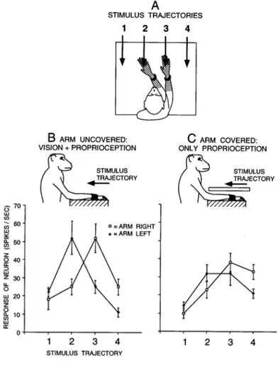
Another characteristic of these bimodal neurons is that they continue to respond to visual stimuli also when these stimuli are no longer present, for instance in complete darkness (Graziano, Hu & Gross, 1997b). Graziano (1999) specifically tested the relative role of vision and proprioception in encoding limb position in the monkey brain. He tested the response of PMv multisensory neurons with a tactile receptive field on the arm and a visual receptive field anchored to the tactile one. The neurons were tested under different configurations, in which both the position on the arm and the visual information regarding the arm were manipulated (see Figure 1.2). Results demonstrated that both visual and proprioceptive information play an important role in building a coherent representation of the space around us. Moreover, results from
this study demonstrated that these neurons responded not only to a visual stimulus presented close to monkeys’ arm, but also close to a fake but realistic arm placed in a realistic posture in front of the monkeys during the testing (Graziano, 1999). Interestingly, if the real arm was hidden from view, and the fake arm was moved, the movement of that fake arm caused a shift in the visual RF of the bimodal neurons.
Figure 1.2 Visual responses of a typical premotor neuron with a tactile RF (hatched) on the forearm and hand, and a visual RF within 10 cm of the tactile RF. (A) On each trial, the arm contralateral to the neuron was fixed in one of two positions and the visual stimulus was advanced along one of four trajectories (1–4). For this neuron, the two arm positions were chosen to align the visual RF near the hand and forearm with trajectories 2 and 3. For other neurons, the arm was moved to different extents depending on the location of the visual RF, to better capture the movement of the visual RF with the arm. (B) Responses of the neuron to the four stimulus trajectories when the arm was visible to the monkey. When the arm was fixed on the right, the response was maximum at position 3. When the arm was fixed on the left, the maximum response moved to the left, to position 2. (C) Responses of the neuron when the arm was covered. The movement of the visual RF with the arm was reduced but not eliminated, indicating that the neuron combined both proprioceptive and visual information about the position of the arm. Each point is a mean of 10 trials. Error bars are standard error. Adapted from Graziano, 1999.
These multisensory neurons are particularly sensitive to moving stimuli. Some neurons are directionally selective, that is they code preferentially for neurons moving along a specific trajectory. They also code for stimulus velocity (Fogassi, Gallese, Fadiga, Luppino, Matelli, & Rizzolatti, 1996).
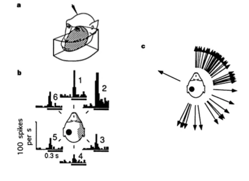
Not only bimodal, but also trimodal neurons are located in PMv. These neurons respond to a tactile stimulus located on the back and the side of the head and to a visual and/or auditory stimulus presented close to the body part where the tactile stimulus is administered (Graziano, Reiss & Gross, 1999). In Figure 1.3, a typical response of a trimodal neuron is shown: this neuron respond to sounds produced near the contralateral side of the head. Specific testing of trimodal neurons properties of an awake monkey revealed that the auditory responses in PMv clearly span the entire contralateral space, and seem to represent the space behind of the head more densely than the space in front of the head (Graziano et al., 1999).
Figure 1.3 Responses of bimodal and trimodal neurons in PMv.
a) Receptive fields of a typical bimodal, visual-tactile neuron. The tactile receptive field (shaded) is on the front of the face contralateral to the recording electrode (indicated by the arrowhead). The visual receptive field (boxed) is confronted to a region of space within about 10 cm of the tactile receptive field. b) Responses of a typical trimodal, visual-tactile-auditory neuron. The tactile receptive field is
contralateral to the recording electrode (indicated by the black spot) and includes the ear and back of the head. The visual receptive field (not shown) extends about 20 cm into the space near the contralateral side of the face. The histograms show the response, summed over ten trials, to a burst of white noise presented 10 cm away at the indicated azimuth angles. c) The calculated preferred direction of the auditory response for 43 trimodal neurons. Each arrow shows the result for one neuron. Adapted from Graziano, Reiss, & Gross, 1999.
Parietal neurons
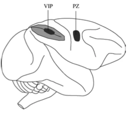
Multisensory bimodal and trimodal neurons with similar properties as compared to the ones in the PMv were found also in the posterior parietal cortex of monkeys’ brain, specifically at the level of the ventral intraparietal sulcus (VIP), and in area 7b. VIP area is located in the fundus of the intraparietal sulcus and it receives projections from the middle temporal visual areas, as well somatosensory, auditory, and vestibular regions (Graziano & Cooke, 2006). Most neurons in area VIP exhibit bimodal visuo-tactile properties, in the sense that they respond to stimuli applied in either sensory modality (Duhamel et al., 1998). The tactile RFs are equally distributed on the top, side, or back of the head, and on the neck, meaning that this area primarily describes spatial area around the face, but they can sometimes be on the chest, shoulder, or arm (Graziano & Cooke, 2006). Some of these neurons are also selective for the distance at which the visual stimulus is presented and they show a strong sensitivity to speed and direction of motion of both visual and tactile stimuli (Duhamel et al., 1998). Some VIP neurons are sensitive to the three-dimensional trajectory of objects. Moreover, some VIP neurons are trimodal, responding to visual, tactile and auditory stimuli, with the three receptive fields usually aligned (Schlack, Hoffman & Bremmer, 2003). Differentially from multisensory neurons described in the PMv, whose RFs are mainly arm centred, bimodal neurons in VIP area have tactile receptive fields encoding a head-centered reference frame, whereas visual receptive fields are widely distributed between eye-to head-centered coordinates.
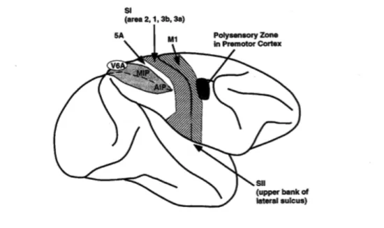
Figure 1.4 Schematic side view of macaque monkey brain showing approximate location of the ventral intraparietal area (VIP) and the polysensory zone (PZ). Intraparietal sulcus is shown opened up, with light shaded area indicating buried cortex. Adapted from Graziano & Cooke, 2006.
Area 7b is prevalently a somatic area, with most of its neurons being somatosensory or somato-motor (Gross & Graziano, 1995; Hyvarinen, 1981), even if a part of the neurons studied in this area is bimodal, responding to both visual and tactile stimulation. Here the majority of the neurons have bilateral RFs located on the limbs often covering the whole body (Leinonen, Hyvarinen, Nyman, & Linnankoski, 1979). Finally at a subcortical level, the putamen has a complete somatotopic map of the body, including a large proportion of bimodal neurons with tactile RFs centred on the head (Graziano & Gross, 1993).
1.1.2 Sensory-to-motor function
These neurons described in the previous paragraph also have a motor function. Most of both premotor (Fogassi et al., 1996) and parietal neurons (Leinonen et al., 1979) are active during movements of the body part where their visuo-tactile receptive fields are anchored. In addition, electrical stimulation of neurons in the ventral premotor cortex (Graziano, Taylor & Moore, 2002) evokes movements of the body part anchoring the aforementioned tactile, visual or acoustic RFs. Similarly, stimulating areas in the fundus of the intraparietal sulcus, corresponding to VIP, evoked different types of movement including included eye movements, reaching, bringing the hand to the mouth, aggressive displays, and defensive movements (Stepniewska, Fang, & Kaas, 2005). Finally, as described before, these neurons encode the location and trajectory of objects, with an emphasis on objects that are near or approaching the body. This specificity for moving stimuli as compared to static one can be considered a hallmark of the clear sensory-to-motor function of peripersonal space representation. Indeed looming stimuli are an essential component of threat (Gibson, 1972; Graziano & Cooke, 2006). For obvious adaptive reason, a stimulus approaching the body need to be detected as fast as possible, in order to plan a proper motor reaction. The neurons inVIP and PMv could be described as looming detectors in general sense (Graziano & Cooke, 2006).
Taken together, all these properties demonstrate the sensory-motor function of PPS representation: coding the spatial position and dynamics of an external stimulus with respect to a part of the body potentially interacting with it, in order to plan an approach toward an interesting object (Rizzolatti et al., 1997) or evade a potential threat (Graziano & Cooke, 2006).
1.1.3 Neuropsychological evidence
Neglect
The first evidence for dissociated spatial representations in humans came from patients affected by hemi spatial neglect. Neglect has long been recognized as a multi-component syndrome, usually observed following brain lesions affecting the right hemisphere, in particular at the level of the right inferior parietal cortex (inferior parietal lobule, angular and supramarginal gyrus) and right temporo-parietal junction (Vallar & Perani, 1986). Neglect patients are characterized by a failure to respond, to attend or to orient voluntarily to objects placed in the contralesional space. This contralesional unawareness may occur selectively for different sectors of space. Specifically, a form of neglect, known as personal neglect, has been described, characterized by the presence of deficits relative to the side of the body contralateral to the lesion. Bisiach and colleagues (1986) investigated the dissociation between different forms of neglect for different sectors of space. They first reported evidence of a double dissociation between patients selectively affected by neglect for personal and extrapersonal space. In addition, Halligan and Marshall (1991) presented a case of a patient selectively affected by neglect limited to the peripersonal space. This patient showed a typical neglect bias in a bisection line task; when he was asked to bisect a horizontal line placed closed to him, in the near space, the patient committed rightward errors. Instead, when the line was positioned in the far space (i.e. at two meters distance), these rightward errors disappeared (see also Berti & Frassinetti, 2000 for a similar case).
Extinction
The study of crossmodal extinction has brought a considerable contribution in investigating how multisensory stimuli are perceived and integrated in order to build the representation of the space around us.
Extinction is a neuropsychological syndrome generally following a right brain lesion, most typically in the posterior parietal region. In these patients, the perception of a contralesional tactile stimulus is affected by the presentation of a concurrent ipsilesional visual stimulus (Bender, 1952), with the almost normal detection of contralesional stimuli presented in isolation. Extinction (and neglect as well) could affect all sensory modalities, either within a single modality i.e. (unimodal extinction) or between different sensory modalities (i.e. crossmodal extinction; Mattingley, Driver, Beschin & Robertson, 1997). Crossmodal extinction has been extensively studied as a model to study the multisensory neural representation of space. Authors initially investigated cases of visuo-tactile extinction. Patients suffering from this syndrome were unable to correctly perceive a tactile stimulus administered at the controlesional hand, when a concurrent visual stimulus was presented in the ipsilesional side of space. Làdavas and colleagues (di Pellegrino, Làdavas, & Farnè, 1997; Farnè & Làdavas, 2000), in a series of studies, demonstrated that crossmodal extinction was also spatially dependent. By using a visuo-tactile stimulation paradigm in right brain damaged patients affected by left tactile extinction, they demonstrated that tactile detection at that hand was inhibited by a visual stimulus presented in the right hemi space. Interestingly, the presence of the visual stimulus produced crossmodal extinction at the same extent as an ipsilesional tactile stimulus in case of unimodal tactile extinction. Critically, this degree of cross modal extinction occurred only when the visual stimulus was presented close to the patient’s hand (i.e. within
the peripersonal space), but was much weaker or absent when the visual stimulus was placed at a distance (in ‘far’ or ‘extrapersonal’ space). Two stimuli are more likely to interact when presented in the same spatial representation: when a visual stimulus is presented far from the body, that is in the extrapersonal space, it does not interact with tactile stimuli presented close to the hand, because they are presented in two different representations. So, crossmodal extinction has been taken as an evidence of the existence of separate representations of space.
A model addresses the competitive dimension of extinction (and neglect as well) by proposing that these phenomena result from a breakdown in the dynamic balance that normally exists in the reciprocal inhibition between homologous areas of the two hemispheres that orient spatial attention in opposing, contralateral directions (Kinsbourne, 1977; Kinsbourne & Bruce, 1987; see Jacobs, Brozzoli, Hadi-Bouziane, Meunier, & Farnè, 2011, for a review).
The same extinction effect previously described for the hand has been demonstrated with tactile stimuli presented at the face (Farnè & Làdavas, 2002; Làdavas, Zeloni, & Farnè, 1998a), suggesting the existence in humans, as in monkeys, of a modular representation of space surrounding different body parts. Farnè and colleagues (Farnè, Demattè & Làdavas, 2005) tested whether these representations effectively operate in a modular way in a group of right brain damaged patients by using cross modal extinction paradigm. They measured the level of cross modal extinction when visual and tactile stimuli were presented on homologous body part (for instance, tactile stimulus at the contralateral hand and visual stimulus near the ipsilesional hand) and non-homologous body parts (i.e. tactile stimulus at the hand and visual stimulus near the face). Results showed a dissociation between the representation of the peripersonal space around the hand and the peripersonal space around the face,
revealing that a visual stimulus presented near the face did not interact with tactile stimulation at the hand and vice-versa. These results confirmed the hypothesis of an organization of space in separated moduli around different body parts.
Interestingly, in order to examine the spatial coordinates used by this multisensory system to code peripersonal space, di Pellegrino and colleagues (di Pellegrino et al., 1997) tested a patient with tactile extinction by manipulating hands’ position in space. They asked the patient to perform the task with the hands crossed; this way, the left hand was placed in the right hemi space and the right hand in the left hemi space. Results showed that a visual stimulus presented near the right hand (that is in the left hemi space) extinguished tactile stimuli at the left hand (that is in the right hemi space). Results of the present studies offered interesting insights on the idea the representation of peripersonal space (peri-hand space, in this case) was anchored to a specific body part, in line with neurophysiological results on monkeys.
Modulation of tactile extinction not only by visual but also auditory stimuli has been investigated in neuropsychological patients (Làdavas, Pavani, & Farnè, 2001; Farnè & Làdavas, 2002). Indeed, in a group of right brain damaged patients Làdavas and colleagues demonstrated that tactile detection at the neck was influenced by the presentation of auditory stimuli presented near, but not far, the patients’ face. Interestingly, spatially dependent interactions between audition and touch were strongest when the auditory stimuli came from the back, rather than from the front, of the patients’ head. This spatial specificity from a particular sector of space, that is the back of the head, is particularly relevant in the case of audio-tactile interaction. Indeed it makes perfectly sense that the backspace, where vision is not available, is more deeply represented through audio-tactile interaction. Moreover, in this study the authors investigated whether this spatial modulation of touch by audition was
dependent on the complexity of the stimuli used. They found that white noise sound, that is a more complex sound, has a stronger effect on tactile perception than pure tones. These results are in line with neurophysiological results in monkeys, showing that multisensory neurons coding for peripersonal space around the head (Graziano et al., 1999) did not respond to pure tones. This specificity could be due to the fact that white noise sounds are more similar to ecologic sounds. Thus, the reduced effect operated by a pure tone might reflect a sort of impenetrability of the integrated auditory – tactile system to a sound that has a little chance to occur in nature (Farnè & Làdavas, 2002).
The near-far modulations of crossmodal extinction here described have been considered as the first behavioural demonstrations of the existence of peripersonal representation in humans (di Pellegrino et al., 1997; Làdavas et al., 1998a; Làdavas, di Pellegrino, Farnè & Zeloni, 1998b).
1.1.4 Behavioural evidence
A series of studies conducted on healthy human subjects confirmed the existence of spatially dependent cross-modal interactions. Most of these studies investigated specifically visuo-tactile interactions. One of the best-known paradigms used to investigate this issue is the cross-modal congruency task. In this task, participants receive a vibrotactile stimulus either at the thumb or the index finger of the hands. At the same time a visual stimulus (a distractor) is presented at four possible positions, corresponding to the four possible locations of the vibrotactile stimuli. Thus, for each trial the visual distractor could be either close to the tactilely stimulated hand or to the other hand. Moreover, the visual distractor could be ”congruent” or ”incongruent in elevation with the tactile target stimuli. Participants are asked to make a speeded
up/down discrimination judgement in response to the tactile stimuli, by pressing a foot pedal, ignoring the visual distractors. A series of studies (Spence, Pavani, & Driver, 2000; Pavani, Spence, & Driver, 2000; Spence, Pavani, Maravita, & Holmes, 2004; see also Brozzoli, Pavani, Urquizar, Cardinali, & Farnè, 2009) using this paradigm revealed that incongruent visual distractors (that is distractors presented at a different elevation as compared to the vibrotactile stimuli) slowed down the judgements about the tactile stimuli and produced more errors. This effect of visual stimuli on tactile one was spatially dependent, since it was stronger from stimuli coming from the same side of space (Spence et al., 2004). When the posture of participants was manipulated, this cross modal effect changed accordingly: for instance, in a crossed hand condition, if the right hand is placed in the left hemi space, a visual distractor in the left space affected tactile detection at the right hand. These results demonstrated that visuo-tactile interactions take changes of posture into account (e.g. Macaluso, Driver, van Velzen, & Eimer, 2005), in agreement with neurophysiological properties of neurons in the parietal and premotor cortex. Interestingly this spatial modulation appeared to be present also when patients were not actually able to see their hand, suggesting the importance of tactile and proprioceptive information in building a strong percept of limb position and, accordingly, of the representation of the space surrounding it.
The particular link between vision and touch for the construction of a congruent representation peripersonal of space is such that even the image of a fake, but spatially congruent, limb can affect tactile perception. Pavani and colleagues (2000) investigated the cross-modal effect of visual distractors on tactile judgments using the cross modal congruency task previously described. Participants were asked to discriminate the location of vibrotactile stimuli administered at the hand (upper, at the
index finger, vs. lower, at the thumb), with the hand occluded under a table, while ignoring distractor lights that could independently be upper or lower with respect to the tactile stimulation. In line with previous results obtained with the same task, an incongruent (with respect to the elevation of the tactile stimuli) visual stimulus presented close to the hand interfered with tactile detection. Critically, the same results was obtained when the visual stimulus was presented close to a fake, realistic hand placed on the table in front of the participants, but only when the hand was placed in an anatomically plausible posture (see also Farnè, Pavani, Meneghello, & Làdavas, 2000, for a similar result on extinction patients). This indicates that, although strongly interrelated, tactile and visual spatial representations are also flexible, and can change to maintain spatial alignment of multisensory signals arising in the peripersonal space. Taken together all these studies support the claim that the human brain represents PPS through an integrated visuo-tactile system.
Cross modal spatially dependent interactions has been described also between audition and touch. Auditory peripersonal space has been first described around the head, both in monkeys (Graziano et al., 1999) and in humans in brain damaged patients (Farné & Làdavas, 2002; see Paragraph 1.1.3). Behavioral studies investigated the existence of auditory-somatosensory interaction around the head (Tajadura-Jimenez, Kitagawa, Väljamäe, Zampini, Murray, & Spence, 2009), particularly in the space behind the head (Kitigawa, Zampini, & Spence, 2005) and around the hand (Murray, Molholm, Michel, Heslenfeld, Ritter, et al., 2005; Zampini, Torresan, Spence, & Murray, 2007) in healthy subjects using different paradigms. In Kitigawa and colleagues’ study (2005) participants performed a temporal order judgment (TOJ) task about pairs of auditory and tactile stimuli presented at different interstimulus interval. Tactile stimuli were applied at participant’s earlobes, while the
auditory stimuli were presented through loudspeakers placed 20 cm behind participant’s head, both in left and right hemispaces. Participants were asked to report the modality (auditory or somatosensory) of the first stimulus they perceived. Results indicated a better performance for stimuli presented from different sides of space. In a second experiment, participants performed a tactile left/right discrimination task while auditory distractors were presented simultaneously from the same or opposite side. Results showed slower (and less accurate) tactile RTs when the auditory distractors were presented on the opposite side from the target tactile stimuli. Interestingly, in this study, in line with previous results both in monkeys (Graziano et al., 1999) and in humans (Farnè & Làdavas, 2000), when bursts of white noise (a more ecologic sound) were used, authors found a greater effect of crossmodal interference when the auditory stimuli were presented close to participant’s head rather then far apart. These results suggested that auditory and tactile stimuli interacted preferentially in the space immediately surrounding the head exhibiting different responses according to the complexity of the auditory stimuli used. These results seem to be in contrast with results obtained by Zampini and colleagues (Zampini, Brown, Shore, Maravita, & Roder, 2005) in a similar experiment in which audio-tactile interaction were investigated through a TOJ task in the front space. In this experiment results showed that participants’ responses to tactile stimuli presented at the finger were not affected by the spatial position of auditory stimuli. However this experiment was conducted in the front space, while a series of evidence in the literature suggested that auditory-tactile interaction are more likely to take place in the back space, where vision is not available and where information coming from audition are more relevant (see also Occelli, Spence, & Zampini, 2011, for a review). Interestingly in a series of experiments, Tajadura-Jimenez and colleagues (2009)
further demonstrated a spatial modulation of auditory–somatosensory interactions when auditory stimuli were presented in the space close to the head, at different distance to the left and the right of the center of participants’ head.
Audio-tactile interactions have been studied also in the space around the hand. Murray and colleagues (2005) investigated the possible interaction between spatially aligned and misaligned (across left and right directions) auditory and somatosensory pairs of stimuli - using a simple reaction time task - and the electrophysiological correlates of this phenomenon - using ERPs. Participants were asked to respond to a tactile stimulation at the hands, while auditory stimuli were presented from loudspeakers placed either close to the left or to the right hand. Results showed that participants’ RTs were facilitated for multisensory stimuli. Additionally, the extent of facilitation did not change as a function of the spatial alignment of the stimuli, showing that auditory stimuli facilitated tactile RTs independently of the alignment of auditory stimuli with respect to the tactile stimuli. Accordingly, ERPs results showed a greater response, at 50-90 ms post stimulus onset, for multisensory audio-tactile stimuli, as compared to the summation of the constituent unisensory ones, at lateral central scalp sites over the hemisphere contralateral to the stimulated hand, for both the aligned and misaligned configurations. The source of these interactions were localized to unimodal rather then multimodal area, at the level of the auditory association cortices in the hemisphere contralateral to the hand being stimulated, independently of the location of the auditory stimulus. Taken together, both behavioral and electrophysiological results from the present study provided evidence for the existence of multisensory auditory-somatosensory interaction across spatial separations. One proposition from these results is that the brain regions involved in processing auditory–somatosensory stimuli contain large spatial RFs, which allow for
the integration of stimuli that happen to be separated by a relatively large distance in external space (Murray et al., 2005; see also Zampini et al., 2007). Zampini and colleagues (2007) further investigated audio-tactile interaction at the hand both in front and rear space. Results confirmed a facilitation for multisensory stimuli as compared to unisensory ones, regardless of the spatial location from which the auditory stimuli were presented or the specific body posture adopted by participants. Taken together, these studies revealed the existence of audio-tactile interaction mechanisms in humans. However, these studies did not clearly establish whether these audio–tactile integrative mechanisms are modulated by the spatial location of auditory stimuli. A study of Serino and colleagues (Serino, Bassolino, Farnè, & Làdavas, 2007), by using an audio-tactile interaction task, demonstrated a space dependent audio-tactile interaction mechanism. In this paradigm participants received tactile stimuli at their index finger, while a concurrent sound was presented. The sound position in space was manipulated, so that in different trials it was presented either close to the participant’s hand (near condition) or far from it, at a distance of around 125 cm (far condition). Participants were asked to verbally respond to the tactile stimulation as quickly as possible, by saying TAH in a microphone. They were also asked to completely ignore the auditory stimuli during the task. Results showed that tactile detection was speeded up when the sound was presented close to the body as compared to when it was presented far apart, revealing the existence of an integrative auditory peri-hand space in humans, in line with studies on visuo-tactile interaction.
1.1.5 Neural basis of PPS in humans
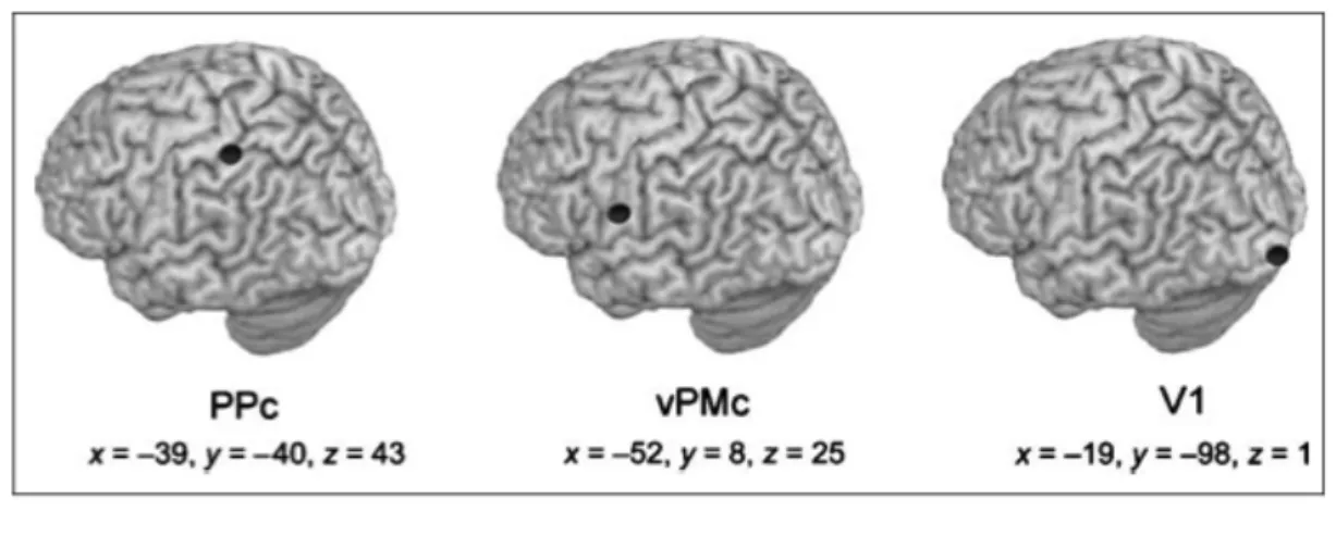
Human imaging studies suggest that a neural system dedicated to multisensory integration in the peripersonal space also exists in the human brain. An increasing number of studies in healthy humans using fMRI (Bremmer, Schlack, Shah, Zafiris, Kubischik, Hoffmann, et al., 2001; Makin, Holmes, Brozzoli, Rossetti, & Farnè, 2009; Brozzoli, Gentile, Petkova, & Ehrsson, 2011) and EEG (Sambo & Forster, 2009) suggested that the multisensory representation of PPS in the human brain is implemented in a fronto-parietal network, encompassing premotor and posterior parietal areas largely corresponding to PMv and VIP areas in the monkey’s brain. Bremmer and colleagues (2001) measured changes in neural activity in healthy volunteers using functional magnetic resonance imaging (fMRI) in order to identify a set of common areas involved in polymodal information processing. Indeed participants were tested in fMRI while presenting moving visual, tactile, or auditory stimuli. They demonstrated that portions of the posterior parietal cortex (PPc), around the intraparietal sulcus and the ventral premotor cortex (vPMc) were activated by tactile and by visual and auditory stimuli moving towards the head (see also Macaluso & Driver, 2001; 2005). The homologous of this set of areas in the monkey’s brain is extensively activated by moving - rather than stationary - multisensory stimuli, preferentially approaching towards the body (Colby, Duhamel & Goldberg, 1993). Moreover, Sereno & Huang (2006) described aligned maps of tactile and peri-face visual stimuli in the ventral part of the intraparietal sulcus, coding the location of visual stimuli with respect to the face, and not with respect to the eye. A different fMRI study in healthy participants (Makin, Holmes, & Zohary, 2007) showed that activity in PPc and vPMc was selectively modulated by a visual stimulus approaching the hand, compared to seeing the same stimulus moving away from the hand. Neural
activity in PPc and vPMc was sensitive to the position of the hand relative to the visual stimulus, as signalled both by visual and proprioceptive information.
These results are in line with a recent fMRI study by Brozzoli and colleagues (Brozzoli et al., 2011) using an adaptation mechanism. They found a consistent adaptation in premotor and parietal areas exclusively for objects near the hand. Finally, Gentile and colleagues (Gentile, Petkova, & Ehrsson, 2011) described an increased activation in response to visuo-tactile stimulation in a set of cortical (premotor and posterior parietal) and subcortical regions matching the neurophysiological literature on multisensory areas in monkeys and humans. Taken together, these results suggested the existence of common neuronal mechanism in humans and nonhuman primates including parietal and premotor cortices for the representation of peripersonal space.
1.2 From the body to its representation(s)
In order to interact with objects in space, in either reaching an interesting stimulus or avoiding potential harm, the human brain needs to concurrently represent not only the position and movements of external stimuli in space, and especially within the PPS, but also the different parts of one’s own body potentially interacting with those stimuli. In the following section I will focus on high-level multisensory representation of the body (Body Representation, BR) in the brain, supporting this function.
Our body represents the centre of our sensations and perceptual experiences and at the same time it mediates every physical interaction with the external stimuli. The brain contains several representations of the physical body. It is fair to state that body representation begins with a tactile map of the body surface, since somatosensation is the most basic property that allows every perceptual interaction with the world. One key function of somatosensory representation is the ability to localise the location of tactile stimuli on the body surface. Indeed, as soon as a fly touches our skin, we immediately know where we have been touched in respect to the skin surface. Somatosensory receptors located all over the skin project to the well-known sensory homunculus of the primary somatosensory cortex (SI, Penfield & Rasmussen, 1950) within the post-central sulcus (see Paragraph 1.2.1). Neurons in this area form a characteristically distorted map of the contralateral body surface: each part of this map selectively responds to mechanical and electrical stimulation of a given part of the body, thus supporting perception of where on the skin a sensory stimulus is located. The localisation based on skin receptors can only take account of the localisation of touch in the receptor field surface. However, in order to perform a successful interaction with an external stimulus, for instance to make a motor
response towards the source of a tactile stimulation (such as for swatting away an insect that might be about to sting) it is important to know different information exceeding the spatial location of touch on the body. Indeed, localising tactile inputs within the somatotopic map is not sufficient by itself to localise them on the body surface. Since our body and, even more prominently, our limbs are constantly moving in the environment, the relative position of a body part in the external space with respect to the other body parts may vary. So, proprioceptive information about the position of a body parts with respect to each other must be taken into account. Moreover, the body changes continuously in position and dimensions throughout life, so, in order to perform a movement, body representations in the brain need to be continuously updated about shape and dimension of the different body parts in each moment. Surprisingly, no periphery receptors provide information about the dimension of the different body parts in the brain. This body referencing processing is especially true for tactile sensation, but not only for that. For instance, in order to localize of an insect flying around us just through auditory information, our brain needs to compute the distance between the two ears. Similarly, for visual depth perception, the spacing between the two eyes must be taken into account (Serino & Haggard, 2010).
So, it is clear that, in order to entirely account for the different aspects of body experience involved in interaction with the environment, more complex higher-level multimodal representations of the body in the brain must exist, supporting complex perceptual, motor and emotional functions, and, ultimately, underling the experience of having a body and the ability of using that body to interact with the external world. In the next paragraphs I will review main evidence about the existence of low-level and high-level body representations.
1.2.1 Unimodal low-level body representations
Much is known about unimodal body representations in the primary somatosensory cortex (SI; Kaas, Nelson, Sur, Lin, & Merzenich et al., 1979) and in the primary motor cortex (MI; Penfield, & Boldrey, 1937), each of which contains a tactile and a motor map of the body.
Penfield and colleagues (Penfield & Rasmussen, 1950), using direct cortical stimulation on patients undergoing neurosurgery, first described a somatosensory map in the post-central gyrus. The somatosensory cortex is divided in two parts, the primary somatosensory (SI) and the secondary somatosensory cortex (SII) at the level of the anterior portion of the parietal cortex in the post central gyrus. Somatosensory cortical areas receive a wide range of somatosensory inputs from different peripheral receptors, such as mechanoreceptors, thermoreceptors, and nociceptors, which activation determines an evoked sensation of different nature. And indeed different somatosensory functions are processed by distinct neural pathways: tactile information are transmissed through the dorsal column medial lemniscal pathway, while the anterolateral pathway conveys pain, and temperature information from the periphery to the brain. For sake of clarity, here I will focus mainly on tactile sensation.
Tactile processing is somatotopically organized, meaning that tactile stimuli administered on a given body part elicit a neural response in a specific portion of the somatosensory cortex, matching the same body part. Adjacent neurons on SI surface tend to have adjacent receptive fields on the body. In this representation, known as “somatosensory homunculus” the legs are represented medially, while the face and hands are more laterally.
Figure 1.5 Schematic somatosensory (on the left) and motor (on the right) homunculus
In the case of the motor system, as for somatosensory perception, it has been demonstrated that movements of a specific body parts depends on neural activity in a matched region of the primary motor cortex (MI). Indeed, electrical stimulation of specific regions of MI evokes movements of a specific body part (Penfield & Boldrey, 1937; Penfield, 1950). As for SI, also MI is organized in a somatotopic way, with the trunk and legs represented more medially, while face, hands and arms are represented more laterally. Few studies have shown a somatotopic organization also at the level of the Supplemental Motor Area (SMA). Recently, Zeharia and colleagues (Zeharia, Hertz, Flash & Amedi, 2012) investigated movement encoding both in the primary motor cortex and supplementary motor area. Results confirmed that MI and SMA are organized in a somatotopic way, and that SMA activity is more associated with movement suppression as compared to M1.
Finally, functional magnetic resonance imaging (fMRI) studies have identified two separate regions, which specifically process body-related visual information: the extrastriate body area (EBA; Downing, Jiang, Shuman, & Kanwisher, 2001), and the fusiform body area (FBA). These two areas selectively respond to images of the whole body (as well as of non facial body parts) compared to other object categories (Downing et al., 2001; Peelen & Downing, 2005; Taylor, Wiggett, & Downing, 2007; for a review, see Peelen & Downing, 2007). In a recent fMRI study of Orlov and colleagues (Orlov, Makin, & Zohary, 2010), participants were scanned when viewing images of body parts. Results showed a consistent activation at the level of the occipital temporal cortex (OTC), with distinguishable clusters for separate body parts. Moreover the authors tested whether the separate clusters of activation within the body map for particular body parts could depend only on the particular shape of the body parts tested. Results show that separate images of the “hand” and the “elbow”, for instance, consistently activated the area that was previously activated for the “upper limb”. This suggested that the specificity of activation at the level of OTC for different body parts cannot be explained with a difference in shape only. Finally, this study showed a partial correspondence in the activation of visual and motor (unseen movements or self generated) body parts, suggesting that action related information about the body converge in this area.
Taken together, these low-level unimodal representations process information related to a single body part and single modalities, and thus they are not sufficient to entirely account for the different aspects of body experience involved in interaction with the environment.
In the next paragraph I will review neuropsychological and behavioral evidence about the existence of higher-level body representations in the brain.
1.2.2 Neuropsychological evidence
Several alterations of body representations, without any impairment at the level of primary somatosensory and motor cortices have been described in neuropsychological patients. These deficits result in alteration of body perception, which cannot be ascribed to pure sensory or motor deficits. For instance, the studies of patients suffering from numbsense, a tactile deficit with preserved tactually guided movements (de Vignemont, 2010; see also Dijkermann & de Haan, 2007) were especially relevant. Paillard and colleagues (Paillard, Michel, & Stelmach, 1983) reported the case of a patient who suffered from deafferentation after a left posterior cortical lesion. Deafferentation is a clinical condition characterized by a loss of somatosensory information that can affect a portion of the body (Dijkerman & De Haan, 2007). This patient was unable to report a tactile stimulus presented at the contralesional hand, but critically the ability of pointing to the same tactile target was preserved. Rossetti and colleagues (see Rossetti, Rode, & Boisson, 2001) reported a case of a patient, suffering from a lesion at the level of the thalamic nucleus VPL, who presented a similar dissociation: indeed he was able to point with the left hand directly to a tactile target at the impaired right hand, while verbal responses (and pointing responses on a drawing of the arm) were affected. The opposite dissociation was revealed by Paillard and colleagues (1985) in a patient with, instead, preserved ability to report a tactile stimulus, but deficit in pointing to it. These phenomena reviewed above supported the idea of two separate somatosensory pathways for action and conscious perception (see Dijkerman & De Haan, 2007, for a review). This dissociation supported the idea of the existence of different body representations, with a representation of the body that is used for action, more precisely for the guidance of movements, while more perceptual judgements about the spatial relations of the different body parts are
supported by a different body representation, as suggested by several authors (see Paragraph 1.2.4 for a discussion on this point).
Other body representation disorders are more properly defined as distortions, like the feeling of having a body of different dimension that its actual size, i.e. macrosomatoagnosia, a” distorted awareness of the size of the whole body of body parts”, de Vignemont, 2010), or the Alice in Wonderland Syndrome, a “distorted awareness of the size, mass, shape of the body or its position in space” (Todd, 1955; see also de Vignemont, 2010).
There are patients exhibiting abnormal belief about their body, not supported by real motor or somatosensory deficits (such as finger agnosia, the inability of recognizing their own finger). These abnormal beliefs can be associated with somatoparaphrenia (Gerstmann, 1942; see also Vallar & Ronchi, 2009), a pathological denial of ownership of contralesional limb that is felt as not belonging to oneself (Bottini, Bisiach, Sterzi, & Vallar, 2002; Critchley, 1953), sometimes in absence of somatosensory deficits. This phenomenon is often associated to hemispatial neglect (Vallar & Perani, 1986). Neglect patients are characterized by a failure to respond, to attend or to orient voluntarily to objects placed in the contralesional space. Neglect can have different manifestations: patients can exhibit a personal neglect, defined as “a lack of attention towards one’s side of the body”: in this case the patient do not voluntarily attend one side of their body, or also a motor manifestation that is the underutilisation of the contralesional side of the body, even in absence of primary motor or somatosensory deficits (Vallar, 1998).
Brain damaged patients with a lesion at the level of the premotor cortices frequently exhibited anosognosia for hemiplegia as a symptom. Anosognosia for hemiplegia is the denial of the contralesional motor deficits that may follow brain damage (Berti,
Bottini, Gandola, Pia, Smania, Stracciari et al., 2005; Carruthers, 2008; Pia, Neppi-Modona, Ricci, & Berti, 2004). In this case patients have a motor problem, but they fail in recognizing the severity of their problem, instead they also claim that they do not have any body related problems. These disorders may be accompanied by reports of supernumerary limbs (Halligan & Marshall, 1995), that is the strong awareness of the existence of non-existent limbs.
The phantom limb, that is the strong awareness of an amputated limb, represents a well-known disorder of body representation (Hunter, Katz, & Davis, 2003; Ramachandran & Hirstein, 1998). This strong awareness of the phantom limb includes a series of sensory phenomena that are perceived as originating from the missing body part. The phantom limb phenomena could include tactile sensations, such as the sensation of being touched on the missing body part, as well as more generic somatic sensations such as tingling, itching, pressure, warmth, or cold (Hunter et al., 2003) or also motor sensations. Indeed amputees can report, generally immediately after amputation, being able to move their missing limb voluntarily, with this ability decreasing over time. These sensations are frequently accompanied by painful sensation originating from the missing limb. Importantly, in this case patient are aware of their amputation and that these sensations are not veridical (Serino & Haggard, 2010; see also Chapter 2). Interestingly, phantom limb phenomena are not limited to a sensory or motor percepts originating from the missing body part (Hunter et al., 2003; Kooijman, Dijkstra, Geertzen, Elzinga, & van der Schans, 2000), but are often referred by patients as conscious awareness of the presence - implying position, shape and size - of the missing limb (Flor, Nikolajsen, & Staehelin Jensen, 2006; Hunter et al., 2003). The complexity and richness of these phantom limb phenomena is hardly explainable as resulting only from cortical reorganization in unimodal
primary cortices. Rather, they suggest an involvement of multisensory body representations, which integrate the continuous flow of information from different sensory modalities in order to give raise to the experience of the body and its parts (Blanke & Metzinger, 2009; Petkova, Bjornsdotter, Gentile, Jonsson, Li, & Ehrsson, 2011a; Ionta, Gassert, & Blanke, 2011; see Paragraph 7.4).
These studies on neuropsychological patients suggested that the experience of the body could be affected independently from primary somatosensory or motor deficits. The diversity and variety of bodily disorders suggested the existence of multiple and more complex body representations that account for a complete body experience.
1.2.3 Behavioural evidence
Different effects of interaction between multisensory body-related signals support the existence of multisensory body representation. For instance, in a study Kennet and colleagues (Kennet, Taylor-Clarke, & Haggard, 2001) showed that tactile information was improved by viewing the body tactilely stimulated, although visual information was totally uninformative about tactile stimulation (tactile stimuli were hidden from view). The authors measured tactile acuity by means of the 2-point discrimination thresholds on the forearm, while participants were looking either at the arm or at a neutral object, presented at the same location of the arm. Results demonstrated that spatial resolution of touch was better when participants could view the arm as compared to when participants viewed the neutral object. These results clearly show that viewing the body boosts tactile processing. The fact that participants did not see any information about the tactile stimulation during the task suggests that the visual information specifically related to the body offers a special context, that is the body itself, to which tactile information is referred to.
Tactile and proprioceptive inputs also interact to build and update high-level BRs: this is revealed for instance through some spectacular illusions, such as the Pinocchio illusion (Lackner, 1988; de Vignemont, Ehrsson & Haggard, 2005). In this illusion, participants held the tip of their nose with the thumb and the index finger while the tendon of the biceps muscle was vibrated. Vibration of this tendon normally elicited the sensation of the arm moving away from the body. When the nose and the finger were in direct contact, in order to solve the mismatching information about constant tactile sensation and dynamic proprioception, participants felt like their nose was elongating. De Vignemont and colleagues (2005) used the Pinocchio illusion to experimentally demonstrate that proprioceptive information is used to update body representation. In this experiment participants held their right index finger with the left index finger while the tendons of either the biceps or the triceps muscles of the right arm were vibrated. When the tendon of the biceps was vibrated, the arm was perceived as extending and it elicited the sensation that the left index finger was elongating. During the vibration participants received a couple of tactile stimuli at the finger and at the forehead as a reference body part, and they were asked to judge whether the tactile distance felt on the finger or the forehead was longer or shorter. Participants perceived the tactile distance as longer when the finger was perceived as elongating (that is, in case of the vibration of the tendon of the biceps of the forearm). The contrary effect, that is the perception of the tactile distance as shorter when the arm was perceived as contracting (so the finger was perceived shortening) was not found. These results suggested that proprioceptive information is used to update Body Representation: the internal representation of the body, such as the perceived body size, affected the perception of an external object. Critically the effect on the
perceived tactile distance was evident only in the case of the perceived elongation, but not contraction, of the arm.
In a different study, Taylor-Clarke and colleagues (Taylor-Clarke, Jacobsen & Haggard, 2004) demonstrated the relevance of visual information in updating body representation. According to an illusion originally provided from Weber (1978), the same tactile distance is perceived larger when presented on a zone with higher tactile acuity, as compared to a zone with low tactile acuity. This illusion suggested that tactile information is processed with reference to tactile receptors density. In the study of Taylor Clarke and colleagues, participants performed a tactile distance perception task for two tactile stimuli presented simultaneously to the finger or the arm after a period of visual experience of these body parts. When subjects viewed an enlarged version of their arm (but not of the hand), the tendency to underestimate tactile distances on the arm relative to the hand was significantly reduced, while the tactile acuity remained unaffected. They demonstrated that perceptual judgements about an object touching the skin depend not only on the primary tactile sensations, but also on the perceiver’s representation of the body part that the object touches. Indeed, in this case the visual size of the body is shown to affect the tactile size of an object touching the skin. These results suggest that tactile signals are processed with reference to an implicit representation of the body (see also Medina & Coslett, 2010). Finally, in a recent study Tajadura-Jimenez and colleagues (Tajadura-Jimenez, Väljamäe, Toshima,
Kimura, Tsakiris, & Kitagawa, 2012) demonstrated that also auditory information contributes to update body representation. In their study participants performed an audio-tactile tapping task. They were asked to tap on a surface with the right arm and, in synchrony with the tap they produced, they listened to a tapping sound that originated at different distances from where they performed the tapping. After the task,