Università degli Studi di Ferrara
DOTTORATO DI RICERCA IN
SCIENZE FARMACEUTICHE
CICLO XXIV
COORDINATORE Prof. Manfredini Stefano
Design, synthesis and evaluation of novel
dualistic molecules provided with UV
filtering and scavenging capabilities
Settore Scientifico Disciplinare CHIM/08
Dottoranda
Tutore
Dott.ssa Scalambra Emanuela Prof. Manfredini Stefano
Cotutore
Index
1 Introduction 1
1.1 Sunlight 2
1.2 The human skin 3
1.2.1 Melanocytes and melanogenesis 6
1.3 Effects of UV rays on the organism 8
2 UV Filters 13
2.1 Inorganic UV filters 14
2.2 Organic UV filters 15
2.2.1 Organic UVB filters 16
2.2.2 Organic UVA filters 20
2.2.3 Organic broad-spectrum UV filters 22
2.3 Evaluation of sunscreen formulations efficacy 24
2.4 Regulatory and labelling 26
2.5 Sunscreens efficacy and safety 28
3 Free radicals and antioxidants 30
3.1 Free radical and other oxidant species 30
3.2 Cellular targets of oxidative stress 31
3.2.1 Lipids 31 3.2.2 Nucleic acids 32 3.2.3 Proteins 32 3.2.4 Carbohydrates 33 3.3 Antioxidants 33 3.3.1 Enzymatic antioxidants 34 3.3.2 Preventive antioxidants 34 3.3.3 Chain-breaking antioxidants 35
3.4 Antioxidants in skin photoprotection 37
4 Aim 40
6 Antioxidant measurements 46 6.1 PCL Test 46 6.2 DPPH Test 47 6.3 FRAP Test 48 6.4 Results 48 7 UV spectra measurement 51 8 Cosmetic formulation evaluation 55
8.1 Analysis of filtering parameters 56
8.2 Antioxidant efficacy of cosmetic formulations 58
9 Cytotoxicity and Phototoxicity tests 60 10 Photostability and Stability studies 64
10.1 Photostability studies 64
10.2 Stability studies 65
11 Conclusions 67
12 Materials and Methods 71
12.1 General 71
12.2 Antioxidant Analyses 71
12.3 Evaluation of filtering parameters 72
12.4 Cytotoxicity and Phototoxicity tests 73
12.5 HPLC analysis 74
12.6 Photostability studies 75
12.7 Synthetic procedures 76
13 References 91
1. Introduction
Characteristics of living organisms are result of gradually evolutive adaptation toward ambient, also humans are exposed to environmental conditions and then during history course they have developed defence mechanisms adapting to habitat.
One of the most variable phenotypes in human is pigmentation; the colour of skin and also of hair and eyes is primarily determined by melanin, a complex group of biopolymers synthesized by specialized cells called melanocytes. Stimulation of melanin synthesis is the main defence against the damaging effects of ultraviolet radiation and skin colour is the principal outcome of adaptation toward UV rays. Melanin acts as an optical and chemical photoprotective filter reducing the penetration of radiation into subepidermal tissues. Skin coloration is strongly correlated with absolute latitude and then with UV radiation levels to which humans are exposed, closer are persons to the equator greater is UV energy that reachs the earth surface and consequently natural selection have favoured in tropical regions dark pigmentation, because highly melanised epidermis affords better protection against UV-induced injuries such as sunburn, skin cancer and sweat glands damages. Lighter skin instead can be explained as adaptation to the lower UV radiation incidence in regions far from the equator and the importance of maintaining UV-induced biosynthesis of vitamin D3, since increasing the melanin in human skin increases the time of exposure to UV light that is needed to maximize synthesis of previtamin D3. Sunlight is not only dangerous for skin, but also degrade some essential nutrients such as folate, a fundamental molecule for nucleotide and, therefore, DNA biosynthesis. Folate deficiency can result in complications during pregnancy and multiple fetal abnormalities, including neural tube defects, such as spina bifida and anencephalus and it was significant cause of perinatal and postnatal mortality in some populations before the introduction of preventive supplementation. Deeply melanised skin protects also folate in the blood from photolysis, another reason that could have favoured positive selection of people with dark skin in areas with high solar intensity together with the lower skin cancer incidence.1,2
Dark skin and light skin were adaptations to environments of high ultraviolet light exposure and low ultraviolet light exposure, respectively; light skin is most vulnerable to UV radiation and fair skinned individuals, living in regions with great incidence of UV rays, are at highest risk of developing skin cancer. Although dark skinned persons are less affected by UV radiation, they are not completely safe and can also develop, even though incidence is lower, UV-induced skin cancers. It is important to remember that exposure to UV radiation is dangerous for people all over the world and then to avoid detrimental effects of UV radiation it must avoid UV overexposure, particularly during the time of the day of higher UV incidence, and protect the skin with sunscreen products when it decide to stay in the sun.
1.1 Sunlight
The electromagnetic spectrum of the sun is composed, from shorter wavelengths to higher, by: cosmic rays, gamma rays, X ray, UV radiation (UVC, UVB, UVA), visible light, infrared rays, microwaves and radio waves (Fig.1). Fortunately higher energy rays, such as cosmic rays (below 10-16 m), gamma rays (10-16-10-11 m), X rays (10-11-10-8 m) and UVC rays (100-280 nm), are filtered by the stratospheric ozone layer;3 while earth’s surface is constantly irradiated by light coming from the sun, composed of 56% infrared waves (wavelength, 780-5000 nm), 39% of visible light (400-780 nm), 4,9% of UVA rays (320-400 nm) and 0,1% of UVB light (290-(320-400 nm).4 Although UVA and UVB rays are a small portion of the total radiation reaching the earth, they are responsible for skin, eyes and hair damages, because of their higher energy content. The following relationship describes the correlation between energy, frequency and wavelength; radiation energy increases proportionally with the frequency and decreases with the increase of wavelength.3
E = energy
v = frequency (Hertz)
h = Planck’scostant = 6.62 x 10⁻²⁷ erg/s)
c = speedof light = 3.0 x 1010 cm/s
Figure 1.1 Sunlight
Besides energy, it must be considered also the intensity and the composition of UV radiation, since these parameters are not constant but change affected by different factors, such as season, ozone layer, transmission, reflexion, altitude, latitude, cloudiness and inclination of the sun, which varies during the time of the day. The highest irradiance is at higher elevations, because the atmosphere is thinner resulting in an increase of the intensity by 4% every 300m of elevation, and at the equator where the sun is most directly so the UV radiation travels the least distance through the atmosphere and there is less ozone to absorb the UV radiation, since ozone is naturally thinner near the equator. Because UVA rays are of longer wavelength compared with UVB, UVA are less affected by altitude or atmospheric conditions; on the earth’s surface the ratio of UVA to UVB is 20:1. Atmospheric agents as fog, haze, clouds, and pollutants can reduce ultraviolet radiation by 10–90% while snow, sand, and metal can reflect up to 90%. Sea water can reflect up to 15% and penetration through water is possible to a depth of 1 m.5,6
1.2 The human skin
The skin is the largest organ of the body, covers the entire body surface and is continuous with the mucous membranes. It has several functions involving protection from the environment (against external physical, chemical and biological aggressions), tactile sensation, immunity defence, regulation of body temperature and secretions.
Human skin consists of three layers (fig. 2); a stratified, cellular epidermis and an underlying dermis of connective tissue, below the dermis there is the hypodermis, the panniculus adiposus, a fatty layer usually designated as ‘subcutaneous’. This is separated from the rest of the body by a vestigial layer of striated muscle, the panniculus carnosus. The skin shows considerable regional variations, concerning its thickness (varying from 1 to 4 mm) and distribution of epidermal appendages, so it can be divided in: glabrous skin and hair-bearing skin. Hairless skin is found on palms and soles and is characterized by thick epidermis divided into several well-marked layers, including compact stratum corneum, by the presence of encapsulated sense organs within the dermis and by lack of hair follicles and sebaceous glands. Hair-bearing skin, on the other hand, has both hair follicles and sebaceous glands but lacks of encapsulated sense organs, there is also wide differences between the body sites.7,8
Figure 1.2 Human skin.
The epidermis is a stratified squamous epithelium, made of various cell types, the great
number of which (90-95%) are keratinocytes that undergoing a specific differentiation process resulting in the production of flattened, anucleate cells (corneocytes). The remaining 5-10% of epidermal cells are mainly melanocytes, which synthesized melanin,
Langerhans’ cells, which have immunological functions and Merkel cells, which seem to function as mechanoreceptors. Keratinocytes moves progressively from the basal layer towards the skin surface, forming several well-defined layers during its transit, so the epidermis can be divided into four distinct layers: stratum basale or stratum germinativum,
stratum spinosum, stratum granulosum and stratum corneum; in some body areas (the
palmoplantar region) an additional layer, the stratum lucidum, can be seen between the granular and the horny layers.
The stratum basal is a continuous layer, generally described as only one cells thick, but may be two to three cells thick in glabrous skin and hyperproliferative epidermis. The basal cells are small and cuboidal (10–14 nm) and have large nuclei, dense cytoplasm containing many ribosomes and dense tonofilament bundles. Immediately above the basal cell layer, there is the stratum spinosum; when a basal cell from the basal layer goes up to the spinous layer begins to differentiate in a keratinocyte. The stratum spinosum is succeeded by the stratum granulosum, here the cells don’t undergo mitotic divisions but produce high quantity of keratohyalin and keratin, basal structural proteins of nails and hairs. The outermost layer of epidermis is the stratum corneum where cells (now corneocytes) have lost nucleus and cytoplasmic organelles. These cells are generally already died, flattened, interdigitated and disposed in sheets, they have filamentous keratin matrix and a thick cornified envelope within the plasma membrane.
Epidermal keratinocytes originate from mitotic divisions of stem cells takes 15-30 days to go from basal layer towards the skin surface and during migration undergoes morphological and biochemical differentiation (keratinisation). The cornified cells remaining in the horny layer for about two week, then they are shed from the skin surface.7,8
The dermis is situated under the epidermis, is a connective tissue compressible and elastic, highly innervated and vascularized. It consists of supporting matrix or ground substance in which polysaccharides and protein are linked to produce macromolecules with a significant capacity in retaining water. Within and associated with this matrix are two kinds of proteins: elastin and collagen fibers, which have great tensile strength and form the main constituent of the dermis. The cells present in the dermis are fibroblasts, mast cells and dermal dendrocytes.
The thickness of the dermis varies considerably with the anatomic location (being much thicker on the back, on palms and soles than on the eyelids) and its fine structure varies
dermis, reticular or deep dermis. The papillary dermis is made of collagen fibers, arranged in loose bundles, and of thin elastic fibers; it forms conic upward projections (dermal papillae) that increase the surface of contact between dermis and epidermis allowing a better adhesion between these layers; it contains also tactile corpuscles, specialized nerve endings acting as mechanoreceptors. The reticular (deep) dermis is made of denser collagen bundles and the elastic network is also thicker.7,8
The hypodermis is a fatty tissue, which plays an important role in thermoregulation, insulation, provision of energy (nutritional store) and protection from mechanical injuries. It consists of loose connective tissue and is mainly constituted of adipocytes, large, rounded cells with a high lipid content in cytoplasm (triglycerides, fatty acids). Adipocytes are arranged in primary and secondary lobules, separated by the connective tissue septa containing fibroblasts, dendrocytes and mast cells.7,8
1.2.1 Melanocytes and melanogenesis
Melanocytes are dendritic cells residing in the epidermis, hair follicle and eyes; their principal task is to produce the pigment melanin that is responsible for skin hair and eyes pigmentation. In the epidermis, melanocytes are approximately 1–2% of epidermal cells; they are located in the basal layer in contact with keratinocytes. Within melanocyte the pigment melanin is synthesized inside membrane-bound organelles termed melanosomes, which at the maturation move to adjacent keratinocytes through dentritic structures. Within the keratinoctyes, melanosomes are typically aggregated over the nucleus, to provide protection against ultraviolet radiation.9
Two kinds of melanin are synthesized within melanosome: eumelanin and pheomelanin; these biopolymers are both deriving from the L-Tyrosine, which is oxidized by the tyrosinase enzyme to dopaquinone, a key intermediate compound of two synthetic pathways, the one leading to eumelanin production and the other to pheomelanin production. Eumelanogenesis involves transformation of dopaquinone by a series of oxidoreduction reactions with production of the intermediates 5,6-dihydroxyindole (DHI) and DHI carboxylic acid (DHICA), that undergo polymerization to form eumelanin consisting of different oxidative states of 5,6-dihydroxyindole (DHI), 5,6-dihydroxyindole-2-carboxylic acid (DHICA) units, and pyrrole units derived from their peroxidative cleavage. Pheomelanogenesis also starts with dopaquinone, here it is conjugated to cysteine to give cysteinyldopa and after further transformation pheomelanin (Fig. 3).10
Comparison of the biochemical melanin pathways revealed that eumelanin requires higher concentrations of tyrosine together with higher activity and protein levels of the enzymes tyrosinase and tyrosinase-related proteins, while pheomelanin synthesis requires availability of cysteine and proceeds in the presence of low tyrosine concentrations, low activity and level of tyrosinase and absence of tyrosinase-related proteins; from these results come out that pheomelanin synthesis is less stringent than those of eumelanin and it seems to be the default pathway.11
Melanin pigments differ for structure, physical and chemical properties; eumelanin is a black to brown color pigment, whereas pheomelanin show a yellow to reddish colour, the first one is able to scavenge free radical, contrary pheomelanin is photolabile and after irradiation generates hydroxyl radicals and superoxide anions, which might contribute to UV rays oxidative damages; additionally pheomelanin increases the histamine release that contributes to erythema and edema induced after sun exposure. Diverse pigmentary phenotypes vary for the amount and the type of melanin synthesized; darkly pigmented skin present larger and more pigmented melanosomes enriched in eumelanin than lightly pigmented skin, which contains mainly pheomelanins and low concentration of eumelanin, furthermore the melanosomes tend to be less pigmented and smaller in size.12
HO NH2 COOH O O NH2 COOH Tyrosine O2 Tyrosinase Dopaquinone (DQ) N H HO HO COOH HO NH2 COOH HO NH2 COOH HO S HOOC H2N + HO NH2 COOH HO S H2N HOOC HO O2 + Cysteine Tyrosinase N H O -O COOH Dopa Cyclodopa 2-S-Cysteinylpoda 5-S-Cysteinylpoda DQ Dopa O NH2 COOH O S H2N HOOC HO NH2 COOH HO S HOOC H2N Cysteinyldopa-quinones + +
Dopachrome Dopachromone tautomerase
N H HO HO - CO2 N H HO HO COOH
5,6-Dihydroxyindole (DHI) 5,6-Dihydroxyindole-2-carboxylic acid (DHICA) DQ Dopa O2 EUMELANIN + NH2 COOH HO N S (HOOC) NH2 COOH HO S N (HOOC) 1,4-Benzothiazine intermediates PHEOMELANIN
1.3 Effects of UV rays on the organism
UV rays are the most dangerous radiation reaching the earth surface; the organs mainly affected by UV radiation are skin and eyes, since they are directly exposed to sunlight and because of the presence in the tissues of chromophores, molecules able to absorb the radiation energy, such as melanin, DNA, RNA, proteins, lipids, trans-urocanic acid, and aromatic amino acids, as tyrosine and tryptophan.13
In the past only UVB radiation was believed to be dangerous for tissues, while UVA radiation was considered harmless; currently it is known that, although they show different properties, also UVA rays are damaging. UVB rays have higher energy content and are more cytotoxic and mutagenic than UVA, however UVA rays are able to penetrate deeper into the skin reaching the dermis and are responsible for indirect damages by generation of reactive oxygen specie. Human exposure to UV radiation causes different acute and chronic effects on the skin; acute responses include photodamage, erythema, synthesis of vitamin D, tanning and most dangerous immunosuppression and mutation, which are responsible of chronic UVR effects such as photocarcinogenesis.14
The most evident effects following exposure to sunlight are appearance of erythema and tanning; the action spectrum for UV-induced tanning and erythema are almost the same, however, UVA rays are more efficient in inducing tanning whereas UVB are in inducing erythema, because the ability of UV to induce erythema decreases going towards longer wavelength, then to produce the same erythemal response its need about 1000 times more UVA dose compared with UVB. UVB-induced erythema occurs in about 4 hours after exposure, peaks around 8 to 24 hours and fades generally over a day. The erythema is due to superficial vasodilatation and is associated with the appearance of apoptotic keratinocytes.6
Tanning is the main auto-protection mechanism of the skin in response to UV radiation
exposure, devised by evolution in order to protect against haemoglobin photo-degradation, and is a process that takes place by step. Within minutes after UVA exposure occurs immediate pigment darkening (IPD), a transient pigmentation not due to new melanin synthesis but to photo-oxidation of pre-existing melanin and to redistribution of melanosomes in a peripheral dendritic location. IPD is followed by a second phase of tanning, the persistent pigment darkening (PPD). PPD appears as brown coloration and is thought to result from the oxidation of melanin (like IPD), occurs within hours after UV exposure and persists at least 3–5 days. The last phase of skin tanning is the delayed
tanning (DT), can be induced by UVB or UVA and becomes apparent 2–3 days after UV exposure. DT is caused by increased tyrosinase activity and then production of new melanin, are involved also increase in the number of melanocytes, melanosomes, and the number of melanosomes transferred to keratinocytes.Tanning is determined directly by the response of melanocytes to UVR, but is also affected indirectly by a complex system of paracrine and autocrine factors such as hormones, cytokines and growth factors, whose synthesis in epidermal cells is influenced by UVR.14
Another mechanism of protection against UV rays is skin thickening; UV rays exposure is immediately followed by the appearance of keratinocyte apoptotic cells, but 48–72 h later occurs keratinocyte hyperproliferation, that leads to an increase in epidermal thickness and particularly to an increase in the stratum corneum thickness. The production of melanin is the most important defence of the organism against UV rays, but also skin thickness of the stratum corneum or of total epidermis contributes to reduce the radiation amount reaching the epidermis basal layer and the dermis.15
A further visible result of sun exposure is photoaging, which is due to chronic sun damage and contributes to accelerate the intrinsic ageing process. The injuries of UV radiation exposure varies depending also on ability to block or repair sun induced damages; generally fair–skinned persons are the most affected by photoaging and develop also areas of total depigmentation owing to absence of melanocytes probably destroyed by UV radiation. Generally, photoaged skin appears with deep wrinkles, laxity, a leathery appearance with coarse skin texture, enlarged pores, impaired wound healing and marked telangiectasia with an increase in number and diameter of small blood vessels. Both UVB and UVA radiation contribute to photoaging and then are interested epidermis as well as dermis for direct effects or for the over-stimulated production of reactive oxygen species. UV-generated reactive oxygen species seems responsible for mitochondrial DNA mutations, protein oxidative modifications, within collagen is particularly affected by oxidation and degradation that is carried out by matrix metalloproteases; the synthesis of these enzymes increases following signalling pathways initiated by reactive oxygen species. In addition, the large collagen degradation products inhibit new collagen synthesis and thus, collagen degradation itself negatively regulates new collagen synthesis and then interstitial collagen are reduced and damaged. Photoaged skin is also characterized by a reduced number of anchoring fibrils connecting the epidermis with the dermis and by an increase in the thickness of the horny layer and in general of epidermis and dermis, while
skin shows yellow discoloration and coarse surface; histologically the dermis displays an overgrowth of degraded elastic fibers, organized in tangled masses, and an increased amount of ground substance, largely composed of glycosaminoglycans and proteoglycans. In photoaged epidermis keratinocytes can be irregular with a loss of polarity, a disorder know as actinic keratosis, clinically perceived as red, rough, hyperkeratotic patches; actinic keratosis has been demonstrates as the initial lesion that can progress to invasive squamous cell carcinoma.16,17,18
Within skin there is the urocanic acid, a chromophore synthesised in keratinocytes by deamination of histidine, which is accumulated in epidermis because here there aren’t catabolic enzymes and then it is removed only by monthly cellular skin renewal or by dissolution in sweat. This molecule exists in two isoforms; trans-urocanic acid is the predominant cutaneous isoform, but upon UV exposure isomerizes to cis-urocanic acid and the isomerization ratio increases with UV dose. The photoisomerization from trans to
cis-urocanic acid is of particular importance because cis-urocanic acid is believed to be
responsible for photo-immunosuppression. Urocanic acid sensitizes and reacts with singlet oxygen species and also reacts with biomolecules such as proteins and DNA; the interaction with DNA involves the formation of cyclobutane adducts of urocanic acid with thymine and the production of pyrimidine dimers and strand breaks, that are implicated in immunosuppression.19,20
Acute and chronic immunosuppression is caused by both UVB and UVA exposure and
is dose dependent. It is believed that the decrease of immune response, observed after UV irradiation, serves to prevent excessive inflammation reaction and damages to the skin following sun exposure. The drawback of this physiological response is the suppression of cell-mediated immunity, result of functional inhibition of Langerhans cells, which leads to an impaired immune defence against neoplastic cells or infectious agent. Furthermore, immunosuppression is not merely an event limited to the skin, but UV radiation suppresses also immune response in internal organ inducing release, by keratinocytes, of immunosuppressive mediators such as cytokines that enter in the circulation.13,21
The most detrimental consequence of exposure to sunlight is skin cancer; broad-spectrum UV radiation is listed within human carcinogens in the report on carcinogens, while UVB and UVA radiations are classified as “reasonably anticipated to be a human carcinogen”.22 Many of the adverse effects of UV radiation can be attributed to DNA damage; while UVB, implicated as primary mutagen, is absorbed directly by DNA inducing base structural DNA damage, UVA is mainly responsible for indirect DNA damage by
generation of reactive oxygen species. Photolesions, with potentially mutagenic properties, induced by UVB are dipyrimidine lesions that include cyclobutane pyrimidine dimers (CPD), especially thymine dimers, and pyrimidine (6-4) pyrimidone photoproducts (64PP). The CPD are the most abundant and probably the most cytotoxic lesions as they block transcription and replication; if not repaired, they can lead to misreading of the genetic code, mutations and cell death. UVA rays also can promote the formation of CPD in keratinocytes and melanocytes in vivo, but requires higher doses than UVB, then UVA rays mainly act inducing indirect damage via absorption by other endogenous chromophores that can generate reactive oxygen species with consequently production of DNA strand breaks and mutagenic changes to purines as formation of 8-oxo-7,8-dihydro-2’-deoxyguanosine. Cells have several DNA repair mechanisms to maintain genetic integrity, including nucleotide excision repair, however, the corrections are not always faithful to the original, irreversible defects can occur leading to mutagenesis. Furthermore cells can only manage low amounts of DNA damage, when DNA damage reaches critical levels repair systems can be overloaded and fail. The consequences of such transient or definitive modifications can be the inability to read and transcribe the vital messages (cell death caused by genotoxicity) or to misinterpret genes, the last occurrence can lead to an abnormal behaviour of the cells, such as hyperproliferation. When DNA mutations interest oncogenes and tumour suppressor genes, they can loose their function; in example p53 is a tumour suppressor gene that accumulates and is activated as a transcription factor, in damaged cells, driving a chain of events that culminates in cell cycle arrest or apoptosis. If p53 undergoes mutation, the ability to manage DNA repair and to remove highly damaged cells get lost, facilitating further mutations and enhancing tumour development. Mutation of p 53 is thought to be the first step in induction of nonmelanoma skin cancer.
The three main types of skin cancers are melanoma and nonmelanoma skin cancer that comprises basal cell carcinoma and squamous cell carcinoma. The cutaneous malignant melanoma stems from melanocytes, it is the most aggressive type of skin cancer since it can metastasise very quickly and is the most responsible for skin cancer death; fortunately it is the least widespread types of skin cancer. The nonmelanoma skin cancers, deriving from keratinocytes, are less aggressive than melanoma; however, they can grow invasively and squamous cell carcinoma can also metastasise, event that occurs very rarely for basal cell carcinoma. People who sunburn easily and never tan have the highest risk of develop all three types of skin cancer; amongst white Caucasians basal cell carcinoma is the most
melanoma. The incidence rises increasing environmental UV exposure for all cancer types, but, while the occurrence of developing nonmelanoma skin cancer seems increase with the long-term chronic UV exposure, the risk of developing melanoma skin cancer enhances with acute intermittent UV exposure.23,24,25,26,27 The low incidence of cutaneous malignancies in darker-skinned persons is primarily a result of photoprotection provided by high amounts of melanin, indeed black epidermis transmits 7.4% of UVB and 17.5% of UVA rays, compared with, respectively, 24% and 55% for caucasian epidermis. Dark skin transmits less ultraviolet light thanks to the larger and more melanized melanosomes hat absorb and scatter more light energy than the smaller, less melanized melanosomes of white skin.28
Maybe the only useful effect of UV rays is the induction of vitamin D synthesis; in fact, humans depend on sun exposure to satisfy their requirements for vitamin D because very few foods naturally contain vitamin D and only few foods are fortified with vitamin D. During exposure to sunlight, 7-dehydrocholesterol, which is present in the skin plasma membranes of both epidermal keratinocytes and dermal fibroblasts, absorbs solar UVB radiation and is converted to previtamin D3 that undergoes thermally induced transformation to vitamin D3 (cholecalciferol). Once formed, vitamin D3 is metabolized in the liver to 25-hydroxy-vitamin-D3 and then in the kidney to its biologically active form, 1α,25-dihydroxy-vitamin-D3 (calcitriol) that regulates mainly calcium homeostasis.29
2. UV Filters
In the Cosmetics Directive 76/768/EEC, annex VII, the council of the European communities defines UV filters as substances which, contained in cosmetic sunscreen products, are specifically intended to filter certain UV rays in order to protect the skin from certain harmful effects of these rays and the filters may be added to other cosmetic products within fixed limits and condition.30
The sunscreens classification changes worldwide, while in Europe they are included in cosmetic category, in USA, in example, they are categorized as OTC31 and in Australia the products labelled as protecting the skin from certain harmful effects of the sun’s UV rays are regulated as therapeutic goods.32 Furthermore differ the list of permitted UV filters; currently in Europe 26 molecules are permitted, in Australia 29 actives and in USA only 18 molecules are authorized, also the maximum allowed concentration of active agents shows variation among national regulatory agencies.30,31,32
Aside the classification of legislative guidelines, the task of sunscreens remains the same and therefore to shield the skin against acute and long-term UV-induced damages. To provide suitable protection an UV filter should:
ü Absorb radiation in both UVB and UVA range; ü be photostable;
ü be stable to heat;
ü be chemically stable and inert to other cosmetic ingredients; ü be photochemically inert;
ü posses a large molar extinction coefficient; ü not show cytotoxicity and phototoxicity;
ü not penetrate through the stratum corneum but remain on the skin surface30,33,34 Generally a single molecule can’t satisfy completely the above mentioned requirements, in particular it can’t ensure a broad spectrum photoprotection, because most of the molecules are or UVB filter or UVA filter; to overcome this problem sunscreen formulations never contain a single UV filter but a combination of active ingredients, where it can find
associations of organic filters or organic filter and inorganic filters.
2.1 Inorganic UV filters
Inorganic UV filters, also called physical filters, work reflecting or scattering visible, UV, and infrared radiation. Common agents for inorganic sunscreens are zinc oxide, titanium dioxide, silicates and iron oxide, nowadays the most used and authorized by competent authorities are zinc oxide (ZnO) and Titanium dioxide (TiO2), in Europe only Titanium
dioxide are permitted in sunscreen formulations.30,35
These actives are photostable, have low allergenic and sensitizing potential, but they are often cosmetically unacceptable because of occlusiveness and production of an opaque and white appearance on skin, due to high refractive index of both ZnO (refractive index = 1.9) and TiO2 (refractive index = 2.6); the last one with its higher refractive index cause a
greater whitening appearance because of higher reflexion of visible light. To reduce
reflection of visible light and give at cosmetic formulations a more transparent appearance,
the particles of inorganic sunscreens were micronized. Micronized titanium dioxide has an absorption profile greater in UVB than micronized zinc oxide, whereas zinc oxide provided a more effective UVA protection (up to 380 nm) than titanium dioxide; the photoprotection range shifts in function of particle size and micronization causes shift towards smaller wavelengths. The greatest UV absorption/scattering properties of titanium dioxide are afforded with particles size between 20 nm to 30 nm, while zinc oxide particles provide better UV protection in the range from 60 nm to 120 nm.36
The particles micronization causes changes in behaviour of the materials, indeed optical, mechanical and electrical properties are different from their conventional-sized counterparts; for this reason there is a growing concern regarding the safety profile of personal care products containing nanomaterials, in particular there are questions regarding the dermal penetration potential, systemic absorption and subsequent toxicity. However, in vitro and in vivo studies using murine, porcine, or human skin have shown that the nano-sized TiO2 and ZnO don’t go beyond the stratum corneum and the level of penetration are
the same of the macrosized counterparts.36
The micronized particles not only reflect and spread light but also at the same time absorb the radiation, in this way the electrons of the metallic oxides are mobilized by absorption of UV radiation, leading to the production of reactive oxygen species, causing DNA
damages. To overcome the problem related to photocatalytic activity, titanium dioxide is coated with dimethicone or silica, this reduce the free radical formation and at the same time allows to keep microparticles in dispersion, since, due to electrostatic effects, nanomaterials tend to agglomerate resulting in loss of the efficacy of the formulation. Despite the concern regarding the microparticles, the inorganic sunscreens are considered safer then organic sunscreens and are preferred for the children and in patients with a history of sunscreen allergy. 37,38
2.2 Organic UV filters
The general structure of organic UV filter consists of an aromatic ring conjugated with an electron-receiving group or a double bond and substituted in ortho or para position with an electron-releasing group (Fig 2.1).
Figure 2.1 General structures of organic UV filters.
These molecules absorb ultraviolet radiation and tend to delocalize electrons to reach a higher energy state, then the excited molecules returns to the ground state emitting energy lower than that absorbed; if loss of energy is great, the emitted radiation lies in infrared region, whereas if a minor quantity of energy is lost the emitted radiation stays in the visible range and are perceived as either fluorescent or phosphorescent effect.3,35 Unfortunately, sometimes after energy absorption the UV filters can undergo structural transformation (cis-trans or keto-enol photochemical isomerization with consecutive λmax
shift) or even worse degradation, resulting in activity loss; these molecules are defined as photo-unstable. Another undesirable condition is the photoreactivity of the filtering molecules that is the interaction of the molecule in its excited state with oxygen or surrounding biomolecules of the skin, leading to the production of dangerous reactive species.3,34 Y X C O R Y C O R para-disubstituted ortho-disubstituted
Organic filters absorb radiation energy within a specific range of wavelength, depending on their chemical structure, then on the basis of the lambda maxima and of bandwidth of absorption spectrum they can be divided in UVB, UVA and broad-spectrum filters.
2.2.1 Organic UVB filters
UVB filters mainly absorb radiation in the wavelengths region between 290 nm and 320 nm; several molecules are permitted for use in sunscreen formulation and they can be divided in different groups according to their chemical structure.
ü PABA and its derivatives (fig 2.2). Para-aminobenzoic acid (PABA) was one of the first widely available UV filter, it is water-soluble and its peak absorption wavelength is 283 nm. The presence on the aromatic ring of an electro-releasing group (-NH2) in para position respect to the carboxylic acid allows an efficient electrons delocalization;
however, at the same time the two polar groups in these positions cause problems that have contributed to make the product less attractive for the formulation, because amine and acid tend to form intermolecular hydrogen bond leading to increased association of the molecules and consequent dissolution problem in the cosmetic vehicles. Moreover PABA can make hydrogen bonds with polar emollients too, this solvent effect leads to shift of λmax from 293 nm in non polar solvent to 266 nm in polar solvent.3 Through
hydrogen bonds with the protein of the keratinocytes, PABA also clings to skin cell, this quality makes its an ideal water resistant UV filter; however same problem about PABA have limited its use, actually up to 4% of the population have photoallergic reaction to it and consumers dislike it for staining effects on clothing.36,38 To overcome the above mentioned problems with PABA and to avoid that pH changes led absorption spectrum variations, due to the presence of free amino and acidic groups, PABA derivatives were designed with both moieties protected.3 The only PABA ester approved for use by the FDA is Padimate O, or octyl dimethyl PABA (2-ethylhexyl 4-dimethylaminobenzoate), it maintains high UVB filtering activity (λmax 311 nm) and
the ability to sticks to keratinocytes, keeping water-resistant properties, like the lead compound, but fortunately with less photoallergenic potential and it is easily to incorporates into cosmetic products.38 In Europe and Australia another PABA derivative is permitted, the PEG-25 PABA (4-bis(polyethoxy)para-aminobenzoic acid polyethoxyethyl ester), a water soluble UVB filter.30,32
Figure 2.2 PABA derivatives
ü Cinnammates (fig. 2.3) have an unsaturation between the aromatic ring and the carbonyl portion, members of this class are: Octinoxate ethylhexyl-p-methoxycinnamate), Cinoxate ethoxyethyl-p-ethylhexyl-p-methoxycinnamate), Octocrylene (2-cyano-3,3-diphenylacrylic acid 2’-ethylhexyl ester), Amiloxate (isopentenyl-4-methoxycinnamate); they have good UVB filtering capacity and low skin irritancy potential. Octinoxate is the most commonly used cinnamate worldwide, it has a high molar extinction coefficient and being insoluble in water it is suitable for water resistant sunscreen formulation, furthermore its systemic absorption is insignificant after whole-body topical application, estimated nearly 0.002%.3,38 On the other hand Octinoxate after irradiation undergoes cis-trans isomerisation with consequently loss of UV absorption efficacy in a short time, it is also reported photoinstability of the filter if used together with Avobenzone (UVA filter), it seems that the photoinstability of Avobenzone may cause the photolysis of the Octinoxate affecting the overall UV protection.36,39 The Octocrylene is the latest molecule approved of this group; it has broad UVB spectrum (peak absorption at 307 nm), but low extinction coefficient so to increase SPF value it is used in association with other UV filter. In the past, this agent was not widely used because of its cost and difficulty in formulation, but the finding that is the best available photostabilizer for Avobenzone has increased the use.3,36
H2N OH O N O O PABA PADIMATE O 4-Amino-benzoic acid 4-Dimethylamino-benzoic
O O O O O O O CINOXATE OCTINOXATE 2-ethoxyethyl-p-methoxycinnamate 2-ethylhexyl-p-methoxycinnamate
2-cyano-3,3-diphenylacrylic acid 2'-ethylhexyl ester
O O N OCTOCRYLENE O O O Isoamyl p-Methoxycinnamate AMILOXATE Figure 2.3 Cinnammates
ü Salicylates (fig. 2.4) are ortho-disubstituted compounds and their spatial arrangement permit intramolecular hydrogen bond, making thus electrons less available for interactions with biological molecules and with other ingredients of the formulation, this contributes to give stability to the filter and an excellent safe profile. They were the first available UV filters, this group include: Homosalate (3,3,5-trimethylcyclohexyl 2-hydroxybenzoate), Octyl salicylate (2-ethylhexyl salicylate), Trolamine salicylate (triethanolamine salicylate). Their absorption maximum range is between 300 nm and 310 nm, they are weak UVB absorber so it needs high concentrations to achieve a high SPF, despite this, salicylates have the advantages to do not penetrate the horny layer and so have low sensitizing potential. Octyl salicylate and Homosalate are water insoluble and then they still remain on the skin after bathing and perspiration. Trolamine salicylate instead is water soluble and has been used in hair products.3,38
Figure 2.4 Salicylates OH O O OH O O 2-ethylhexyl salicylate OCTYL SALICILATE 3,3,5-trimethylcyclohexyl 2-hydroxybenzoate HOMOSALATE
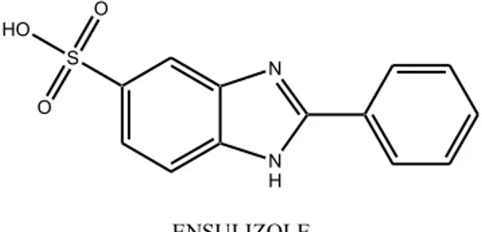
ü Ensulizole (fig 2.5) or 2-Phenylbenzimidazole-5-sulfonic acid (PBSA) is widely used in sunscreen formulations and because of its strong absorption in the UVB region within 290 nm and 320 nm, it is included in the list of authorized filters in Europe,30 USA31 and Australia.32 PBSA is a water soluble filter, this makes it desirable for the use in cosmetic formulation and if used in association with lipophilic filters achieves a synergic increase of the sun protection factor. It is considered efficient to prevent erythema and safe, because few events are reported of skin irritation, sensitization phototoxicity or photoallergy.38 Even though PBSA protects skin cells from UVB radiation and is considered photostable, studies have assessed that it can generate reactive oxygen species and cause photoinduced damage to DNA in vitro. PBSA was found to induce the production of singlet oxygen, as demonstrated by the generation of the diagnostic compound 4,8-dihydro-4-hydroxy-8-oxo-2′-deoxy-guanosine when it is irradiated with UVB or natural sunlight in oxygenated solution in the presence of 2′-deoxyguanosine, and to photoinduce the formation of alkali-labile cleavage sites in both single and double-stranded DNA. Although no phototoxic effects have yet been reported in vivo for PBSA, its proven capacity to generate singlet oxygen upon UV irradiation could be a threat of oxidative damage to adjacent skin tissue and to cell membranes, but nuclear DNA molecules would be at risk only if PBSA was able to enter the cells.40,41
Figure 2.5 Ensulizole
ü Camphor derivatives have a high molar extinction coefficient (> 20.000) and absorb in the UVB range within 290-300nm, they are considered rather photostable, because upon irradiation they undergo cis-trans photoisomerisation but it is reversible and the two isomer have similar spectra, then photoprotective ability doesn’t change significantly.42 In Europe six camphor derivatives are approved for the use in sunscreen formulation, instead Food and Drug administration has approved only Ecamsule (UVA absorber) and Enzacamene (4-methylbenzylidene camphor) is waiting for approval.30,31
N N H S O O HO 2-Phenylbenzimidazole-5-sulfonic acid ENSULIZOLE
Figure 2.6 Enzacamene
2.2.2 Organic UVA filters
UVA filters essentially absorb wavelengths between 320 and 400 nm; most of commercially available filters provide excellent protection against UVB but they are ineffective against UVA and there aren’t on market as many molecules effective against UVA; moreover usually these filters absorb in UVA II region (320-340 nm) and only few molecules provide protection in the UVA I range (340-400 nm).4 Within UVA filters are
included different classes of molecules.
ü Benzophenones (fig. 2.7) are aromatic ketones with a relative broad absorption profile that goes from 270 to 350 nm, hence they are active against UVB and UVA II rays. There are three benzophenones: Oxybenzone, Sulisobenzone, and Dioxybenzone, Oxybenzone mainly offers protection toward UVA II rays, it is the most commonly used although it presents some drawbacks as high incidence of contact photoallergic dermatitis, systemic absorption and it is also photounstable, initiating free radical production upon UV exposure.31,43
Figure 2.7 Benzophenones
ü Anthranilates or ortho-aminobenzoates are among the oldest available UVA filters, they may absorb up to about 350 nm.44 The absorption spectrum in the UVA region is
principally due to the presence of two functional groups in ortho, which can easily
O ENZACAMENE 4-Methylbenzylidene camphor O OH O OXYBENZONE SULISOBENZONE O OH O OH DIOXYBENZONE
(2-Hydroxy-4-methoxy-phenyl)-phenylmethanone 5-Benzoyl-4-hydroxy-2-methoxy-benzenesulfonic acid
S O O HO O O OH (2-Hydroxy-4-methoxy-phenyl)-(2-hydroxy-phenyl)-methanone
delocalize the electrons, but at the same time the ortho disubstitution is responsible of the low molar extinction coefficient of these filters. They are considered stable and safe compounds and in cosmetic formulations don’t exhibit significant solvent shift effects. The filter most commonly used of this category is Meradimate (Methyl anthranilate), its lambda maximum is 340 nm in ethanol and it is mainly a UVA II filter.45
NH2 O O MERADIMATE 2-Amino-benzoic acid 2-isopropyl-5-methyl-cyclohexyl ester Figure 2.8 Meradimate
ü Dibenzoylmethanes or substituted diketones are a relatively new class of UV filters; only one molecule of this group, Avobenzone (butyl methoxydibenzoylmethane) is permitted for use; in Europe was permitted also 4-isopropyldibenzoylmethane but in 1993 was withdrawn from the market because of high associated incidence of contact and photocontact dermatitis.44 Avobenzone exhibits absorption properties resulting
from keto-enol tautomerism, the two structural isomers have their own maximum absorption peak; the keto form absorbs in UVC range from 260 to 280 nm while the enol form absorbs in UVA range within 310 and 400 nm, with peak absorption around 360 nm. The relative amounts of the isomers are solvent dependent but usually in solution and in sunscreen formulations Avobenzone exists predominantly in the enol form and then it can provide good protection against UVA mainly in UVA I range. Unfortunately this filter is photounstable, upon irradiation photoisomerisation occurs from the enol to keto form with consequently absorption loss46; in one hour of sun exposure it undergoes 50-60%.photodegradation37 Avobenzone is also able to generate free radicals after irradiation and in vitro studies proved that it can cause DNA strand breaks and oxidative protein damage.46 As said above, the photoinstability of Avobenzone can affect the stability of the others filters present in the formulation leading to complete loss of activity of sunscreen formulation; as a consequence, greatly efforts have been made to stabilize this compound and on market there are stabilized
formulation where Avobenzone is formulated with other sunscreen ingredients such as Octocrylene and Oxybenzone with diethylhexyl 2,6- naphthalate ( non-filter).38
O O O AVOBENZONE 1-(4-tert-Butyl-phenyl)-3-(4-methoxy-phenyl)-propane-1,3-dione Figure 2.9 Avobenzone
ü Ecamsule (terephthalylidene dicamphor sulphoic acid) is a camphor derivative, a broad-spectrum UVA filter with an absorption profile ranging from 290 and 390 nm and peak absorption at 345 nm. It is photostable and has low systemic absorption, less then 1% of the compound passes through the horny layer. In keratinocytes irradiated with UV rays it prevents DNA breakage, pyrimidine dimer formation and p53 protein expression compared to unprotected cells and in clinical studies, Ecamsule have demonstrated to prevent photodermatoses, to block UV induced pigmentation and immunosuppression and to preserve skin elasticity slowing down photoaging.47
Figure 2.10 Ecamsule
2.2.3 Organic broad-spectrum UV filters
An ideal UV filter should provide a widespread protection against damaging UV rays, however only few of available molecules are able to filter effectively both UVA and UVB rays. These filters are the new generation of UV absorbers and are permitted in Europe and Australia, while in the USA are waiting for approval from the FDA via the Time and Extent Application (TEA) process.
HO3S O O SO3H ECAMSULE {3-[4-(7,7-Dimethyl-3-oxo-4-sulfomethyl-bicyclo[2.2.1] hept-2-ylidenemethyl)-benzylidene]-7,7-dimethy l-2-oxo-bicyclo[2.2.1]hept-1-yl}-methanesulfonic acid
ü Methylene-bis-benzotriazolyl tetramethylbutylphenol, known under the trade name of Tinosorb M (Ciba Specialty Chemicals, Basel, Switzerland), is a broad-spectrum UV filter with two absorption peaks at 303 nm and 360 nm. As a result of its molecular structure, that facilitates energy dissipation by intramolecular heat transfer and vibrational relaxation, it is a photostable molecule and helps also to stabilize other UV filters such as Avobenzone. The large size of the molecules minimizes the skin penetration and systemic absorption. It protects from UV rays by absorption, scattering and reflection because, although it is an organic filter, it behaves also like an inorganic one due to the micronized particles that are dispersed in the aqueous phase of sunscreen emulsions.36,38,48
Figure 2.11 Tinosorb M
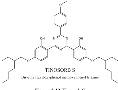
ü Bis-ethylhexyloxyphenol methoxyphenyl triazine or Tinosorb S (Ciba Specialty Chemicals, Basel, Switzerland) is an oil-soluble filter with an absorption spectrum that goes from 280 to 380 nm and two absorption peaks at 310 nm and 343 nm. By the presence of two hydroxyl groups in ortho position, which facilitate the return of the molecule to ground stable state by rapid energy release, Tinosorb S is endowed with great stability and then, like Tinosorb M, is used to stabilize other filters.36,38
N N N OH N N N OH TINOSORB M Methylene-bis-benzotriazolyl tetramethylbutylphenol
O N N N OH OH O O TINOSORB S
Bis-ethylhexyloxyphenol methoxyphenyl triazine
Figure 2.12 Tinosorb S
ü Drometriazole trisiloxane (Mexoryl XL, L’Oréal) belongs to the class of hydroxybenzotriazoles, its absorption spectrum covers both UVB and UVA rays with an absorption peak in the UVB range at 303 nm and the other in UVA range at 344 nm. Two structural groups constitute this molecule: the hydroxyphenylbenzotriazole group that provides wide range UV absorption and siloxane chain, which renders the molecule lipophilic. It is a photostable filter and rarely causes intollerances.38,48
N N N OH Si O Si O Si MEXORYL XL Drometrizole trisiloxane Figure 2.13 Mexoryl XL
2.3 Evaluation of sunscreen formulations efficacy
An international standard to measure efficacy of sunscreen is the Sun Protection Factor
(SPF) that is the value for a product, determined under solar simulated radiation, defined
as the ratio of the Minimal Erythemal Dose (MED) on product protected skin to the Minimal Erythemal Dose on unprotected skin of the same subject. MED is defined as the lowest ultraviolet (UV) dose that produces the first perceptible unambiguous erythema with defined borders appearing over most of the field of UV exposure, 16 to 24 hours after
UV exposure. The amount of test product applied to the skin shall be 2 mg/cm2 before
spreading.49
Since UVB is approximately 1000 times more erythemogenic then UVA, SPF value gives information on protection towards UVB and UVA II (320-340 nm) but not regarding UVA I (340-400 nm) and therefore SPF is of poor utility in understanding the UVA protection of a sunscreen. Because also UVA rays play a significant role in cellular damaging, it is important to establish, not only SPF value of a sunscreen formulation but, also the UVA protection to ensure a wide defence toward all UV radiation. Commonly used in vivo methods are IPD (immediate pigment darkening) and PPD (persistent pigment darkening), instead, for in vitro determination it is used the critical wavelength.
The IPD response occurs during UVA exposure, appears as a transient gray-brown pigmentation and fades within few minutes after the exposure is completed. The threshold dose for the IPD response is used with and without sunscreen protection to assess the UVA protection index; since pigmentation develops relative early after UVA exposure the test response is immediate but pigmentation disappears rapidly after irradiation leading to evaluation errors, along with wide individual variability response.50
PPD test is more used than IPD to verify UVA protection, it is also included in European
Commission Recommendation to assess UVA protection. PPD is a skin response linearly dependent on the amount of UVA that enters the epidermis and the response is equally sensitive throughout the UVA range. The UVA protection factor of a product is calculated on the Minimal Persistent pigment darkening Dose of protected skin (MPDp) divided by that of unprotected skin (MPDu); MPDu and MPDp are defined as the quantity of radiant energy required to produce the first unambiguous pigmented reaction.
The test product is applied in amount of 2mg/cm2, as for SPF test, and the UVA dose required to induce minimal pigmentation (MPD) is greater than 10 J/cm2 (approximately 40 minutes of midday summer sunlight), thus also the stability of sunscreens is challenged during this test.51a,b
SPF =
MED
product protected skinMED
unprotected skinUVA protection factor = MPDp MPDu
The critical wavelength method is an in vitro test based on the absorption spectrum of a sunscreen product, measured applying the formulation on a substrate; polymethylmethacrylate plates are a satisfactory substrate for this method because they are UVR-transparent, non-fluorescent, photostable and inert to all potential sunscreen formulation ingredients. The critical wavelength (λc) value of a product is defined as the
wavelength at which the integral of the spectral absorbance curve reaches 90% of the area under the curve from 290 to 400 nm. It is important to note that the critical wavelength value is based on the inherent shape of the absorbance curve, not on its peak absorbance, therefore is independent by application thickness of the tested sunscreen. The products reaching a critical wavelength of 370 nm or greater are considered broad-spectrum sunscreens.52,53
The absorption spectrum measurement by means of UV spectrophotometer is also used to calculate the SPF in vitro.
2.3 Regulatory and labelling
Before became available on the market, sunscreen formulations are tested to determine the filtering capacity and then properly labelled, trying to give clear information on the product efficacy at the consumer; the competent authorities for UV filters define effectiveness tests and labelling, these rule may vary among countries. In USA the Food and Drug Administration (FDA) have recently published the latest guideline (June 2011) for testing and labelling UV filters, where it is given much attention to testing and labelling of UVA protection. In the past there were limited requirement and guideline for assessing UVA protection, and if a sunscreen formulation contained one or more UVA filter it was labelled as “broad spectrum”. In the current FDA rules, instead to measure UVA protection by means of persistent pigment darkening test, the in vitro critical wavelength is adopted as pass/fail test to evaluate the UVA or broad-spectrum protection. To pass the broad spectrum test the amount of UVA protection must increase as the SPF value and only products with a critical wavelength of 370 nm or greater can be labelled as “broad spectrum”. Only sunscreen products that pass the broad-spectrum test and have SPF of 15 or higher can include the statement: “decreases the risk of skin cancer and early skin aging caused by the sun.” Both in USA and in Europe the SPF is estimated by means of an in vivo method; European commission recommends an in vivo method, the persistent
pigment darkening test, also to assess the UVA protection factor in addition to critical wavelength method. In Europe a sunscreen product, to satisfy the minimum efficacy requirements, should provide a minimum UVB and UVA protection and when the SPF increase also the UVA protection should increase; the ratio of UVA protection, measured by persistent pigment darkening test, should be at least 1/3 of the SPF; moreover to ensure a broad protection it is recommended a critical wavelength at least of 370 nm. In USA it is not required in vivo tests method to evaluate the UVA protection, as FDA have identified disadvantages in the persistent pigment darkening test such are: the skin darkening, measured in this test, is mainly due to UVA II (320-340 nm), human subjects are exposed to high dose of UVA radiation, it is expensive and time consuming, exposure to UVA radiation alone does not occur in nature, the method is poor reproducible and also the interpretation of pigmentation can lead to valuation errors.
Regarding labelling, the SPF number have been reduced in recent years to facilitate the comparison between different products, in USA values are expressed in multiples of 5 and about UVA protection, FDA recommends that products, providing UVB and UVA protection, are labelled as broad spectrum and no mention of the word UVA is allowed. In Europe the sunscreens are also divided within categories of protection according to SPF (see Table 2.1) and to attest the UVA protection all sunscreens have to display a simple logo: the letters “UVA” printed in a circular shape.54,55,56
2.4 Sunscreens efficacy and safety
Sunscreens provide effective defence by decreasing the amount of UV radiation to which biomolecules are exposed and therefore preventing both acute and chronic damages due to UV rays. The first visible benefit is the prevention of sunburns but they are useful also to prevent more detrimental consequences; indeed in vitro and in vivo studies have demonstrated that sunscreens protect against UV-induced photoaging, immunosuppression and mutations and then decrease the risk of developing melanoma and non-melanoma skin cancer.57
The first step in developing a new sunscreen molecule is to assess the UV filtering capacity and if the molecule has a good absorption profile is then evaluated for the photostability; these assays are performed by means of instrumental analyses (UV spectrophotometer, solar simulator and HPLC analyses). When a molecule display interesting filtering profile and photostability, before becoming available on the market, it undergoes safety and efficacy testing that are very rigorous and comparable to those of dermatological drugs. The safety dossier for a new UV filter includes in vitro as well as in vivo investigation of their potential to produce local toxicity, such are irritation, sensitization, photo-toxicity, photo-genotoxicity, as well as systemic toxicity, such as long-term toxicity, reproductive toxicity, carcinogenicity and photo-carcinogenicity. The first level of safety evaluation consists exclusively of in vitro tests (i.e. MTT test), in order to exclude cytotoxicity on human and/or murine fibroblasts or keratinocytes and then, if they are non cytotoxic, genotoxicity assay, photo-toxicity and photo-genotoxicity challenges, irradiating the cell in presence of the new substance are performed. A new sunscreen should not only be devoid of genotoxic activity, but also demonstrate its potential to protect cells from the genotoxic activity of UV radiation. Another aspect to be verified is the percutaneous absorption of the filtering molecule, because it should remain on the surface of the skin, where it is most effective. Ideally, a sunscreen should impregnate the stratum corneum creating a barrier against UV radiation, but not penetrate into the underlying tissue; UV filters that deeply penetrate the skin are of little value, since they would leave the skin unprotected. Penetration test are performed previously in vitro by means of diffusion cells (Franz cells) using skin of pig, human reconstructed epidermis or human epidermis deriving from surgical interventions. After in vitro studies, the product is tested on animals to determine NOAEL (Non Observable Adverse Effects Levels) for oral or dermal administration in sub-chronic or chronic toxicity studies. If, after these tests, a product is considered
harmless it goes to safety and efficacy trials on humans; these investigations includes confirming studies for absence of skin irritation, photo-irritation, skin sensitization, and photosensitization. Also trials for human systemic exposure dose are required, after topical application of an ultraviolet filter in a typical sunscreen formulation.58,59
Although sunscreens available on the market have passed the above-mentioned clinical trials, it doesn’t mean that they are completely free of adverse effects or relating problems. They may induce adverse effects such as irritant, allergic contact reactions, photoallergy, and phototoxic effects; contact and photocontact sensitivity to sunscreens has been reviewed for filters such as PABA, padimate O, enzacamene, octinoxate, and ensulizole. Maybe the most important problem to resolve, regarding sunscreen products, is the photostability; during UV exposure some of these molecules may change spectral performance or act as photo-oxidants via generation of free radicals and reactive oxygen species or may degrade producing toxic by-products. Moreover photounstable products give a false sense of safety, because photoprotection is guaranteed only when the UV filters remain stable throughout the entire period of exposure to sunlight, but in labelling the sunscreens there are no informations regarding the photostability of the product. Some filtering molecule currently used suffers of photostability problems that can be overcome adding to the formulation photostable sunscreens or stabilizing agents.
Another undesirable effects is the sunscreens penetration in the underlying tissues, since they can reach the circulatory system.60,61
Sunscreen in some cases can display same adverse effects but they offer undeniable protection against dangerous UV rays and the benefit or potential risk in using UV filtering molecules is weighted against the hazard of skin exposure to a carcinogen.
3. Free radicals and antioxidants
The skin, acting as a barrier toward the environment, is the most exposed organ to external agents, including ulraviolet rays. As above mentioned, detrimental effects of UV radiation is also due to production of reactive oxygen species, which are responsible of early ageing, inflammatory disorders, immunosuppression and skin cancers; for these reason free radicals and antioxidants are significant topics in photoprotection.
3.1 Free radical and other oxidant species
Free radicals are very reactive species, capable of independent existence that contain one or more unpaired electrons in the outermost orbital; the energetic situation is responsible for highly instability and short half-life of these substances. To gain stability, free radicals react with surrounding molecules subtracting electrons to form an electrons pair; in this way also the injured molecule becomes a free radical beginning a chain reaction. Usually radical reactions can be divided into three processes; the first is initiation, when occurs radical formation; the second is propagation, with increase of radicals production because of chain reaction; the last one is termination, the radical number decrease because two radicals combine or dismutation occurs with following formation of less reactive species. Human organism is subjected to the action of different kinds of oxidant species known as ROS (Reactive Oxygen Species) an RNS (Reactive Nitrogen Species), which comprise radicals and non-radical species within there are: superoxide anion radical (O2-·), singlet
oxygen (1O
2), hydroxyl radical (OH·), hydrogen peroxide (H2O2), lipid peroxyl radical
(LOO·), nitric oxide radical (NO·), nitrogen dioxide radical (NO2·). Under normal
condition, these species are present in small quantity during cellular activity, such as cellular respiration, apoptosis and immune response by macrophages and neutrophils to kill pathogens; unfortunately, in same situations take place an imbalance because of
overproduction of reactive species. Oxidative stress in human body can be enhanced by pathologies or external factors, among which there are:
ü Pathologies as rheumatoid arthritis, cardiovascular diseases, neurodegenerative diseases (Parkinson and Alzheimer) and inflammation in general;
ü Ischemia and reperfusion injury;
ü Excess of transition metals, as iron and copper that promote the production of hydroxyl radical;
ü UV rays and X rays, because of photooxidation; ü Drugs;
ü Smoke;
ü Alcohol abuse;
ü Environmental pollution;
ü Intense physical exercise, because increases oxygen consumption ; ü Diet too rich in proteins or saturated fats.62,63
3.2 Cellular targets of oxidative stress
At the base of free radicals damages there are biomolecules disruption; the essential cellular constituents impaired are nucleic acids, proteins, lipids and carbohydrates.
3.2.1 Lipids
Oxidative action regarding lipids can initiate a radical chain reaction, the lipoperoxidation that lead mainly to destruction of polyunsaturated fatty acids, which are components also of phospholipids of cellular membrane. Lipoperoxidation proceeds through the three steps; a hydroxyl radical initiate the process taking a hydrogen atom from a saturated carbon of the alchylic chain of the fatty acid, this becomes a radical too (L) and stabilize its structure through formation of conjugated diene, which are within the first detectable product of lipid peroxidation. In aerobic condition a fatty acid with an unpaired electron reacts with an oxygen molecules to generate a lipoperoxyl radical (LOO), a very reactive species that can undergo two different kinds of reaction.64 For molecules like arachidonic
and eicosapentanoic acid, peroxylic group can do cyclization producing a cyclic lipoperoxyde that by fragmentation gives aliphatic chain with two carbonyl groups,
can react with free amine moieties of proteins, phospholipids or nucleic acid forming covalent bonds as Schiff bases; crosslinks proteins-MDA-phospholipid, protein-MDA-protein or phospholipid-MDA-phospholipid cause loss of membrane fluidity.66
Another kind of reaction occurs when lipoperoxyl radical (LOO) takes a hydrogen atom from close fatty acid, producing a hydroperoxide lipid and promoting lipid peroxidation. Aldehydic products, originating from lipid peroxidation, can also react with functional groups of the proteins, as sulfhydryl moieties causing break of peptidic bonds or formation of disulphide intramolecular bonds; in this way can be inactivated proteins essential for the cell life.
In the last phase of lipoperoxidation take place the formation of stable compounds because of reaction between two radical species or by means of chain breaking substances; these events terminate the radical chain reaction.
3.2.2 Nucleic acids
About cellular injuries caused by reactive oxygen species, damages to DNA are the most harmful. Almost all altered molecules of DNA are replaced and repaired, however this safeguard system can fail and if modified DNA molecules were not corrected, the mutation could be transmitted during replication to daughter cells. Most commonly oxidative alterations of DNA are related to oxygen inclusion in double bond of DNA bases and sugar removing from bases, intermolecular DNA-DNA or DNA-protein binding.67 Pyrimidines (cytosine and thymine) are most sensitive than purines and can undergo saturation, ring open and therefore loss of aromaticity and planarity with DNA filaments distortion; photocatalized reactions of thymine produce thymine dimers. These DNA transformations are responsible for mutagenesis68, carcinogenesis69 and play a role also in aging,70 diabetes mellitus,71 inflammatory diseases and liver diseases72.
3.2.3 Proteins
Damages produced by free radicals to proteins can be distinguish in reversible and irreversible; among reversible there are oxidation of thiolic groups and of methionine, whereas between irreversible there are ring’s break of histidine and tryptophan and peptide bond hydrolyzation in presence of proline; the last event affects particularly the collagen because it is rich in proline and hydroxyproline. Oxidation of sulfhydryl moieties of the cysteine to thiyl radical can lead to dimerization or oxidation to sulphide dioxide group;