Materials
33
2. Materials and Methods
2.1. Materials
- AG, pharmaceutical grade, was supplied by Opocrin S.p.A..
- MUC1 from submaxillary glands, MUC2 and MUC3 from porcine stomach, 1,4-butanediol
diglycidyl ether, 3-aminopropyltrimethoxysilane, anthrone, bovine serum albumine, chymotrypsinogen, cytochrome c, dibutyltin dilaurate, ethanolamine, ferritin, fluorescein isothiocyanate, glycine, ligroin, methyl sulfoxide, sodium borohydride, thiourea and thyroglobulin were purchased from Sigma-Aldrich.
- Ethanol 95%, hydrochloric acid 36-38%, potassium phosphate monobasic, sodium chloride,
sodium hydroxide, sulphuric acid, toluene and TRIS were purchased from JT Baker.
- Acrylamide 99.9% and N,N’-methylene-bis-acrylamide were purchased from BioRad
Laboratories.
- Concentration membranes Amicon YM10 and YM30 were purchased from Millipore.
- Glass fiber filters (GF/F, 4 Ø) and diethylaminoethyl cellulose (DE52, DEAE) were
purchased from Whatman.
- Polystyrene plates and polyvinyl chloride thick layers were purchased from Wako.
- Dialysis polyvinylidene fluoride (PVDF) tubing was purchased from Spectrum.
- Dialysis cellulose tubing was purchased from Medicell International.
- Sephacryl S300 was purchased from GE Healtcare.
- Polylysine coated glass was purchased from Thermo Scientific.
- Dowex 50WX2 and Dowex 88 were purchased from Dow Chemical Company.
- Mouse monoclonal MUC1 antibody [HMFG1 (aka 1.10F3)] and mouse monoclonal MUC3
antibody (M3.1) were purchased from Abcam.
- Micro BCATM Protein Assay Kit was purchased from Pierce.
Methods
34
2.2. Methods
2.2.1. Determination of protein concentration
The protein concentration was measured with a Micro BCA Protein Assay kit. This method combines the reduction of Cu2+ to Cu1+ by protein in an alkaline medium with the selective colorimetric detection of the cuprous cation using a reagent containing bicinchoninic acid (BCA). The reaction product of the assay is purple-coloured and can be read at 562 nm. It is formed by
the chelation of two molecules of BCA with one cuprous ion.The test was performed by adding
500 µl of the protein sample to 500µl of the kit’s working solution obtained mixing together the three Micro BCA Reagents following the kit information. The mixture was incubated for 1 hour at 60°C and read at 562 nm in a spectrophotometers DU7 (Beckman). The amount of protein in the sample was evaluated using a calibration curve obtained with a series of standards of known bovine serum albumin concentrations ranging between 0–4,000 µg/ml.
Methods
35
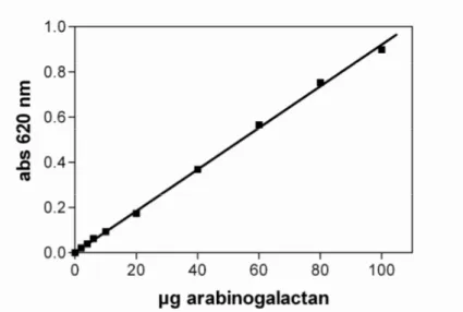
2.2.2. Determination of carbohydrates by anthrone method
Carbohydrates content was evaluated spectrophotometrically upon reaction with the anthrone reagent. Sugars under acidic conditions are dehydrated to hydroxymethyl furfurals. These compounds forms with anthrone a blue-green coloured product with an absorption maximum at 620 nm. The test was performed mixing 250 µl of sample with 1250 µl of anthrone reagent (70%
H2SO4, 2,5 mM anthrone, 130 mM thiourea) and then boiled 15 minutes until the reaction is
completed. The solution is then allowed to cool and the absorbance is measured at 620 nm. A linear relationship exist between the absorbance and the amount of sugar present in the sample. The amount of sugars in the sample was evaluated using a calibration curve obtained with a series of standards of known arabinogalactan concentration ranging between 0–66,667 µg/ml.
Fig. 2.2. Example of calibration curve for determination of carbohydrate content.
2.2.3. Preparation of fluorescent labelled AG
Fluorescein isothiocyanate-labelled AG (AG-FITC) was synthesized according to Burgalassi et al. (Burgalassi et al., 2007); AG was dissolved in methyl sulfoxide (0,1 g/ml), the isothiocyanatefluorescein (60 mM) was added, followed by dibutyltin dilaurate (3 mM), and the mixture was heated for 2 h at 95°C. After several precipitations in ethanol to remove free dye, FITC-AG was dried at 80°C. Fluorescent signal was detected by spectrofluorometer FP 6500 (Jasco), with an excitation of 494 nm and an emission of 514 nm.
Methods
36
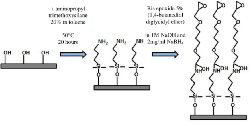
2.2.4. Glass fiber filters modification
Glass fiber filters were modified by reaction with 20% 3-aminopropyltrimethoxysilane in dehydrated toluene at 50°C for 20 h. Glass fiber filters were rinsed 4 times with ligroin and dried. The silanized filters were immersed in butanediol diglycidyl ether solution (5%
1,4-butanediol diglycidyl ether, 1M NaBH4, 1M NaOH, 10 mM sodium phosphate buffer pH 7) for 6
h at 37°C. Filters were rinsed 4 times with ethanol and dried.
Fig. 2.3. Glass fiber filters modification.
2.2.5. ELISA test
Elisa test was performed as described in “Methods in Molecular Biology vol. 125, Glycoprotein
Methods and Protocols, The Mucins, A.P. Corfield,Humana Press”.
Bis epoxide 5% (1,4-butanediol diglycidyl ether) in 1M NaOH and 2mg/ml NaBH4 + aminopropyl trimethoxysilane 20% in toluene 50°C 20 hours