UNIVERSITY OF SASSARI
PhD School in Biomolecular and Biotechnological Sciences
Curriculum in Physiology, Biochemistry and Molecular Biology
XXVIII cycle
Epigenetic modifications associated
with Amyotrophic Lateral Sclerosis
(ALS) onset and progression
Supervisor: Prof. Claudia Crosio
PhD School Director: Prof. Leonardo Sechi
ABSTRACT ... 4
1. INTRODUCTION ... 5
1.1 AMYOTHOPHIC LATERAL SCLEROSIS (ALS) ... 6
1.1.1 ALS etiology: a multifactorial perspective ... 7
1.1.2 Genetic factors... 8
1.1.2.1 ALS1: Superoxide dismutase 1 (SOD1) ... 9
1.1.2.2 ALS6: FUS ... 10
1.1.2.3 ALS10: TDP43 ... 12
1.1.3 Environmental factors ... 14
1.1.4 ALS experimental models ... 16
1.1.5 Pathogenic mechanisms ... 18
1.2 EPIGENETICS ...21
1.2.1 The epigenetic machinery ... 21
1.2.2 Covalent histone-tails post-translational modifications ... 24
1.2.2.1 Histone acetylation ... 27
1.2.2.2 Histone methylation ... 29
1.2.2.3 Histone phosphorylation ... 30
1.2.3 DNA methylation ... 31
1.3 EPIGENETICS AND ALS ...34
1.3.1 Acetylation and deacetylation of histonic and non histonic proteins ... 34
1.3.1.1 Targets ... 34
1.3.1.2 Histone acetyltransferase ... 35
1.3.1.3 Histone deacetylases ... 35
1.3.1.4 HDAC inhibitors and other ALS therapy approaches ... 36
1.3.2 Methylation and demethylation ... 38
1.3.3 Histone phosphorylation ... 39
1.3.4 DNA methylation ... 39
2. AIMS OF THE WORK ... 40
3. MATERIALS AND METHODS ... 42
4. RESULTS... 52
4.1 ANIMAL AND CELLULAR MODELS FOR ALS ...53
4.1.1 Characterization of ALS cellular models ... 54
4.2 ANALYSIS OF HISTONE POST-TRANSLATIONAL MODIFICATIONS H3-K14ac-S10ph, H3K14me2 AND H3K9me3 ...57
4.2.1 SOD1 induce an in vitro decrease in transcriptional activation markers and an increase in repression markers ... 58
4.2.2 SOD1 reduction of H3-14Ac-S10Ph marker is in proportion with the time of infection ... 59 4.2.3 SOD1 induce a decrease in transcriptional activation markers in SOD1 G93A spinal cord mice
4.2.4 FUS and TDP43 overexpression induce an increase in transcriptional repression marker ... 64
4.3 SOD1G93A OVER EXPRESSION INDUCES A GLOBAL INCREASE IN DNA METHYLATION ...67
4.4 TDP43 INTERACTS WITH HDAC1 ...68
4.4.1 TDP43WT interacts with HDAC1 in vitro ... 68
4.4.2 Pathological mutant TDP43M337V and TDP43A382T interact with HDAC1 in vitro ... 70
4.4.3 HDAC1 localizes in the nucleus and partly co-localizes with TDP43 ... 72
4.4.4 Development of TDP43 deletion mutants ... 75
4.4.5 RRM1 and RRM2 domains are both necessary to mediate TDP43-HDAC1 interaction ... 76
4.5 TDP43 DOESN’T ALTER HDAC1 ACTIVITY IN VITRO...78
4.6 TDP43 AND HDAC1 HAVE A SYNERGIC EFFECT IN DECREASING CELLS VITALITY ...79
4.6.1 Possible therapeutic effect of HDAC inhibitors ... 81
4.6.1.1 HDACi Sodyum butyrate and Trichostatin A reduce TDP43 induced mortality ... 81
5. DISCUSSION ... 83
5.1 ALS AND EPIGENETIC ALTERATIONS ...84
5.2 TDP43 AND HDAC1: POSSIBLE FUNCTIONAL ROLE OF THEIR INTERACTION ...85
5.3 HDAC INHIBITORS IMPROVE VITALITY IN ALS CELLULAR MODELS ...87
5.4 CONCLUSIONS ...88
ABSTRACT
In the recent years a growing amount of evidence indicates the functional significance of epigenetics in various aspects of neural function and dysfunction. Alterations in chromatin structure resulting in long lasting changes in gene expression have been associated to many different aspects of neuronal biology, including neurodegenerative disorders such Amyotrophic Lateral Sclerosis (ALS). ALS is predominantly sporadic and environmental triggers may be involved in disease initiation. In this respect, the epigenome can provide the key to transform the genetic information into phenotype. Alterations in the epigenetic machinery and/or epigenetic modifications can be considered a readout of disease onset and progression and an ideal target for therapeutic interventions. My PhD thesis is, indeed, focused on different aspect of epigenetic modifications in ALS cellular and animal models. By in vitro and in vivo assays I demonstrate that some epigenetic markers, linked to transcriptional activation or repression, are selectively altered in cellular and animal model of ALS. Moreover I observed a physical interaction between the ALS-causing gene TDP43 and the histone deacetylases 1 (HDAC1). Finally, I demonstrate that perturbation of HDAC1 level or activity affects TDP-43-induced cell damage. Although preliminary, these data can be extended by testing more specific HDAC1 inhibitors which can be chemically optimized by computational biology approaches.
1.1 AMYOTHOPHIC LATERAL SCLEROSIS (ALS)
Amyotrophic lateral sclerosis (ALS) is a neurodegenerative disorder characterized by the death of motor neurons in the brain, brainstem, and spinal cord, resulting in fatal paralysis. ALS usually begins with asymmetric involvement of the muscles of one or more limbs, speech or deglutition in middle adult life. The involvement appears to spread in an anatomically contiguous manner and eventually becomes bilateral and symmetrical, progressing to paralysis and death. Mean survival is 3 years after the onset of symptoms (Chio, Logroscino et al. 2009). Classical clinical classification proposed three phenotypic forms of familial ALS, each inherited as an autosomal dominant disorder. The first form they delineated is characterized by rapidly progressive loss of motor function with predominantly lower motor neuron manifestations and a course of less than 5 years. Pathologic changes are limited to the anterior horn cells and pyramidal tracts. The second form is clinically identical to the first, but at autopsy additional changes are found in the posterior columns, Clarke column, and spinocerebellar tracts. The third form is similar to the second except for a much longer survival, usually beyond 10 and often 20 years (Meyer, Schwan et al. 2005).
Figure 1. Motor neurons selectively affected in ALS. Degeneration of motor neurons in the motor
cortex leads to clinically apparent signs of upper motor neuron abnormalities. Degeneration of motor neurons in the brain stem and spinal cord causes muscle atrophy, weakness and fasciculation.
Anyway, more recently, clinicians prefer to analyze the complete phenotypic criteria and to formulate a diagnosis basing on continuous variations on the severity of the symptoms, without distinguishing in ALS forms (Rutter-Locher, Turner et al. 2016).
Al Chalabi and colleagues proposed a new classification method that combines the classical diagnostic approach based on El Escorial categories with the rich variety of phenotypic descriptions (Al-Chalabi, Hardiman et al. 2016).
Figure 2. Classification system for amyotrophic lateral sclerosis (Al-Chalabi, Hardiman et al. 2016)
Population based studies have established that the incidence of ALS in Europe is fairly uniform at 2,16 per 100 000 person-years (Logroscino, Traynor et al. 2010). Although ALS affects people worldwide, an exact incidence of this disease is not yet known. Men have a higher incidence of disease (3,0 per 100 000 person-years; 95% CI 2,8–3,3) than do women (2,4 per 100 000 person-years; 95% CI 2,2–2,6), although the incidence between men and women is about the same in familial disease.
1.1.1 ALS etiology: a multifactorial perspective
ALS etiology has been recognized to be multifactorial (Vucic and Kiernan 2009). It occurs as an apparently sporadic disease (sALS) in 90% of cases; while the residual10% of ALS cases are familial (fALS) and linked to a specific gene mutation
(Beleza-Meireles and Al-Chalabi 2009). In sALS, environmental factors may contribute to the pathology, i.e. lesions in frontotemporal lobes or exposition to some pesticides like organochlorine insecticides aldrin or toxaphene have been linked to the development of the disease (Kamel, Umbach et al. 2012).
1.1.2 Genetic factors
Approximately 10% of ALS is classified as familial, whereas the remaining 90% of the cases are considered sporadic, as they appear to occur randomly throughout the community.
Since now, 22 locus have been linked to the development of ALS. Among them, the most studied are those displayed in table 1.
Genes known to carry ALS-causing mutations
Percentage explained Putative protein function Gene Location Inheritance Familial ALS Sporadic ALS
TARDBP 1p36 AD 4 1 RNA metabolism
SQSTM1 5q35 AD 1 <1 Ubiquitination;
autophagy
C9ORF72 9p21 AD 40 7 DENN protein
VCP 9p13 AD 1 1 Proteasome;vesicle trafficking
OPTN 10p13 AR and AD <1 <1 Vesicle trafficking
FUS 16p11 AD and AR 4 1 RNA metabolism
PFN1 17p13 AD <1 <1 Cytoskeletal dynamics
SOD1 21q22 AD and AR 12 1–2 Superoxide metabolism
UBQLN2 Xp11 XD <1 <1 Proteasome
Others<1
Table1.Principal gene locus involved in ALS and relative mutations.
SOD1 is the first historically discovered ALS causing gene (Rosen, Siddique et al. 1993). Over than 200 point mutations have been reported on SOD1 gene. Nevertheless, SOD1 mutations account for the 12% of fALS and for the 1% of sALS.
Locus 16p11 and locus 1p36 encode respectively for FUS and TDP43 protein, both involved in RNA metabolism. Together they are linked with the 8% of all fALS.
In 2009 two research groups reported that a massive hexanucleotide repeat expansion in
C9ORF72 is the cause of chromosome 9p21–linked ALS and FTD
(DeJesus-Hernandez, Mackenzie et al. 2011, Renton, Majounie et al. 2011). This pathogenic expansion accounts for a remarkable percentage of both familial ALS (~40%) and
familial FTD (~25%). In addition, the repeat expansion has been found to account ~7% of apparently sporadic ALS cases in people of European ancestry, marking the first time that a genetic etiology has been identified for more than just the occasional sporadic case (Renton, Chio et al. 2014).
1.1.2.1 ALS1: Superoxide dismutase 1 (SOD1)
21q22 gene locus encodes for a 32 kD homodimeric protein called Cu/Zn SOD1. It is the major cytoplasmic antioxidant enzyme that metabolizes superoxide radicals to molecular oxygen and hydrogen peroxide, thus providing a defense against oxygen toxicity. It forms a β-barrel and contains an intramolecular disulfide bond and a binuclear Cu/Zn site in each subunit. This Cu/Zn site holds the copper and a zinc ion and is responsible for catalyzing the disproportion of superoxide to hydrogen peroxide and dioxygen (Niwa, Yamada et al. 2007).
Figure 3.Structure and function of SOD1.A) SOD1 gene position on chromosome 21. B)
Tridimensional structure of human SOD1. C) Active site of Cu/Zn SOD1. D) Mechanism cyclic oxidation-reduction catalyzed by SOD1.
Since the first description in 1993, more than 200 point mutations have been described, even if only a small percentage of them have been clearly linked to the pathology. In fALS linked to SOD1 mutations, the majority has a dominant inheritance, like G93A or H80R (Rosen, Siddique et al. 1993, Alexander, Traynor et al. 2002).
The pathological mechanism linked to SOD1 is complex. Since some mutations concern the metal binding residues at the active site, while others may concern correct folding or stability of the homodimer, the biochemical and biophysical properties of ALS-associated mutant SOD1 proteins are rather heterogeneous. In fact, studies on recombinant mutant SOD1 proteins have been proved that ALS-associated SOD1 mutations can be attributed at two mutant classes, the "wild-type like" (WTL) SOD1 mutants which retain the ability to bind copper and zinc and exhibit normal specific activity, indicate a native-like structure with only subtle changes to the backbone fold, in contrast the "metal-binding region" (MBR) SOD1 mutants that are deficient in copper and zinc and exhibit severe thermal destabilization and structural disorder of conserved loops near the metal-binding sites. G93A and H80R-SOD1 mutants belong respectively to the two classes of mutants described above (Tiwari and Hayward 2005).
To date, different hypothesis have been made to explain SOD1 toxicity. The aggregation hypothesis is particularly attractive because protein aggregates are frequently associated with neurodegenerative diseases such as ALS. Numerous studies revealed that SOD1 mutant is incline to misfolding and to form cytoplasmic aggregates (Rotunno and Bosco 2013). In turn, these aggregates could lead to cell death by sequestering other cytoplasmic proteins essential for neuronal survival, by clogging the ubiquitin/proteasome system, by chaperones depletion, or by disrupting mitochondria, cytoskeleton and/or axonal transport. Another hypothesis is that SOD1 misfolding induced by mutations would allow the access of abnormal substrates such as peroxynitrite to the catalytic site leading to the nitration of tyrosine residues (Beckman, Carson et al. 1993, Rotunno and Bosco 2013).
1.1.2.2 ALS6: FUS
Mutations in 16p11.2 locus encoding for FUS protein have been linked to a familiar form of ALS (Kwiatkowski, Bosco et al. 2009). FUS is a 75 kDa nuclear protein. It is ubiquitously expressed. FUS/TLS is a 526 amino acid protein encoded by 15 exons and
characterized by an N-terminal domain enriched in glutamine, glycine, serine and tyrosine residues (QGSY region), a glycine-rich region, an RRM domain, multiple arginine/glycine/glycine (RGG) repeats in an arginine and glycine-rich region and a C-terminal zinc finger motif. Most of the mutations are clustered in the glycine-rich region and in the extreme C-terminal part of the protein with evidence for mutations in each of the five arginine residues (Deng, Gao et al. 2014). Thirty mutations have now been reported in 4% of familial ALS and in rare sporadic patients with no apparent familial history. The inheritance pattern is dominant except for one recessive mutation (H517Q) found in a family of Cape Verdean origin. Most are missense mutations with a few exceptions (Kwiatkowski, Bosco et al. 2009).
Figure 4.Genomic and structural organization of human FUS gene and protein.FUS gene is encoded
by 15 exons that cover an 11.6 kb region on chromosome 16p11.2. QGSY rich: serine–tyrosine–glycine– glutamine rich domain; RRM: RNA recognition motif; ZnF: cysteine2/cysteine2 zinc finger motif; RGG rich: arginine–glycine–glycine rich domain.(Adapted from(Deng, Gao et al. 2014)
FUS functions are multiple and not completely understood. Many studies enlighten that FUS binds to single-strand and double-strand RNA; FUS seems to act at various levels in RNA metabolism, including transcription, splicing and translation (Prasad, Ouchida et al. 1994). In addition, FUS acts as a transcriptional regulator. FUS associates with products of RNA polymerase II transcription, forms complexes with hnRNPs, and represses RNA polymerase III transcription. Furthermore, FUS inhibits the acetyltransferase activities of CREB-binding protein (CBP) and p300 on cyclin D1 (CCND1) (Wang, Arai et al. 2008) and regulates the transcription factor nuclear factor kB (NF-kB) (Goransson, Andersson et al. 2009). FUS also engages in rapid nucleo-cytoplasmic shuttling, associates with actin-dependent motor protein myosin Va
(MyoVa) (Yoshimura, Fujii et al. 2006), and is a component of RNA granules that transport mRNAs (Belly, Moreau-Gachelin et al. 2005).
Many insights into FUS function come out studying FUS interactome with different experimental approach in a variety of models. FUS has been demonstrated to directly bind (i) to the U1-snRNP, reducing RNA processing, and SMN complexes activity; (Sun, Ling et al. 2015), (ii) to the C-terminal domain (CTD) of RNA polymerase II (RNA Pol II) in an RNA-dependent manner (Schwartz, Ebmeier et al. 2012), (iii) to the heterogeneous nuclear ribonucleoprotein (hnRNP) A1 and hnRNPA2B1 (Takanashi and Yamaguchi 2014), (iv) to histone deacetylase 1 (HDCA1) during DNA repair (Wang, Pan et al. 2013).
1.1.2.3 ALS10: TDP43
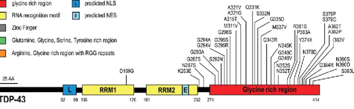
TDP-43 is a multifunctional protein involved in gene expression and regulation, including transcription, RNA splicing, transport, and translation. It is ubiquitously expressed, nuclear, 43 kDa. It is a multidomain protein: it has a N-terminal domain, with a NLS signal, two RRM domains (RRM1 and RRM2) and a glycine-rich domain, in the terminus of the protein. The majority of the mutations are localized in the C-terminal part of TDP43 (Sreedharan, Blair et al. 2008).
TDP-43 is also involved in the processing of small regulatory RNAs (micro RNAs) and in RNA maturation and splicing. TDP43 is a major component of the ubiquitin-positive neuronal inclusions that are the pathological hallmark of both ALS and frontotemporal dementia (FTD) (Neumann, Sampathu et al. 2006).
Mutations in TDP43 are responsible of 4–6% of familial and 0.7–2% of sporadic ALS. Although TDP43 mutations are found in ALS families across the globe, some regional variability does exist. For example, the A382T mutation of the protein is particularly frequent in Sardinia, reflecting the conserved nature of that island population combined with a historical founder effect (Chio, Borghero et al. 2011).
Mutations in TDP43 are mainly located with-in the C-terminal glycine-rich domain of the protein. Although the pathophysiological mechanisms by which TDP43 gene mutations result in neurodegeneration remain to be defined fully, emerging evidence suggests multiple mechanisms including gain of toxicity, loss of nuclear function, and the formation of large stress granules. Support for a toxic gain of function has been provided by studies in transgenic mouse models wherein increased expression of the
mutated TDP43 proteins leads to neurodegeneration through dysfunction of cellular organelles and proteins (Lee, Lee et al. 2011). The severity of cortical and spinal motor neuron degeneration appears to be proportional to TDP43 protein levels, suggesting a potential role for TDP-43 in regulating disease severity (Wils, Kleinberger et al. 2010). Alternatively, loss of nuclear TDP43 accompanied by accumulation of TDP43 aggregates in the cytoplasm has been well established in ALS patients, implying a potential role for a TDP43 loss of nuclear function mechanism in ALS pathogenesis (Lee, Lee et al. 2011). Of further relevance, TDP43 associates with cytoplasmic stress granules (McDonald, Aulas et al. 2011). Specifically, stress granules function to suppress mRNA translation temporarily and store pre-RNA complexes during periods of cellular stress, thereby safeguarding the coded RNA information from deleterious chemicals. Pathological TDP43 mutant protein appears to exhibit a greater propensity to associate with cytoplasmic stress granules and form larger stress granules with altered dynamics.
Figure 5.TDP43 mutations in ALS. Forty-four mutations have been identified in TDP43 in sporadic and
familial ALS patients, with most lying in the C-terminal glycine-rich region. All are missense mutations, except for the truncating mutation TDP43Y374X. Adapted from (Lagier-Tourenne, Polymenidou et al. 2010).
TDP43 interacts with a large amount of RNAs and proteins. For example, the glycine-rich C-terminus of TDP-43 has been shown to mediate interaction with several hnRNP proteins, specifically hnRNPs A1, A2/B1, C1/C2, and A3 (Buratti, Brindisi et al. 2005). TDP-43 interacts with nuclear and cytoplasmic proteins, and these interactions are often RNA-dependent. Freibaum and Chitta elaborated an interactome of TDP43, recognizing two classes of TDP43 binding protein: the first is a network of nuclear proteins that regulate RNA splicing and other aspects of nuclear RNA metabolism, and the second is a network of cytoplasmic proteins that regulate mRNA translation (Freibaum, Chitta et al. 2010). Moreover, they demonstrated that TDP43 pathological mutations M337V and
A315T do not alter the interaction. TDP43 interacts also with FUS (Ling, Albuquerque et al. 2010). TDP43 and FUS directly interact in a RNA/protein complex with HDAC6, regulating its expression levels (Kim, Shanware et al. 2010). TDP43 and HDAC6 interaction was demonstrated by Hebron and colleagues in 2013 (Hebron, Lonskaya et al. 2013).
Another interesting point to discuss is TDP43 changing functions when acetylated. As for others transcription factor, acetylation is one of the most important control mechanism. A well studied example is p53, whom acetylation has many important effects. It increases p53 protein stability, binding to low affinity promoters, association with other proteins, antiviral activities, and is required for its checkpoint responses to DNA damage and activated oncogenes (Reed and Quelle 2014). Since now, few is known about TDP43 acetylation. Cohen and colleagues demonstrated that TDP-43 acetylation impairs RNA binding and promotes accumulation of insoluble, hyper-phosphorylated TDP-43 species that largely resemble pathological inclusions in ALS and FTLD-TDP (Cohen, Hwang et al. 2015).
1.1.3 Environmental factors
In ALS onset and progression the interaction between genetic background and external factors is thought to play a major role (Bozzoni, Pansarasa et al. 2016). Exposure to heavy metals, such as lead, selenium, mercury, cadmium and iron, as a risk factor for ALS has long been studied, and the results produced are contradictory. Exposure to selenium has been largely studied and results indicate that there are in vitro evidences but not any statically significant association. Similar results emerged in studies about mercury and lead (Trojsi, Monsurro et al. 2013). Another factor that has recently been linked to sALS is the exposure to electromagnetic fields (EFMs), especially to low frequency EFMs. Despite interesting initial evidences, no any significant association has emerged in the latest studies (Vergara, Mezei et al. 2015). Exposure to a various spectrum of substances has been largely investigated. Cyanobacteria produce several cyanotoxins, divided into neurotropic (e.g. BMAA) and hepatotropic, such as cycasin, whose carcinogenic potential is well documented. The hypothesis that BMAA may have a role in neurodegenerative diseases was initially based on reports of elevated rates of ALS, Parkinson‟s disease and dementia in the island of Guam, where cycad seeds are
used to produce flour containing a remarkable amount of BMAA. Increased brain levels of BMAA were found in Guam ALS patients and similar BMAA levels were also found in the brains of ALS patient in Florida (Pablo, Banack et al. 2009). The latest in vivo studies confirm that BMAA can be incorporated into nerve cell proteins, causing misfolding, aggregates and cell death (Dunlop, Cox et al. 2013). BMAA role in causing worldwide sALS needs to be more detailed studied. Pesticides and precisely insecticides are the earlier studied fALS cause. Some of them, particularly organophosphate pesticides, can cause neurological damage, due to inhibition of acetylcholinesterase, the enzyme responsible for terminating the biological activity of acetylcholine. Moreover, most of these chemical compounds are known for their ability to induce oxidative stress, mitochondrial dysfunction, α-synuclein storage and neuronal loss.
Environmental toxins Effect
Cigarette smoke Increase the probability of developing ALS through inflammation, oxidative stress, and neurotoxicity by heavy metals contained in cigarettes.
Heavy metals Lead: greater tibia and blood Pb levels were found to be associated with
increased survival of ALS patients
Mercury: Diet organic mercury, through fish and derivates, and
occupational exposure have been shown to be linked to ALS onset.
Selenium: Se exposure may considerably influence SOD1 accumulation
into mitochondria, a somatic feature occurring in neurons during ALS pathogenesis.
Physical activity Several genes (i.e., ciliary neurotrophic factor, leukemia inhibitory factor, and vascular endothelial growth factor 2) related to exercise have been recognized as possible ALS risks factors.
Agricultural chemicals ALS risk is associated with use of organochlorine insecticides (e.g., dichloro-diphenyl-trichloroethane), pyrethroids, herbicides, and fumigants but not with other pesticide classes
Radiation/electromagnetic fields
Electromagnetic fields cause DNA strands to break in brain cells, leading to cell death and such reaction could be the reason for the association between electromagnetic fields and ALS risk.
Diet Consuming high level of glutamate and fat can have adverse effects on ALS patients while Omega 3 fatty acids, Vitamin E, and fiber can have defensive impact.
Table 2. Environmental factors linked to ALS risk and pathophysiology. Adapted from (Zarei, Carr et al. 2015).
This data emerges by the more elevated risk to develop ALS in farmers, employees of chemical industries, inhabitants in rural areas (McGuire, Garrison et al. 1997). A big amount of case-control studies confirm that pesticides exposure is a risk factor for sALS (Sutedja, Fischer et al. 2009). ALS owes its colloquial name to baseball player Lou Gehrig, who contracted ALS at the age of 27 in 1939. A cohort study conducted on 24000 soccer player Italian players evidences that there is a high prevalence of ALS, approximately 5 or 6 folds more than global population. Causes are unclear yet, but many were postulated. Football players may be exposed to various risk factors that could contribute to neurodegenerative processes, namely excessive physical activity, repeated head injuries, exposure to pesticides and dietary supplements or illegal substances.
1.1.4 ALS experimental models
In order to better understand the pathophysiological processes of the ALS onset, several model systems have been created over the years such as cell lines and transgenic animals, expressing different ALS-causative genes (SOD1, FUS and TDP43) in the wild type form or with the several pathological mutations. Among the mammalian animal and cellular models Table 3 summarize the most relevant to this thesis.
TARGETED
GENE DESCRIPTION PHENOTYPE (Y/N) NEURONAL DEGENERATION (Y/N) PROTEINOPATHY / AGGREGATES (Y/N) An ima l m od el
SOD1 At least three mutations: SOD1 G93A, G87R, G85R
G93A fALS mice: 20copies, hSOD1 promoter
Y; paralysis and
premature death Y; degeneration, motor neuron muscle denervation; implication of non-cell autonomous mechanisms;
inflammation
characterized by astroglial and microglial activation; muscle atrophy
Y: aggregated SOD1 can be detected with specific antibodies TDP-43 Overexpression of WT and mutated forms Y; Motor deficits but no paralysis Y; +/- MN loss, depending on the line. Overall mild effects
Not in all cases Sometimes Ub+, TDP43 nuclear and cytosol aggregates TDP-43 Conditional KO Weight loss and
age-dependent motor impairment
Y; MN degeneration N
FUS PrP-hFUS WT
overexpression Y; impairment, motor paralysis and death Y: MN loss Y Transgenic mutated FUS rats Motor axon degeneration, muscle denervation Y; Motor axon degeneration, muscle denervation Y Pharmacologi
cal model Treatment BMAA with Some phenotypes motor reported ? ? Ce ll ular M od els iPS cells (SOD1, TDP43, FUS C9ORF72) Co-cultures MN/astrocytes/mi croglial cells Y; cells
degeneration Y; used to evaluate non-cell autonomous processes by co-cultivating cells carrying or not-carrying the pathogenic mutation
Y
Primary MN Primary cultures with or w/o astrocytes Y; cells degeneration Y Y SH-SY5Y Human neuroblastoma cells Apoptosis and necrosis Y Y NSC-34 Motoneuronal cells Apoptosis and necrosis Y Y
1.1.5 Pathogenic mechanisms
Understanding ALS pathogenesis appears to be central for future development of diagnostic and therapeutic strategies in ALS. Pathogenesis of ALS is complex and many processes seem to contribute to the development of the disease. In vitro studies show that synaptosomes in neural tissue display a marked decrease in the maximal velocity of transport for high-affinity glutamate uptake in spinal cord, motor cortex, and somatosensory cortex compared to controls. The decrease in glutamate uptake was not observed in tissue from visual cortex, striatum, or hippocampus (Rothstein, Martin et al. 1992).Oxidative stress is another pathway lined to neurodegeneration in ALS. SODl mutations have been shown to enhance in vitro generation of hydroxyl radical from hydrogen peroxide. In addition to the dismutation of superoxide to hydrogen peroxide, wild-type CuZn-SOD1 can use hydrogen peroxide as a substrate to initiate a Fenton-like reaction with production in vitro of hydroxyl radical (Liu, Althaus et al. 1998). Several evidences report a link between protein misfolding and ALS. In particular, SOD1 mutations
induce conformational instability and misfolding of the SOD1 peptide, resulting in formation of intracellular aggregates that inhibit normal proteosomic function, disrupting axonal transport systems and vital cellular functions (Zetterstrom, Stewart et al. 2007). Recently, a functional study demonstrated that mutant ineffective Hsp104 impairs SOD1 or TDP43 aggregation and lead to the disgregation of misfolded aggregates (Jackrel and Shorter 2014). Of further relevance, TDP-43 was recognized as a major component of ubiquitinated cytoplasmic protein aggregates in almost all patients with sporadic ALS, but not in the nucleus, as in normal neurons. Given that TDP43 binds both DNA and RNA, mutations in TDP43 could result in deregulation of RNA processing (Arnold, Ling et al. 2013). As with TDP43, the finding of cytoplasmic FUS-positive inclusions in ALS patients also implies loss of nuclear function as a potential pathogenic mechanism, and is supported by FUS expression studies in transgenic mouse models. Conversely, a toxic-gain of function has also been inferred from FUS expression studies. Of further relevance, TDP-43 and FUS associate with cytoplasmic stress granules. Specifically, stress granules function to suppress mRNA translation temporarily and store pre-RNA complexes during periods of cellular stress, thereby safeguarding the coded RNA information from deleterious chemicals (Van Deerlin, Leverenz et al. 2008). The role of RAN-proteins, for example the possible
translation of C9Orf72 RNA, have recently been studied in ALS context. (Cleary and Ranum 2014). RAN translation of the sense GGGGCC expansion is predicted to result in the expression of three dipeptide proteins: GlyPro (GP), GlyArg (GR) and GlyAla (GA). Support for the accumulation of RAN-proteins in C9ORF72 ALS/FTD autopsy brains was reported using antibodies against the predicted dipeptide repeat motifs (GP, GR and GA).Structural and functional abnormalities of mitochondria, impairment of axonal transport systems and endosomal trafficking, together with neuroinflammation and induction of the endoplasmic reticulum stress response, have all been implicated in ALS pathogenesis. Although these mechanisms contribute to neurodegeneration, they appear to be secondary events in ALS (Ferraiuolo, Kirby et al. 2011).
Figure 6.ALS in a multifactorial disease. Pathway involved are formation of SOD1 and TDP43
misfolded aggregates, increased oxidative stress, neuroinflammation, impaired glutamate uptake, glutamate excitotoxicity (Vucic, Rothstein et al. 2014).
In addition, alterations in epigenetic regulation are implicated in ALS pathogenesis (Al-Chalabi, Kwak et al. 2013). In fact in the last decade, the field of epigenomics has emerged, revealing that DNA modifications, including DNA-bound histones, DNA methylation, and chromatin remodeling, which may depend from environmental clues, such as lifestyle, diet and toxin exposure, also provide levels of gene regulation and alter gene expression. Epigenetic factors are probably much more suited than genetic
factors to explain disease onset and progression in ALS, since aberrant epigenetic, patterns may be acquired throughout life. In the next paragraphs I will review epigenetic modifications, focusing on their role in ALS.
1.2 EPIGENETICS
1.2.1 The epigenetic machinery
Historically, the word “epigenetics” was used to describe events that could not be explained by genetic principles. Conrad Waddington (1905–1975), who is given credit for coining the term, defined epigenetics as “the branch of biology which studies the causal interactions between genes and their products, which bring the phenotype into being” (Waddington 1959).The term epigenetics in its contemporary usage emerged in the 1990s, but for some years has been used in somewhat variable meanings (Berger, Kouzarides et al. 2009). A consensus definition of the concept of epigenetic trait as "stably heritable phenotype resulting from changes in a chromosome without alterations in the DNA sequence" was formulated at a Cold Spring Harbor meeting in 2008 (Berger, Kouzarides et al. 2009).
Epigenetics refers to patterns of gene expression that are heritable through cellular division (i.e., mitosis and meiosis) and are not directly attributed to any changes in the primary DNA sequence. Such effects on cellular and physiological phenotypic traits may result from external or environmental factors that switch genes on and off and affect how cells express genes. A wide variety of illnesses, behaviors, and other health indicators already have some level of evidence linking them with epigenetic mechanisms, including cancers of almost all types, cognitive dysfunction, and respiratory, cardiovascular, reproductive, autoimmune, and neurobehavioral illnesses (Goldberg, Allis et al. 2007). Known or suspected drivers behind epigenetic processes include many agents, including heavy metals, pesticides, diesel exhaust, tobacco smoke, polycyclic aromatic hydrocarbons, hormones, radioactivity, viruses, bacteria, and basic nutrients.
Many types of epigenetic processes have been identified. The most well-known epigenetic mechanisms in humans are DNA methylation and post-translational modifications of histone proteins. Other epigenetic mechanisms and considerations are likely to surface as work proceeds. Epigenetic processes are natural and essential to many organism functions, but if they occur improperly, there can be major adverse health and behavioral effects. Perhaps the best known epigenetic process, in part because it has been easiest to study with existing technology, is DNA methylation, namely the addition or removal of a methyl group (CH3). DNA methylation was first confirmed to occur in human cancer in 1983, and has since then been observed in many other illnesses and health conditions.
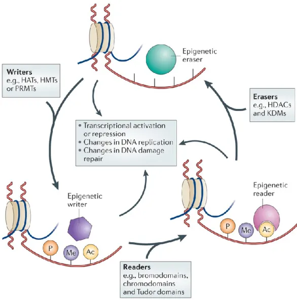
Figure 8. Epigenetic regulation is a dynamic process. A class of epigenetic regulators are “the writers”,
which modify chromatin laying down epigenetic marks on amino acid residues on histone tails. They are histone acetyltransferases (HATs), histone methyltransferases (HMTs), protein arginine methyltransferases (PRMTs) and kinases. The “epigenetic readers” such as proteins containing bromodomains, chromodomains and Tudor domains bind to these epigenetic marks and translate the messages. Finally, “epigenetic erasers” such as histone deacetylases (HDACs), lysine demethylases (KDMs) and phosphatases catalyse the removal of epigenetic marks. Addition and removal of these post-translational modifications of histone tails leads to the addition and/or removal of other marks in a highly complicated histone code. Together, histone modifications regulate various DNA-dependent processes, including transcription, DNA replication and DNA repair. Adapted from (Falkenberg and Johnstone 2014).
Histone modifications involve multiple post-transcriptional alterations of the N-terminal tails of histone proteins such as acetylation, methylation and phosphorylation, which can contribute to the 'open' or 'closed' transcriptional state of the chromatin. DNA methylation and histone-modification events are tightly interconnected.
Histone-modifying enzymes can recruit DNA-methylransferases to the genomic loci bearing a specific 'histone code' and, in turn, methylated DNA binding proteins can recruit histone-modifying enzymes to the hypermethylated loci, thus establishing a combined DNA/histone epigenetic mark self-perpetuated in cellular divisions.
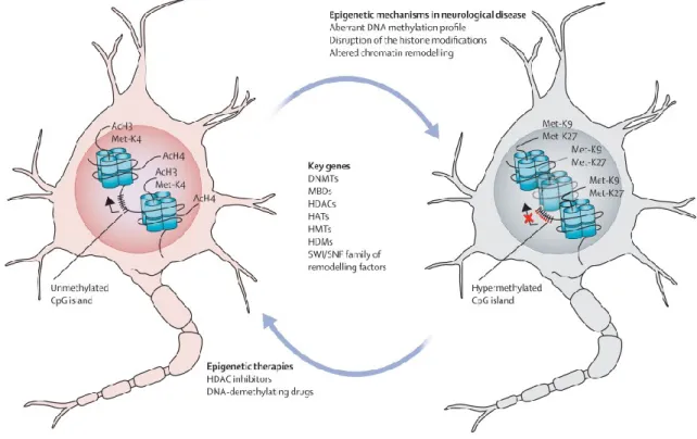
Figure 9. Epigenetic machinery in neurons. In healthy neurons or glia (left), transcriptionlly active
gene promoter have unmethylated CpG island and a set of histone modifications associated with open “chromatin” conformation (eg, hyperacetylation and methylation of lysine 4 of histone H3). One the left, a neurodegenerated neuron carries “closed” chromatin conformation with dense hypermethylation of the CpG island promoter, repressive histone chemical modifications such as methylation of lysines 9 and 27 of histone H3. Epigenetic drugs such as DNA-demethylating drugs and HDAC inhibitors can partially rescue the distorted epigenetic processes. Ac=acetylation. DNMT=DNA methyltransferase. HAT=histone acetyltransferase. HDAC=histone deacetylase. HDM=histone demethylase. HMT=histone methyltransferase. MBD=methyl-CpG binding domain protein. K4=methylation of lysine 4. Met-K9=methylation of lysine 9. Met-K27=methylation of lysine 27. SWI/SNF=switching/sucrose non fermenting chromatin-remodelling complex. Adapted from (Urdinguio, Sanchez-Mut et al. 2009).
1.2.2 Covalent histone-tails post-translational modifications
At the heart of chromatin structure are highly conserved histone proteins (H3, H4, H2A, H2B and H1) that function as building blocks to package eukaryotic DNA into repeating nucleosomal units that are folded into higher-order chromatin fibers. Covalent modifications of histone proteins play central roles in many types of epigenetic inheritance. The most common histone tails modifications are lysine acetylation, lysine
and arginine methylation, serine and threonine phosphorylation, but also lysine ubiquitination and sumoylation. The majority of this covalent modifications occurs in histone H3 and H4 tail (Strahl and Allis 2000). This modifications acts as an alphabet, a code that is read by protein complexes, which slide on DNA molecules and translate the code into a biological and physical response, which may be the switch to an active into a repressed state of the chromatin or vice versa. In addition, histone code differentially marks chromatin that is constitutively condensed, or facultative eterochromatin, which can be converted into a non-condensed form in certain moments of cell cycle.
Every single modification has a determined significance if considered in the context in which it is localized. That is, a modification that acts as a repressive message, changing context, can change its meaning. In general, considering the electrostatic requirements for folding the chromatin polymer, histone acetylation, through the neutralization of positive charge, and histone phosphorylation, through the addition of negative charge, would probably cause decondensation of the chromatin. On the contrary, lysine and arginine methylation usually causes chromatin condensation.
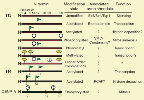
Figure 10. The most frequent covalent modifications on histone H3 and H4 tails and their relative meaning. S10 phosphorylation associated with K14 acetylation lead to transcriptional activation; K4
dimethylation is associated with transcriptional activation; K9 and K27 methylation are associated with transcriptional repression. From (Strahl and Allis 2000).
As histone code is written by a variety of enzymes (see below), it is read by complex of proteins. Recent evidence shows that the bromodomain of human PCAF (P300/CBP-associated factor), a domain of little known function which is shared between many, but not all HATs, binds acetylated lysine in the context of H3 and H4 tail sequences (Dhalluin, Carlson et al. 1999). For example, ATP-dependent chromatin remodeling complexes are specialized protein machinery able to restructure the nucleosome to make its DNA accessible during transcription, replication and DNA repair. ATP-dependent chromatin remodeling complexes specifically recognize these histones marks, and through ATP hydrolysis unwrap, mobilize, exchange or eject the nucleosome, and subsequently recruiting a transcriptional apparatus to nucleosomal DNA (Tang, Nogales et al. 2010). Among them, there is the SWI/SNF complex. The SWI/SNF family of chromatin remodeling complexes was initially discovered in yeast by two independent screenings aimed at identifying mutations in genes that affect the mating-type switching (SWI) and sucrose fermentation (Sucrose Non Fermenting - SNF) pathways. Chromatin remodeling complexes use the energy of ATP hydrolysis to slide the DNA around the nucleosome. The first step consists in the binding between the remodeler and the nucleosome. This binding occurs with nano molar affinity and reduces the digestion of nucleosomal DNA by nucleases. Upon ATP hydrolysis, the torsion subdomain carries out a directional DNA translocation. This event destroys histone-DNA contacts and creates a transient DNA loop that propagates around the nucleosome and resolves when it reaches the exit site on the other side of the nucleosome resulting in nucleosome repositioning. The tracking domain ensures that the waves of DNA loops can move only in one direction blocking any backward movement. The remodeler then resets its original position ready for a new remodeling cycle.
Figure 11. Schematic representation of the SWI/SNF dependent nucleosome remodeling process.
During Step1, the translocase domain binds the nucleosome two turns away from the dyad. Upon ATP-dependent hydrolysis, the torsion sub-domain generates a DNA loop that translocates through the tracking subdomain and the dyad, continuing in the second gyre (Step 2-3). The loop resolves when it reaches the exit site on the other side of the nucleosome (Step 4). The combination of these steps results in nucleosome repositioning. The complex is then ready for a new remodeling cycle (Step1). Adapted from (Tang, Nogales et al. 2010).
1.2.2.1 Histone acetylation
Histone acetylation occurs on lysine residues on H3 and H4 histone tails. It is the well-studied histone modification. The mechanism for acetylation and deacetylation takes place on the NH3+ groups of Lysine amino acid residues. These residues are located on the tails of histones that make up the nucleosome of packaged dsDNA. The process is aided by factors known as Histone Acetyltransferases (HATs). HAT molecules facilitate the transfer of an acetyl group from a molecule of Acetyl Coenzyme-A (Acetyl-CoA) to the NH3+ group on Lysine. When a Lysine has to be deacetylated, factors known as Histone Deacetylases (HDACs) catalyze the removal of the acetyl group with a molecule of H2O. Acetylation has the effect of changing the overall charge of the histone tail from positive to neutral. Nucleosome formation is dependent on the positive charges of the H4 histones and the negative charge on the surface of H2A histone fold domains. Acetylation of the histone tails disrupts this association, leading to weaker
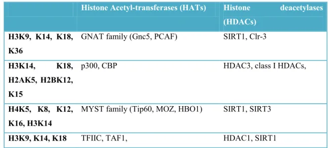
binding of the nucleosomal components. By doing this, the DNA is more accessible and leads to more transcription factors being able to reach the DNA. Thus, acetylation of histones is known to increase the expression of genes through transcription activation. Deacetylation performed by HDAC molecules has the opposite effect. By deacetylating the histone tails, the DNA becomes more tightly wrapped around the histone cores, making it harder for transcription factors to bind to the DNA. This leads to decreased levels of gene expression and is known as gene silencing (Verdone, Agricola et al. 2006). Histone acetyl-transferases (HATs) are a protein family that mediate the acetylation of lysine residues on histone tails. The most important are CBP/p300, MYST family and GNAT family. Histone deacetylase (HDACs) mediate the opposite reaction, that is the removal of acetyl group from lysine residues on histone tails. HDACs are divided into 4 families. Class I includes HDAC1, 2, 3 and HDAC8. Class IIa includes HDAC 4, 5, 7 and 9, whereas Class IIb includes HDAC 6 and 10. HDAC Class III includes the Sirtuins and Class IV contains HDAC 11 (Gallinari, Di Marco et al. 2007).
Histone Acetyl-transferases (HATs) Histone deacetylases (HDACs)
H3K9, K14, K18, K36
GNAT family (Gnc5, PCAF) SIRT1, Clr-3
H3K14, K18, H2AK5, H2BK12, K15
p300, CBP HDAC3, class I HDACs,
H4K5, K8, K12, K16, H3K14
MYST family (Tip60, MOZ, HBO1) SIRT1, SIRT3
H3K9, K14, K18 TFIIC, TAF1, HDAC1, SIRT1
Table 4. Principal histone acetyltransferase (HATs) and deacetylases (HDACs) and their relative target. (Seto and Yoshida 2014).
The most studied histone acetylation are acetylation of lysine 9, 14, 18 and 23 on H3 tail and lysine 5, 8, 12 and 16 on H4 tail (Strahl and Allis 2000). Every of this signals correlate with transcriptional activation.
1.2.2.2 Histone methylation
Histone methylation consists in the addition of a methyl group to lysine or arginine residues. It preferentially occurs on histone H3 and H4 tails. Lysine and arginine residues both contain amino groups, which confer basic and hydrophobic characteristics. Lysine is able to be mono-, di-, or trimethylated with a methyl group replacing each hydrogen of its NH3+ group. With a free NH2 and NH2+ group, arginine can be mono-, di- or tri-methylated. Methylation is not studied as well as acetylation, because it‟s technically more difficult to detect (doesn‟t change the total charge of the histone). The majority of methylation occurs on lysine 4, 9 and 27 on H3 tail. Histone methylation can be associated with either transcriptional repression or activation. For example, dimethylation and trimethylation of histone H3 at lysine 4 (H3K4me3) is an active mark for transcription and is upregulated in hippocampus one hour after contextual fear conditioning in rats. However, dimethylation or trimethylation of histone H3 at lysine 9 (H3K9me2), a signal for transcriptional silencing, is increased after exposure to either the fear conditioning or a novel environment alone (Gupta, Kim et al. 2010).
Histone methylation is mediated by enzymes that are specific for a single lysine or arginine residue. They catalyze the transfer of one, two, or three methyl groups to lysine and arginine residues of histoneproteins. Lysine methyltransferase can be divided into two groups: the ones which possess a SET domain (Su(var)3-9, Enhancer of Zeste, Trithorax), and the ones who don‟t have a SET domain. Both use S-adenosyl-methionine as cofactor. There are two different types of protein arginine methyltransferases (PRMTs) and three types of methylation that can occur at arginine residues on histone tails. The first type of PRMTs (PRMT1,PRMT3,CARM1/PRMT4, and Rmt1/Hmt1) produces monomethyl arginine and asymmetric dimethyl arginine. The second type (JBP1/PRMT5) produces monomethyl or symmetric dimethyl arginine. The differences in the two types of PRMTs arise from restrictions in the arginine binding pocket (Branscombe, Frankel et al. 2001).
Histone Methyltransferase (HMTs) Histone demethylase (HDMs)
H3K4me1/2/4 SET1, MLL PHF8
H3K9me3 Suv3-9H1/H2, SETDB1 KDM2B
H3K27 EZH1, EZH2 KDM6
Table 5. Principal histone methyltransferases (HMTs) and demetylase (HDMs) and their relative target (Khare, Habib et al. 2012)
1.2.2.3 Histone phosphorylation
Phosphorylation, particularly that of histones H1 and H3, has long been implicated in chromosome condensation during mitosis (Koshland and Strunnikov 1996). However, converging evidence suggests that H3 phosphorylation (specifically serine 10) is also directly correlated with the induction of immediate-early genes such as c-jun, c-fos and
c-myc (Mahadevan, Willis et al. 1991). Histone phosphorylation can occur on serine,
threonine and tyrosine residues and constitutes an essential part of the histone code. Phosphorylation of H2A(X) is an important histone modification that plays a major role in DNA damage response (Rossetto, Truman et al. 2010). A substantial number of phosphorylated histone residues are associated with gene expression. Interestingly, these are often related to regulation of proliferative genes. Phosphorylation of serines 10 and 28 of H3 and serine 32 of H2B has been associated with regulation of epidermal growth factor (EGF)-responsive gene transcription. H3S10ph and H2BS32ph have been linked to the expression of proto-oncogenes such as c-fos, c-jun and c-myc (Lau, Lee et al. 2011). Indeed, in EGF-stimulated cells, phosphorylation of H3S10 is tightly coupled to H3 K9ac and K14ac, both marks of transcriptional activation. It has been shown that phosphorylation of H3S10 promotes acetylation of H3K14 by the Gcn5 acetyltransferase in vitro and allows Gcn5-regulated gene transcription in vivo (Lo, Trievel et al. 2000). Phosphorylation is a chimeric modification, and its meaning depends on the chromatin conformation. It is interesting to observe that the same phosphorylation events can be implicated in multiple cellular processes involving chromatin modulation. The same phosphorylated residue can have significantly distinct effects on chromatin structure depending on the context in which it occurs. Phosphorylation of H3S10 and H3S28 is a good example of this duality: both phosphorylated residues are involved in chromatin condensation associated with mitosis and meiosis, as well as in chromatin relaxation linked to transcription activation.
1.2.3 DNA methylation
DNA methylation is a process by which methyl groups are added to DNA. Methylation modifies the function of the DNA, altering its expression. When located in a gene promoter, DNA methylation typically acts to repress gene transcription. DNA methylation is essential for normal development and is associated with a number of key processes including genomic imprinting, X-chromosome inactivation, repression of repetitive elements, aging and carcinogenesis (Jin, Li et al. 2011).
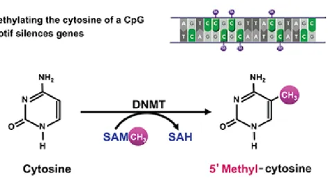
DNA methylation occurs at the cytosine bases of eukaryotic DNA, which are converted to 5-methylcytosine by DNA methyltransferase (DNMT) enzymes. The altered cytosine residues are usually immediately adjacent to a guanine nucleotide, resulting in two methylated cytosine residues sitting diagonally to each other on opposing DNA strands. The addition of methyl groups is controlled at several different levels in cells and is carried out by a family of enzymes called DNA methyltransferases (DNMTs). Three DNMTs (DNMT1, DNMT3a and DNMT3b) are required for establishment and maintenance of DNA methylation patterns. Two additional enzymes (DNMT2 and DNMT3L) may also have more specialized but related functions. DNMT1 appears to be responsible for the maintenance of established patterns of DNA methylation, while DNMT3a and 3b seem to mediate establishment of new or de novo DNA methylation patterns (Bestor 2000). Methylation can be directly observed by staining cells with an immunofluorescently labeled antibody for 5-methylcytosine. In mammals, methylation is found sparsely but globally, distributed in definite CpG sequences throughout the entire genome, with the exception of CpG islands, or certain stretches (approximately 1 kilobase in length) where high CpG contents are found. In human DNA, 5-methylcytosine is found in approximately 1.5% of genomic DNA, on the contrary, In embryonic stem (ES) cells, a substantial amount of 5-mC is also observed in non-CpG contexts (Lister, Pelizzola et al. 2009).
Figure 12. Methylation of cytosine residues in 5’-methyl cytosine happens thanks to DNA methyl transferases (DNMTs) which uses S-adenosylmethyonine as a cofactor.
Although patterns of DNA methylation appear to be relatively stable in somatic cells, patterns of histone methylation can change rapidly during the course of the cell cycle. Despite this difference, several studies have indicated that DNA methylation and histone methylation at certain positions are connected. Indeed, evidence has been presented that in some organisms, such as Neurosporacrassa (Tamaru and Selker 2001) and Arabidopsis thaliana (Soppe, Jasencakova et al. 2002), H3-K9 methylation is required in order for DNA methylation to take place. Given the critical role of DNA methylation in gene expression and cell differentiation, it seems obvious that errors in methylation could give rise to a number of devastating consequences, including various diseases. Indeed, medical scientists are currently studying the connections between methylation abnormalities and diseases such as cancer, lupus, muscular dystrophy, and a range of birth defects that appear to be caused by defective imprinting mechanisms (Robertson 2005).
DNA methylation can alter gene expression in two ways. First, the methylation of DNA itself may physically impede the binding of transcriptional proteins to the gene, and second, and likely more important, methylated DNA may be bound by proteins known as methyl-CpG-binding domain proteins (MBDs). MBD proteins then recruit additional proteins to the locus, such as histone deacetylases and other chromatin remodeling proteins that can modify histones, thereby forming compact, inactive chromatin, termed
heterochromatin. This link between DNA methylation and chromatin structure is very important. In particular, loss of methyl-CpG-binding protein 2(MeCP2) has been implicated in Rett syndrome; and methyl-CpG-binding domain protein 2 (MBD2) mediates the transcriptional silencing of hypermethylated genes in cancer (Soppe, Jasencakova et al. 2002).
1.3 EPIGENETICS AND ALS
Since epigenetic mechanisms were discovered and subsequently well described, a variety of physiological and pathological conditions were linked to alterations in the epigenetic regulation. Beyond the well-known epigenetic imprinting, that lead for example to Angelman and Prader-Willis disease (Knoll, Nicholls et al. 1989), several studies had clarified the connection between many human diseases and epigenetics. Among them, cancer and several neurodegenerative disorders had been linked to epigenetics alterations (Sharma, Kelly et al. 2010, Landgrave-Gomez, Mercado-Gomez et al. 2015). Addiction from abuse of drugs had been correlated to changes in normal epigenetic regulation (Renthal and Nestler 2008).
ALS is one of the neurodegenerative diseases that has been linked to epigenetics. In the next paragraphs I will discuss what is known until now about this link.
1.3.1 Acetylation and deacetylation of histonic and non histonic proteins
1.3.1.1 Targets
Beyond the most studied role of histone acetylation, lysine acetylation of non histonic proteins has emerged as a major covalent modification controlling diverse cellular processes and has been implicated in Alzheimer‟s disease (AD) and other neurodegenerative disorders. In particular, acetylation of misfolded tau proteins marks mature neurofibrillary tangles (NFTs) in AD and related tauopathies and represents a disease-specific marker of AD pathology (Cohen, Guo et al. 2011).
A recent global proteomics study identified ~1750 proteins that are subject to lysine acetylation, including a distinct subset of RNA-binding proteins and associated factors (Choudhary, Kumar et al. 2009). Among them, the case of p53 is the well studied. Acetylation of p53 is important in regulating DNA damage response. The acetylation of p53 regulates its stability through crosstalk with the ubiquitination machinery, modulates interactions with TAF1, and regulates its transcriptional activity (Yang and Seto 2008).
Moreover, recent evidences demonstrated that TDP43 functions are regulated by acetylation. Cohen and colleagues demonstrated that TDP-43 acetylation impairs RNA binding and promotes accumulation of insoluble, hyper-phosphorylated TDP-43 species that largely resemble pathological inclusions in ALS and FTLD-TDP43 (Cohen, Hwang et al. 2015).
1.3.1.2 Histone acetyltransferase
Rouaux et al. observed that in a mouse model of ALS, it is present a severe decrease in the levels of histone acetyltransferase CBP (CREB-cAMP response element-binding protein-binding protein) (Rouaux, Jokic et al. 2003) in lumbar spinal cord motor neurons. Even if a decrease in H3 acetylation level was not revealed in the ALS mouse model, it was detected in cellular model of apoptosis. This imbalance induce a relative increase in global histone deacetylation, leading to the silencing of several anti-apoptotic genes and the contemporary overexpression of pro-anti-apoptotic ones (Saha and Pahan 2006).
1.3.1.3 Histone deacetylases
Several HDACs alterations have been linked to ALS. Among them, HDAC4 mRNA and protein greater levels in ALS patients have been linked to a severe phenotype a negatively correlates with re innervation processes (Bruneteau, Simonet et al. 2013). HDAC6 directly interacts with TDP43, and is deacetylated by it (Cohen, Hwang et al. 2015), and, in association with FUS; regulates its mRNA (Kim, Shanware et al. 2010). Valle and colleagues reported the global deregulation of HDACs in two mouse models of ALS (Valle, Salvatori et al. 2014). Interestingly, they demonstrated that HDACs expression pattern changes from spinal cord to muscle.
HDAC1 is a nuclear ubiquitous histone deacetylases class I of 55 kDa. It deacetylatesHDAC1 knockout embryos are lethal (Lagger, O'Carroll et al. 2002). Silencing of HDAC1 via RNAi lead to inhibition of proliferation associated with the upregulation of cyclin-dependent kinase (cdk) inhibitors. In addition, HDAC1 along with HDAC2 are recruited at the double stranded break sites and play an important role in DNA damage repair to promote non homologous end joining (NHEJ) (Miller, Tjeertes et al. 2010) . HDAC1 directly interacts with FUS in DNA damage sites (Wang, Pan et al. 2013). In my work, I demonstrated that HDAC1 interacts also with TDP43.
1.3.1.4 HDAC inhibitors and other ALS therapy approaches
There are several evidences that restoring the correct acetylation/deacetylation balance using histone deacetylases inhibitors (HDACi) like Sodium Valproate (VPA), Sodium Butyrate (NaB) and Trichostatin A (TSA), contrasts MNs death and the neurodegenerative process (Sugai, Yamamoto et al. 2004). Anyway, their efficacy hasn‟t been proved in clinical yet. In my work, I tested the efficacy two HDACi, sodium 4-phenylbutyrate and trichostatin A, in improving cell viability in a cellular model of ALS.
Table 6.Principal HDAC inhibitors and their relative target.
Sodium 4-phenylbutyrate (PBA) is an orally bioavailable, blood brain barrier (BBB) permeable, short-chain fatty acid that has been approved by the Food and Drug Administration (FDA) for treatment of urea cycle disorders. PBA has potential benefit for a wide variety of diseases like cancer, cystic fibrosis, thalassemia, spinal muscular atrophy as well as protein folding diseases such as type 2 diabetes mellitus, ALS and other neurodegenerative disesases. PBA and sodium valproate promoted motorneuron survival in mouse models of ALS, and compound efficacy was at least partially
attributed to the amelioration of abnormal histone hypoacetylation and transcriptional dysregulation, which is implicated in ALS (Ryu, Smith et al. 2005) (Rouaux, Panteleeva et al. 2007). Similarly, combined lithium and valproate treatment delayed disease onset, reduced neurological deficits and prolonged survival (Feng, Leng et al. 2008), and treatment with trichostatin A (Yoo and Ko 2011), or valproate (Sugai, Yamamoto et al. 2004), delayed disease progression and/or increased survival in the SOD1-G93A mice. A phase II study in ALS individuals revealed that sodium phenylbutyrate was safe and tolerable, and histone acetylation was significantly increased after sodium phenylbutyrate administration (Cudkowicz, Andres et al. 2009). Conversely, a trial using valproic acid did not show a beneficial effect on survival or disease progression in patients with ALS (Piepers, Veldink et al. 2009). An open-label study, involving 40 patients with ALS, has been performed to establish the safety and pharmacodynamics of escalating dosages of phenylbutyrate in 26 participants who completed a 20-week treatment phase. From this study emerges that phenylbutyrate is safe and the majority of subjects tolerated higher dosages (21 g/day) of this drug, but the lowest dose (9 g/day) was therapeutically efficient in improving histone acetylation levels. No deaths or clinically relevant laboratory changes occurred with phenylbutyrate treatment. Histone acetylation decreased by approximately 50% in blood buffy-coat specimens at screening and significantly increased after phenylbutyrate administration. In addition, blood levels of phenylbutyrate and the primary metabolite (PAA) increased with dosage (Piepers, Veldink et al. 2009).
At present there is a clinical trials from Duke University (USA) recruiting patients to test the effect of Lunasin, a 43-amino acid polypeptide originally discovered in soy (ClinicalTrials.gov Identifier: NCT02709330). Lunasin can modify the epigenome and using an HAT assays, with acid-extracted histones as templates, it has been demonstrated that Lunasin specifically inhibited H4K8 acetylation while enhanced H4K16 acetylation catalyzed by HAT enzymes p300, PCAF, and HAT1A (Galvez, Huang et al. 2011).
HDAC inhibition is not the only therapeutic strategy that is in course of study. Rodriguez-Paredes and colleagues reviewed all the molecules that have an effect in
targeting HATs, HMT, HDM, and other epigenetic modifiers (Rodriguez-Paredes and Esteller 2011).
Figure 13. A summary of all molecules targeting DNMTs, HMT, HDMs, SIRTs, HDACs and HATs.
Adapted from (Rodriguez-Paredes and Esteller 2011)
1.3.2 Methylation and demethylation
Histone methylation of some particular residues is involved in the development of ALS. In particular, the trimethylation of lysine residues within histones H3 and H4 is a mechanism involved in reducing C9orf72 mRNA expression in expanded repeat carriers of c9FTD/ALS patients blood. The residues involved are lysine 9 and 27 on histone H3 (H3K9, H3K27) (Belzil, Bauer et al. 2013).
Recent evidences reported that FUS in methylated on a specific arginine residue. Arginine methylation modulates nuclear import of FUS via a TRN-binding epitope and chemical or genetic inhibition of arginine methylation restores TRN-mediated nuclear import of ALS-associated FUS mutants (Dormann, Madl et al. 2012).
1.3.3 Histone phosphorylation
Very few data are available on histone phosphorylation and neurodegenerative mechanisms. Phosphorylation of histone H2AX on serine 139 is correlated to DNA damage response (Rogakou, Nieves-Neira et al. 2000) in a variety of conditions, such as in Alzheimer disease (Myung, Zhu et al. 2008). No any alterations in histone phosphorylation pattern has been demonstrated since now in ALS patients. Nevertheless, several evidences induce to speculate that histone phosphorylation may be involved in ALS pathogenesis, considering that DNA damage is one of the marker of ALS (Qiu, Lee et al. 2014).
1.3.4 DNA methylation
Another epigenetic hypothesis of ALS pathogenesis involves DNA methylation. Recent genome-wide analyses have found differential gene methylation in human ALS. Neuropathologic assessments have revealed that motor neurons in human ALS show significant abnormalities in Dnmt1, Dnmt3a, and 5-methylcytosine. Similar changes are seen in mice with motor neuron degeneration, and Dnmt3a was found abundantly at synapses and in mitochondria (Martin and Wong 2013). Furthermore, Chestnut and colleagues demonstrated that during apoptosis of cultured motor neuron-like cells, Dnmt1 and Dnmt3a protein levels increase, and 5-methylcytosine accumulates. In addition, enforced expression of Dnmt3a, but not Dnmt1, induces degeneration of cultured neurons (Chestnut, Chang et al. 2011). These data suggest that DNA methylation may be a therapeutic target in ALS. As a matter of fact, inhibition of Dnmt catalytic activity with small molecules RG108 and procainamide protects motor neurons from excessive DNA methylation and apoptosis in cell culture and in a mouse model of ALS (Chestnut, Chang et al. 2011).
Up to date, several experimental evidences underline the link between epigenetic and neurodegenerative diseases, such ALS. In particular, it is widely demonstrated that both in sporadic and in familiar ALS there is a severe decreasing in acetylation of N-terminal tail of histone H3 and H4. In parallel, many evidences suggest how there is a contemporary decreasing in the expression of histone-acetyl transferases such as CBP. Finally, in ALS patients several studies show an increase in DNA methylation. This data lead me to speculate on the role of ALS causative genes SOD1, FUS and TDP43 on epigenetic deregulations. For this reason, I focused my PhD project on 3 main objectives:
1) Understand the role of SOD1, FUS and TDP43 in regulating histone post-translational modifications and the possible pathological implications. In this sense, I performed western blot analysis on cells lysates expressing SOD1, FUS or TDP43, with primary antibodies targeting histonic modifications linked to transcriptional activation or to transcriptional repression; in addition, we measured DNA methylation levels.
2) Investigate the possible interaction between TDP43 and HDAC1. We performed co-immunoprecipitation experiments to evidence the interaction, the role of point pathological mutations and which is TDP43 interacting domain.
3) Analyze the possible therapeutic effect of HDAC inhibitors such as Sodyum butyrate and Trichostatin A, performing cells vitality assays.
Bacterial strain.
Competent cells used are E. coli DH5-α. Bacterial cells are defective for the restriction and have mutations in relA1 and recA1 genes, to improve the stability and quality of recombinant plasmids.
Cell lines.
SH-SY5Y cell line: SH-SY5Y cells (ATCC, Rockville, MD, CRL-2266) are human cells
derived from neuroblastoma cell line.
Adeno-X 293 cell line: Adenovirus 5-transformed Human Embryonic Kidney 293 cell
line (HEK 293; ATCC, Rockville, MD, CRL 1573) is used to package and propagate the recombinant adenoviral- based vectors produced with the BD Adeno-X Expression System.
SH-SY5Y-HDAC1 cell line: SHSY-5Y cell line stably expressing HDAC1-FLAG
created in our lab. Selection is maintained with 200 ug/ml of G1418.
Adenoviral vectors.
All adenoviral vectors (pAdenoX-hFUSWT/R495X/H517Q/R521G/P525L; pAdenoX-hTDP43WT/Q331K/M337V/A382Tand pAdenoX-hSOD1WT/H80R/G93A) were generated using the Adeno-X Expression System 1 (Clontech). Their production is completed in two stages. First, generation of mammalian expression cassette by cloning gene of interest into pShuttle2.Second, excision of expression cassette from pShuttle2 and insertion into I-Ceu I and PI-Sce I sites of BD Adeno-X Viral DNA. All constructions were verified by automated sequencing.
Plasmids and oligonucleotides.
Name Expression cassette Resistance Tag
pMTK-hTDP43WT,M337V,A382T hTDP43WT,M337V,A382T Ampicillin 5x-myc
pMTK-hFUSWT,R521G,P525L,R495X hFUSWT,R521G,P525L,R495X Ampicillin 5x-myc
pcDNA3-hHDAC1 hHDAC1 Ampicillin
1x-FLAG
pMTK-hTDP43-ΔN-term hTDP43-ΔN-term Ampicillin 5x-myc
pMTK-hTDP43-ΔRRM1 hTDP43-ΔRRM1 Ampicillin 5x-myc
pMTK-hTDP43-ΔRRM2 hTDP43-ΔRRM2 Ampicillin 5x-myc
pMTK-hTDP43-ΔG-rich hTDP43-ΔG-rich Ampicillin 5x-myc
pMTK-hTDP43-ΔRRM1/RRM2 hTDP43-ΔRRM1/RRM2 Ampicillin 5x-myc