Alma Mater Studiorum
Alma Mater Studiorum –
– Università di Bologna
Università di Bologna
DOTTORATO DI RICERCA IN
“S
CIENZEP
NEUMO-C
ARDIO-T
ORACICHE DII
NTERESSE MEDICO EC
HIRURGICO”
Ciclo XXV
Settore Concorsuale di afferenza: 06/D1 Settore Scientifico disciplinare: MED/10
TITOLO TESI
Airway Basal Cell Vascular Endothelial Growth Factor-mediated
Cross-Talk Regulates Endothelial Cell Dependent Growth Support of Human
Airway Basal Cells
Presentata da: Dr.G.Curradi
Coordinatore Dottorato
Relatore
Prof. S.Mattioli
Prof.C.Saltini
INDEX
Summary ………. 1
Introduction ……… 2
Methods ………... 4
Sampling the Airway Epithelium ……….. 4
Culture and Characterization of Primary Human Airway Basal Cells ……….. 4
Gene Expression ……… 6
Immunohistochemistry Analysis of VEGFA Expression ………...……….. 7
ELISA ………... 7
Air-liquid Interface Culture ………... 8
Basal Cell Proliferation ………. 9
Western Analysis ……….. 9
Analysis of MAPK Activation ……….. 11
Culture and Maintenance of HUVEC Cells ……….. 11
Co-culture Proliferation Assays ……… 12
Results ………. 13
Expression of VEGFA in Human Airway Basal Cells ………. 13
VEGFA is Actively Secreted by Human Airway Basal Cells ……….. 14
Expression of VEGFA-associated Receptors in Human Airway Basal Cells ………... 15
Inhibition of VEGFA Has No Effect on Proliferation of Airway Basal Cells ……….. 16
Activation of Endothelial Cells by Basal Cell-derived VEGFA ………... 17
Activated Endothelial Cells Support the Growth of Airway Basal Cells in the Ab-sence of Growth Factors ……… 19 Inhibition of VEGFR2 Signaling Suppresses Endothelial Cell-dependent Prolifera-tion of Airway Basal Cells ……… 20
Discussion ……… 22
Expression and Function of VEGFA in the Lung ………. 22
Basal Cell – Endothelial Cell Cross Talk ……….. 23
Possible Roles in Health and Disease ………... 24
Figures ………. 27
1
Title of the scientific research project:
Airway Basal Cell Vascular Endothelial Growth Factor-mediated Cross-Talk Regulates Endothelial Cell Dependent Growth Support of Human Airway Basal Cells
Summary
The human airway epithelium is a pseudostratified heterogenous layer comprised of cili-ated, secretory, intermediate and basal cells. As the stem/progenitor population of the airway epi-thelium, airway basal cells differentiate into ciliated and secretory cells to replenish the airway epithelium during physiological turnover and repair. Transcriptome analysis of airway basal cells revealed high expression of vascular endothelial growth factor A (VEGFA), a gene not typically associated with the function of this cell type. Using cultures of primary human airway basal cells, we demonstrate that basal cells express all of the 3 major isoforms of VEGFA (121, 165 and 189) but lack functional expression of the classical VEGFA receptors VEGFR1 and VEGFR2. The VEGFA is actively secreted by basal cells and while it appears to have no direct autocrine function on basal cell growth and proliferation, it functions in a paracrine manner to activate MAPK signaling cascades in endothelium via VEGFR2 dependent signaling pathways. Using a cytokine- and serum-free co-culture system of primary human airway basal cells and human endothelial cells revealed that basal cell secreted VEGFA activated endothelium to ex-press mediators that, in turn, stimulate and support basal cell proliferation and growth. These data demonstrate novel VEGFA mediated cross-talk between airway basal cells and endothe-lium, the purpose of which is to modulate endothelial activation and in turn stimulate and sustain basal cell growth.
2
Introduction
The human bronchial tree is a branching structure of up to 23 generations that functions as a conduit of air to and from the alveoli [1,2]. The bronchial tree is lined with a pseudostrati-fied heterogeneous epithelium composed of 4 major cell types: ciliated, secretory, intermediate and basal cells [3-5]. The classic role of the basal cell population is to function as stem/progenitor cells that, with appropriate signals, differentiate into intermediate cells and fi-nally the specialized ciliated and secretory cells [6-11]. Utilizing methodology developed in our laboratory to culture pure populations of human airway basal cells from the complete airway epi-thelium obtained by brushing the airway epiepi-thelium of healthy nonsmokers, we recently charac-terized the transcriptome of basal cells of healthy individuals [11].
Analysis of the human airway basal cell transcriptome uncovered expression of a variety of genes/pathways linked to the known stem/progenitor cell function of these cells, but also iden-tified that basal cells express genes coding for molecules not typically associated with epithelial structure and function [11]. Among these genes was vascular endothelial growth factor A (VEGFA), the product of which is primarily associated with vascular endothelial growth and function [12,13]. The VEGF family of receptors and ligands are critical regulators of vascular and lymphatic function during development and in health and disease [13-16]. There are five structurally related mammalian VEGF ligands (VEGFA, B, C, and D, and placenta growth fac-tor; PLGF), three receptors (VEGFR1, 2 and 3) and two co-receptors (neuropilin-1 and 2) that interact in various combinations to modulate vascular-related biological processes [12-14,17]. VEGFA functions as a highly potent pro-angiogenic factor [12], and its signaling is mediated through direct binding of the ligand to the tyrosine kinase receptors VEGFR1 and VEGFR2 and subsequent activation of downstream kinase signaling cascades [13,16,18].
3 Together, these observations lead to the hypothesis that airway basal cells may have a novel function beyond the role as stem/progenitor cells, i.e., do human airway basal cells support the structure and function of lung endothelial cells by expressing and secreting VEGFA? Using cultures of primary human airway basal cells and human endothelial cells alone and together, the data demonstrate that human airway basal cells express all of the 3 major isoforms of VEGFA (121, 165 and 189) but lack functional expression of the classical VEGFA receptors VEGFR1 and 2. The VEGFA is actively secreted by basal cells and while it appears to have no direct autocrine function on basal cell growth and proliferation, it functions in a paracrine manner to activate MAPK signaling cascades in endothelium via VEGFR2 dependent signaling pathways, with consequent endothelial cell-mediated reciprocal activation of basal cell proliferation. Over-all, these data suggest a novel function of human airway basal cells to regulate activation of en-dothelium in a paracrine manner via secretion of VEGFA. In turn, activated enen-dothelium express mediators that stimulate and support basal cell proliferation. Regulation of this molecular cross-talk between basal and endothelial cells may play an important role in health and disease.
4
Methods Sampling the Airway Epithelium
Subjects were evaluated at the Department of Genetic Medicine Clinical Research Facil-ity and the Weill Cornell NIH Clinical Translational Science Center (CTSC) or the Rockefeller University CTSC using Institutional Review Board-approved clinical protocols. All subjects were confirmed to be nonsmokers by urine levels of nicotine (<2 ng/ml) and cotinine (<5 ng/ml) with normal pulmonary functions tests and chest X-ray. After obtaining written informed con-sent, flexible bronchoscopy was used to collect large airway epithelial cells by brushing the epi-thelium as previously described [19-21]. Cells were detached from the brush by flicking into 5 ml of ice-cold Bronchial Epithelium Basal Medium (BEGM, Lonza, Basel, Switzerland). An ali-quot of 0.5 ml was used for differential cell count. The remainder (4.5 ml) was processed imme-diately for either immediate RNA extraction or basal cell culture. The number of cells recovered by brushing was determined by counting on a hemocytometer. To quantify the percentage of epithelial and inflammatory cells and the proportions of basal, ciliated, secretory and intermedi-ate cells recovered, cells were prepared by centrifugation (Cytospin 11, Shandon Instruments, Pittsburgh, PA) and stained with Diff-Quik (Baxter Healthcare, Miami, FL). In all samples the epithelial cells represented >97% of the cell population. The proportions of epithelial cells were as previously reported [19,21].
Culture and Characterization of Primary Human Airway Basal Cells
Pure populations of human airway basal cells were obtained and characterized using the detailed protocols described in Hackett et al [11]. Briefly, airway epithelial cells collected by brushing were pelleted by centrifugation (250 x g, 5 min) and disaggregated by resuspension in 0.05% trypsin-ethylenediaminetetraacetic acid (EDTA) for 5 min, at 37°C. Trypsinization was
5 stopped by addition of HEPES buffered saline (Lonza, Basel, Switzerland), supplemented with 15% fetal bovine serum (FBS; GIBCO-Invitrogen, Carlsbad, CA), and the cells were again pel-leted at 250 x g, 5 min. The pellet was resuspended with 5 ml of phosphate buffered saline, pH 7.4 (PBS), at 23°C, then centrifuged at 250 x g, 5 min. Following centrifugation, the PBS was
removed, the cells resuspended in 5 ml of BEGM and 5 x 10⁵ cells were cultured in T25 flasks in
BEGM (Lonza, Basel, Switzerland), supplemented with growth factors according to the manu-facturer's instructions. The antibiotics supplied by the manufacturer were replaced with gen-tamycin (50 µg/ml; Sigma, St Louis, MO), amphotericin B (1.25 µg/ml; Invitrogen, Carlsbad, CA), and penicillin-streptomycin (50 µg/ml; Invitrogen, Carlsbad, CA). Cultures were
main-tained in a humidified atmosphere of 5% CO2, 37°C. Unattached cells were removed by
chang-ing medium after 12 hr. Thereafter, media was changed every 2 days until time of harvest at day 7 of culture, when the cells had reached 70 to 80% confluence. For sub-culturing, the cells were seeded at a density of 104 cells/cm2 and maintained in an identical manner. At day 7 of the initial culture, the basal cells were trypsinized and cytospin slides prepared for characterization by im-munohistochemistry, as described below, using the following cell-type specific markers: cy-tokeratin 5 (basal cell; 1/50; Thermo Scientific, Rockford, IL); p63 (basal cell; 1/50; Santa Cruz Biotechnology, Inc., Santa Cruz, CA); CD151 (basal cell; 1/200; Leica Microsystems, Inc., Ban-nockburn, IL); N-cadherin (mesenchymal cell; 1/2500; Invitrogen, Carlsbad, CA); mucin 5AC (secretory cell; 1/50; Vector Laboratories, Burlingame, CA); TFF3 (secretory cell; 1/1000; Santa Cruz Biotechnology, Inc., Santa Cruz, CA); β-tubulin IV (ciliated cell; 1/2000 dilution; Bio-genex, San Ramon, CA); chromogranin A (neuroendocrine cell; 1/5000; Thermo Scientific, Rockford, IL) and CGRP (neuroendocrine cell; 1/500; Sigma, St Loius MO). Only cultures that were >95% positive for basal cell markers and negative for other cell types were used in this
6 study. To maintain consistency, air-liquid interface (ALI) and proliferation experiments were performed using passage 2 cells.
Gene Expression
Genome wide gene expression analysis of basal cells and large airway epithelium was assessed using the HG-U133 Plus 2.0 array (Affymetrix, Santa Clara, CA) as previously de-scribed [11]. TaqMan real-time RT-PCR was performed on RNA samples from the complete large airway epithelium of healthy non-smokers, cultured basal cells derived from the same re-gion and human umbilical cord vein endothelial cells (HUVECs). cDNA was synthesized from 1 µg RNA in a 50 µl reaction volume, using the TaqMan Reverse Transcriptase Reaction Kit (Ap-plied Biosystems, Foster City, CA), with random hexamers as primers. Two dilutions, 1:20 and 1:200 were made from each sample, and duplicate wells were run for each sample. TaqMan PCR reactions were carried out using the following gene-specific expression kits from Applied Bio-systems: VEGFA all isoforms (Hs00900054_m1); 121 (Hs03929005_m1); VEGFA-165 (Hs00900057_m1); VEGFA-189 (Hs00903129_m1); VEGFR1 (Hs01052961_m1); VEGFR2 (Hs 00911700_m1) and NRP-1 (Hs 00826128_m1). The endogenous control was hu-man 18S rRNA (Applied Biosystems). Relative expression levels were determined using the ΔΔCt method, with the average value of expression in complete airway epithelium as the nor-malizer [22]. The PCR reactions were run in an Applied Biosystems Sequence Detection System 7500, and the relative quantity was determined using the algorithm provided by the manufac-turer.
The mRNA levels of specific VEGFA isoforms were assessed by RT-PCR using forward
(5’-TGCAGACCAAAGAAAGATAGAGCAAGA-3’) and reverse
re-7 spectively. These primers give rise to an expected product of 86 bp for VEGFA-121, 218 bp for VEGFA-165 and 290 bp for VEGFA-189. Basal cell cDNA was synthesized using the method described above. All reactions were performed using Platinum® PCR Supermix (Invitrogen, Carlsbad, CA) in a 50 µl reaction volume and 40 cycles of amplification as follows: denature 94°C, 30 sec; anneal 55°C, 30 sec; and extend 72°C, 1 min.
Immunohistochemistry Analysis of VEGFA Expression
To analyze VEGFA expression in basal cell cultures by immunohistochemistry, the cells were trypsinized, and cytospin slide preparation fixed in 4% paraformaldehyde for 15 min. To enhance staining, an antigen recovery step was carried out by steaming the samples for 15 min in citrate buffer solution (Labvision, Fremont, CA) followed by cooling at 23°C, 20 min.
Endoge-nous peroxidase activity was quenched using 0.3% H2O2, and normal serum matched secondary
antibody was used for 20 min to reduce background staining. Samples were incubated overnight at 4°C with the primary antibody, mouse monoclonal anti-human VEGF antibody (1/50; MAB293, R&D Biosystems, Minneapolis, MN). Isotype matched IgG (Jackson Immunoresearch Laboratories, Inc, West Grove, PA) was the negative control. Vectastain Elite ABC kit and AEC substrate kit (Dako North America, Inc, Carpinteria, CA) were used to visualize antibody bind-ing. The sections were counterstained with Mayer’s hematoxylin (Polysciences, Inc, Warrington, PA) and mounted using Faramount mounting medium (Dako North America, Inc.). Brightfield microscopy was done using a Nikon Microphot microscope equipped with a Plan x40 numerical aperture (NA) 0.70 objective lens. Images were captured with an Olympus DP70 CCD camera.
ELISA
Min-8 neapolis, MN). BEGM growth media exposed to basal cell cultures for 2 days was removed and then centrifuged at 250 x g, 5 min to pellet cellular debris. The supernatant was then analyzed by ELISA using the manufacturer’s instructions. Basal (BEBM) and growth (BEGM) media not ex-posed to basal cells were used as a negative control. To determine whether basal cell derived VEGFA is secreted apically or basolaterally, ALI cultures of basal cells were established as de-scribed below. At day 12 of ALI culture, when tight junctions are established, fresh media was exclusively added to either the lower or upper chamber of the cultures. Two days post-incubation, the media was removed and processed to determine the levels of secreted VEGFA as described above. ALI media not exposed to basal cells were used as a negative control.
Air-liquid Interface Culture
Primary airway basal cells were trypsinized and seeded at a density of 6 x 10⁵ cells/cm2 onto a 0.4 µm pore-sized Costar Transwells inserts (Corning Incorporated, Corning, NY) pre-coated with type IV collagen (Sigma, St Louis, MO). The initial culture medium consisted of a 1:1 mixture of DMEM and Ham's F-12 medium (GIBCO-Invitrogen, Carlsbad, CA) containing 100 U/ml penicillin, 5% fetal bovine serum 100 µg/ml streptomycin, 0.1% gentamycin, and 0.5% amphotericin B. The following day, the medium was changed to 1:1 DMEM/Ham's F12 (including antibiotics described above) with 2% Ultroser G serum substitute (BioSerpa S.A., Cergy-Saint-Christophe, France). Two days post seeding once the cells had reached confluence on the membrane, the media was removed from the upper chamber to expose the apical surface to air and establish the ALI (referred to as ALI “day 0”). The cells were then grown at 37°C, 8%
CO2, and the culture medium was changed every other day. Following 5 days on ALI, the cells
9
Basal Cell Proliferation
Proliferation assays were used to assess the ability of specific blocking antibodies to in-hibit basal cell proliferation under growth factor rich culture conditions. Basal cells were (2 x 10⁴) seeded into each well of a 12-well plate in BEGM growth media. The next day (termed day 0) the media was removed and the cells washed twice with PBS before addition of fresh BEGM. For inhibition of basal cell proliferation, anti-VEGFA antibody (Bevacizumab; 0.1 µg/ml; Genentech), human VEGFR2 neutralizing antibody 1121 (1 µg/m; ImClone, New York, NY) or IgG isotype control was added to the media at the desired concentration. Fresh media and antibody was added every 2 days of culture throughout the course of the experiment. At the desired time points, cells were trypsinized and total cell numbers were measured with a hemocytometer and the viability assessed by counting of trypan blue dye-excluded cells.
Western Analysis
Cells were trypsinized and lysed in radioimmunoprecipitationlysis (RIPA) buffer (Sigma,
St Louis, MO) plus Complete Protease Inhibitor Cocktail (Roche, Mannheim,Germany) and Halt
phosphataseinhibitor cocktail (Pierce, Rockford, IL), and incubated on ice for 30 min. Lysates
wereclarified by centrifugation at 22,500 x g for 10 min in an Eppendorf 5415C microcentrifuge
at 4°C. Total proteinconcentration was measured using the Bio-Rad (Hercules, CA) protein
as-say to the manufacturer’s guidelines. NuPAGE® LDS Sample Buffer (4X; Invitrogen, Carlsbad, CA) supplemented with 200 mM dithiothreitol (DTT) was added to each sample before boiling
for 10 min and sodium dodecyl sulfate-polyacrylamide gel electrophoresis (SDS-PAGE). For
analysis of protein phosphorylation, cells were lysed directly in the dish using 1X NuPAGE LDS Sample Buffer (diluted in RIPA buffer containing Complete Protease Inhibitor Cocktail (Roche, Mannheim, Germany), Halt phosphatase inhibitor cocktail (Pierce, Rockford, IL) and 50 mM
10 DTT). Once lysed, the samples were then transferred to a 1.5 ml Eppendorf tube before boiling
for 10min and SDS-PAGE. All proteins were analyzed usingNuPAGE 4 to 12% Bis-Tris
gradi-ent gels (Invitrogen, Carlsbad, CA) and subsequgradi-ently transferred onto nitrocellulose membranes with a Bio-Rad Semi-Dry apparatus before Western analysis. The membranes were then blocked overnight at 4°C in 4% blocking reagent made in PBS containing 0.1% Tween-20 (PBST). Non-fat milk was used as a standard blocking reagent for general protein analysis. This was replaced with bovine serum albumin for analysis of protein phosphorylation. After blocking the mem-branes overnight, immobilized proteins were reacted with cell type specific antibodies in 4% blocking reagent for 1 hr, 23°C with shaking. Following the primary antibody incubation,
mem-branes were washed three times for5 min each with PBST, incubated with an rabbit or
anti-mouseantibody conjugated to horseradish peroxidase in 4% blocking reagent for 1 hr, at 23°C
with shaking. Upon completion of secondary antibody incubation, the membranes werewashed
again three times for 5 min with PBST and twice withPBS, and antibodies were visualized after
the addition of ECL Western Blotting Detection Reagents (GE Healthcare Biosciences, Pitts-burgh, PA) by exposure to X-ray film.
The primary antibodies used for Western analysis included: rabbit polyclonal anti-human VEGFR1 (1/1000, #2893, Cell Signaling Technology, Danvers, MA); rabbit monoclonal anti-human VEGFR2 (55B11; 1/1000, #2479, Cell Signaling Technology, Danvers, MA); rabbit monoclonal anti-human Phospho-VEGFR2 (Tyr1175; 19A10; 1/1000, #2478, Cell Signaling Technology, Danvers, MA); rabbit monoclonal anti-human neuropilin-1 (NRP-1; 1/1000, ab81321, Abcam, Cambridge, MA); rabbit polyclonal anti-human p44/42 MAPK (Erk1/2; 1/1000, #9102, Cell Signaling Technology, Danvers, MA); rabbit polyclonal anti-human phos-pho-p44/42 MAPK (Erk1/2; Thr202/Tyr204; 1/1000, #9101, Cell Signaling Technology, Danvers, MA); rabbit polyclonal anti-human p38 MAPK (1/1000, #9212, Cell Signaling Technology, Danvers, MA); rabbit polyclonal anti-human Phospho-p38 MAPK (Thr180/Tyr182;
11 nology, Danvers, MA); rabbit polyclonal anti-human Phospho-p38 MAPK (Thr180/Tyr182; 1/1000, #9211, Cell Signaling Technology, Danvers, MA); mouse monoclonal anti-human β-actin (1/10000; Santa Cruz Biotechnology) and mouse monoclonal anti-human α-tubulin (1/10000; Santa Cruz Biotechnology).
Analysis of MAPK Activation
Equal numbers of HUVEC and basal cells were seeded per dish in the appropriate growth media. The following day, cells were washed twice with PBS and then serum starved for at least 6 hr in the appropriate cell type specific serum free media. Following serum starvation, the cells were stimulated for 15 min with either: (1) base media (BEBM); (2) conditioned media (BEBM exposed overnight to cultured basal cells) or (3) base media supplemented with recombinant VEGFA-165 (50 ng/ml; R&D Biosystems). After stimulation, the media was aspirated from the cells and the cells were washed once with PBS. Following removal of the PBS, the cells were lysed directly in the dish with 1X NuPAGE LDS Sample Buffer (diluted in RIPA buffer
contain-ing Complete Protease Inhibitor Cocktail, Halt phosphataseinhibitor cocktail and 50 mM DTT)
and processed for Western analysis as described above. VEGFR2 dependent MAPK activation was evaluated using both phosphor- and pan-specific antibodies targeted against VEGFR2, p44/p42 MAPK and p38 MAPK. The data shown are representative of four independent experi-ments.
Culture and Maintenance of HUVEC Cells
Human umbilical cord vein endothelial cells (HUVECs) were isolated as previously de-scribed [23]. Cells were cultured in endothelial cell growth medium (Medium 199; Sigma, St
(Hall-12
way), 1% (v/v) antibiotics (Hallway), and 20 units/mlheparin (Sigma, St Louis, MO).
HUVEC-Akt cells were generated as previously described [23]. Briefly, HUVEC cells were transduced with a lentivirus expressing myristoylated Akt and a GFP marker at a multiplicity of infection of 10 and maintained in an identical manner to HUVEC cells.
Co-culture Proliferation Assays
Co-culture proliferation assays were used to assess the ability of endothelial cells to
sup-port basal cell proliferation in cytokine- and serum-free conditions. HUVEC-Akt cells (5 x 10⁴)
were seeded into each well of a 12-well plate in HUVEC growth media. The next day the media
was removed and the cells washed twice with PBS followed by seeding 2 x 10⁴ basal cells into
each well in BEGM growth media. The next day (termed day 0) the media was removed and the cells washed twice with PBS before addition of cytokine- and serum-free BEBM media. The cells were then incubated and subsequently harvested and counted at the desired time points. As a control, basal cells were seeded into wells containing no HUVEC-Akt controls and subse-quently treated in an identical manner to those in co-culture. For inhibition of basal cell prolifera-tion in co-culture, human VEGFR2 neutralizing antibody 1121 (ImClone, New York, NY) or IgG isotype control was added to the media at a concentration of 1 µg/ml. Fresh media and anti-body was added every 2 days of culture throughout the course of the experiment.
At the desired time points, cells were trypsinized and total cell numbers were measured with a hemocytometer and the viability assessed by counting of trypan blue dye-excluded cells. The population of GFP-labeled HUVEC-Akt cells in the harvested sample was determined as the GFP+VE-cadherin+ population by flow cytometric analysis, and the GFP-VE-cadherin- popula-tion quantified as expanded basal cells.
13
Results
Expression of VEGFA in Human Airway Basal Cells
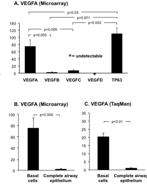
Microarray analysis of VEGF ligand (VEGFA, VEGFB, VEGFC and VEGFD) expres-sion in airway basal cells revealed differential abundance of each. VEGFA was the most highly expressed ligand, while both VEGFB (p<0.005) and VEGFC (p<0.005) were expressed at sig-nificantly lower levels compared to VEGFA (Figure 1A). Analysis of multiple probe sets for VEGFD confirmed the ligand was not expressed in basal cells. Comparison of each ligand’s ex-pression level with that of the basal cell specific gene TP63 revealed each was expressed at sig-nificantly lower levels (VEGFA, 1.5-fold lower, p<0.03; VEGFB, 43.5-fold lower, p<0.001 and VEGFC, 14.7-fold lower, p<0.002; Figure 1A). Due to the high expression level of VEGFA in basal cells compared to the other ligands and its known role in lung biology, we focused the re-mainder of our study on this ligand.
Further analysis of VEGFA revealed that it was highly expressed in cultured human basal cells relative to the complete airway epithelium, with a relative difference of 75.2-fold (p<0.004; Figure 1B). TaqMan quantitative PCR using specific primers and probe for VEGFA confirmed the microarray data and demonstrated VEGFA was expressed 20.2-fold (p<0.01) higher in basal cells relative to the complete airway epithelium (Figure 1C).
The gene encoding VEGFA has 3 major splice variants, including VEGFA-121, 165 and 189 [12,17]. To determine which VEGFA isoforms were expressed by human airway basal cells, two approaches were employed. First, forward and reverse PCR primers were designed to am-plify products of a unique size for each isoform. RT-PCR analysis revealed that human airway basal cells predominantly express all three major VEGFA isoforms 121, 165 and 189 (Figure 2A, lanes 1-4). No PCR products were present in the negative control, confirming the specificity of
14 the result (Figure 2A, lane 5). Second, TaqMan quantitative PCR was carried out using specific primers and probe for each VEGFA isoform (Figure 2B-D). As expected, VEGFA-121 (Figure 2B), 165 (Figure 2C) and 189 (Figure 2D) were highly expressed in basal cells relative to the complete airway epithelium. VEGFA-121 expression was 24.4-fold higher in basal cells relative to the complete airway epithelium (p<0.01). Whereas, VEGFA-165 and 189 were 17.3-fold (p<0.05) and 27.5-fold (p<0.01) higher, respectively.
To further confirm the gene expression data, anti-VEGFA immunohistochemistry was carried out on cytospin preparations of cultured basal cells with the appropriate isotype used as a negative control (Figure 3A). Consistent with the gene expression, immunohistochemical stain-ing confirmed expression of VEGFA in human airway basal cells with all cells stainstain-ing positive.
VEGFA is Actively Secreted by Human Airway Basal Cells
The secretion of VEGFA by human airway basal cells was assessed using ELISA (Figure 3B). Growth media exposed to basal cell cultures for 2 days was removed and processed as de-scribed in the materials and methods. While basal and growth media were negative for VEGFA, growth media exposed to basal cell cultures contained high levels of VEGFA. Analysis for nine independent cultures revealed an average level of 1600 pg/ml of VEGFA. To determine whether VEGFA was secreted from the apical or basolateral surface of basal cells, air-liquid interface (ALI) cultures of basal cells were established as described in the materials and methods. At day 12 of ALI culture, when tight junctions had been established, ALI media was added exclusively to the apical surface (upper chamber) or basolateral surface (lower chamber) of basal cells and following 2 days of incubation, removed and processed. As expected, the ALI media was nega-tive for VEGFA; however, media exposed to both the apical and basolateral surface of basal cells were positive for VEGFA (Figure 3C). Analysis of three independent cultures revealed an
aver-15 age level of 830 pg/ml of VEGFA on the apical surface versus an average level of 381 pg/ml of VEGFA on the basolateral surface.
Expression of VEGFA-associated Receptors in Human Airway Basal Cells
To further characterize the VEGF signaling pathway in basal cells, we assessed the ex-pression of the VEGF receptors (VEGFR1, VEGFR2 and VEGFR3) and co-receptors (NRP-1 and NRP-2). Microarray analysis revealed moderate expression of NRP-1 and NRP-2 in cultured human basal cells, whereas VEGFR1 and VEGFR2 were almost undetectable, with each demon-strating a normalized expression value of less than 1. VEGFR3 was undetected in all the probe-sets analyzed (Figure 4A). Compared to the basal cell specific gene TP63, all receptors and co-receptors were expressed at significantly lower levels (VEGFR1, 173-fold lower, p<0.0009; VEGFR2, 176-fold lower, p<0.0009, NRP-1, 10-fold lower, p<0.0008 and NRP-2, 22.3-fold lower, p<0.001; Figure 4A).
VEGFA signaling is mediated through direct binding of the ligand to the tyrosine kinase receptors VEGFR1 and VEGFR2 and subsequent activation of downstream kinase signaling cas-cades [12,13,16-18]. The ability of VEGFA to bind and activate these receptors is regulated by the co-receptor neuropilin-1 (NRP-1) which can directly bind VEGFA and function as a bridging molecule between ligand and receptor [14,24]. To further validate the microarray expression data for the VEGFA dependent receptors and co-receptors, TaqMan quantitative PCR using specific primers and probe for VEGFR1, VEGFR2 and NRP-1 was performed (Figure 4B-C). The results showed that VEGFR2 was expressed in basal cells; however, as expected, the levels were ex-tremely low and significantly lower relative to the complete airway epithelium (18.7-fold lower, p<0.04, Figure 4B) and human umbilical vein endothelial cells (HUVEC) (2912–fold lower, p<0.02, Figure 4B). Direct comparison of VEGFR2 expression in the complete airway
epithe-16 lium with HUVEC cells demonstrated a significantly lower level of epithelial expression (156-fold lower, p<0.02). VEGFR1 expression was undetected in most complete airway epithelium samples tested (three out of four tested) and undetected in all basal cell samples (not shown). These low levels of VEGFR1 expression in complete airway epithelium are consistent with a previous study [25]. In contrast to VEGFR1 and VEGFR2, NRP-1 was expressed significantly higher in basal cells compared to the complete airway epithelium (17.2-fold higher, p<0.03; Fig-ure 4C). However, when compared to HUVEC cells, the levels were significantly lower (9.5-fold lower, p<0.002, Figure 4C). Western analysis of human airway basal cells using antibodies di-rected against VEGFR1, VEGFR2 and NRP-1, with HUVEC as a positive control, demonstrated that VEGFR1 and VEGFR2 protein were undetected in airway basal cells even following long exposures, whereas NRP-1 was expressed and readily detectable (Figure 4D, lane 1). The VEGFR2 Western analysis data suggests that even though basal cells express a low level of VEGFR2 mRNA, the resulting transcripts are not translated into detectable levels of protein. As expected HUVEC cells expressed high levels of all three proteins (Figure 4D, lane 2). Overall, the gene expression and Western analysis data demonstrate that even though basal cells express moderate levels of the co-receptor NRP-1, they lack functional expression of the VEGFA de-pendent signaling receptors VEGFR1 and VEGFR2.
Inhibition of VEGFA Has No Effect on Proliferation of Airway Basal Cells
To further dissect the role of VEGFA in basal cell biology, we next investigated the ef-fect of specifically inhibiting VEGFA on basal cell growth and proliferation. Basal cells were cultured alone in regular growth media in the absence and presence of VEGFA blocking anti-body or IgG control (Figure 5). In three independent experiments, there was a minor insignificant decrease in basal cell growth following 4 days of culture in the presence of control IgG relative
17 to untreated cells (8-fold vs 9.3-fold increase in cell numbers compared to day 0, p>0.7). Treat-ment of cells with anti-VEGFA antibody also resulted in a minor insignificant decrease in basal cell growth compared to untreated cells (8.3-fold vs 9.3-fold increase in cell numbers at day 4 compared to day 0, p>0.8); however, this was identical to that observed with IgG control. There-fore, inhibition of VEGFA signaling has no specific direct effects on basal cell growth and pro-liferation, suggesting that basal cell derived VEGFA functions in a paracrine rather than auto-crine manner.
Activation of Endothelial Cells by Basal Cell-derived VEGFA
If the function of basal cell secreted VEGFA is to regulate activation of signaling in other cell types in a paracrine manner, then which cell types are targeted? Basal cells secrete VEGFA both apically and basolaterally (Figure 3C). Therefore, it is possible that basal cells can signal to additional cell types of the complete airway epithelium or the underlying stroma. Considering the anatomical location of airway basal cells and their close proximity with the vasculature [6,9], we investigated whether basal cell derived VEGFA could activate signaling cascades in endothe-lium. To assess this question, HUVEC exposed to basal cell-derived media were analyzed for the activation of the VEGFR2 receptor by examining the phosphorylation of VEGFR2 and its down-stream activated kinases p44/42 MAPK and p38 MAPK [13,16,18]. Following serum starvation, HUVEC were stimulated with either basal media, conditioned media (basal media exposed over-night to cultured basal cells) or basal media supplemented with recombinant VEGFA-165. After stimulation, the media was aspirated from the cells and total cell lysates were generated for sub-sequent Western analysis. In cells stimulated with basal media, no phosphorylated VEGFR2 was detected, demonstrating that classical VEGFA signaling was not initiated (Figure 6, lane 1). This was further confirmed by low levels of basal phosphorylation of the downstream activated
18 kinases p44/p42 MAPK and p38 MAPK. In contrast, HUVEC stimulated with basal cell condi-tioned media (Figure 6, lane 2) or basal media containing recombinant VEGFA-165 (Figure 6, lane 3) showed robust levels of phosphorylated VEGFR2, p44/p42 MAPK and p38 relative to the control. To confirm the differences in phosphorylated protein levels between samples were not the result of differences in total proteins levels, each membrane was stripped and re-probed with antibodies that recognize total cellular VEGFR2, p42/p44 MAPK and p38 MAPK. In addi-tion, α-tubulin levels were analyzed as a loading control. As expected, the levels of total protein for each were equal for each sample.
To further investigate the specificity and function of basal cell derived VEGFA, the above experiment was repeated, but this time stimulating serum starved cultured basal cells un-der the same conditions. The data demonstrated that stimulation of basal cells with recombinant VEGFA (Figure 6, lane 6) showed no increase in the levels of phosphorylated p44/p42 MAPK and p38 MAPK relative to the basal media stimulated control (Figure 6, lane 4). As expected, no VEGFR2 or phosphorylated VEGFR2 was detected under any condition (Figure 6, lanes 4 to 6). These data show that VEGFA does not activate classical VEGFR2 mediated signaling cascades in basal cells. Interestingly, in cells stimulated with basal cell conditioned media (Figure 6, lane 5), a small increase in the level of phosphorylated p44/p42 MAPK relative to control (Figure 6, lane 4) was observed, suggesting airway basal cells secrete additional factors that stimulate MAPK activation via VEGFR2 independent mechanisms. Overall, the data confirms that basal cell secreted VEGFA is biologically active and functions in a paracrine, rather than autocrine, manner to activate VEGFR2 mediated signaling cascades of the endothelium.
19
Activated Endothelial Cells Support the Growth of Airway Basal Cells in the Absence of Growth Factors
Following the confirmation that basal cell-derived VEGFA activates endothelium via VEGFR2 dependent signaling, we next asked if this activation was reciprocal and whether acti-vated endothelium could support the growth of basal cells. To answer this question, a cytokine- and serum-free co-culture system was used to examine the growth of airway basal cell in the ab-sence and preab-sence of endothelial cells. Primary endothelial cells require growth factor enriched media for maintenance in vitro, the deprivation of which results in rapid cell death [26]. To cir-cumvent this issue, we utilized modified HUVEC constitutively expressing Akt activity (HUVEC-Akt) that are capable of surviving cytokine- and serum-free conditions for extended periods of time [23]. Basal cells were cultured alone or in co-culture with HUVEC-Akt cells (in growth factor negative media) and proliferation was quantified every two days. When cultured alone in the absence of growth factors, no basal cell proliferation was observed over the course of 4 days, and cell numbers remained constant relative to day 0 (Figure 7A). However, when co-cultured with HUVEC-Akt cells, at 4 days post-culture, basal cell proliferation was observed and the total cell number increased 12.7-fold relative to day 0. This increase in cell number was sta-tistically significant when compared with the number of basal cells at day 4 when grown alone (p<0.006). Expansion of the basal cell population at day 4 of culture vs day 0 was demonstrated using flow cytometric analysis (Figure 7B). To investigate if the mitogenic effects of HUVEC cells on basal cell growth are secreted, we performed co-culture experiments in the absence of cell to cell contact, using the ALI system. In growth factor negative media, basal cells were cul-tured in the upper chamber with HUVEC-Akt cells in the lower chamber and proliferation was quantified every two days. From two independent experiments, no endothelial dependent growth support of basal cells was observed (not shown). These data suggest the mitogenic effects of
20 HUVEC cells are not secreted; however, the distance between the cells in these experiments is large and non-physiological. Therefore, it is possible that factors are secreted but the concentra-tions are too low to function over this distance.
Overall, these data demonstrate that activated endothelial cells can support and sustain airway basal cell proliferation in the absence of exogenous growth factors.
Inhibition of VEGFR2 Signaling Suppresses Endothelial Cell-dependent Proliferation of Airway Basal Cells
To further characterize the mechanisms regulating endothelial cell dependent prolifera-tion of airway basal cells, we asked if VEGFA dependent activaprolifera-tion of endothelium cells was required. Using the co-culture system described above, co-cultures of basal and HUVEC-Akt cells were untreated, or incubated with an antibody against VEGFR2 which blocks VEGFA de-pendent signaling through this receptor. To control for non-specific off target effects of the anti-body, cells were also incubated with an appropriate IgG isotype. As expected, over 4 days of cul-ture untreated basal cells proliferated with a 14-fold increase in cell numbers compared to day 0 (Figure 8A). Incubation of cells with IgG had no significant effect (p>0.6) on basal cell prolifera-tion over 4 days (12.1-fold increase at day 4 compared to day 0) relative to untreated cells (Fig-ure 8A). However, incubation of cells with a blocking antibody against VEGFR2 significantly suppressed basal cell growth compared to untreated (p<0.02) and IgG treated cells (p<0.02). We next analyzed the effect of VEGFR2 inhibition on the HUVEC-Akt cell population in the same experiments. Over 4 days of culture, the HUVEC-Akt cell population proliferated in co-culture with airway basal cells with a 1.3-fold increase in cell numbers compared to day 0 (Figure 8B). Incubation of cells with IgG and anti-VEGFR2 resulted in a small decrease in HUVEC-Akt cells numbers (1.1-fold increase at day 4 compared to day 0 for IgG and 1.2-fold increase at day 4
21 compared to day 0 for anti-VEGFR2, Figure 8B) relative to untreated cells. However, in both cases, the decreases were insignificant (p>0.05 for IgG and p>0.07 for anti-VEGFR2). A reduc-tion in basal cell numbers following treatment with VEGFR2 blocking antibody vs IgG control at day 4 of culture was demonstrated using flow cytometric analysis (Figure 8C).
To confirm the effect is due to specific inhibition of VEGFR2 signaling cascades in endo-thelium, and not due to direct effects of the VEGFR2 antibody on basal cells, basal cells were cultured alone in regular growth media in the absence and presence of VEGFR2 blocking anti-body or IgG control (Figure 8D). In three independent experiments, there was a minor insignifi-cant decrease in basal cell growth following 4 days of culture in the presence of control IgG rela-tive to untreated cells (19.7-fold vs 23.3-fold increase in cell numbers compared to day 0, p>0.6). Treatment of cells with anti-VEGFR2 antibody also resulted in a minor insignificant decrease in basal cell growth compared to untreated cells (19.8-fold vs 23.3-fold increase in cell numbers at day 4 compared to day 0, p>0.6), however this was identical to that observed with IgG control. Therefore, anti-VEGFR2 treatment has no specific direct effects on basal cell proliferation and growth, further confirming the endothelial specific targeting of the antibody in the co-culture system. Overall, these data demonstrate that activation of endothelial cells via VEGFR2 depend-ent signaling cascades is essdepend-ential for efficidepend-ent endothelial cell dependdepend-ent proliferation of airway basal cells.
22
Discussion
Human airway basal cells function as the stem/progenitor population of the airway epi-thelium, capable of differentiation into specialized ciliated and secretory cells during physiologi-cal turnover and repair [6-11]. A previous study in our laboratory characterizing the transcrip-tome of human airway basal cells identified that basal cells likely have other functions, including the expression of vascular endothelial growth factor A (VEGFA) [11]. The focus of the present study was to characterize the expression of VEGFA in human airway basal cells and elucidate its function.
Expression and Function of VEGFA in the Lung
VEGFA levels in respiratory epithelial lining fluid are 500 times higher than in plasma [27]. Consistent with this observation, a variety of studies have shown that VEGFA is expressed in several types of pulmonary cells, including endothelium, smooth muscle, fibroblasts, alveolar type II epithelial cells and airway epithelium [15,28-33]. Our study characterizing the human airway basal cells transcriptome demonstrated that, on a per cell basis, airway basal cells express 8.3-fold greater VEGFA than the complete airway epithelium [11]. The present study extends these observations, demonstrating that human airway basal cells express high levels of all three major VEGFA isoforms 121, 165 and 189, and that VEGFA is actively secreted by the basal cells. Interestingly, however, although the airway basal cells express high levels of VEGFA, they lack functionally detectable levels of the receptors VEGFR1 and VEGFR2, thus preventing their activation upon exposure to VEGFA. Based on this observation, we hypothesized that basal cell-derived VEGFA functions via a paracrine, rather than autocrine, manner to signal other cell types of the airway, i.e., that basal cells have other functions beyond their role as stem/progenitor cells for the airway epithelium.
23
Basal Cell – Endothelial Cell Cross Talk
Airway basal cells play a central role in anchoring the epithelium to the basement mem-brane and matrix, helping to protect the underlying airway cell types (including endothelium, smooth muscle and fibroblasts) from the external environment [3-5]. The positioning of basal cells allows for potential paracrine signaling of these other airway cell types, as well as the other airway cell types regulating basal cell function. Considering the anatomical location of airway basal cells, their close proximity with the vasculature [6,9], and the knowledge that endothelial cells express VEGFA receptors [12], we investigated whether basal cell-derived VEGFA could activate endothelium. The data demonstrates that basal cell-derived VEGFA activates VEGFR2-mediated signaling cascades within the endothelium, confirming that basal cell secreted VEGFA functions in a paracrine manner. Utilizing a cytokine- and serum-free basal cell-endothelial cell co-culture system, we observed that endothelial cells can support and sustain airway basal cell proliferation in the absence of exogenous growth factors. Interestingly, the endothelium activated by basal cell-derived VEGFA talks back to the basal cells, supporting basal cell growth. In this context, inhibition of VEGFR2 activation on endothelium significantly suppressing endothelial dependent basal cell growth, i.e., VEGFA-VEGFR2 cross-talk between these two cell types plays a role in activating the endothelium, but also regulating the growth of basal cells.
Consistent with these observations, we recently demonstrated in mice that epithelial-endothelial cross-talk plays an important role in promoting alveologenesis following unilateral pneumonectomy [34]. The study showed that pneumonectomy stimulates pulmonary capillary endothelial cells via VEGFR2 and FGFR1 signaling dependent mechanisms to produce an-giocrine growth factors that induce proliferation of epithelial progenitor cells supporting al-veologenesis. In addition to its role in repair processes, murine studies have demonstrated that
24 epithelial-endothelial cross-talk plays an important role in lung morphogenesis [35,36]. In these studies, epithelial expression of VEGFA is critical in mediating branching morphogenesis and distal septae formation in the developing lung [35,36]. In concordance with these observations, Franzdottir et al. [37], have demonstrated that, in the presence of endothelial cells, an immortal-ized human lung epithelial cell line can be induced to form branching bronchioalvelor-like struc-tures in 3D culture. Studies from other organ systems including bone marrow, brain, liver and pancreas suggest endothelial cell interactions play a pivotal role in regulating organogenesis, tis-sue maintenance and stem cell fate [38]. In this context, studies of both neural and hematopoietic stem cells demonstrate that endothelial cells stimulate self-renewal of stem cells at the expense of differentiation [23,39,40]. These data, together with the data from the present study, suggest that the adult lung basal to endothelial cross-talk may play a role in the maintenance of basal cell growth in the steady state and also in the initial stages of the repair process, whereby there is an initial expansion of the basal cell population to re-populate the injured site, followed by subse-quent differentiation to replenish the pseudostratified epithelium [8,10].
Possible Roles in Health and Disease
It is well recognized that airway basal cells have a central role in homeostasis of the nor-mal airway epithelium and regeneration following injury [8,10]. However, there is increasing evidence that airway basal cells contribute to the pathogenesis of chronic obstructive pulmonary disease (COPD) and lung cancer [9]. Smoking is the most common insult to the airways and a known contributor to the risk of developing both COPD and lung cancer [41-43]. The earliest abnormality in the airway epithelium associated with smoking is hyperplasia of basal cells [44]. The identification of airway VEGFA/endothelial cell VEGFR2-mediated cross-talk between air-way basal and endothelial cells raises the question regarding the role of this process in
smoking-25 induced lung disorders. In support of this concept, there is evidence of over-expression of VEGFA in bronchial dysplasias of smokers and smokers with COPD relative to the normal epi-thelium [28,45]. In addition to the epithelial remodeling associated with COPD [46], the airways in COPD have an increased capillary number compared to healthy controls [47]. Therefore, one may postulate that if the elevated levels of VEGFA observed in the bronchial epithelium is basal cell-derived, it may increase activation of endothelium, resulting in enhanced proliferation and angiogenesis. In addition to the direct effects of VEGFA, additional indirect factors may regulate cross-talk between basal and endothelial cells during homeostasis and the disease state. Soltani et al. [48], has demonstrated increased fragmentation of the basement membrane and altered distri-bution of vessels in the airway of smokers and smokers with COPD compared to healthy non-smokers. In such circumstances, it is likely that increased fragmentation of the basement mem-brane alters the local microenvironment, allowing closer interaction of the basal and endothelial cells, further enhancing the cross-talk. Based on these concepts, if indeed enhanced basal-endothelial cell cross-talk influences the development of smoking-induced diseases such as basal cell hyperplasia, then inhibition of this cross-talk via specific targeting of VEGFA or VEGFR2 may result in therapeutic benefits and a slowing of disease progression.
26
Acknowledgments. We thank R. Zwick for technical assistance, M. Staudt and J. Fuller for
co-ordinating sample collection and N. Mohamed, D.N. McCarthy and R. Hamid for help in prepar-ing this manuscript. These studies were supported, in part, by P50 HL084936, 1R01HL107882, UL1 – RR024996 and UL1 – RR024143.
27
Figures
Figure 1. VEGFA is highly expressed in cultured human airway basal cells. A. Microarray
28 203683_s_at ), VEGFC (probeset: 209946_at ) and VEGFD (probset: 206742_at ) in basal cells (n=4). For comparison, expression levels of the basal cell specific gene TP63 (probeset: 209863_s_at) is included. B. Microarray analysis of VEGFA expression (probeset: 212171_x_at) in basal cells (n=4) compared to complete airway epithelium (n=22). C. TaqMan analysis of VEGFA expression (all isoforms) in basal cells (n=3) compared to complete airway epithelium (n=3).
29
Figure 2. Expression of VEGFA isoforms 121, 165 and 189 in cultured human airway basal
cells. A. PCR amplification of VEGFA isoforms in basal cells (n=4). Lanes 1-4. Examples of 4 independent basal cell cultures. VEGFA-121, VEGFA-165 and VEGFA-189 are expressed. Lane 5 – Negative control (no DNA added). B-D. TaqMan analysis of VEGFA isoform expression in
30 basal cells (n=3) compared to complete airway epithelium (n=3) using isoform specific probes.
32
Figure 3. Expression and secretion of VEGFA by cultured human airway basal cells. A.
Immu-nohistochemical staining of VEGFA in human airway basal cells. Bar = 10 µm. B. VEGFA se-cretion by human airway basal cells. VEGFA levels assessed by ELISA in basal media, growth media and growth media from basal cell cultures (n=9). C. VEGFA secretion by human airway basal cells during air liquid interface culture (ALI). VEGFA levels assessed by ELISA in ALI media, and ALI media exclusively exposed to the apical surface (upper chamber) or basolateral surface (lower chamber) of basal cells during ALI culture (n=3).
34
Figure 4. Expression of VEGF receptors and co-receptors in cultured human airway basal cells. A. Microarray analysis of VEGFR1 (probeset: 226497_s_at), VEGFR2 (probeset: 203934_at),
VEGFR3 (probeset: 210316_at), NRP-1 (probeset: 212298_at) and NRP-2 (probeset: 229225_at) expression in basal cells (n=4). For comparison, expression levels of the basal cell specific gene TP63 (probeset: 209863_s_at) are included. B-C. TaqMan analysis of VEGFR2 and NRP-1 ex-pression in basal cells (n=4) and human umbilical vein endothelial cells (HUVEC) (n=4) com-pared to complete airway epithelium (n=4) using specific probes. B. VEGFR2; C. NRP-1. D. Western analysis of VEGFR1, VEGFR2 and NRP-1 in basal cells compared to HUVEC. Lane 1 – Basal cells; lane 2 – HUVEC. For both cell types, shown is expression of VEGFR1, VEGFR2, NRP-1 (short and long exposure) and β-actin as a loading control.
35
Figure 5. Inhibition of VEGFA signaling has no effect on proliferation of airway basal cells.
Human airway basal cells were cultured in growth media and incubated with control IgG or blocking monoclonal antibody against VEGFA. Data shown is the average of 3 independent ex-periments. Untreated (black), IgG control (gray), and anti-VEGFR2 (white).
36
Figure 6. Secreted VEGFA from airway basal cells activates endothelium via
VEGFR2-mediated signaling. Human umbilical vein endothelial cells (HUVEC) and human airway basal cells were serum starved for 6 hr and then stimulated with basal media (without serum or cytoki-nes), basal media conditioned with basal cells, or, as a positive control, basal media containing recombinant VEGFA-165 (50 ng/ml). Following stimulation, cell lysates were prepared and the activation of endothelium and basal cells was evaluated by Western analysis and staining for
37 phosphorylated VEGFR2 (Phos-VEGFR2), phosphorylated p44/42 MAPK (Phos-p44/44 MAPK) and phosphorylated p38 MAPK (Phos-p38 MAPK). The levels of total VEGFR2, p44/42 MAPK and p38 MAPK were also evaluated. α-tubulin was used a loading control. Lane 1 – lysates of HUVEC exposed to basal cell media alone; lane 2 – HUVEC exposed to conditioned media; lane 3 – recombinant VEGFA-165. Lane 4-6, identical to lanes 1-3, but with lysates of basal cells.
38
Figure 7. Endothelial cells support the growth of airway basal cells in the absence of growth
fac-tors. A. Human airway basal cells were cultured alone or in co-culture with Akt-activated human umbilical vein endothelial cells (HUVEC-Akt) in cytokine- and serum-free conditions. At the
39 desired time points, cells were harvested and the GFP-labeled HUVEC-Akt cells was determined as the GFP+VE-cadherin+ population by flow cytometric analysis, and the GFP-VE-cadherin -population quantified as expanded basal cells. Data shown is the average of 4 independent ex-periments. B. Representative flow cytometric analysis of human airway basal cell and HUVEC-Akt populations at day 0 and day 4 of co-culture. HUVEC-HUVEC-Akt cells were determined as the GFP+VE-cadherin+ population, and the GFP-VE-cadherin- population quantified as expanded basal cells.
41
Figure 8. Inhibition of VEGFR2 signaling suppresses endothelial cell dependent proliferation of
airway basal cells. A-B. Human airway basal cells were co-cultured with human umbilical vein endothelial cells activated with Akt (HUVEC-Akt) in cytokine- and serum-free conditions and incubated with control IgG or blocking monoclonal antibodies against VEGFR2. Data shown is the average of 3 independent experiments. A. Basal cells; B. HUVEC-Akt cells. C. Representa-tive flow cytometric analysis of human airway basal cell and HUVEC-Akt cell populations at day 4 of co-culture following incubation with control IgG or anti-VEGFR2. HUVEC-Akt cells were determined as the GFP+VE-cadherin+ population, and the GFP-VE-cadherin- population quantified as basal cells. D. Human airway basal cells were cultured in growth media and incu-bated with control IgG or blocking monoclonal antibodies against VEGFR2. Data shown is the average of 3 independent experiments. For all panels, shown is untreated (black), IgG control (gray), and anti-VEGFR2 (white).
42
References
1. Breeze RG, Wheeldon EB (1977) The cells of the pulmonary airways. Am Rev Respir Dis 116: 705-777
2. Mercer RR, Russell ML, Roggli VL, Crapo JD (1994) Cell number and distribution in hu-man and rat airways. Am J Respir Cell Mol Biol 10: 613-624
3. Knight DA, Holgate ST (2003) The airway epithelium: structural and functional properties in health and disease. Respirology 8: 432-446
4. Thompson AB, Robbins RA, Romberger DJ, Sisson JH, Spurzem JR, Teschler H, et al (1995) Immunological functions of the pulmonary epithelium. Eur Respir J 8: 127-149 5. Crystal RG, Randell SH, Engelhardt JF, Voynow J, Sunday ME (2008) Airway epithelial
cells: current concepts and challenges. Proc Am Thorac Soc 5: 772-777
6. Evans MJ, Van Winkle LS, Fanucchi MV, Plopper CG (2001) Cellular and molecular cha-racteristics of basal cells in airway epithelium. Exp Lung Res 27: 401-415
7. Hajj R, Baranek T, Le NR, Lesimple P, Puchelle E, Coraux C (2007) Basal cells of the human adult airway surface epithelium retain transit-amplifying cell properties. Stem Cells 25: 139-148
8. Rock JR, Onaitis MW, Rawlins EL, Lu Y, Clark CP, Xue Y, et al (2009) Basal cells as stem cells of the mouse trachea and human airway epithelium. Proc Natl Acad Sci U S A 106: 12771-12775
43 9. Rock JR, Randell SH, Hogan BL (2010) Airway basal stem cells: a perspective on their
roles in epithelial homeostasis and remodeling. Dis Model Mech 3: 545-556
10. Rock JR, Gao X, Xue Y, Randell SH, Kong YY, Hogan BL (2011) Notch-dependent dif-ferentiation of adult airway basal stem cells. Cell Stem Cell 8: 639-648
11. Hackett NR, Shaykhiev R, Walters MS, Wang R, Zwick RK, Ferris B, et al (2011) The human airway epithelial basal cell transcriptome. PLoS One 6: e18378-
12. Ferrara N, Gerber HP, LeCouter J (2003) The biology of VEGF and its receptors. Nat Med 9: 669-676
13. Grunewald FS, Prota AE, Giese A, Ballmer-Hofer K (2010) Structure-function analysis of VEGF receptor activation and the role of coreceptors in angiogenic signaling. Biochim Bi-ophys Acta 1804: 567-580
14. Tugues S, Koch S, Gualandi L, Li X, Claesson-Welsh L (2011) Vascular endothelial growth factors and receptors: anti-angiogenic therapy in the treatment of cancer. Mol As-pects Med 32: 88-111
15. Mura M, dos Santos CC, Stewart D, Liu M (2004) Vascular endothelial growth factor and related molecules in acute lung injury. J Appl Physiol 97: 1605-1617
16. Roy H, Bhardwaj S, Yla-Herttuala S (2006) Biology of vascular endothelial growth factors. FEBS Lett 580: 2879-2887
17. Woolard J, Bevan HS, Harper SJ, Bates DO (2009) Molecular diversity of VEGF-A as a regulator of its biological activity. Microcirculation 16: 572-592
44 18. Akeson A, Herman A, Wiginton D, Greenberg J (2010) Endothelial cell activation in a
VEGF-A gradient: relevance to cell fate decisions. Microvasc Res 80: 65-74
19. Hackett NR, Heguy A, Harvey BG, O'Connor TP, Luettich K, Flieder DB, et al (2003) Va-riability of antioxidant-related gene expression in the airway epithelium of cigarette smoke-rs. Am J Respir Cell Mol Biol 29: 331-343
20. Harvey BG, Heguy A, Leopold PL, Carolan BJ, Ferris B, Crystal RG (2007) Modification of gene expression of the small airway epithelium in response to cigarette smoking. J Mol Med (Berl) 85: 39-53
21. Heguy A, Harvey BG, Leopold PL, Dolgalev I, Raman T, Crystal RG (2007) Responses of the human airway epithelium transcriptome to in vivo injury. Physiol Genomics 29: 139-148
22. Livak KJ, Schmittgen TD (2001) Analysis of relative gene expression data using real-time quantitative PCR and the 2(-Delta Delta C(T)) Method. Methods 25: 402-408
23. Kobayashi H, Butler JM, O'Donnell R, Kobayashi M, Ding BS, Bonner B, et al (2010) Angiocrine factors from Akt-activated endothelial cells balance self-renewal and differen-tiation of haematopoietic stem cells. Nat Cell Biol 12: 1046-1056
24. Soker S, Takashima S, Miao HQ, Neufeld G, Klagsbrun M (1998) Neuropilin-1 is ex-pressed by endothelial and tumor cells as an isoform-specific receptor for vascular endothe-lial growth factor. Cell 92: 735-745
45 25. Suzuki M, Betsuyaku T, Nagai K, Fuke S, Nasuhara Y, Kaga K, et al (2008) Decreased
airway expression of vascular endothelial growth factor in cigarette smoke-induced emphy-sema in mice and COPD patients. Inhal Toxicol 20: 349-359
26. Seandel M, Butler JM, Kobayashi H, Hooper AT, White IA, Zhang F, et al (2008) Genera-tion of a funcGenera-tional and durable vascular niche by the adenoviral E4ORF1 gene. Proc Natl Acad Sci U S A 105: 19288-19293
27. Kaner RJ, Crystal RG (2001) Compartmentalization of vascular endothelial growth factor to the epithelial surface of the human lung. Mol Med 7: 240-246
28. Kranenburg AR, de Boer WI, Alagappan VK, Sterk PJ, Sharma HS (2005) Enhanced bron-chial expression of vascular endothelial growth factor and receptors (Flk-1 and Flt-1) in pa-tients with chronic obstructive pulmonary disease. Thorax 60: 106-113
29. Boussat S, Eddahibi S, Coste A, Fataccioli V, Gouge M, Housset B, et al (2000) Expres-sion and regulation of vascular endothelial growth factor in human pulmonary epithelial cells. Am J Physiol Lung Cell Mol Physiol 279: L371-L378
30. Medford AR, Douglas SK, Godinho SI, Uppington KM, Armstrong L, Gillespie KM, et al (2009) Vascular Endothelial Growth Factor (VEGF) isoform expression and activity in human and murine lung injury. Respir Res 10: 27-
31. Alagappan VK, Willems-Widyastuti A, Seynhaeve AL, Garrelds IM, ten Hagen TL, Sax-ena PR, et al (2007) Vasoactive peptides upregulate mRNA expression and secretion of vascular endothelial growth factor in human airway smooth muscle cells. Cell Biochem Bi-ophys 47: 109-118
46 32. Kamio K, Sato T, Liu X, Sugiura H, Togo S, Kobayashi T, et al (2008) Prostacyclin
ana-logs stimulate VEGF production from human lung fibroblasts in culture. Am J Physiol Lung Cell Mol Physiol 294: L1226-L1232
33. Voelkel NF, Vandivier RW, Tuder RM (2006) Vascular endothelial growth factor in the lung. Am J Physiol Lung Cell Mol Physiol 290: L209-L221
34. Ding BS, Nolan DJ, Guo P, Babazadeh AO, Cao Z, Rosenwaks Z, et al (2011) Endothe-lial-derived angiocrine signals induce and sustain regenerative lung alveolarization. Cell 147: 539-553
35. Del Moral PM, Sala FG, Tefft D, Shi W, Keshet E, Bellusci S, et al (2006) VEGF-A sig-naling through Flk-1 is a critical facilitator of early embryonic lung epithelial to endothelial crosstalk and branching morphogenesis. Dev Biol 290: 177-188
36. Yamamoto H, Yun EJ, Gerber HP, Ferrara N, Whitsett JA, Vu TH (2007) Epithelial-vascular cross talk mediated by VEGF-A and HGF signaling directs primary septae forma-tion during distal lung morphogenesis. Dev Biol 308: 44-53
37. Franzdottir SR, Axelsson IT, Arason AJ, Baldursson O, Gudjonsson T, Magnusson MK (2010) Airway branching morphogenesis in three dimensional culture. Respir Res 11: 162- 38. Red-Horse K, Crawford Y, Shojaei F, Ferrara N (2007) Endothelium-microenvironment
interactions in the developing embryo and in the adult. Dev Cell 12: 181-194
39. Butler JM, Nolan DJ, Vertes EL, Varnum-Finney B, Kobayashi H, Hooper AT, et al (2010) Endothelial cells are essential for the self-renewal and repopulation of Notch-dependent hematopoietic stem cells. Cell Stem Cell 6: 251-264
47 40. Shen Q, Goderie SK, Jin L, Karanth N, Sun Y, Abramova N, et al (2004) Endothelial cells
stimulate self-renewal and expand neurogenesis of neural stem cells. Science 304: 1338-1340
41. Shopland DR (1995) Tobacco use and its contribution to early cancer mortality with a spe-cial emphasis on cigarette smoking. Environ Health Perspect 103 Suppl 8: 131-142 42. Hylkema MN, Sterk PJ, de Boer WI, Postma DS (2007) Tobacco use in relation to COPD
and asthma. Eur Respir J 29: 438-445
43. Barnes PJ (2007) Chronic obstructive pulmonary disease: a growing but neglected global epidemic. PLoS Med 4: e112-
44. Auerbach O, Gere JB, Forman JB, Petrick TG, Smolin HJ, Muehsam GE, et al (1957) Changes in the bronchial epithelium in relation to smoking and cancer of the lung; a report of progress. N Engl J Med 256: 97-104
45. Merrick DT, Haney J, Petrunich S, Sugita M, Miller YE, Keith RL, et al (2005) Overex-pression of vascular endothelial growth factor and its receptors in bronchial dypslasia dem-onstrated by quantitative RT-PCR analysis. Lung Cancer 48: 31-45
46. Zanini A, Chetta A, Imperatori AS, Spanevello A, Olivieri D (2010) The role of the bron-chial microvasculature in the airway remodelling in asthma and COPD. Respir Res 11: 132-
47. Calabrese C, Bocchino V, Vatrella A, Marzo C, Guarino C, Mascitti S, et al (2006) Evi-dence of angiogenesis in bronchial biopsies of smokers with and without airway obstruc-tion. Respir Med 100: 1415-1422
48 48. Soltani A, Reid DW, Sohal SS, Wood-Baker R, Weston S, Muller HK, et al (2010)
Base-ment membrane and vascular remodelling in smokers and chronic obstructive pulmonary disease: a cross-sectional study. Respir Res 11: 105