Coronary Flow Reserve of the
Angiographically Normal Left Anterior
Descending Coronary Artery in Patients
With Remote Coronary Artery Disease
Francesco Pizzuto,
MD, Paolo Voci,
MD,
PhD, Enrica Mariano,
MD,
Paolo Emilio Puddu,
MD, Patrizia Spedicato,
MD, and Francesco Romeo,
MDCoronary artery disease (CAD) has been suggested to alter coronary flow reserve (CFR; the ratio between hy-peremic and baseline coronary flow velocities) not only in territories supplied by stenotic arteries but also in angiographically normal, remote regions. However, few data exist regarding the left anterior descending (LAD) coronary artery as the normal index artery. The influ-ence of remote CAD on CFR of the angiographically normal LAD was evaluated with transthoracic Doppler ultrasound to measure CFR in the LAD during 90 seconds of venous adenosine infusion (140 g/kg/min) in 122
subjects who were assigned to 1 group; group 1 com-prised 49 controls without angiographically detectable CAD, and group 2 consisted of 73 patients with an angiographically normal LAD and remote CAD. Group 2 was divided into 4 subgroups: 16 patients with previous remote percutaneous coronary intervention (group 2A); 13 patients with significant remote stenosis (group 2B);
23 patients with previous remote myocardial infarction and percutaneous coronary intervention (group 2C); and 21 patients with previous remote myocardial infarction but no percutaneous coronary intervention (group 2D). CFR in the LAD was not significantly different in groups 1 and 2 (3.08ⴞ 0.61 and 3.03 ⴞ 0.69, respectively, p ⴝ NS). Decreased ejection fraction and increased wall
motion score index in patients with remote CAD (p <0.00001) and multivessel CAD did not affect CFR in the LAD (group 2A 3.18 ⴞ 0.77; group 2B 3.05 ⴞ 0.65;
group 2C 3.07ⴞ 0.79; group 2D 2.86 ⴞ 0.50,
respec-tively; F ⴝ 0.63, p ⴝ NS). In conclusion, CFR of an
angiographically normal LAD is preserved in patients with remote CAD, even in the presence of previous remote myocardial infarction and wall motion abnormalities. 䊚2004 by Excerpta Medica, Inc.
(Am J Cardiol 2004;94:577–582)
E
xperimental and clinical reports have suggestedthat acute myocardial infarction decreases coro-nary flow reserve (CFR; the ratio between hyperemic and baseline flow velocities) not only in the infarct-related artery but also in remote, angiographically normal coronary arteries.1–5 Discordant results have
been reported in patients without previous myocardial infarction,6 – 8and it is unclear what the interaction is
when the normal vessel is the left anterior descending (LAD) coronary artery. Recent advances in color Doppler technology have allowed imaging of the dis-tal LAD9,10 by transthoracic echocardiography,
thereby opening the way to the noninvasive detection of recanalization in acute anterior myocardial infarc-tion,11 diagnosis of LAD disease by measurement of
CFR,12–15 and monitoring of changes in CFR after
stenting.16,17 The aim of this study was to assess
whether CFR in an angiographically normal LAD is preserved in patients with remote coronary artery dis-ease (CAD), including those with myocardial infarc-tion. CFR was prospectively measured by transtho-racic coronary Doppler ultrasound.
METHODS
Study population: Patients were selected from a population undergoing diagnostic or therapeutic car-diac catheterization at our institution. The requisite to be enrolled in this study was the presence of an angiographically normal LAD with or without remote CAD. From June 2002 to August 2003, 122 consec-utive subjects (105 men and 17 women; mean age 58 ⫾ 10, range 31 to 77) were recruited and assigned to 1 of 2 groups; group 1 comprised 49 controls with chest pain but no angiographically detectable CAD (i.e., coronary arteries with angiographically smooth silhouettes), and group 2 consisted of 73 patients with an angiographically normal LAD and remote CAD. Group 2 was further divided into 4 subgroups: 16 patients with remote percutaneous coronary interven-tion but no evidence of myocardial infarcinterven-tion (group 2A); 13 patients without remote myocardial infarction but significant (ⱖ70%) remote coronary stenosis (group 2B); 23 patients with remote myocardial in-farction and previous remote percutaneous coronary intervention (group 2C); and 21 patients with remote myocardial infarction but no remote percutaneous cor-onary intervention (group 2D). Data on hypercholes-terolemia (total cholesterol levels ⬎220 mg/dl), hy-pertension (blood pressure⬎140/90 mm Hg), diabetes (fasting glycemia ⬎126 mg/dl), active smoking, and left ventricular hypertrophy (thickness of septum and left ventricular free wallⱖ12 mm) were collected. From the Section of Cardiology, “La Sapienza” University; and the
Section of Cardiology, “Tor Vergata” University, Rome, Italy. Manu-script received February 4, 2004; revised manuManu-script received and accepted May 18, 2004.
Address for reprints: Paolo Voci, MD, Via San Giovanni Eudes, 27, 00163 Rome, Italy. E-mail: [email protected].
Transthoracic coronary Doppler echocardiography: All subjects underwent transthoracic echocardiogra-phy and noninvasive Doppler ultrasound assessment of CFR in the LAD 1 to 2 days within diagnostic coronary angiography, 5.58 ⫾ 2.13 days (range 2 to 10) after percutaneous coronary intervention, and 37.33 ⫾ 18.97 days (range 2 to 70) after myocardial infarction. Patients with acute coronary syndromes, congestive heart failure, significant valvular heart dis-eases, and contraindications to adenosine administra-tion (second- to third-degree atrioventricular block, severe chronic obstructive pulmonary disease, and bronchospasm) were excluded. All subjects were in sinus rhythm, stable condition, and fasting state when CFR was assessed. All coronary active medications were withdrawn the day before the Doppler ultrasound examination. The study was approved by our institu-tional review committee. All subjects were informed on the purpose and nature of the study and provided written informed consent before participation.
Transthoracic coronary Doppler echocardiography was performed as previously described9,10,15–17with a
small multihertz transducer that allowed independent change of frequency between 2-dimensional (3.5 to 7.0 MHz) and color (3.5 to 6.0 MHz) Doppler and was connected to an ultrasound system (Sequoia C256, Siemens-Acuson, Mountain View, California). Coro-nary flow velocity was measured by pulsed Doppler ultrasound under a color-coding guide. The best long-axis view in color flow imaging was obtained to maintain a ⬍30° angle between flow and Doppler beams. All studies were continuously recorded on a half-inch VHS videotape for off-line analysis.
End-diastolic and end-systolic volume indexes and ejection fraction were measured by the biplane method of disks. Wall motion score index was calcu-lated using the 16-segment model proposed by the American Society of Echocardiography,18 (1 ⫽
nor-mal, 2 ⫽ hypokinetic, 3 ⫽ akinetic, and 4 ⫽ dyskinetic).
Coronary flow reserve: Each subject underwent Doppler echocardiographic recordings of LAD blood flow velocity at baseline and during 90 seconds of adenosine infusion (140 g/kg/min). Heart rate and electrocardiogram were continuously monitored. Blood pressure was recorded at baseline, during aden-osine infusion, and at recovery. Peak and mean dia-stolic flow velocities were measured before and during adenosine infusion. For each test, the 3 highest Dopp-ler velocities were computed and averaged. CFR was calculated by the same operator who performed the test using peak diastolic values and who was blinded to the angiographic data.
Feasibility and reproducibility of CFR assessment: Feasibility of coronary imaging and Doppler ultra-sound recordings was evaluated by consensus of 2 experienced observers. Inter- and intraobserver vari-abilities in measurements of Doppler velocity in our laboratory were 3.2% and 2%, respectively,16whereas
intraindividual variability never exceeded (in absolute average values) 2 cm/s, thus providing a maximal ⫾6% difference in relative terms.16
Coronary angiography:Cardiac catheterization was performed in all patients by the percutaneous femoral approach. Coronary lumen diameter was measured online with electronic calipers by 2 expert operators performing angiography who were blinded to the Doppler results. The outer diameter of the fluid-filled diagnostic catheter, which was centered, was used as a scaling device to obtain absolute arterial dimensions. Two orthogonal projections of the coronary artery lesion at end-diastole were used to measure coronary stenosis, and percent diameter stenosis was derived from the angiographic view best depicting the narrow-ing. In this study, stenosis ⱖ70% was considered significant.
Statistical analysis: Data are expressed as mean⫾ SD except for data expressed as percent or proportion, where⫾ SE were used. One-way analysis of variance using Bonferroni’s correction to assess intergroup dif-ferences (BMDP-7D, University of California Press, Berkeley, California) was used to analyze data, and 2-way analysis of variance was used to assess inter-group differences of hemodynamic variables (BMDP-2V, University of California Press). Confidence inter-vals (95%) were calculated by standard formulas. A p value ⬍0.05 was considered statistically significant.
RESULTS
Patient characteristics: There was, as expected, more severe CAD, lower ejection fractions, and more wall motion abnormalities in group 2 and a higher prevalence of smoking in groups 2A and 2D (Table 1). Coronary flow reserve: CFRs of the angiographi-cally normal LAD were 3.08 ⫾ 0.61 in group 1 and 3.03⫾ 0.69 in group 2 (p ⫽ NS). When patients were grouped according to CAD characteristics outside the LAD (previous percutaneous coronary intervention, ⱖ70% coronary stenosis, and previous myocardial infarction), a slight but nonsignificant decrease of CFR in the LAD was observed only in those with previous myocardial infarction who did not undergo percutaneous coronary intervention (Table 1and Fig-ure 1). Neither decreased ejection fraction nor a higher wall motion score index affected CFR in the LAD (Table 1).Figure 2shows a subject with angiographi-cally normal coronary arteries and a CFR of 3.3, and
Figures 3 to 5 show patients with angiographically normal LAD and different types of remote CAD. The CFR in the LAD of these patients was always within the range of group 1 (controls), even in the case of recent (Figure 4) or old (Figure 5) remote myocardial infarctions, with or without revascularization of the infarct-related artery. The presence of 1- or 2-vessel CAD did not influence CFR in the LAD (F⫽ 0.15, p ⫽ NS) when all patients with remote CAD were analyzed and when patients with previous remote myocardial infarction were excluded (F ⫽ 2.17, p ⫽ NS).
Hemodynamic variables: Adenosine infusion in-duced similar changes in all groups (2-way analysis of variance interaction term, p⫽ 0.13 and 0.59 in groups 1 and 2, respectively) with respect to heart rate, which increased (p ⬍0.0001), and mean arterial pressure,
which decreased (p⬍0.0001). Rate–pressure product slightly decreased during adenosine infusion com-pared with baseline values (p ⬍0.02). However, groups behaved moderately differently, and the 2-way analysis of variance interaction term showed border-line significance (p ⬍0.06).
DISCUSSION
This quite large study shows that remote CAD, in its different forms, including myocardial infarction, has no significant influence over CFR of an angio-graphically normal LAD. The mean value of 3.03 ⫾ 0.69 found in group 2 (all patients with remote CAD) was largely above the proposed cut-off values of 2.519
and 2.24,20below which a microvascular dysfunction
should be considered in the absence of a significant epicardial stenosis.
Acute coronary occlusion may influence dynamics and flow reserve in remote areas not directly involved by the ischemic event,1–5but it is possible that the problem
has been overestimated. Uren et al1used positron
emis-sion tomography in 13 patients 1 week after acute myo-cardial infarction and found an impaired CFR of 1.53⫾ 0.36 in non–infarct-related areas, which increased to 2.19 ⫾ 0.69 at 6 months. This finding conflicts with other reports that used intracoronary Doppler ultrasound dur-ing myocardial infarction.21,22 Lepper et al21 found a
CFR ofⱖ1.6 even in the infarct-related artery immedi-ately after successful primary percutaneous coronary in-tervention, followed by good reperfusion as assessed by myocardial contrast echocardiography. Neumann et al22
obtained a similar CFR (1.56 ⫾ 0.51) in the infarct-related artery after primary percutaneous coronary inter-vention, which increased to 2.04⫾ 0.65 at 1 hour and to 2.66 ⫾ 0.72 at 2 weeks. Therefore, it is difficult to reconcile these contrasting findings, obtained in few pa-tients and with overlapping confidence intervals,1
whereby CFR in the revascularized infarct-related artery returned toward normal values (2.66 ⫾ 0.72) at 2 weeks,22 but in the angiographically normal artery
re-mained low (2.19⫾ 0.69) at 6 months.1If the impaired
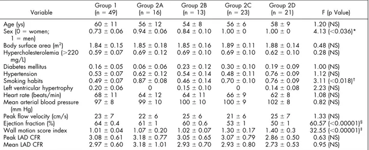
TABLE 1 Study Population, Risk Factors, and Hemodynamic Data
Variable
Group 1
(n⫽ 49) Group 2A(n⫽ 16) Group 2B(n⫽ 13) Group 2C(n⫽ 23) Group 2D(n⫽ 21) F (p Value)
Age (ys) 60⫾ 11 56⫾ 12 54⫾ 8 56⫾ 6 58⫾ 9 1.20 (NS)
Sex (0⫽ women;
1⫽ men) 0.73⫾ 0.06 0.94⫾ 0.06 0.84⫾ 0.10 1.00⫾ 0 1.00⫾ 0 4.13 (⬍0.036)*
Body surface area (m2) 1.84⫾ 0.15 1.85⫾ 0.18 1.85⫾ 0.16 1.89⫾ 0.11 1.88⫾ 0.14 0.48 (NS) Hypercholesterolemia (⬎220 mg/L) 0.59⫾ 0.07 0.69⫾ 0.12 0.69⫾ 0.10 0.69⫾ 0.10 0.62⫾ 0.10 0.28 (NS) Diabetes mellitus 0.16⫾ 0.05 0.06⫾ 0.06 0.23⫾ 0.12 0.30⫾ 0.10 0.19⫾ 0.09 1.00 (NS) Hypertension 0.53⫾ 0.07 0.62⫾ 0.12 0.54⫾ 0.14 0.48⫾ 0.11 0.76⫾ 0.09 1.12 (NS) Smoking habits 0.49⫾ 0.07 0.87⫾ 0.08 0.46⫾ 0.14 0.70⫾ 0.10 0.76⫾ 0.09 3.11 (⬍0.018)†
Left ventricular hypertrophy 0.20⫾ 0.06 0 0.15⫾ 0.10 0 0.14⫾ 0.08 2.23 (NS)
Heart rate (beats/min) 68⫾ 11 64⫾ 12 64⫾ 11 66⫾ 9 62⫾ 8 1.08 (NS)
Mean arterial blood pressure (mm Hg)
97⫾ 8 99⫾ 10 100⫾ 10 100⫾ 9 102⫾ 8 0.82 (NS)
Peak flow velocity (cm/s) 23⫾ 7 22⫾ 6 25⫾ 6 21⫾ 6 25⫾ 7 1.33 (NS)
Ejection fraction (%) 64⫾ 0.4 61⫾ 1 60⫾ 0.6 53⫾ 1 50⫾ 1 60.57 (⬍0.00001)§
Wall motion score index 1.01⫾ 0.04 1.07⫾ 0.20 1.02⫾ 0.07 1.30⫾ 0.17 1.40⫾ 0.3 32.55 (⬍0.00001)‡
Peak LAD CFR 3.08⫾ 0.61 3.18⫾ 0.77 3.05⫾ 0.65 3.07⫾ 0.79 2.86⫾ 0.50 0.63 (NS)
Mean LAD CFR 2.97⫾ 0.60 3.18⫾ 1.01 2.93⫾ 0.70 2.93⫾ 0.80 2.73⫾ 0.53 0.95 (NS)
One-factor analysis of variance was used to test significance. F statistics (and corresponding p values) are shown. In the presence of a significant (p⬍0.05) F, pairwise comparisons were performed using Bonferroni’s correction.
*p⬍0.05, group 1 versus groups 2C and 2D, p ⫽ NS in remaining comparisons.
†p⬍0.10, group 1 versus group 2A, only.
§p⬍0.01, group 1 versus groups 2B, 2C, and 2D, group 2A versus groups 2C and 2D, group 2B versus groups 2C and 2D; and p ⬍0.05, group 2C versus group
2D.
‡p⬍0.01, group 1 versus groups 2C and 2D, group 2A versus groups 2C and 2D, group 2B versus groups 2C and 2D only.
FIGURE 1. CFR is shown individually in control subjects (group 1) and patients with angiographically normal LAD coronary arteries and different types of remote ischemic heart disease (groups 2A, 2B, 2C, and 2D). Confidence intervals of the average value ob-tained in control subjects without risk factors (nⴝ 7, CFR 2.95 ⴞ
0.24, 95% confidence interval 2.77 to 3.13) are indicated by shading to provide a direct comparison with visually estimates of how CFR distributes according to the groups considered. Thick lines represent average values in each subgroup, none of which lies distant from the distribution of control subjects, considered overall or as those without risk factors (see Results and Discus-sion for further details).
remote CFR is a reality, it should trans-late into an unusual clinical scenario in which virtually all patients with acute myocardial infarction have symptoms and positive diagnostic tests for remote ischemia before discharge. Of note, Si-cari et al23 found remote ischemia in
only 96 of 778 patients (10%) who underwent dobutamine stress echocar-diography 12 ⫾ 5 days after myocar-dial infarction.
Other factors may influence re-mote flow in acute myocardial infarc-tion, namely: (1) the culprit artery (right and/or circumflex coronary ar-tery affects LAD flow less than the reverse4), (2) wall motion
abnormal-ities of the jeopardized myocardium,4
(3) tight stenosis of the culprit ar-tery,4 (4) the time elapsed between
the acute event and flow assessment,5
and (5) the reopening of the culprit artery.24In our experience, only wall
motion abnormalities and the reopen-ing of the culprit artery had a slight but nonsignificant influence (Table 1). An impaired CFR in an angio-graphically normal coronary artery has been described in small series of patients with stable remote CAD without previous myocardial infarc-tion.6,7It was mainly attributed to an
early impairment of microvascular function in the presence of angio-graphically undetectable coronary atherosclerosis or to a longstanding adaptation to the increased load in remote regions with structural remod-eling of the coronary vasculature.6,7
Other studies have described an im-pairment of vasomotor tone of the an-giographically normal coronary artery and remote CAD only in patients with coronary risk factors.8
In our series, all patients with an-giographically normal LAD, remote CAD, and no myocardial infarction had a CFR in the LAD within the range of the control group (Figure 1
andTable 1), which was also indepen-dent of risk factors (see confidence intervals in Figure 1) and number of vessels with disease.
We recognize 4 main limitations of our study. (1) The control group consisted of subjects with angio-graphically normal coronary arteries who for some reason underwent diag-nostic coronary angiography. Of note, the CFR of 3.08⫾ 0.61 of this group was similar to that reported by Kern et al25(2.81⫾ 0.61) and Wieneke et al26
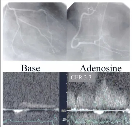
FIGURE 2. (Top) Angiographically normal coronary arteries in a patient in group 1. (Bottom) CFR of the LAD coronary artery, as measured by transthoracic coronary Doppler ultrasound, is 3.3.
FIGURE 3. (Top) Coronary angiography of a patient in group 2B (angiographically normal LAD and remote significant coronary stenosis) shows 95% stenosis of the first obtuse marginal branch (open arrow) and 95% stenosis of the middle tract (solid arrow) of the right coronary artery. (Bottom) CFR of the LAD coronary artery, measured by transthoracic Doppler ultrasound, shows a CFR of 2.9, within the range of the control group.
(3.0 ⫾ 0.8) who used intracoronary Doppler ultrasound in subjects without angiographically detectable CAD and who were similar ages. CFRs of 4.07 ⫾ 0.98 and 3.95 ⫾ 0.68 have been found by positron emission tomogra-phy in healthy volunteers with low risk of CAD, but these series included younger subjects.6,27Therefore, it seems
that advancing age more than subclinical angiographically undetectable athero-sclerosis may affect CFR.26,28 (2) Risk
factors did not significantly affect our results, based on standard cut-off values. However, extremely high levels of cho-lesterol or other risk factors may play a role. (3) This is a single-center study that requires confirmation by data from other laboratories. (4) CFR was measured only in the LAD territory. Recent data have shown that imaging of the posterior descending coronary artery, regardless of its origin from the right or circumflex artery, is feasible.29,30
In conclusion, focal factors in each territory are responsible for CFR, and impaired CFR in 1 region is not a general phenomenon of the coronary circulation.
1.Uren NG, Crake T, Lefroy DC, de Silva R, Davies GJ, Maseri A. Reduced coronary vasodilator function in in-farcted and normal myocardium after myocardial infarc-tion. N Engl J Med 1994;331:222–227.
2.Heras M, Sanz G, Roig E, Perez-Villa F, Recasens L, Serra A, Betriu A. Endothelial dysfunction of the non-infarct related, angiographically normal, coronary artery in patients with an acute myocardial infarction. Eur Heart J 1996;17:715–720.
3.Daher E, Dione DP, Heller EN, Holahan J, DeMan P, Shen M, Hu J, Sinusas AJ. Acute ischemic dysfunction alters coronary flow reserve in remote nonischemic re-gions: potential mechanical etiology identified in an acute canine model. J Nucl Cardiol 2000;7:112–122.
4.Gibson CM, Ryan KA, Murphy SA, Mesley R, Marble SJ, Giugliano RP, Cannon CP, Antman EM, Braunwald E. Impaired coronary blood flow in nonculprit arteries in the setting of acute myocardial infarction. J Am Coll Cardiol 1999;34:974 –982.
5.French JK, Straznicky IT, Webber BJ, Aylward PE, Frey MJ, Adgey AA, Williams BF, McLaughlin SC, White HD, for the HERO-1 Investigators. Angiographic frame counts 90 minutes after streptokinase predict left ventricular function at 48 hours following myocardial infarction. Heart 1999;81:128 –133.
6.Uren NG, Marraccini P, Gistri R, de Silva R, Camici PG. Altered coronary vasodilator reserve and metabolism in myocardium subtended by normal arteries in patients with coronary artery disease. J Am Coll Cardiol 1993;22: 650 – 658.
7. Sambuceti G, Marzullo O, Giorgetti A, Neglia D, Marzilli M, Salvadori P, L’Abbate A, Parodi O. Global alteration in perfusion response to increasing oxygen con-sumption in patients with single vessel coronary artery disease. Circulation 1994;90:1696 –1705.
8. Fujiwara M, Tamura T, Yoshida K, Nakagawa K, Nakao M, Yamanouchi M, Shikama N, Himi T, Masuda Y. Coronary flow reserve in angiographically normal cor-onary arteries with one-vessel corcor-onary artery disease without traditional risk factors. Eur Heart J 2001;22:479 – 487.
FIGURE 4. (Top) Coronary angiography of a patient in group 2C (angiographically normal LAD coronary artery and revascularized remote acute myocardial infarc-tion) shows (1) absence of luminal narrowing of the LAD coronary artery, (2) distal occlusion of the right coronary artery, and (3) reopening of the right coronary ar-tery by primary percutaneous transluminal coronary angioplasty and stenting. (Bot-tom) CFR of the LAD coronary artery, measured by transthoracic Doppler ultra-sound 2 days after reopening and stenting of the right coronary artery, was 2.8, within the range of the control group.
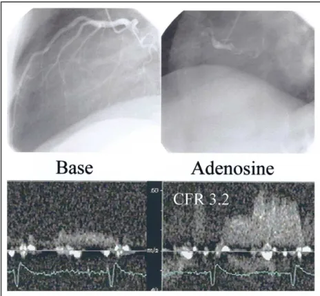
FIGURE 5. (Top) Coronary angiography of a patient in group 2D (angiographically normal LAD coronary artery and non-revascularized remote myocardial infarction) shows absence of luminal narrowing of the LAD coronary artery and a proximally occluded right coronary artery. (Bottom) CFR of the LAD coronary artery, measured by transthoracic Doppler ultrasound, shows a CFR of 3.2, within the range of the control group.
9.Voci P, Testa G, Plaustro G, Campa PP, Marino B. Studio del flusso coronarico con ecocardiografia transtoracica ad alta risoluzione e Doppler non direzionale. Cardiologia 1997;42:849 – 853.
10.Voci P, Testa G, Plaustro G, Caretta Q. Coronary Doppler intensity changes during handgrip: a new method to detect coronary vasomotor tone in coronary artery disease. J Am Coll Cardiol 1999;34:428 – 434.
11.Voci P, Mariano E, Pizzuto F, Puddu PE, Romeo F. Coronary recanalization in anterior myocardial infarction. The open perforator hypothesis. J Am Coll Cardiol 2002;40:1205–1213.
12.Hozumi T, Yoshida K, Ogata Y, Akasaka T, Asami Y, Takagi T, Morioka S. Noninvasive assessment of significant left anterior descending coronary artery stenosis by coronary flow velocity reserve with transthoracic color Doppler echocardiography. Circulation 1998;97:1557–1562.
13.Hozumi T, Yoshida K, Akasaka T, Asami Y, Ogata Y, Takagi T, Kaji S, Kawamoto T, Ueda Y, Morioka S. Noninvasive assessment of coronary flow velocity and coronary flow velocity reserve in the left anterior descending coronary artery by Doppler echocardiography. J Am Coll Cardiol 1998;32:1251– 1259.
14.Caiati C, Montaldo C, Zedda N, Bina A, Iliceto S. New noninvasive method for coronary flow reserve assessment. Contrast-enhanced transthoracic second harmonic echo Doppler. Circulation 1999;99:771–778.
15.Voci P, Pizzuto F, Mariano E, Puddu PE, Sardella G, Romeo F. Coronary flow reserve measured by transthoracic coronary Doppler ultrasound accurately detects severe left anterior descending coronary artery stenosis. Am J Cardiol 2003;92:1320 –1324.
16.Pizzuto F, Voci P, Mariano E, Puddu PE, Sardella G, Nigri A. Assessment of flow velocity reserve by transthoracic Doppler echocardiography and venous adenosine infusion before and after left anterior descending coronary artery stenting. J Am Coll Cardiol 2001;38:155–162.
17.Pizzuto F, Voci P, Mariano E, Puddu PE, Chiavari PA, Romeo F. Noninva-sive coronary flow reserve assessed by transthoracic coronary Doppler ultrasound in patients with a left anterior descending coronary artery stent. Am J Cardiol 2003;91:522–526.
18.Schiller N, Shah P, Crawford M, De Maria A, Devereux R, Feigenbaum H, Gutgesell R, Reichek N, Sahn D, Schnittger I, et al, for the American Society of Echocardiography Committee of Standards, Subcommittee on Quantification of 2-dimensional Echocardiograms. Recommendation for quantification of the left ventricle by 2-dimensional echocardiography. J Am Soc Echocardiogr 1989;5: 358 –367.
19.Bergmann SR, Herrero P, Markham P, et al. Noninvasive quantification of myocardial blood flow in human subjects with15O-labeled water and positron
emission tomography. J Am Coll Cardiol 1989;14:639 – 652.
20.Reis ES, Holubkov R, Lee JS, Sharaf B, Reichek N, Rogers WJ, Walsh EG, Fuisz AR, Kerensky R, Detre KM, et al. Coronary flow velocity response to
adenosine characterizes coronary microvascular function in women with chest pain and no obstructive coronary disease: results from the pilot phase of Women’s Ischemia Syndrome Evaluation (WISE) Study. J Am Coll Cardiol 1999;33:1469 –1475.
21. Lepper W, Hoffmann R, Kamp O, Franke A, de Cock CC, Kuhl HP, Sierswerda G, vom Dahl J, Janssens U, Voci P, et al. Assessment of myocardial reperfusion by intravenous myocardial contrast echocardiography and coronary flow reserve after primary percutaneous transluminal coronary angioplasty in patients with acute myocardial infarction. Circulation 2000;101:2368 –2374.
22. Neumann FJ, Kosa I, Dickfeld T, Blasini R, Gawaz M, Hausleiter J, Schwaiger M, Schömig A. Recovery of myocardial perfusion in acute myocardial infarction after successful balloon angioplasty and stent placement in the infarct-related coronary artery. J Am Coll Cardiol 1997;30:1270 –1276.
23.Sicari R, Picano E, Landi P, Pingitore A, Bigi R, Coletta C, Heyman J, Casazza F, Previtali M, Mathias W, et al, on the behalf of the EDIC Study. Prognostic value of dobutamine-atropine stress echocardiography early after acute myocardial infarction. J Am Coll Cardiol 1997;29:254 –260.
24.Gregorini L, Marco J, Kozakova M, Palombo C, Anguissola GB, Marco I, Bernies M, Cassagneau B, Distante A, Bossi IM, et al. Alpha-adrenergic blockade improves recovery of myocardial perfusion and function after coronary stenting in patients with acute myocardial infarction. Circulation 1999;99:482– 490.
25.Kern MJ, Bach RG, Mechem CJ, Caracciolo EA, Aguirre FV, Miller LW, Donhoue TJ. Variations in normal coronary vasodilatory reserve stratified by artery, gender, heart transplantation and coronary artery disease. J Am Coll Cardiol 1996;28:1154 –1160.
26.Wieneke H, Haude M, Ge J, Altmann C, Kaiser S, Baumgart D, von Birgelen C, Welge D, Erbel R. Corrected coronary flow velocity reserve: a new concept for assessing coronary perfusion. J Am Coll Cardiol 2000;35:1713–1720.
27.Kubo S, Tadamura E, Toyoda H, Malcero M, Mukai T, Magata Y, Kitano H, Iida H, Tamaki N, Konishi J. Comparison of myocardial blood flow during adenosine triphosphate and dobutamine infusion and after dipyridamole admin-istration in normal men. Circulation 2002;106:II-618 –II-619.
28.Czernin J, Müller P, Chan S, Brunken RC, Porenta G, Krivokapich J, Chen K, Chan A, Phelps ME, Schelbert HR. Influence of age and hemodynamics on myocardial blood flow and flow reserve. Circulation 1993;88:62– 69.
29.Voci P, Pizzuto F, Mariano E, Puddu PE, Chiavari PA, Romeo F. Measure-ment of coronary flow reserve in the anterior and posterior descending coronary arteries by transthoracic Doppler ultrasound. Am J Cardiol 2002;90:988 –991.
30.Lethen H, Tries HP, Kersting S, Lambertz H. Validation of noninvasive assessment of coronary flow velocity reserve in the right coronary artery. A comparison of transthoracic echocardiographic results with intracoronary Dopp-ler flow wire measurements. Eur Heart J 2003;24:1567–1575.