doi: 10.3389/fnagi.2020.00068
Edited by: Simone Engelender, Technion Israel Institute of Technology, Israel Reviewed by: Anna Maria Colangelo, University of Milano-Bicocca, Italy Weili Yang, Jinan University, China *Correspondence: Arianna Bellucci [email protected] Received: 18 December 2019 Accepted: 25 February 2020 Published: 24 March 2020 Citation: Bellucci A, Bubacco L, Longhena F, Parrella E, Faustini G, Porrini V, Bono F, Missale C and Pizzi M (2020) Nuclear Factor-κB Dysregulation andα-Synuclein Pathology: Critical Interplay in the Pathogenesis of Parkinson’s Disease. Front. Aging Neurosci. 12:68. doi: 10.3389/fnagi.2020.00068
Nuclear Factor-
κB Dysregulation and
α-Synuclein Pathology: Critical
Interplay in the Pathogenesis of
Parkinson’s Disease
Arianna Bellucci
1* , Luigi Bubacco
2, Francesca Longhena
1, Edoardo Parrella
1,
Gaia Faustini
1, Vanessa Porrini
1, Federica Bono
1, Cristina Missale
1and Marina Pizzi
11Division of Pharmacology, Department of Molecular and Translational Medicine, University of Brescia, Brescia, Italy, 2Department of Biology, University of Padua, Padua, Italy
The loss of dopaminergic neurons of the nigrostriatal system underlies the onset of
the typical motor symptoms of Parkinson’s disease (PD). Lewy bodies (LB) and Lewy
neurites (LN), proteinaceous inclusions mainly composed of insoluble
α-synuclein (α-syn)
fibrils are key neuropathological hallmarks of the brain of affected patients. Compelling
evidence supports that in the early prodromal phases of PD, synaptic terminal and
axonal alterations initiate and drive a retrograde degeneration process culminating
with the loss of nigral dopaminergic neurons. This notwithstanding, the molecular
triggers remain to be fully elucidated. Although it has been shown that
α-syn fibrillary
aggregation can induce early synaptic and axonal impairment and cause nigrostriatal
degeneration, we still ignore how and why
α-syn fibrillation begins. Nuclear
factor-κB (NF-factor-κB) transcription factors, key regulators of inflammation and apoptosis, are
involved in the brain programming of systemic aging as well as in the pathogenesis of
several neurodegenerative diseases. The NF-
κB family of factors consists of five different
subunits (c-Rel, p65/RelA, p50, RelB, and p52), which combine to form transcriptionally
active dimers. Different findings point out a role of RelA in PD. Interestingly, the nuclear
content of RelA is abnormally increased in nigral dopamine (DA) neurons and glial cells of
PD patients. Inhibition of RelA exert neuroprotection against
(1-methyl-4-phenyl-1,2,3,6-tetrahydropyridine) MPTP and 1-methyl-4-phenylpyridinium (MPP+) toxicity, suggesting
that this factor decreases neuronal resilience. Conversely, the c-Rel subunit can exert
neuroprotective actions. We recently described that mice deficient for c-Rel develop
a PD-like motor and non-motor phenotype characterized by progressive brain
α-syn
accumulation and early synaptic changes preceding the frank loss of nigrostriatal
neurons. This evidence supports that dysregulations in this transcription factors may be
involved in the onset of PD. This review highlights observations supporting a possible
interplay between NF-
κB dysregulation and α-syn pathology in PD, with the aim to
disclose novel potential mechanisms involved in the pathogenesis of this disorder.
INTRODUCTION
The therapeutic management of Parkinson’s disease (PD) patients
is one of the major challenges in the field of neurodegenerative
diseases nowadays (
Orayj and Lane, 2019
). This is mostly
related to two determinants. (1) Although since many years
the administration of levodopa (
L-Dopa) has allowed motor
symptom recovery, its efficacy has a limited time window and
associates with severe adverse effects, thus gathering the unmet
need of novel interventions (
Olanow and Stocchi, 2018
). (2) In
spite of more than 200 years of research in the field of PD, we
still have poor discernment on the main pathogenic mechanisms.
The only certainties we own relate to the neuropathological
features of post-mortem PD brains. These present a prominent
loss of dopaminergic nigrostriatal neurons and contain profuse
insoluble proteinaceous aggregates named Lewy bodies (LB) and
Lewy neurites (LN), likely originating from fibrillary
α-synuclein
(
α-syn) protein accumulation in cell bodies and neurites,
respectively (
Spillantini et al., 1997
;
Spillantini and Goedert,
2000
). This notwithstanding, we ignore the key molecular
determinants initiating the degeneration of nigrostriatal neurons
and/or
α-syn aggregation and deposition. The molecular features
of the toxic
α-syn species driving neurodegeneration are also an
unsolved conundrum (
Longhena et al., 2019
).
Investigations on experimental models reproducing human
α-syn aggregate accumulation, or transgenic expression of
mutant forms of the protein associated with familial PD as
well as more extensive neuropathological examinations and
brain imaging studies in patients, have disclosed that the most
probable initiation sites for neuronal degeneration in PD are
synapses and axonal projections (for review, please see
Bellucci
et al., 2017
;
Uchihara, 2017
;
Wong et al., 2019
). Moreover,
the selective neuronal vulnerability of nigral dopaminergic
neurons in PD has been proposed to derive from their massive
neuronal arborization and peculiar metabolic and functional
profile (
Surmeier et al., 2017
;
Wong et al., 2019
). In this
scenario,
α-syn pathological aggregation at synapses appears to
play a major role in triggering dopaminergic neuron dysfunction,
flowing in a retrograde degeneration pattern involving axonal
projections first and culminating with neuronal cell death
(
Bellucci et al., 2016, 2017
;
Wong et al., 2019
). In particular,
the pivotal role exerted by
α-syn within dopaminergic neurons
terminals as a regulator of neurotransmitter synthesis, reuptake
and vesicle storage or motility (
Bellucci et al., 2012b
;
Longhena
et al., 2019
), hints that this neuronal population may be more
vulnerable to minimal perturbations of functional homeostasis
induced by
α-syn accumulation and aggregation. Moreover, the
massive energy consumption rate of dopaminergic neurons has
to be combined with an enhanced mitochondrial bioenergetics
support. This is lost upon synapse degeneration in the brain
of PD patients, although it appears to be upregulated with a
compensatory fashion in the remaining terminals (
Reeve et al.,
2018
). On this line, findings indicating that
α-syn physiologically
regulates mitochondrial homeostasis (
Guardia-Laguarta et al.,
2014
;
Faustini et al., 2019
), while its aggregation prompts
mitochondrial dysfunction (
Nakamura et al., 2011
;
Tapias et al.,
2017
;
Wang et al., 2019
), support that early pathological shifts
in the conformation of this protein, leading to its loss of
function and accumulation, may hinder the bioenergetics profile
of dopaminergic neurons, compromising their resilience along
aging. This hypothesis offers a bridge between two key biological
processes that are thought to participate in PD pathogenesis:
mitochondrial alterations and
α-syn deposition, though it is
still arduous to determine who is on first, as mitochondrial
impairment can also trigger
α-syn pathological aggregation
(
Betarbet et al., 2002
;
Zaltieri et al., 2015
;
Faustini et al., 2018
;
Grunewald et al., 2019
).
Beside
α-syn pathology and mitochondrial impairment,
numerous other mechanisms have been proposed to contribute
to sporadic PD pathogenesis, including neuroinflammation,
impaired autophagy, and oxidative stress (
Pang et al., 2019
).
Interestingly, some of these molecular pathways appear to be
cross-linked and can be regulated by nuclear factor
κB
(NF-κB) transcription factors (
Kratsovnik et al., 2005
;
Djavaheri-Mergny et al., 2007
;
Sarnico et al., 2009a
;
Morgan and Liu,
2011
;
Lanzillotta et al., 2015
;
Kaminska et al., 2016
;
Nivon
et al., 2016
;
Lingappan, 2018
;
Nandy et al., 2018
). In particular,
oxidative stress can activate NF-
κB-mediated protective signaling
(
Kratsovnik et al., 2005
;
Lingappan, 2018
), which can repress
autophagy as well as autophagy-dependent apoptosis (
Djavaheri-Mergny et al., 2007
;
Nandy et al., 2018
). On the other hand,
NF-κB-regulated genes play a major role in controlling the amount
of ROS in the cell (
Morgan and Liu, 2011
;
Lingappan, 2018
),
and by modulating autophagic activity, this factor appears as a
key regulator of protein aggregate clearance (
Nivon et al., 2016
).
Upon exposure of microglia to lipopolysaccharide (LPS),
NF-κB is among the predominantly activated signaling pathways
and initiates the transcription of proinflammatory gene coding
for cytokines and proteolytic enzymes (
Kaminska et al., 2016
).
However, the different composition of NF-
κB dimers imprints
either the protective or the noxious action of this factor. While
p50/RelA dimers induce pro-apoptotic Bim and Noxa genes,
c-Rel-containing dimers exert neuroprotective actions (
Inta et al.,
2006
;
Sarnico et al., 2009b
).
NF-
κB factors play a crucial role in the regulation of
inflammation and apoptosis, are involved in the brain
programming of systemic aging, as well as in the pathogenesis
of several neurodegenerative diseases (
Sarnico et al., 2009a
;
Mincheva-Tasheva and Soler, 2013
;
Lanzillotta et al., 2015
).
The NF-
κB family of transcription factors consists of five
different subunits (c-Rel, p65/RelA, p50, RelB, and p52),
which interact to form transcriptionally active homo and
heterodimers (
Perkins, 1997
;
Chen and Greene, 2004
;
Ghosh
et al., 2012
). Different findings point to a role of NF-
κB/RelA
in PD. Interestingly, the nuclear content of RelA is abnormally
increased in nigral dopaminergic neurons and glial cells
of PD patients. Inhibition of RelA prevents dopaminergic
neuron loss in a 1-methyl-4-phenyl-1,2,3,6-tetrahydropyridine
(MPTP)-mouse model of PD, and downregulation of RelA
protects neurons from 1-methyl-4-phenylpyridinium (MPP+)
toxicity, suggesting that RelA upregulation, may play a role
in dopaminergic neuron degeneration (
Ghosh et al., 2007
).
Moreover, we recently described that mice deficient for c-Rel
subunit, which can exert pro-survival effects (
Pizzi et al., 2002
;
Sarnico et al., 2009b
), develop a PD-like motor and
non-motor phenotype characterized by progressive brain
α-syn
accumulation and early synaptic changes preceding the frank
loss of nigrostriatal neurons (
Baiguera et al., 2012
;
Parrella et al.,
2019
). This evidence hints that a reduction in the protective
function of c-Rel may render dopaminergic neurons more
vulnerable to aging, the primary risk factor for PD (
Collier
et al., 2011
;
Reeve et al., 2014
), thus predisposing toward the
development of this disorder.
This review provides an updated critical overview of findings
supporting a possible interplay between NF-
κB dysregulation and
α-syn pathology in PD pathogenesis, with the aim to uncover
and discuss novel potential molecular mechanisms involved
in this process.
α-SYNUCLEIN: PHYSIOLOGICAL
FUNCTION AND ROLE IN PD
α-synuclein is a neuronal protein mainly localized at synaptic
sites. Its physiological functions seem to be mostly related
with regulation of neurotransmitter release and recycle, as it
modulates the size, assembly, and release of synaptic vesicle
pools (
Murphy et al., 2000
;
Burre, 2015
;
Fusco et al., 2016
),
neurotransmitter reuptake (
Burre, 2015
;
Longhena et al., 2019
),
exocytotic fusion pore dilation (
Logan et al., 2017
), and
neurotransmitter vesicular uptake (
Guo et al., 2008
;
Pifl et al.,
2014
;
Phan et al., 2017
). Remarkably, the multiplicity of
α-syn
interactions at synaptic sites, coupled with its intrinsic structural
plasticity, can account for the cardinal role of the protein at
terminals (
Bendor et al., 2013
;
Longhena et al., 2019
;
Sulzer and
Edwards, 2019
). Studies in PD brains and experimental models
have shown that
α-syn overexpression and aggregation induce
significant alterations of synaptic proteins, neuronal dysfunction
and degeneration as well as motor deficits (
Fleming et al., 2004
;
Garcia-Reitbock et al., 2010
;
Bellucci et al., 2011
;
Lam et al., 2011
;
Lundblad et al., 2012
;
Wang et al., 2014
;
Visanji et al., 2016
).
Nonetheless,
α-syn has been found to localize in and
affect other cellular compartments aside synapses. In the
nucleus, the protein physiologically interacts with and regulates
histones (
Goers et al., 2003
;
Kontopoulos et al., 2006
;
Schaser
et al., 2019
). On the other hand, its overexpression and
phosphorylation modulates gene expression (
Pinho et al., 2019
),
impairs the neuroprotective NF-
κB signaling pathway (
Yuan
et al., 2008
), and regulates the promoter of proliferator-activated
receptor gamma coactivator 1
α (PGC1α), a transcription factor
governing mitochondria biogenesis, to inhibit its transcription
(
Siddiqui et al., 2012
).
The overexpression of wild-type (wt)
α-syn, and even more
of its A53T mutated form, affects endoplasmic reticulum
(ER)/Golgi transport by direct binding of the soluble proteins to
ER/Golgi soluble
N-ethylmaleimide-sensitive factor attachment
protein receptors (SNAREs), resulting in their inhibition
(
Thayanidhi et al., 2010
). Interestingly, this effect appears to be
rescued by Rab1 overexpression and is responsible for lysosomal
dysfunction onset (
Cooper et al., 2006
;
Martinez-Vicente et al.,
2008
;
Mazzulli et al., 2016
;
Hoffmann et al., 2019
). Alterations in
Golgi morphology and increased susceptibility to ER stress have
been also found to occur in dopaminergic cells overexpressing
A30P
α-syn (
Paiva et al., 2018
).
In addition, we showed that overexpression of wt
α-syn,
leading to formation of insoluble aggregates within the ER,
activates the protein kinase RNA-like ER kinase (PERK)-related
pathway of the unfolded protein response (UPR) (
Bellucci
et al., 2011
). Upon misfolded protein accumulation in the ER,
the induced UPR inhibits protein synthesis and generation
of molecular chaperones implicated in protein folding, whose
activation may ultimately lead to apoptotic cell death. These
findings have been further corroborated by other studies
supporting that ER stress is relevant for the manifestations of
synucleinopathy
in vivo (
Colla et al., 2012a,b
;
Mercado et al.,
2013
). For these main reasons, therapeutic strategies targeting the
UPR have been proposed as PD treatments (
Bellucci et al., 2012a
;
Mercado et al., 2018
;
Martinez et al., 2019
).
α-synuclein can also bind mitochondria-associated ER
membranes (MAM) (
Guardia-Laguarta et al., 2014
), and
evidence indicating that its absence impairs mitochondria lipid
composition, function, fusion, and trafficking (
Ellis et al.,
2005
;
Faustini et al., 2019
) supports that the protein can
play a physiological role in the regulation of mitochondrial
homeostasis. This hypothesis is in line with findings showing
that pathological
α-syn aggregates or oligomers produce
mitochondria fragmentation, impair mitochondrial trafficking,
and lead to respiration failure (
Devi et al., 2008
;
Nakamura et al.,
2011
;
Tapias et al., 2017
;
Prots et al., 2018
;
Wang et al., 2019
).
Therefore, although synapses, which result the sites where
α-syn is most abundant, are likely the first sites to be affected
by and suffer from its pathological changes, it is reasonable
to prospect that
α-syn aggregation and toxicity may proceed
progressively through the induction of dysfunctional alterations
to other cellular compartments. This view implies that a finer
characterization of the pathological changes occurring after
the initiation of
α-syn aggregation may bring new insight
into PD pathogenesis. On the other hand, it reinforces that
studies exploring the factors leading or predisposing to
α-syn
accumulation and aggregation along aging are even more
important, as they may help us to identify new targets for the
development of therapies halting PD progression.
Another peculiarity of
α-syn relates to its transmission from
cell-to-cell and to the spreading of its pathological aggregates
from the peripheral nervous system (PNS) to the central nervous
system (CNS), or vice versa, in experimental models (
Luk et al.,
2012
;
Ulusoy et al., 2013, 2017
;
Recasens and Dehay, 2014
;
Dehay
et al., 2016
;
Emmanouilidou and Vekrellis, 2016
;
Helwig et al.,
2016
;
Cavaliere et al., 2017
;
Rutherford et al., 2017
;
Grozdanov
and Danzer, 2018
). The capability of pathological forms of
α-syn to spread seems to be corroborated by neuropathological
examination of post-mortem brains from patients who received
fetal neuron grafts over one decade prior to death and showing
the development of LB within grafted neuronal cells (
Kordower
et al., 2008a,b
;
Chu and Kordower, 2010
;
Li et al., 2010
;
Kurowska et al., 2011
). These findings, together with the Braak
hypothesis, suggesting that the progression of PD symptoms
relates with the caudo-rostral diffusion of LB pathology in the
brain (
Braak et al., 2003, 2018
), fed the shoot up of the
prion-like hypothesis of PD, emphasizing the multiple similarities
between
α-syn and prion protein (
Olanow and Brundin, 2013
;
Brundin et al., 2016
;
Brundin and Melki, 2017
). However,
studies showing that PD patients exhibit a systemic
α-syn
neuropathology within both PNS and CNS (
Gelpi et al., 2014
;
Surguchov, 2016
) and a critical analysis of neuronal vulnerability
to
α-syn accumulation, were supportive for the development of
alternative hypotheses as presented by
Engelender and Isacson
(2017)
and
Surmeier et al. (2017)
. Very recent findings showing
that systemic delivery of
α-syn synthetic pre-formed fibrils in
rats trigger pathological transformation of endogenous
α-syn,
leading to neurodegeneration in discrete CNS and PNS neuronal
populations (
Kuan et al., 2019
), bring novel insights into this
subject, opening the way to a more exhaustive comprehension of
the role and relevance of
α-syn spreading in PD.
NF-
κB FACTORS: TRANSCRIPTIONAL
REGULATORS GOVERNING
NEUROINFLAMMATION, APOPTOSIS,
NEURONAL FUNCTION AND
RESILIENCE
NF-
κB is expressed in both the CNS and PNS and localizes
in neurons, glial cells, and Schwann cells mostly as p50/p50
homodimers and p50/RelA heterodimers (
Meffert and Baltimore,
2005
). By regulating synaptic signaling and behavior, or pivotally
controlling cell survival and glial cell activation, the NF-κB family
of transcription factors is crucially involved in the regulation of
CNS and PNS response to physiological and pathological stimuli
(
Meffert et al., 2003
;
Meffert and Baltimore, 2005
;
Mattson and
Meffert, 2006
;
Mincheva-Tasheva and Soler, 2013
).
NF-κB factors are ubiquitously expressed in mammalian cells,
although they were first identified in lymphocytes (
Meffert and
Baltimore, 2005
). All the NF-
κB subunits, highly conserved
across species, show a Rel homology domain containing the
key functional regions for DNA binding, dimerization, nuclear
translocation, and interaction with their inhibitory elements
named I
κB. Only RelA, c-Rel, and RelB show the C-terminal
transactivation domain (TAD) that allows the dimer to initiate
transcription (
Hayden and Ghosh, 2012
). Although p52 and
p50 lack TADs, their heterodimerization with TAD-containing
NF-
κB subunits, or interaction with non-Rel proteins that have
trans-activating capability, allows them to positively regulate
transcription. The homodimers composed by p50 and p52 can
also negatively regulate transcription by competing with
TAD-containing dimers for binding to
κB sites or by constitutively
occupying some
κB sites to increase the activation threshold for
certain NF-κB target genes (
Hayden and Ghosh, 2012
).
Inactive NF-
κB dimers localize in the cytoplasm bound to
the I
κB inhibitory proteins. NF-κB-inducing stimuli activate the
I
κB kinase complex, IKKα and IKKβ, with the regulatory
IKK
γ/NEMO, resulting in sequential phosphorylation,
ubiquitination, and degradation of I
κB. Upon the detachment
of I
κB, the exposure of DNA-binding domain and nuclear
localization sequence allows the NF-
κB dimer to translocate to
the nucleus to bind the target gene promoter regions (
Karin
et al., 2004
;
Meffert and Baltimore, 2005
).
A body of evidence has shown that NF-
κB plays a relevant
role in regulating the function of immune system by driving
both the diverse inflammatory phases and the host defense
(
Li and Verma, 2002
).
In neurons, the transcription of both p50/RelA and p50/p50
NF-
κB dimers can be activated by glutamatergic synaptic
inputs through Ca
2+/calmodulin-dependent protein kinase II
(CaMKII) and local submembranous Ca
2+increase (
Meffert
et al., 2003
). While the p50/p50 dimers localize in the cytosol, the
p50/RelA dimers are found within synaptic boutons from where,
upon glutamate or
N-methyl-
D-aspartate (NMDA) stimulation,
they translocate to reach the nucleus and translate synaptic
signals into altered gene expression (
Meffert et al., 2003
).
Consistently, RelA knockout (ko) mice exhibit spatial learning
deficits, thus supporting that NF-
κB nuclear translocation and
gene activation govern long-term changes to adult neuronal
function caused by synaptic stimulation.
NF-
κB controls adult neurogenesis in CNS (
Rolls et al.,
2007
;
Koo et al., 2010
), and evidence supporting that these
transcriptional regulators are important for ensuring Schwann
cell differentiation and myelination of peripheral axons suggests
that NF-
κB factors are essential differentiation signals with a
prominent role also in PNS development and plasticity (
Nickols
et al., 2003
;
Tang et al., 2013
).
It has been found that most of the stimuli that activate
NF-κB in the immune system, such as tumor necrosis factor-α
(TNF-α) or interleukin 1 (IL-1), viral infections, and oxidative
stress, exert the same effect in the CNS. Though surprisingly,
TNF-
α-mediated NF-κB activation plays a unique role in
mediating neuronal plasticity in the hippocampus without
inducing neuroinflammatory changes (
Albensi and Mattson,
2000
;
Beattie et al., 2002
). The NF-κB target genes in the CNS
are only partially characterized, but it is predictable that these
may display significant differences in their promoter organization
when compared to the canonical genes affected in the immune
system. On this line, we previously showed the presence of two
NF-
κB sites within the regulatory region of the DA D2 receptor
(
Bontempi et al., 2007
) as a proof of the NF-
κB involvement in the
regulation of neuronal responses to DA-mediated transmission.
In glial cells, basal NF-
κB activity is very low. For this reason,
most studies have focused glial NF-
κB in models of inflammation,
injury, or disease (
Dresselhaus and Meffert, 2019
) where it is
activated in its predominant form, the p50/RelA dimer (
Kiebala
et al., 2010
;
Simmons et al., 2016
;
Gupta et al., 2019
). In
inflammation, microglia activation results in the transcription
of NF-
κB-target genes, nitric oxide, IL-1β, and TNFα that in
turn induce NF-
κB signaling with consequent enhancement of
inflammatory mediators that exacerbate neuronal cell death.
NF-κB pathway in microglia seems also to actively participate in
plasticity mechanisms and neuronal homeostasis in response
to injury (
Dresselhaus and Meffert, 2019
). Likewise, beside
inducing pro-inflammatory gene expression, astrocytic NF-
κB
appears to play a role in the central control of metabolism
(
Zhang et al., 2017
) and, by promoting the clearing of glutamate
from the synapses, in the termination of excitatory signals
(
Ghosh et al., 2011
).
NF-κB has been reported to be essential for systemic and brain
aging (
Adler et al., 2008
;
Zhang et al., 2013
), with RelA subunit
mediating the most significant contribution to degenerative
changes associated with senescence (
Tilstra et al., 2012
). On
this line, NF-
κB dysregulation has been found to participate in
brain neurodegenerative mechanisms occurring in PD (
Hunot
et al., 1997
), Alzheimer’s disease (AD) (
Kaltschmidt et al., 1997
;
Chen et al., 2012
;
Jones and Kounatidis, 2017
), as well as in
post-traumatic or post-ischemic brain injury (
Bethea et al., 1998
;
Schneider et al., 1999
). With regard to the regulation of neuronal
cell death, diverse NF-
κB dimers in response to specific stimuli
can mediate distinct responses (
Pizzi et al., 2005
;
Lanzillotta
et al., 2015
). We showed that the selective inhibition of RelA or
c-Rel expression produces opposite effects on neuron survival.
Whereas the over-activation of p50/RelA dimers promotes
apoptosis, activation of c-Rel-containing dimers improves the
resilience of neuronal cells after injury (
Pizzi et al., 2002, 2005
;
Sarnico et al., 2009b
). Neurotoxic stimuli, such as ischemia,
high glutamate concentrations,
β-amyloid, or MPP+, induce the
activation of p50/RelA dimers improving the transcription of
proapoptotic genes (
Pizzi et al., 2002, 2005
;
Inta et al., 2006
;
Valerio et al., 2006
;
Sarnico et al., 2008, 2009b
;
Yang et al.,
2010
). Conversely, c-Rel-containing dimers favor the expression
of anti-apoptotic genes by signals promoting neuroprotection in
diverse cell-based neurotoxic settings, such as IL-1
β in
NMDA-mediated excitotoxicity, mGlu5 receptor agonists in
β-amyloid-or MPP+-mediated toxicity and adipocyte-derived hβ-amyloid-ormone
leptin in oxygen and glucose deprivation (OGD)-mediated
apoptosis (
Pizzi et al., 2002, 2005
;
Kogel et al., 2004
;
Valerio et al.,
2006
;
Sarnico et al., 2008
). Furthermore, the over-expression
of c-Rel in cultured neurons promotes anti-apoptotic effects by
inducing manganese superoxide dismutase (MnSOD) and Bcl-xL
(
Bernard et al., 2001
;
Pizzi et al., 2005
). Overabundance of c-Rel
also limits the generation of reactive oxygen species (ROS) by
inducing transcription of the mitochondrial uncoupling proteins
4 (UCP4) (
Ho et al., 2012
), a brain-specific mitochondrial ion
channel producing mild reduction in mitochondrial membrane
potential and neuroprotection (
Echtay, 2007
).
The dual effects of NF-
κB activation on neuron survival
were corroborated by studies in severe brain ischemia models.
Indeed, a rapid activation of p50/RelA in both neurons and
glial cells has been implicated in the pathogenesis of
post-ischemic injury (
Herrmann et al., 2005
;
Crack et al., 2006
). In
ischemic brain tissue of mice subjected to permanent middle
cerebral artery occlusion (MCAO) and in primary cortical
neurons exposed to OGD, NF-
κB followed a similar pattern of
activation (
Pizzi et al., 2009
;
Lanzillotta et al., 2010
) characterized
by increased nuclear translocation of p50/RelA dimers (
Inta
et al., 2006
;
Pizzi et al., 2009
) and decreased translocation
of c-Rel-containing dimers (
Sarnico et al., 2009b
). In these
conditions, NF-
κB activity was associated with an unbalanced
expression of pro-apoptotic RelA target genes, with increased
expression of the pro-apoptotic members of Bcl-2 family genes,
Bim and Noxa, and parallel reduction of the anti-apoptotic
member Bcl-xL (
Cao et al., 2002
;
Inta et al., 2006
;
Sarnico et al.,
2009b
). During brain ischemia, RelA induced the expression
of the 1B isoform of divalent metal transporter-1 (1B/DMT1),
the membrane carrier responsible for iron accumulation and
brain damage after injury (
Ingrassia et al., 2012
). This acted
as an upstream mechanism responsible for iron accumulation
and contributing to neuronal cell death. Knocking-down c-Rel
expression exacerbated neuronal susceptibility to OGD-mediated
damage and c-Rel ko mice exposed to cerebral ischemia resulted
insensitive to the neuroprotective activity of leptin, a c-Rel
inducer capable of limiting cortical damage in wt mice (
Valerio
et al., 2006, 2009
). These data strongly suggested that inhibition
of c-Rel-containing dimers and activation of p50/RelA are key
events in the pathogenesis of post-ischemic brain injury.
Despite these premises, p50/RelA activation
per se appeared to
be insufficient to drive pro-apoptotic transcription during brain
ischemia. Site-specific acetylation of RelA at the Lys 310 residue
was necessary to switch anti-apoptotic p50/RelA, activated after
a brief preconditioning ischemia and leading to brain tolerance
toward the pro-apoptotic p50/RelA activated after a prolonged
harmful brain ischemia (
Blondeau et al., 2001
;
Lanzillotta et al.,
2010
). In this regard, studies showing that post-stroke induction
of
α-syn mediates ischemic brain damage (
Ishimaru et al., 1998
;
Hu et al., 2006
;
Yoon et al., 2006
;
Unal-Cevik et al., 2011
;
Surgucheva et al., 2014
;
Kim T. et al., 2016
) and that the levels
of oligomeric form of
α-syn of red blood cells in ischemic stroke
and PD patients are both significantly higher than in controls
(
Zhao et al., 2016
), seem to shed light on a possible link between
NF-
κB dysregulation and α-syn accumulation that deserves to be
addressed by
ad hoc studies.
NF-
κB ALTERATIONS IN PD AND THEIR
LINK TO
α-SYN PATHOLOGY
A body of evidence has highlighted the occurrence of changes
and dysregulation of NF-
κB in PD. Post-mortem studies showed
that in the brain of patients, there is an increased RelA nuclear
translocation in melanized neurons of the
substantia nigra that
is supportive of NF-
κB activation in PD (
Hunot et al., 1997
).
Later studies, addressing glial involvement in the degeneration
process of LB-bearing neurons on the post-mortem brain of
patients affected by dementia with LB (DLB), found that some LB
present NF-
κB immunopositivity (
Togo et al., 2001
), thus hinting
that NF-
κB may participate in the α-syn-deposition-dependent
neuronal loss. These findings were corroborated by other studies
showing that either RelA or active phosphorylated NF-κB can
be detected in the nucleus of subpopulations of neurons and
glial cells of the
substantia nigra of PD patients (
Ghosh et al.,
2007
;
Garcia-Esparcia et al., 2014
). On this line, it has been
described that
α-syn can be internalized by microglia and
induces nuclear accumulation of RelA (
Cao et al., 2010, 2012
).
Moreover, Ghosh and co-authors found NF-
κB activation in the
substantia nigra of MPTP-treated mice. Mice treatment with a
peptide corresponding to the NEMO-binding domain (NBD)
of IKK
α or IKKβ, which acts as a selective NF-κB inhibitor,
reduced microglia activation in the
substantia nigra, prevented
both nigrostriatal axis degeneration, DA loss and improved
motor functions (
Ghosh et al., 2007
). These findings confirmed
that, by modulating microglia activation, NF-
κB deregulation
may play a role in MPTP mouse model. Consistently,
Wang
et al. (2020)
have recently corroborated that c-Rel is rapidly
upregulated in the
substantia nigra and striatum of mice acutely
exposed to MPTP treatment. They also found that c-Rel can
maintain neuronal survival by promoting antiapoptotic gene
expression in MPP+-exposed SH-SY5Y cells and inhibits
LPS-induced BV2 cell activation by suppressing inflammatory gene
expression, while the c-Rel inhibitor IT901 aggravated neuronal
damage and microglia activation in the acute MPTP mouse
model (
Wang et al., 2020
). Interestingly, they also reported a
significant reduction in c-Rel expression in whole blood samples
from PD patients (
Wang et al., 2020
), thus supporting that the
loss of the protective role of c-Rel could play a role in PD-related
neurodegeneration. It has also been shown that the expression
of RING finger protein 11 (RNF11), a negative regulator of
NF-κB signaling pathway localizing in LB (
Anderson et al., 2007
), is
reduced in PD brains, and this feature correlates with increased
phosphorylated form of activated RelA. Other studies support
that NF-
κB dysregulation may play a role in the control of
α-syn expression (
Liu et al., 2014
). In particular, blockade of
NF-κB signaling in a rat MPTP model was found to significantly
decrease histone H3 acetylation in the
α-syn SNCA promoter
region, thus blunting
α-syn in the substantia nigra and allowing
recovery of the motor dysfunction (
Liu et al., 2014
). We recently
described that mice deficient for c-Rel factor model most of
the features of human PD (
Baiguera et al., 2012
;
Parrella et al.,
2019
). In particular, at 18 months of age, they develop an
L-Dopa-responsive parkinsonism, which associates with significant
loss of nigral neurons and striatal fibers, reduction of DA levels,
increased immunoreactivity for fibrillary
α-syn, and cluster
of differentiation 11b (CD11b)-positive microglia as well as
iron accumulation with augmented DMT1 expression in the
substantia nigra (
Baiguera et al., 2012
;
Parrella et al., 2019
).
At 18 months of age, c-Rel ko mice also display a striatal
increase in the proapoptotic form of RelA, carrying the
site-specific acetylation at Lys 310 residue (
Lanzillotta et al., 2015
)
that, in light of our previous findings (
Sarnico et al., 2009b
),
supports the induction of neuronal damage. From 2 months
of age, c-Rel-deficient mice suffer olfactory impairment and
intestinal constipation associated with
α-syn accumulation in the
distal colon. At 5 months, they also start to exhibit progressive
age-dependent deposition of fibrillary
α-syn in the olfactory
bulbs,
dorsal motor nucleus of vagus, and locus coeruleus.
The
substantia nigra is affected by
α-syn accumulation only
from 12 months of age, when the striatal DA transporter
drops, anticipating by 6 months the dopaminergic fiber
degeneration. Finally, from 12 months onward, c-Rel ko mice
exhibit oxidative/nitrosative stress in the
striatum that parallels
the altered expression of mitochondrial homeostasis regulators
in the
substantia nigra (
Parrella et al., 2019
). To assess whether
inflammation and microglia activation accompany the onset and
the progression of such PD-like pathology, we investigated the
expression of cytokines and microglia/macrophage activation
markers, together with microglial ionized calcium-binding
adapter molecule 1 (Iba1) and astrocyte glial fibrillary acidic
protein (GFAP) immunoreactivity, in the
substantia nigra
of c-Rel-deficient mice at both premotor and motor phase
(
Porrini et al., 2017
). We observed increased expression
of markers for alternative microglia/macrophage activation
[mannose receptor C-type 1 (MRC 1) and arginase 1 (Arg1)]
in 4-month-old c-Rel ko mice that, however, dropped in
13-month-old mice. At this age, they rather exhibited an increased
expression of pro-inflammatory IL-1β, but not IL-6 or the
microglia/macrophage phagocytic marker receptor for the Fc
region of complexed immunoglobulins gamma (Fcgr3)/CD16,
when compared to wt. At 18 months, c-Rel-deficient mice did not
show significant variations in the transcription of inflammatory
and microglia/macrophage activation genes when compared to
age-matched wt littermates. Immunofluorescence analysis of
Iba1-positive and GFAP-positive cells in the
substantia nigra
revealed no morphological changes in microglia and astrocytes in
c-Rel ko mice along aging, while MRC1-Iba1-positive cells were
identified as non-parenchymal macrophages only in
4-month-old animals. These observations indicate that c-Rel-deficient mice
exhibit a mild brain inflammatory profile at a premotor phase
(12 months) without evident signs of gliosis. Unexpectedly, this
finding supports that a PD-like pathology can develop also in the
absence of concomitant severe brain inflammatory process.
Taken together, our studies on c-Rel ko mice offer new insights
into the pathogenesis of PD. They support that NF-
κB unbalance
increases the susceptibility of neurons to aging by promoting
pathological changes, such as accumulation of
α-syn and nigral
neuronal loss, which are peculiar features of frail aged subjects
over and above PD patients’ brain (
Buchman et al., 2013, 2014
).
Indeed, the LB pathology incidentally detected in brain of people
dying from non-neurological causes support that PD is an
exacerbated version of aging, and if subjects could live longer,
they would all develop the disease (
Hawkes, 2008
;
Reeve et al.,
2014
). Therefore, alterations in c-Rel function may predispose
people to PD development. What we miss are the mechanistic
insights linking this phenomenon to
α-syn accumulation and
neurodegeneration. This notwithstanding, multiple pathways
could be responsible for the onset of synucleinopathy in the
brain of c-Rel ko mice. The peculiar energy-demanding profile
of dopaminergic neurons, which depends on their distinctive
morphological/structural and physiological features, predisposes
them to mitochondrial shortfalls (
Surmeier, 2018
). The energy
production by mitochondria and ER in the dopaminergic
neurons of the
substantia nigra associates with the generation
of large amounts of ROS (
Pacelli et al., 2015
). ROS can
be constantly neutralized by anti-oxidant systems, including
superoxide dismutases (SODs), catalases, glutathione peroxidase,
and UCP4 and UCP5. Remarkably, UCP4 and MnSOD are
known to mediate the protective effects of c-Rel (
Chen et al.,
2000
;
Ho et al., 2012
), supporting that, by blunting the expression
of these genes, the absence of c-Rel might enhance ROS
accumulation during aging in
substantia nigra neurons. This
process can synergize with reduced c-Rel-dependent expression
of anti-apoptotic Bcl-xL (
Sarnico et al., 2009a
) to affect neuronal
resilience. It is also conceivable that mitochondria impairment
associated with c-Rel deficiency may first switch the acetylation
state of RelA during aging to elevate Bim, DMT1, iron, as well as
α-syn intracellular levels (
Halliwell, 2006
;
Surmeier, 2018
), which
result into deposition of
α-syn aggregates and neuronal damage.
It is worth mentioning that NFκB appears as a central and
major regulator of protein aggregate clearance by modulating
autophagic activity. Misincorporation of amino acid analogs into
proteins as well as inhibition of proteasomal activity or expression
of mutated SOD1 can induce a non-canonical NF
κB activation
that, in turn, upregulates the expression of two activators of
selective autophagy, Bcl-2-associated athanogene 3 (BAG3), and
heat shock protein B8 (HspB8), thus allowing clearance of
protein aggregates (
Nivon et al., 2016
). Therefore, we may
advance the hypothesis that a dysregulation of NF-
κB deriving
from c-Rel deficiency may predispose cells to misfolded protein
accumulation, an effect that in the context of aging and PD may
produce enhancement of
α-syn accumulation as a main fall-out.
In addition to these possible explanations to justify how
c-Rel deficiency may lead to PD, relevant clues may also derive
from the fact that this factor is a key regulator of immunity
by affecting T-cell differentiation and regulatory T-cell (Treg)
function (
Hilliard et al., 2002
;
Visekruna et al., 2012
;
Grinberg-Bleyer et al., 2017
;
Luu et al., 2017
). Either innate or adaptive
immune responses are implicated in PD (
Kannarkat et al., 2013
;
Sulzer et al., 2017
;
Kustrimovic et al., 2019
;
Li et al., 2019
;
Tansey and Romero-Ramos, 2019
). PD patients exhibit both
brain and peripheral inflammation, and it has been found that
inflammatory bowel disease predisposes to PD (
Brudek, 2019
).
Notably, neuroinflammation promotes
α-syn prion-like behavior,
and along with aging, both the gastrointestinal tract and
olfactory epithelium, which have been proposed as the initiation
sites for
α-syn spreading in the prion hypothesis, are mostly
vulnerable to inflammation (
Lema Tome et al., 2013
). A body
of evidence shows that by secreting antimicrobial peptides
such as mucin or defensins or sensing pathogens via Toll-like
receptors, enterocytes or intestinal epithelial cells represent the
first barrier against gut microbiota (
Magrone and Jirillo, 2013
;
Nagpal et al., 2018
). Intestinal microbiota and gut immune system
interact with each other, maintaining a condition of homeostasis
in the context of the intestinal habitat. In daily interplay
between normal microbiota (
Balfour Sartor, 1997
) and innate
and adaptive immune cells, the more harmful bacteria species
induce the release of pro-inflammatory IL-17 by T helper 17
(Th17) cells, which lead to IL-8 production by intestinal epithelial
cells (
Wu et al., 2009
). Conversely, protective bacteria stimulate
anti-inflammatory IL-10 by Treg cells that, by counteracting the
activity of Th17 cells, avoid the noxious reaction to the host
(
Magrone and Jirillo, 2013
).
As c-Rel is a crucial determinant of Treg identity and function
(
Grinberg-Bleyer et al., 2017
), it may be easily conceivable that
compromised Treg-mediated activity make c-Rel-deficient mice
more vulnerable to microbiota-dependent bowel inflammation.
The enteric inflammatory microenvironment, together with LPS,
present at the outer membrane of Gram-negative bacteria, would,
in turn, promote
α-syn conformational shifts, aggregation,
and spreading (
Kim C. et al., 2016
;
Fitzgerald et al., 2019
).
Our observations, showing that young c-Rel ko mice exhibit
α-syn accumulation in the distal colon (
Parrella et al., 2019
)
anticipating the progressive
α-syn brain deposition, sound in
line with this hypothesis. Studies on patients with early stage
diagnosed PD, showing that
α-syn staining in the enteric nervous
system correlates with compromised intestinal barrier integrity
(
Lema Tome et al., 2013
) seem to further corroborate the
above postulate.
CONCLUSION
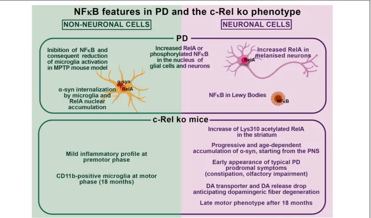
Collectively, these findings support the occurrence of a
possible interplay between NF-
κB dysregulation and α-syn
pathology in PD as observed in c-Rel ko mice (Figure 1).
In particular, by affecting key molecular pathways driving
neuronal resilience to stressors, neuroinflammation, and protein
misfolding, unbalances in the activation of NF-
κB factors may
significantly contribute to PD pathogenesis by favoring
α-syn
accumulation, aggregation and spreading, and promoting glial
cell activation and neuronal cell death. These same processes may
boost NF-
κB activation, initiating a vicious circle perpetrating
disease progression. Further studies are compelling to elucidate
the causes and features of NF-κB alterations as well as whether
and how NF-
κB unbalance may lead to α-syn pathology and
neurodegeneration in PD.
AUTHOR CONTRIBUTIONS
AB wrote the first draft of the manuscript. AB, LB, FL, GF, CM,
EP, FB, VP, and MP revised the manuscript. AB, FL, FB, and EP
prepared the figure and integrated it in the manuscript.
FUNDING
We are grateful to the Italian MIUR PRIN 2017-1065 and the
MIUR PNR 2015-2020 PerMedNet. VP Researcher Fellowship is
covered by Fondazione Cariplo–Giovani Ricercatori – Research
Support GR 2018-0391.
REFERENCES
Adler, A. S., Kawahara, T. L., Segal, E., and Chang, H. Y. (2008). Reversal of aging by NFkappaB blockade.Cell Cycle 7, 556–559. doi: 10.4161/cc.7.5.5490 Albensi, B. C., and Mattson, M. P. (2000). Evidence for the involvement of TNF
and NF-kappaB in hippocampal synaptic plasticity.Synapse 35, 151–159. doi: 10.1002/(sici)1098-2396(200002)35:2<151::aid-syn8>3.0.co;2-p
Anderson, L. R., Betarbet, R., Gearing, M., Gulcher, J., Hicks, A. A., Stefansson, K., et al. (2007). PARK10 candidate RNF11 is expressed by vulnerable neurons and localizes to Lewy bodies in Parkinson disease brain.J. Neuropathol. Exp. Neurol. 66, 955–964. doi: 10.1097/nen.0b013e3181567f17
Baiguera, C., Alghisi, M., Pinna, A., Bellucci, A., De Luca, M. A., Frau, L., et al. (2012). Late-onset Parkinsonism in NFkappaB/c-Rel-deficient mice.Brain 135(Pt 9), 2750–2765. doi: 10.1093/brain/aws193
Balfour Sartor, R. (1997). Enteric microflora in IBD: pathogens or commensals? Inflamm. Bowel Dis. 3, 230–235. doi: 10.1097/00054725-199709000-00008 Beattie, E. C., Stellwagen, D., Morishita, W., Bresnahan, J. C., Ha, B. K., Von
Zastrow, M., et al. (2002). Control of synaptic strength by glial TNFalpha. Science 295, 2282–2285. doi: 10.1126/science.1067859
Bellucci, A., Antonini, A., Pizzi, M., and Spano, P. (2017). The end is the beginning: Parkinson’s disease in the light of brain imaging.Front. Aging Neurosci. 9:330. doi: 10.3389/fnagi.2017.00330
Bellucci, A., Mercuri, N. B., Venneri, A., Faustini, G., Longhena, F., Pizzi, M., et al. (2016). Review: Parkinson’s disease: from synaptic loss to connectome dysfunction.Neuropathol. Appl. Neurobiol. 42, 77–94. doi: 10.1111/nan.12297 Bellucci, A., Navarria, L., Zaltieri, M., Falarti, E., Bodei, S., Sigala, S., et al. (2011).
Induction of the unfolded protein response byα-synuclein in experimental models of Parkinson’s disease.J. Neurochem. 116, 588–605. doi: 10.1111/j.1471-4159.2010.07143.x
Bellucci, A., Navarria, L., Zaltieri, M., Missale, C., and Spano, P. (2012a). alpha-synuclein synaptic pathology and its implications in the development of novel therapeutic approaches to cure Parkinson’s disease.Brain Res. 1432, 95–113. doi: 10.1016/j.brainres.2011.11.031
Bellucci, A., Zaltieri, M., Navarria, L., Grigoletto, J., Missale, C., and Spano, P. (2012b). Fromα-synuclein to synaptic dysfunctions: new insights into the pathophysiology of Parkinson’s disease.Brain Res. 1476, 183–202. doi: 10.1016/ j.brainres.2012.04.014
Bendor, J. T., Logan, T. P., and Edwards, R. H. (2013). The function of alpha-synuclein.Neuron 79, 1044–1066. doi: 10.1016/j.neuron.2013.09.004 Bernard, D., Quatannens, B., Begue, A., Vandenbunder, B., and Abbadie, C. (2001).
Antiproliferative and antiapoptotic effects of crel may occur within the same
cells via the up-regulation of manganese superoxide dismutase.Cancer Res. 61, 2656–2664.
Betarbet, R., Sherer, T. B., Di Monte, D. A., and Greenamyre, J. T. (2002). Mechanistic approaches to Parkinson’s disease pathogenesis.Brain Pathol. 12, 499–510. doi: 10.1111/j.1750-3639.2002.tb00468.x
Bethea, J. R., Castro, M., Keane, R. W., Lee, T. T., Dietrich, W. D., and Yezierski, R. P. (1998). Traumatic spinal cord injury induces nuclear factor-kappaB activation.J. Neurosci. 18, 3251–3260. doi: 10.1523/jneurosci.18-09-03251. 1998
Blondeau, N., Widmann, C., Lazdunski, M., and Heurteaux, C. (2001). Activation of the nuclear factor-kappaB is a key event in brain tolerance.J. Neurosci. 21, 4668–4677. doi: 10.1523/jneurosci.21-13-04668.2001
Bontempi, S., Fiorentini, C., Busi, C., Guerra, N., Spano, P., and Missale, C. (2007). Identification and characterization of two nuclear factor-kappaB sites in the regulatory region of the dopamine D2 receptor.Endocrinology 148, 2563–2570. doi: 10.1210/en.2006-1618
Braak, H., Del Tredici, K., Rub, U., de Vos, R. A., Jansen Steur, E. N., and Braak, E. (2003). Staging of brain pathology related to sporadic Parkinson’s disease. Neurobiol. Aging 24, 197–211. doi: 10.1016/s0197-4580(02)00065-9 Braak, H., Del Tredici-Braak, K., and Gasser, T. (2018). Special issue "Parkinson’s
disease".Cell Tissue Res. 373, 1–7.
Brudek, T. (2019). Inflammatory bowel diseases and Parkinson’s disease. J. Parkinsons Dis. 9, S331–S344. doi: 10.3233/JPD-191729
Brundin, P., Ma, J., and Kordower, J. H. (2016). How strong is the evidence that Parkinson’s disease is a prion disorder?Curr. Opin. Neurol. 29, 459–466. doi: 10.1097/WCO.0000000000000349
Brundin, P., and Melki, R. (2017). Prying into the prion hypothesis for Parkinson’s disease.J. Neurosci. 37, 9808–9818. doi: 10.1523/JNEUROSCI.1788-16.2017 Buchman, A. S., Yu, L., Wilson, R. S., Boyle, P. A., Schneider, J. A., and Bennett,
D. A. (2014). Brain pathology contributes to simultaneous change in physical frailty and cognition in old age.J. Gerontol. A Biol. Sci. Med. Sci. 69, 1536–1544. doi: 10.1093/gerona/glu117
Buchman, A. S., Yu, L., Wilson, R. S., Schneider, J. A., and Bennett, D. A. (2013). Association of brain pathology with the progression of frailty in older adults. Neurology 80, 2055–2061. doi: 10.1212/WNL.0b013e318294b462
Burre, J. (2015). The synaptic function of alpha-synuclein.J. Parkinsons Dis. 5, 699–713. doi: 10.3233/JPD-150642
Cao, G., Pei, W., Ge, H., Liang, Q., Luo, Y., Sharp, F. R., et al. (2002). In vivo delivery of a Bcl-xL fusion protein containing the TAT protein transduction domain protects against ischemic brain injury and neuronal apoptosis.J. Neurosci. 22, 5423–5431. doi: 10.1523/jneurosci.22-13-05423.2002
Cao, S., Standaert, D. G., and Harms, A. S. (2012). The gamma chain subunit of Fc receptors is required for alpha-synuclein-induced pro-inflammatory signaling in microglia.J. Neuroinflammation 9:259. doi: 10.1186/1742-2094-9-259 Cao, S., Theodore, S., and Standaert, D. G. (2010). Fcgamma receptors are
required for NF-kappaB signaling, microglial activation and dopaminergic neurodegeneration in an AAV-synuclein mouse model of Parkinson’s disease. Mol. Neurodegener. 5:42. doi: 10.1186/1750-1326-5-42
Cavaliere, F., Cerf, L., Dehay, B., Ramos-Gonzalez, P., De Giorgi, F., Bourdenx, M., et al. (2017). In vitro alpha-synuclein neurotoxicity and spreading among neurons and astrocytes using Lewy body extracts from Parkinson disease brains. Neurobiol. Dis. 103, 101–112. doi: 10.1016/j.nbd.2017.04.011
Chen, C., Edelstein, L. C., and Gelinas, C. (2000). The Rel/NF-kappaB family directly activates expression of the apoptosis inhibitor Bcl-x(L).Mol. Cell. Biol. 20, 2687–2695. doi: 10.1128/mcb.20.8.2687-2695.2000
Chen, C. H., Zhou, W., Liu, S., Deng, Y., Cai, F., Tone, M., et al. (2012). Increased NF-kappaB signalling up-regulates BACE1 expression and its therapeutic potential in Alzheimer’s disease.Int. J. Neuropsychopharmacol. 15, 77–90. doi: 10.1017/S1461145711000149
Chen, L. F., and Greene, W. C. (2004). Shaping the nuclear action of NF-kappaB. Nat. Rev. Mol. Cell Biol. 5, 392–401. doi: 10.1038/nrm1368
Chu, Y., and Kordower, J. H. (2010). Lewy body pathology in fetal grafts.Ann. N. Y. Acad. Sci. 1184, 55–67. doi: 10.1111/j.1749-6632.2009.05229.x
Colla, E., Coune, P., Liu, Y., Pletnikova, O., Troncoso, J. C., Iwatsubo, T., et al. (2012a). Endoplasmic reticulum stress is important for the manifestations of alpha-synucleinopathy in vivo. J. Neurosci. 32, 3306–3320. doi: 10.1523/ JNEUROSCI.5367-11.2012
Colla, E., Jensen, P. H., Pletnikova, O., Troncoso, J. C., Glabe, C., and Lee, M. K. (2012b). Accumulation of toxic alpha-synuclein oligomer within endoplasmic reticulum occurs in alpha-synucleinopathy in vivo.J. Neurosci. 32, 3301–3305. doi: 10.1523/JNEUROSCI.5368-11.2012
Collier, T. J., Kanaan, N. M., and Kordower, J. H. (2011). Ageing as a primary risk factor for Parkinson’s disease: evidence from studies of non-human primates. Nat. Rev. Neurosci. 12, 359–366. doi: 10.1038/nrn3039
Cooper, A. A., Gitler, A. D., Cashikar, A., Haynes, C. M., Hill, K. J., Bhullar, B., et al. (2006). Alpha-synuclein blocks ER-Golgi traffic and Rab1 rescues neuron loss in Parkinson’s models.Science 313, 324–328. doi: 10.1126/science.1129462 Crack, P. J., Taylor, J. M., Ali, U., Mansell, A., and Hertzog, P. J. (2006).
Potential contribution of NF-kappaB in neuronal cell death in the glutathione peroxidase-1 knockout mouse in response to ischemia-reperfusion injury. Stroke 37, 1533–1538. doi: 10.1161/01.STR.0000221708.17159.64
Dehay, B., Vila, M., Bezard, E., Brundin, P., and Kordower, J. H. (2016). Alpha-synuclein propagation: new insights from animal models. Mov. Disord. 31, 161–168. doi: 10.1002/mds.26370
Devi, L., Raghavendran, V., Prabhu, B. M., Avadhani, N. G., and Anandatheerthavarada, H. K. (2008). Mitochondrial import and accumulation of alpha-synuclein impair complex I in human dopaminergic neuronal cultures and Parkinson disease brain. J. Biol. Chem. 283, 9089–9100. doi: 10.1074/jbc.M710012200
Djavaheri-Mergny, M., Amelotti, M., Mathieu, J., Besancon, F., Bauvy, C., and Codogno, P. (2007). Regulation of autophagy by NFkappaB transcription factor and reactives oxygen species.Autophagy 3, 390–392. doi: 10.4161/auto.4248 Dresselhaus, E. C., and Meffert, M. K. (2019). Cellular specificity of NF-kappaB
function in the nervous system.Front. Immunol. 10:1043. doi: 10.3389/fimmu. 2019.01043
Echtay, K. S. (2007). Mitochondrial uncoupling proteins–what is their physiological role? Free Radic. Biol. Med. 43, 1351–1371. doi: 10.1016/j. freeradbiomed.2007.08.011
Ellis, C. E., Murphy, E. J., Mitchell, D. C., Golovko, M. Y., Scaglia, F., Barcelo-Coblijn, G. C., et al. (2005). Mitochondrial lipid abnormality and electron transport chain impairment in mice lacking alpha-synuclein.Mol. Cell. Biol. 25, 10190–10201. doi: 10.1128/MCB.25.22.10190-10201.2005
Emmanouilidou, E., and Vekrellis, K. (2016). Exocytosis and spreading of normal and aberrant alpha-synuclein.Brain Pathol. 26, 398–403. doi: 10.1111/bpa. 12373
Engelender, S., and Isacson, O. (2017). The threshold theory for Parkinson’s disease. Trends Neurosci. 40, 4–14. doi: 10.1016/j.tins.2016.10.008
Faustini, G., Longhena, F., Varanita, T., Bubacco, L., Pizzi, M., Missale, C., et al. (2018). Synapsin III deficiency hampers alpha-synuclein aggregation,
striatal synaptic damage and nigral cell loss in an AAV-based mouse model of Parkinson’s disease.Acta Neuropathol. 136, 621–639. doi: 10.1007/s00401-018-1892-1
Faustini, G., Marchesan, E., Zonta, L., Bono, F., Bottani, E., Longhena, F., et al. (2019). Alpha-synuclein preserves mitochondrial fusion and function in neuronal cells.Oxid. Med. Cell. Longev. 2019:4246350. doi: 10.1155/2019/ 4246350
Fitzgerald, E., Murphy, S., and Martinson, H. A. (2019). Alpha-synuclein pathology and the role of the microbiota in Parkinson’s disease.Front. Neurosci. 13:369. doi: 10.3389/fnins.2019.00369
Fleming, S. M., Salcedo, J., Fernagut, P. O., Rockenstein, E., Masliah, E., Levine, M. S., et al. (2004). Early and progressive sensorimotor anomalies in mice overexpressing wild-type human alpha-synuclein.J. Neurosci. 24, 9434–9440. doi: 10.1523/JNEUROSCI.3080-04.2004
Fusco, G., De Simone, A., Arosio, P., Vendruscolo, M., Veglia, G., and Dobson, C. M. (2016). Structural ensembles of membrane-bound alpha-synuclein reveal the molecular determinants of synaptic vesicle affinity.Sci. Rep. 6:27125. doi: 10.1038/srep27125
Garcia-Esparcia, P., Llorens, F., Carmona, M., and Ferrer, I. (2014). Complex deregulation and expression of cytokines and mediators of the immune response in Parkinson’s disease brain is region dependent.Brain Pathol. 24, 584–598. doi: 10.1111/bpa.12137
Garcia-Reitbock, P., Anichtchik, O., Bellucci, A., Iovino, M., Ballini, C., Fineberg, E., et al. (2010). SNARE protein redistribution and synaptic failure in a transgenic mouse model of Parkinson’s disease.Brain 133(Pt 7), 2032–2044. doi: 10.1093/brain/awq132
Gelpi, E., Navarro-Otano, J., Tolosa, E., Gaig, C., Compta, Y., Rey, M. J., et al. (2014). Multiple organ involvement by alpha-synuclein pathology in Lewy body disorders.Mov. Disord. 29, 1010–1018. doi: 10.1002/mds.25776
Ghosh, A., Roy, A., Liu, X., Kordower, J. H., Mufson, E. J., Hartley, D. M., et al. (2007). Selective inhibition of NF-kappaB activation prevents dopaminergic neuronal loss in a mouse model of Parkinson’s disease.Proc. Natl. Acad. Sci. U.S.A. 104, 18754–18759. doi: 10.1073/pnas.0704908104
Ghosh, G., Wang, V. Y., Huang, D. B., and Fusco, A. (2012). NF-kappaB regulation: lessons from structures.Immunol. Rev. 246, 36–58. doi: 10.1111/j.1600-065X. 2012.01097.x
Ghosh, M., Yang, Y., Rothstein, J. D., and Robinson, M. B. (2011). Nuclear factor-kappaB contributes to neuron-dependent induction of glutamate transporter-1 expression in astrocytes.J. Neurosci. 31, 9159–9169. doi: 10.1523/JNEUROSCI. 0302-11.2011
Goers, J., Manning-Bog, A. B., McCormack, A. L., Millett, I. S., Doniach, S., Di Monte, D. A., et al. (2003). Nuclear localization of alpha-synuclein and its interaction with histones.Biochemistry 42, 8465–8471. doi: 10.1021/bi0341152 Grinberg-Bleyer, Y., Oh, H., Desrichard, A., Bhatt, D. M., Caron, R., Chan, T. A.,
et al. (2017). NF-kappaB c-Rel is crucial for the regulatory T cell immune checkpoint in cancer.Cell 170, 1096–1108.e13. doi: 10.1016/j.cell.2017.08.004 Grozdanov, V., and Danzer, K. M. (2018). Release and uptake of pathologic
alpha-synuclein.Cell Tissue Res. 373, 175–182. doi: 10.1007/s00441-017-2775-9 Grunewald, A., Kumar, K. R., and Sue, C. M. (2019). New insights into the complex
role of mitochondria in Parkinson’s disease.Prog. Neurobiol. 177, 73–93. doi: 10.1016/j.pneurobio.2018.09.003
Guardia-Laguarta, C., Area-Gomez, E., Rub, C., Liu, Y., Magrane, J., Becker, D., et al. (2014). alpha-synuclein is localized to mitochondria-associated ER membranes.J. Neurosci. 34, 249–259. doi: 10.1523/JNEUROSCI.2507-13.2014 Guo, J. T., Chen, A. Q., Kong, Q., Zhu, H., Ma, C. M., and Qin, C. (2008).
Inhibition of vesicular monoamine transporter-2 activity in alpha-synuclein stably transfected SH-SY5Y cells.Cell. Mol. Neurobiol. 28, 35–47. doi: 10.1007/ s10571-007-9227-0
Gupta, A. S., Waters, M. R., Biswas, D. D., Brown, L. N., Surace, M. J., Floros, C., et al. (2019). RelB controls adaptive responses of astrocytes during sterile inflammation.Glia 67, 1449–1461. doi: 10.1002/glia.23619
Halliwell, B. (2006). Oxidative stress and neurodegeneration: where are we now? J. Neurochem. 97, 1634–1658. doi: 10.1111/j.1471-4159.2006.03907.x Hawkes, C. H. (2008). Parkinson’s disease and aging: same or different process?
Mov. Disord. 23, 47–53. doi: 10.1002/mds.21766
Hayden, M. S., and Ghosh, S. (2012). NF-kappaB, the first quarter-century: remarkable progress and outstanding questions.Genes Dev. 26, 203–234. doi: 10.1101/gad.183434.111