Università degli Studi di Ferrara
DOTTORATO DI RICERCA IN
BIOLOGIA EVOLUZIONISTICA E AMBIENTALE
CICLO XXVII
COORDINATORE Prof. GUIDO BARBUJANI
Molecular and Bioinformatic Analysis of the Circadian
Clock in Phreatichthys andruzzii
Settore Scientifico Disciplinare BIO/05
Dottorando Tutore
Dott. NEGRINI PIETRO Prof. BERTOLUCCI CRISTIANO
3
TABLE OF CONTENTS
PageAbstract (English)
...………...7Abstract (Italiano)
...9I. Introduction
………...111.1
General aspects of the circadian clock
……….131.1.1 Circadian pacemakers………...13
1.1.2 Molecular clock mechanism……….…15
1.1.3 Light input pathways………...…….…19
1.1.4 Temperature input pathway………...………..21
1.1.5 Output pathways………...22
1.2 Zebrafish as a circadian clock model system
………...………221.2.1 Zebrafish circadian clock genes……….……..24
1.2.2 Zebrafish light input pathway………..24
1.2.3 Zebrafish temperature input pathway…………..………..……27
1.2.4 The role of the SCN and pineal gland in zebrafish…….………..…….27
1.2.5 Circadian clock outputs in zebrafish……….…..28
1.3 Cavefish as a new circadian clock model system
………..…….….291.3.1 Life in the darkness: special features of cave inhabitants……….…....29
1.3.2 Astyanax mexicanus: a teleost cavefish model………....30
1.3.3 The Somalian cavefish Phreatichthys andruzzii………..33
1.4 Aim of the study
………..35II. Materials and methods
...372.1 Animals
………...372.2 Cell lines
...372.3 Genomic DNA extraction
...384
2.5 Cloning Period2 sequence
...392.6 RT-PCR reactions
………..392.7 Quantitative PCR (qPCR
)………...402.8 Recording of adult locomotor activity
………412.9 Deep Sequencing analysis
………..…..422.9.1 Stranded RNA sequencing libraries construction………....……..42
2.9.2 Paired-end DNA sequencing construction………..42
2.9.3 Mate pair libraries construction………..………….….……..43
2.9.4 Sequencing of libraries………....……..43
2.9.5 De novo transcriptome assembly……….…….44
2.9.6 De novo genome assembly……….……....44
III. Results
...463.1 Cavefish Period2 analysis
………....463.1.1 Cloning cavefish Period2 gene………...….……..46
3.1.2 Transposon validation and expression levels of cavefish Per2/Cry1a…………...50
3.1.3 Next Generation Sequencing: a new global approach………..….52
3.2 RNAseq analysis in Phreatichthys andruzzii
……..…...………....523.2.1 General statistics on Phreatichthys andruzzii RNAseq……….……….………….52
3.2.2 The characterization of cavefish circadian oscillator from RNAseq…..………..54
3.3 Intron retention in cavefish mRNA
………….………..………..………563.4 DNAseq analysis in Phreatichthys andruzzii
……….………....623.4.1 General statistics on Phreatichthys andruzzii DNAseq……….………....………..62
5
IV. Discussion
...684.1 Cavefish Period2: a candidate underlying
the aberrant circadian clock properties
………….………...…...684.2 RNAseq analysis and intron retention
in Phreatichthys andruzzii mRNA
………...……….694.3 Genomic sequencing and visual/non-visual opsins analysis
in Phreatichthys andruzzii
……….………..…….…...724.4 Phreatichthys andruzzii and its aberrant clock phenotype
………...…...74V. References
...777
Abstract (english)
Daily cycles of light and temperature imposed by the rotation of the Earth on its axis have had a major impact on the evolution of all living organisms. Fascinating demonstrations of this fact can be seen in extreme environments such as caves or in the deep sea, where some species have evolved in complete isolation from daily light-dark cycles for millions of years, sharing a range of striking physical characters acquired by convergent evolution such as loss of eyes and pigmentation. One fundamental issue is to investigate whether these “hypogean” species still retain a functional circadian clock, which is a highly conserved self-sustaining timing system that allows organisms to anticipate daily environmental changes and is synchronized primarily by light. In this study, we have performed a comparative analysis of the circadian clock between the Somalian cavefish
Phreatichthys andruzzii, which has evolved in perpetual darkness, and the model species Danio rerio (the zebrafish) that is evolved under natural daily light-dark cycles. It has been
demonstrated that P. andruzzii retains a food-entrainable clock that is synchronized in response to regular feeding time, but does not respond to light-dark cycles. Moreover, under constant conditions, the cavefish clock oscillates with an extremely long period and also lacks normal temperature compensation. Based on these previous results, we started to analyze in detail one specific clock gene that is light-induced in zebrafish, Period2, where we encountered significant mutations in cavefish. We characterized the coding sequence and the genomic structure of this mutated cavefish gene and we analyzed its expression levels in comparison with zebrafish. Subsequently, for the first time in this species we performed a detailed characterization of clock and visual/non-visual photoreceptor genes as part of a complete P. andruzzii genome and transcriptome analysis. Our RNAseq analysis revealed a surprising phenomenon in cavefish: P. andruzzii mRNA sequences present an unusually high level of retained introns, leading to premature stop codons being introduced into the coding sequences of many transcripts. This mechanism may contribute to the aberrant clock phenotype of this species as well as, from a wider perspective, to other fascinating cavefish adaptations to life in constant darkness. We analyzed in detail this aberrant intron splicing phenomenon in two cavefish transcriptomes, from the brain and from a fin-derived cell line. Finally, the creation of a first P. andruzzii genome assembly by DNAseq analysis allowed us to map and characterize the group of visual/non-visual opsins, the main candidate genes involved in the circadian and non- photoreception
8 system. We used this sequence data in order to perform a comparative analysis of the levels of expression of these genes between cavefish and zebrafish. Our results suggest that the loss of photoreceptor function in cavefish may result either from mutations affecting the coding regions of the opsins, as documented in TMT-opsin and Opn4m2, or, alternatively, from mutations affecting the regulation of the expression levels of this group of photoreceptive genes.
Keywords: circadian clock, cavefish, zebrafish, Period2 gene, transcriptome, genome, de
9
Abstract (Italiano)
I cicli giornalieri di luce e temperatura imposti dalla rotazione della Terra sul suo asse hanno avuto un forte impatto sull'evoluzione di tutti gli organismi viventi. Dimostrazioni affascinanti di questo fatto si possono trovare in ambienti estremi come grotte o abissi marini, in cui alcune specie si sono evolute durante milioni di anni in completo isolamento dai cicli giornalieri di luce e buio, acquisendo un’ampia gamma di caratteri fisici come la perdita degli occhi e della pigmentazione attraverso un’evoluzione convergente. Un problema fondamentale è quello di verificare se queste specie ipogee posseggano ancora un orologio circadiano funzionale, cioè un sistema endogeno altamente conservato che permette agli organismi di anticipare i cambiamenti ambientali quotidiani e che è sincronizzato principalmente dalla luce. In questo studio, abbiamo eseguito un'analisi comparativa del sistema circadiano tra il pesce ipogeo della Somalia Phreatichthys
andruzzii, evolutosi in buio costante, e la specie modello Danio rerio (lo zebrafish) la
quale è normalmente esposta ai cicli giornalieri di luce e buio. È stato dimostrato che P.
andruzzii possiede un orologio circadiano sincronizzabile in risposta a somministrazione
regolare di cibo, ma non a cicli di luce-buio. Inoltre, in condizioni costanti, l'orologio di questo pesce ipogeo si esprime con un periodo estremamente lungo e manca della normale compensazione del periodo al variare della temperatura. Partendo da questi risultati precedentemente ottenuti, abbiamo analizzato in dettaglio uno specifico gene orologio la cui espressione in zebrafish è indotta dalla luce, Period2, in cui abbiamo scoperto mutazioni significative in P. andruzzii. Abbiamo caratterizzato questo gene a partire dalla sequenza codificante fino alla struttura genomica, includendo anche l'analisi dei suoi livelli di espressione confrontandoli con zebrafish. Successivamente, abbiamo sviluppato per la prima volta in questa specie una caratterizzazione dettagliata dello stato dei geni orologio e dei fotorecettori visivi e non visivi attraverso un’analisi completa del trascrittoma e del genoma di P. andruzzii. La RNAseq analisi ha inoltre rivelato in questa specie un fenomeno molto particolare: le sequenze di mRNA di P. andruzzii presentano un livello insolitamente alto di introni ritenuti al loro interno, i quali introducono codoni di stop prematuri nelle sequenze codificanti di molti trascritti. Questo fenomeno potrebbe contribuire alla spiegazione dell’orologio anormale di questa specie e, in una prospettiva più ampia, agli affascinanti adattamenti acquisiti da questo pesce ipogeo alla vita in buio costante. Abbiamo analizzato in dettaglio questi fenomeno di splicing anormale in due
10 trascrittomi diversi, da cervello e da linea cellulare di pinna. Infine, la creazione di un primo assemblaggio del genoma di P. andruzzii attraverso la DNAseq analisi ci ha permesso infine di mappare e caratterizzare il gruppo di opsine visive e non visive, i principali geni candidati coinvolti nel sistema di fotorecezione circadiana e non. Abbiamo potuto utilizzare questi dati al fine di effettuare un'analisi comparativa dei livelli di espressione di questa famiglia di geni tra P. andruzzii e zebrafish. I risultati ottenuti suggeriscono che la perdita della funzione dei fotorecettori in questa specie ipogea può derivare sia da mutazioni che interessano le regioni codificanti delle opsine, come documentato nei casi di TMT-opsin e Opn4m2, o, in alternativa, da mutazioni che interessano la regolazione dei livelli di espressione di questi fotorecettori. Parole chiave: orologio circadiano, pesce ipogeo, zebrafish, gene Period2, trascrittoma, genoma, assemblaggio de novo, splicing degli introni, opsine visive e non visive.
11
I. Introduction
The rotation of the planet earth on its axis creates regular periods of sunlight and darkness, which have had a significant impact on the evolution of life on earth. Light is well known to be a major source of energy, as well as a fundamental signal that allows animals to interact with their environment; furthermore, most organisms regulate their physiology, behavior and biochemistry according to the time of the day.
Daily rhythms in physiology and behavior are controlled and coordinated by the circadian clock mechanism or circadian oscillator (Pittendrigh, 1993), which is an endogenous, self-sustaining time-keeping system. It is a highly conserved mechanism which continues to function even under constant conditions (so called “free-running” conditions), where it generates a rhythm with a period length of around 24 hours (Pittendrigh, 1960). For this reason it is named “circadian”, from Latin circa-diem, that means “around a day”.
One of the key features of the circadian clock is its daily resetting and synchronization by environmental signals such as light-dark cycle, food availability and temperature; these external signals are termed zeitgebers (from German Zeit: time, geben: givers) (Pittendrigh, 1993). Additionally, the circadian clock has been shown to be a temperature compensated mechanism (Tsuchiya et al., 2003), which means that the period length of a rhythm produced by the clock remains relatively constant over a range of temperatures. The circadian timing system in all organisms is essentially composed of three distinct elements (Figure 1) (Menaker et al., 1978):
1. The core oscillator, that is the central cell-autonomous pacemaker which generates the circadian rhythm.
2. The input pathway, which detects the zeitgebers and entrains/resets the core oscillator. 3. The output pathway, which allows the pacemaker to control many behavioral and physiological activities.
The first reports on circadian rhythms date back to 1729, when the French scientist Jean-Jacques d’Ortous de Mairan documented the daily leaf movement of the plant Mimosa
pudica. Later, the major properties of circadian rhythms were described by Bünning
(Bünning, 1935) and Pittendrigh (Pittendrigh, 1967), using plants and insects as models. Experiments performed by Jürgen Aschhoff in humans also contributed to the emerging of the chronobiology field. The modern era of genetic analysis of the clock started in 1971 with the isolation of the Period (Per) mutant in Drosophila melanogaster (Konopka &
12 Benzer, 1971), the first clock mutant ever identified. Subsequently, genetic studies in a wide range of organisms including Neurospora, Cyanobacteria, Arabidopsis, Drosophila and more recently in mouse, helped to identify several clock mutants and to isolate the so-called “clock genes”, the fundamental components of the core clock mechanism. As already mentioned, the basic organization of the central clock mechanism appears to be highly conserved through evolution, even though the sequences of individual clock components between certain phylogenetic groups appear to be divergent. This suggests that circadian clocks may have evolved numerous times independently during the course of evolution (van Ooijen & Millar, 2012).
In many cases, clock components serve as transcriptional regulators in the context of a transcription-translation feedback loop, which takes approximately 24 hours (circadian rhythm) to complete one cycle (Wager-Smith & Kay, 2000). Light, food and temperature changes represent the strongest entraining stimuli for the circadian clock, but how these environmental signals are detected and communicate with the core oscillator is still incompletely understood. Furthermore, the complex systemic and cell autonomous mechanisms that constitute clock output pathways are also poorly understood.
Figure 1. Schematic representation of the circadian timekeeping mechanism. By daily environmental
signals, the input pathways synchronize the circadian pacemaker with natural cycles; this generates cell autonomous circadian rhythms. Through multiple output pathways, the circadian pacemaker is able to control and regulate various behavioral and physiological aspects in all living organisms.
13
1.1 General aspects of the circadian clock
Thanks to the early studies of the circadian clock which identified the physical localization and cellular organization of the circadian oscillator, we now know that in vertebrates the circadian clock consists of a hierarchy of multiple pacemakers. The “central” pacemakers are located in the brain and play an important role by coordinating the function of the “peripheral” clocks, which are distributed throughout tissues, organs and cells. The central pacemakers control and communicate with the peripheral clocks via complex systemic signals, which are still poorly understood. Major challenges are now to understand how all these multiple clocks communicate with each other and remain synchronized throughout the whole body.
This section will describe the general features of the circadian clock and how light and temperature can be perceived and entrain the clock.
1.1.1 Circadian pacemakers
Classically, the circadian pacemaker was considered as a single, centralized clock that regulates all the physiological rhythms in the body (Pittendrigh, 1993). For example, in
Drosophila, pacemaker neurons located in the brain control locomotor activity rhythms.
However, the spatial distribution of clock genes expression revealed the existence of multiple pacemakers located in many tissues of the fly; furthermore, studies with transgenic flies expressing luciferase or GFP under per or tim regulatory clock gene promoters, showed rhythmic expression in various tissues of this insect (Plautz et al., 1997). Rhythmic clock gene expression in all these tissues has been demonstrated even in
vitro, underlining the existence of brain-independent autonomous clocks in many cells. For
this reason, the centralized function of the circadian clock system has been updated with the notion of multiple peripheral clocks spread throughout the whole organism (Plautz et al., 1997; Yamazaki et al., 2000) and that, theoretically, every single cell or tissue can contain a circadian clock (Balsalobre et al., 1998; Tamai et al., 2003).
In mammals, the suprachiasmatic nucleus (SCN) has been shown to act as the master circadian pacemaker. Through lesioning and transplantation experiments (Moore & Eichler, 1972; Ralph et al., 1990) it has been demonstrated that the SCN is responsible for the generation and regulation of rhythms in behavior, hormonal secretion and other physiological functions (Klein & Reppert, 1991). This pacemaking ability of the SCN, as well as its response to environmental time signals, neuropeptides expression and control of
14 rhythms, is organized in a small network involving around 20,000 neurons (Antle & Silver, 2005) which is located in the anterior part of the hypothalamus, immediately dorsal to the optic chiasma and bilateral to the third ventricle. Within the SCN, two sub-regions have been identified, the dorsal shell and a ventral core, by their expression of clock genes (Yamaguchi et al., 2003) as well as neural peptides such as Arginine Vasopressin (AVP) and Vasoactive Intestinal Polypeptide (VIP) (Klein & Reppert, 1991). Furthermore, in vivo experiments revealed that rhythmic clock gene expression in the ventral core neurons is more rapidly synchronized to light-dark (LD) cycles shifts, compared to the clock gene expression from the dorsal shell neurons (Albus et al., 2005). Communication between SCN neurons involves neurotransmitters such as GABA (Liu & Reppert, 2000), VIP (Aton et al., 2005) and also gap junctions (Colwell, 2000). Another central pacemaker in vertebrates has been identified in the retina, that is responsible for controlling the local synthesis of the hormone melatonin (Tosini & Menaker, 1996), while, in non-mammalian vertebrates, a central circadian pacemaker is located in the pineal gland, driving rhythms of melatonin release. These rhythms are directly regulated by exposure to light even in cell culture (Menaker et al., 1997; Takahashi et al., 1980).
Circadian clocks do not only exist in SCN neurons, but they appear to be a property of most peripheral tissues (Dibner et al., 2010). Peripheral clock rhythms in mice and rats have been studied in many tissues such as lung, liver and skeletal muscles, using transgenic models. Explanted and cultured peripheral tissues of transgenic rats, expressing a
per1::luciferase reporter construct, show circadian oscillations of bioluminescence that
persist over several days, while explanted cultures of the SCN can continue to oscillate over a time period of at least 32 days (Yamazaki et al., 2000). Similar experiments have been performed in mice carrying a luciferase reporter gene integrated into the endogenous
Period2 locus by “knock-in” recombination (Yoo et al., 2004). In this experiments,
rhythms of luciferase expression could be measured in various organs for a longer period, suggesting that peripheral clocks are more robust than supposed by the original Period1 transgenic rat studies (Yoo et al., 2004). From these findings, the prediction would be that dysfunction of the central core clock does not necessarily inactivate the peripheral clocks, which may be instead forced to become temporally uncoupled (Nagoshi et al., 2004; M. P. Pando et al., 2002).
Peripheral clocks of mammals are not directly influenced by light, so the entrainment of peripheral clocks by light-dark cycles is indirect and occurs via the central clock in the SCN. Peripheral clocks seem to be synchronized with the central clock through a complex combination of signals, including metabolites and body temperature (Brown et al., 2002;
15 Damiola et al., 2000; Schibler, 2007). However, the physiological significance of peripheral clock responses independently from the SCN is not yet completely clear (Uchida et al., 2010). The situation is different in Drosophila and zebrafish, where the clocks in peripheral tissues and in cell cultures are directly entrainable by light (Underwood & Groos, 1982; Whitmore et al., 1998).
1.1.2 Molecular clock mechanism
In many model organisms, such as Drosophila, Neurospora, Cyanobacteria, mouse and fish, genetic analysis has identified a certain number of clock genes which are fundamental for the function of the molecular oscillator. Many of these clock genes are regulatory elements of transcription-translation feedback loops and act with a transcription repression or activation function (Wager-Smith & Kay, 2000).
In the vertebrate molecular core loop, Clock (Clk) and Bmal genes encode transcription factors with a basic-helix-loop-helix DNA binding domain (bHLH) (Crews & Fan, 1999) and a PAS (PER-ARNT-SIM) domain, a domain encountered in many signaling proteins where it serves as a signal sensor (Ponting & Aravind, 1997) and mediates protein–protein interactions. These two bHLH-PAS transcription factors constitute the positive elements in the vertebrate clock. Genetic experiments in mice generated by N-ethyl-N-Nitrosourea (ENU) mutagenesis, contributed to identify the Clock (Clk) gene. Clock mutant mice showed aberrant locomotor activity rhythms under constant darkness conditions (DD) in wheel running assays (Vitaterna et al., 1994). Subsequently, positional cloning led to the characterization of the mClk locus, homologous to the Drosophila Clock gene (King et al., 1997). BMAL was first identified because to its capacity to interact with CLK (Gekakis et al., 1998; Hogenesch et al., 1998). In addition, gene knock-out of Bmal in mice caused an immediate loss of locomotor activity rhythms under constant darkness (Bunger et al., 2000). CLK and BMAL proteins heterodimerize in a transcription-activating complex; this complex binds to conserved E-box enhancer elements (5´-CACGTG-3´) that are present in the promoter region of clock-controlled genes (CCGs). These other clock genes constitute the negative regulatory elements of the core clock loop and include three Period genes (mPer1, mPer2, mPer3) and two Cryptochrome genes (mCry1, mCry2) (Dunlap, 1999). The PER proteins are central elements of the mammalian circadian clock. They contain two PAS (PER-ARNT-SIM) domains (PAS-A and PAS-B), which can mediate homodimeric and heterodimeric mPER-mPER interactions, as well as interactions with transcription factors and kinases. These three mammalian Period gene homologs have been
16 identified by their amino acid similarity with the Drosophila PER protein (Shearman et al., 1997; Tei et al., 1997), and the discovery of multiple Period genes suggested either specialization or redundancy of function of these family members. All three PER proteins function as negative regulators of CLK and BMAL; however, in mammals, this inhibition is not as strong as in Drosophila (Kume et al., 1999). Systematic knock-out studies have revealed distinct roles of the Period genes in the circadian clock mechanism (Bae et al., 2001; Shearman et al., 2000). Whereas mPer1 and mPer2 seem to be essential, mPer3 is not necessary for circadian rhythmicity (Shearman et al., 2000). Mutations in single mPer genes do not result in loss of circadian clock function, while double mutants of mPer1 and mPer2 lead to a complete loss of rhythmicity (Zheng et al., 2001). MPer1 and mPer2 are expressed with a circadian rhythm and rapidly induced in the SCN by administration of light pulses during the subjective night, but not during the subjective day (Shigeyoshi et al., 1997; Zylka et al., 1998). Furthermore, phosphorylation of the mPER proteins regulates the rate of turnover, ensuring a delay in their accumulation and in their negative action within the feedback loop (Maier et al., 2009). The importance of the phosphorylation sites in the PER proteins is demonstrated by a mutation affecting the phospho-acceptor site for CK1ε in the hPER2 protein, which is responsible for the familial advanced sleep syndrome (Toh et al., 2001). However, our understanding of the precise contribution of these genes to the entrainment of the clock by light and their molecular function in general, is still incomplete (Albrecht et al., 2007; Albrecht et al., 2001).
The mammalian Cry1 and Cry2 genes are also rhythmically expressed and can form complexes with PER proteins. Knock-out mice deficient in Cry1 exhibit shorter free-running circadian oscillators, whereas mice deficient for Cry2 show a longer free-free-running circadian oscillator (van der Horst et al., 1999; Vitaterna et al., 1999). Interestingly, double knock-out mice for mPer1 and mPer2 (Zheng et al., 2001), as well as double knock-out mice for mCry1 and mCry2 (van der Horst et al., 1999), show nearly the same phenotype, with an immediate arrhythmic locomotor activity after transfer from light-dark cycle (LD) to constant darkness (DD) conditions. Thus, both the cryptochrome and period proteins appear to function in combination as negative elements in the core feedback loop. Following the activation of Per and Cry gene expression by CLK:BMAL, the complexes of PERIOD (PER) and CRYPTOCHROME (CRY) proteins subsequently enter the nucleus as heterodimers and inhibit their own transcription by blocking the transactivation of the CLK:BMAL heterodimer, thereby closing the core loop (Reppert & Weaver, 2001). Thanks to the ability of the CLK:BMAL complex to directly regulate the expression of
17 other non-clock genes, this feedback loop can control many physiological and behavioral functions.
An additional feedback loop involves the nuclear orphan receptors REV-ERBα and RORα. REV-ERBα and ß are transcription factors belonging to the retinoic acid-related orphan receptor family. The expression of REV-ERBα is activated by CLK and BMAL and repressed by factors from the negative limb. REV-ERBα, in turn, represses the transcription of the Bmal gene (Preitner et al., 2002). Furthermore, RORα can compete with REV-ERBα for the binding of shared DNA-binding elements present in the promoter region of Bmal (Sato et al., 2004). Genes including these REV-ERB/RORE elements in their promoters are repressed by REV-ERBα, whereas RORα activates their transcription. Thus, the REV-ERBα/RORα feedback loop plays a major role by interconnecting the positive and negative limbs of the clock (Duez & Staels, 2008). The presence of this loop is predicted to confer a much higher stability on the core loop in the mammalian clock (Emery & Reppert, 2004; Preitner et al., 2002) (Figure 2). In the case of REV-ERBα, a heme group has been implicated as its ligand (Raghuram et al., 2007; Yin et al., 2007). This finding suggests that heme regulation of REV-ERBα might link the mammalian core clock with the control of metabolism (Raghuram et al., 2007). Recent results have also revealed that double knock-out mice for the REV-ERBα and ß genes show a phenotype with a strongly disrupted circadian clock function (Cho et al., 2012), and mice treated with synthetic REV-ERB ligands have a significantly altered rhythmic clock, as well as disruptions in metabolic gene expression and abnormal circadian behavior (Solt & Burris, 2012).
In addition to the core loop, a complex system of post-translational modifications of clock proteins regulates their turn over and their sub-cellular localization during the circadian cycle (Mehra et al., 2009). Furthermore, transcriptional regulation within the clock gene machinery requires the rhythmic recruitment and assembly of multi-protein complexes in a circadian time dependent manner. These events are connected with rhythmic changes in epigenetic regulation, normally carried out by chromatin modifying enzymes (Borrelli et al., 2008; Masri & Sassone-Corsi, 2010). In the case of the CLK:BMAL complex, in addition to its ability to bind to chromatin via the E-boxes in a circadian manner, CLK itself possesses an histone acetyl-transferase (HAT) activity, which is predicted to contribute to the opening of chromatin fibers and promoting rhythmic transcription of clock controlled proteins, through the histone modifications H3, K9 and K14 (Nakahata et al., 2007). In addition, the CREB binding protein (CBP) as well as p300 coactivators both have HAT function and are rhythmically recruited by the CKL:BMAL complex. However,
18 while p300 seems to function as a co-activator of CLK:BMAL by influencing the acetylation state of histones in a circadian fashion, the function of CBP within this process is not yet fully understood. CBP also interacts with PER2, thereby participating in the negative limb of the clock feedback loop (Etchegaray et al., 2003; Koike et al., 2012).
Figure 2. The core of the molecular clock mechanism in mammals. The clock is composed by two interacting loops. The core loop is formed by the positive elements CLOCK and BMAL, which heterodimerize and activate the transcription of the negative elements of the loop, the Per and Cry genes. Subsequently, PER and CRY proteins enter the nucleus as heterodimers and repress their own transcription by the inhibition of CLOCK:BMAL action. This mechanism is stabilized by a secondary loop, in which the CLOCK:BMAL complex induces the expression of Rev-Erbα and Rorα, regulating in this way the transcription of the Bmal gene.
Another transcriptional activator that interacts with CLK is the methyltransferase mixed lineage leukemia 1 (MLL1) protein, that specifically tri-methylates histone H3K4 in the context of circadian clock gene promoters and is also associated with activation (Katada & Sassone-Corsi, 2010). Conversely, methylation of H3K27 on circadian promoters by EZH2 is associated with CRY dependent inhibition of transcription (Etchegaray et al., 2006). Recently a histone deacetylase HDAC3 and its co-activator NCoR were also demonstrated
19 to play an important role in the maintenance of the epigenetic properties of circadian genes in the mouse liver (Feng et al., 2011). All these findings reveal the importance of the clock-directed epigenetic landscape, in order to generate a rhythmic clock gene expression. The core clock mechanism of transcription-translation feedback loops is very similar in flies and mammals. There are only two main differences in the Drosophila clock, compared to mammals: the function of the CRY protein and the TIM protein. In
Drosophila, in addition to its function in controlling transcriptional activity as in the case
of the mammalian CRYs, the Drosophila CRY protein acts also as a blue-light photoreceptor that interacts with TIM to support its degradation (Hardin, 2011). In other insect species, a CRY2-like protein with a primary function as transcriptional repressor was also found. The TIM protein is essential for clock function in flies, since it forms a complex with PER and inhibits the positive elements of the feedback loop. Differently, the mammalian TIM protein is a closer homolog of the Drosophila gene Timeout, and its precise function in the circadian oscillator remains unclear (Tomioka & Matsumoto, 2010). As in mammals, phosphorylation of clock proteins also represents a key mechanism to regulate the timing of nuclear entry and protein turnover in Drosophila. Phosphorylation of PER by the DBT kinase affects the protein stability of PER and the nuclear entry of the PER-TIM complex (Nawathean & Rosbash, 2004).
1.1.3 Light input pathways
Regular cycles of light and darkness represent the most important zeitgebers for synchronizing the circadian clock (Roenneberg et al., 2003; Roenneberg & Foster, 1997) and for this reason the light input pathways have been extensively studied. Other zeitgebers such as feeding time (Damiola et al., 2000), temperature changes (Lahiri et al., 2005) and social interactions (Levine et al., 2002) are also very important in regulating the circadian system.
The mammalian circadian clock detects light exclusively through the retina (Foster, 1998). To entrain the circadian clock by light, rods or cones, which were for a long time considered to be the only retinal photoreceptors, are not required. Instead, the circadian clock detects light via intrinsically photosensitive retinal ganglion cells (ipRGC), a group of non-image forming photoreceptors which innervate the SCN (Berson et al., 2002) and also play important roles in the pupillary light reflex (PLR) and sleep (Berson et al., 2002; Guler et al., 2008; Hatori et al., 2008). Genetic disruption of retinal rods and cones failed to eliminate light entrainment of the circadian clock (Freedman et al., 1999; Lucas et al.,
20 1999), suggesting that these photoreceptive cells are essential for vision, but they do not have a direct circadian photoreceptive role. These findings were confirmed also in humans and mice which suffer blindness caused by degradation of the outer retinal layer, but are still able to synchronize their circadian clocks by exposure to LD cycles (Ebihara & Tsuji, 1980).
Light detection in the retina is mediated by opsin photoreceptors. Opsins belong to a large group of G-protein-coupled membrane receptors, normally around 350 amino acids (AA) in length. These photoreceptors consist of palisades of seven α-helical transmembrane regions enclosing a ligand binding pocket. In this pocket, the chromophore is bound via a Schiff-base linkage to a lysine residue in the seventh helix. Absorption of a photon by the chromophore leads to the photoisomerization of this molecule from a 11-cis to an all-trans state, which constitutes a key first step in opsins’ response to light. Vertebrate visual pigments have a spectral sensitivity determined primarily by the structure of the opsins (Bowmaker, 2008). Beside the rod class of pigment, there are four spectrally different classes of cone pigments encoded by separate opsin gene families: a long-wavelength class (LW) sensitive from red to green spectral region (490-570nm), a middle-wavelength class (MW), a short-wavelength opsin (SW2) sensitive for blue-violet (410-490nm) and a second short-wavelength opsin (SW1) sensitive for the violet-ultraviolet region (355-440nm) (Yokoyama, 2000).
In 1998, melanopsin was discovered by Provencio and colleges (Provencio et al., 1998), who cloned this novel opsin from the dermal melanophores of Xenopus (Provencio et al., 2000). Knock-out of the melanopsin gene alone showed a reducing effect on entrainment of the circadian clock by light, but not a complete loss of circadian clock rhythms (Hattar et al., 2002; Ruby et al., 2002). Interestingly, a complete loss of circadian photoreception was only detected after the combined ablation of melanopsin together with the rod and cone cells (Lucas et al., 2003; Panda et al., 2003), showing that the rod and cone cells also play some supporting role in circadian photoreception (Hattar et al., 2003). The photopigment melanopsin is a typical opsin-like seven transmembrane protein bound to a vitamin A chromophore, that is in most animals a 11-cis-retinaldehyde (Sexton et al., 2012). When light activates the melanopsin, it interacts with a G-protein of the Gq family and tiggers a phototransduction cascade (Panda et al., 2005), whereby nerve impulses from the melanopsin-containing ganglion cells are transduced through their axons via the retino-hypothalamic tract (RHT) to specific brain targets, including the olivary pretectal nucleus (OPN), that is a centre responsible for pupil size, and the SCN (Hattar et al., 2002). The ipRGCs influence these target regions by releasing from their pre-synapses the
21 neurotransmitters glutamate and pituitary adenylate cyclase activating polypeptide (PACAP) (Hannibal et al., 2004). The release of these neurotransmitters activates various signalling pathways, initiates chromatin re-modelling (Crosio et al., 2000) as well as inducing clock genes (Zylka et al., 1998) and immediate-early genes (Dziema et al., 2003). However, the best-studied light activated pathway in target SCN neurons is the extracellular signal-regulated kinase (ERK) pathway. Activated ERK phosphorylates cyclic adenosine monophosphate (cAMP) response element binding protein (CREB) (Dziema et al., 2003). In turn, phosphorylated CREB binds to cAMP response elements (CRE) located in the promoters of target genes and activates their transcription.
1.1.4 Temperature input pathway
In most organisms, environmental temperature cycles can also act as an efficient zeitgeber for the circadian clock. However in mammals, which are homeotherms being able to regulate their body temperature independently from environmental changes, external environmental temperature cycles represent only a very weak entraining stimuli (Refinetti, 2010). Nevertheless, core body temperature in mammals is rhythmic and under circadian control by the SCN master clock. Peripheral oscillators in various organs such as liver, kidney and lung are highly sensitive to temperature cycles (Brown et al., 2002; Buhr et al., 2010). Temperature pulses can effectively reset these oscillators and temperature cycles that match circadian body temperature rhythms can efficiently entrain peripheral clocks. Temperature appears to regulate peripheral clocks via the transcription factor heat-shock factor 1 (HSF1). It is documented that expression of HSF1 oscillates with a circadian rhythm in the liver of mice, which can be entrained by temperature cycles. Furthermore, the blocking of HSF1 protein by a specific chemical inhibitor also blocks temperature-induced resetting of liver and lung cell culture clocks, implicating a key role for this protein in temperature entrainment of mammalian clocks (Buhr et al., 2010).
Recently, a subset of central pacemaker neurons have been shown to contribute to temperature-induced synchronization of locomotor activity in Drosophila (Glaser & Stanewsky, 2005, 2007; Sehadova et al., 2009). These cells depend on input from peripheral tissues (Picot et al., 2009; Sehadova et al., 2009). Members of the transient receptor potential (TRP) channel family include a well-established class of temperature detectors (Ramsey et al., 2006), and consistently a role for Drosophila trpA1 in temperature synchronization of circadian activity was recently identified (Y. Lee & Montell, 2013).
22 1.1.5 Output pathways
Most animals show circadian rhythms in many, diverse physiological processes such as metabolic regulation, hormonal releases (Dickmeis, 2009) and muscle tone, as well as in behaviour such as locomotor activity, alertness, and feeding (Dunlap & Loros, 2004). It is assumed that circadian regulation of these physiological and behavioral processes can confer survival advantages. Therefore, other fundamental components of the circadian timing system are the output pathways, through which various physiological and behavioral aspects are linked with the core clock machinery. Clock outputs allow the whole organism to adapt to environmental conditions and act also at the cellular level, where peripheral clocks are able to differently regulate the expression of non-clock genes (Schibler & Sassone-Corsi, 2002). For example, in mammals, circadian rhythmicity of sleep-wake cycles and hormone production is linked to the functionality of the SCN (Reppert & Weaver, 2001). The SCN controls the nocturnal synthesis of melatonin in the mammalian pineal gland via indirect adrenergic innervations and in turn, circadian rhythms of circulating melatonin also affect many aspects of physiology (Pevet & Challet, 2011). Another important clock regulatory target is the cell cycle. Circadian-dependent cell cycle regulation has been clearly documented in many different peripheral tissues such as skin, intestine, liver, gut and the heart (Bjarnason & Jordan, 2000), where there is evidence that the circadian clock can regulate steps in DNA synthesis and mitosis. Daily timing of cell cycle processes has been predicted to represent a strategy which can avoid the potentially damaging effects of UV exposure to sunlight (Mori et al., 1996).
1.2 Zebrafish as a circadian clock model system
The zebrafish (Danio rerio) (Figure 3) is a subtropical fish belonging to the Cyprinidae family and is considered one of the most important models for studying vertebrate embryogenesis, early development and toxicology. In the last decade zebrafish has also become an important genetic model for studying the vertebrate circadian timing system. Zebrafish are easy to maintain and breed, they show high fecundity, short generation time and their embryonic development occurs outside the mother. The transparency of the embryos makes possible the observation of individual cells during the earliest stages of development as well as embryogenesis; for this reason zebrafish represent an ideal model
23 for in vivo imaging of various biological processes (Dahm & Geisler, 2006). Large scale forward genetic screens have been successfully applied, the zebrafish genome is fully sequenced and many tools are available for generating transgenic lines (Chatterjee & Lufkin, 2012; Haffter et al., 1996; Mullins & Nusslein-Volhard, 1993). Zebrafish also show some a fascinating capacity to regenerate many tissues upon injury, including the fin, skin, heart and brain. This aspect has become an important focus of biomedical research and makes this model very attractive to study the origin and potential therapies for cardiac and neurodegenerative diseases, as well as cancer (Brittijn et al., 2009; Ingham, 2009).
Figure 3. The cyprinid Danio rerio. The zebrafish (D. rerio) is a tropical freshwater fish belonging to the
Cyprinidae family. It is considered an important model organism in many research fields, including the study of the circadian timing system in vertebrates.
Zebrafish in the last years has also become an attractive species for the study of the origin and function of the vertebrate circadian clock. Grieg Cahill originally studied melatonin production in cultured zebrafish pineal and retina, detecting circadian rhythms of melatonin production in pineal cultures for up to five days under constant conditions (Cahill, 1996). At a stage when the molecular components of the vertebrate clock were completely unknown, it was predicted that, combined with large scale forward genetic analysis, this species would represent an attractive model for identifying vertebrate circadian clock genes as well as genes related to clock input/output pathways (Cahill, 2002; Haffter et al., 1996). However, early studies of clock gene expression in zebrafish revealed an additional attractive feature of this model for studying the circadian timing system in vertebrates. In particular, it was shown that zebrafish cells and tissues are intrinsically light responsive, meaning that direct light exposure can reset and synchronize their peripheral circadian clocks. Light sensitive oscillators in peripheral tissues imply that the zebrafish circadian system is organized as a distributed set of pacemakers which are independently entrained by light (M. P. Pando et al., 2001; Whitmore et al., 1998).
24 Furthermore, this observation predicted the widespread expression of photoreceptors in zebrafish tissues and cell lines, thus making zebrafish an important model to study the regulation of the vertebrate circadian clock by light.
1.2.1 Zebrafish circadian clock genes
After the characterization of the clock genes in mouse by cloning (King et al., 1997), rapid progresses were made in the exploration of the core clock mechanisms in vertebrates, including zebrafish (Shearman et al., 1999). During the evolution of the teleost group, a genome duplication event occurred and led to many cases of gene duplications, including several genes involved in circadian clock regulation (Meyer & Schartl, 1999; Postlethwait et al., 1998; Wang, 2008).
Many zebrafish clock genes have been isolated either by sequence homology with their mammalian counterparts or by two-hybrid screens for interacting partners of the CLOCK protein. In detail, the following zebrafish clock genes have been so far identified: three
Clock genes (Clk1a, Clk1b, Clk2) (Whitmore et al., 1998), three Bmal genes (Bmal1a, Bmal1b, Bmal2) (Cermakian et al., 2000), four Period genes (Per1a, Per1b, Per2, Per3)
(Delaunay et al., 2003; Vallone et al., 2004), six Cryptochrome genes (Cry1a, Cry1b,
Cry2a, Cry2b, Cry3, Cry4) (Kobayashi et al., 2000), three Ror genes (Rorα, Rorß, Rorγ)
(Flores et al., 2007) and one Rev-erbα gene (Kakizawa et al., 2007). Cryptochrome1a, 1b,
2a and 2b share high sequence homology with the mammalian mCry1 gene and they also
repress CLK:BMAL complex directed transcriptional activation, while Cry3 and Cry4 do not. Cry3 shares sequence homology with the mammalian Crys, whereas Cry4 shows higher homology with Drosophila Cry (Kobayashi et al., 2000). Interestingly, the zebrafish
Clk gene exhibits a rhythmic expression profile while the mammalian temporal expression
of Clk is relatively constant (Whitmore et al., 1998). Furthermore, expression of a broad range of genes, including the DNA-repair enzyme gene Cry5 (alias 6-4 photolyase), the key clock genes Cry1a, Per2 and numerous other genes with different cellular functions, are directly activated by light exposure of zebrafish tissues as well as in zebrafish cell lines (Gavriouchkina et al., 2010; Weger et al., 2011).
1.2.2 Zebrafish light input pathway
In non-mammalian vertebrates including fish, specialized photoreceptive organs that develop from the embryonic forebrain, such as the eyes, the pineal gland and deep brain
25 photoreceptors, play an important role in circadian photoreception (Peirson et al., 2009) (Figure 4). However, most zebrafish tissues and cells are directly light responsive (Kaneko et al., 2006; Whitmore et al., 2000), exhibiting then a general tissue photosensitivity and thereby making this species a powerful model to study light input to the vertebrate clock (Vatine et al., 2009) and peripheral clock regulation in general (Vallone et al., 2004).
Figure 4. Main photoreceptive organs in non-mammalian vertebrates. In non-mammalian species the
pineal complex, as well as deep brain structures, contain photoreceptors. Direct photoreception in isolated tissues, besides the classical photoreceptors in the retina of the eye, have been also described. Moreover, in zebrafish, the circadian clocks in peripheral tissues have been shown to be directly light entrainable.
A fundamental discovery in zebrafish circadian clock studies was that the exposure of zebrafish explanted tissues or cell lines to LD cycles, triggers robust rhythmic expression of clock genes. Subsequently, under constant conditions, rhythmicity is lost over the course of several days, due to progressive asynchrony of the individual cell clocks (Carr & Whitmore, 2005; Kaneko et al., 2006; M. P. Pando & Sassone- Corsi, 2002; P. Pando, Cermakian, Sassone-Corsi, 2001; Whitmore et al., 2000). The precise nature of the widely expressed photoreceptors still remains unclear, however three possible candidates have been implicated:
1. The zebrafish Cryptochromes (Crys) act mainly as repressors of the CLK:BMAL complex and share high homology with their mammalian counterparts. However, one zebrafish Cry, namely Cry4, shows higher homology to the Drosophila Cry (Kobayashi et
26 al., 2000), which functions as a blue light photoreceptor. For this reason, it has been speculated that zebrafish Cry4 might also function as a photoreceptor (Kobayashi et al., 2000; Ozturk et al., 2009).
2. H2O2 production by a phototransducing flavin-containing oxidase is considered to be a
potential light detecting mechanism, given the fact that light exposure triggers increases in intracellular levels of H2O2 in different model systems (Hirayama et al., 2007; Hockberger
et al., 1999).
3. Opsins, as well as extra retinal non-visual opsins, are also candidates for peripheral tissue photoreceptors. While the visual opsins are expressed exclusively in the retina and contribute to image formation, recent studies have revealed an impressive diversity of opsin genes in fish species, many of which are also expressed in peripheral tissues, such as melanopsin (Opn4m2), teleost multiple opsin (TMT-opsin) and exorhodopsin (Bellingham et al., 2002; Davies et al., 2011; Mano et al., 1999; Moutsaki et al., 2003; Peirson et al., 2009; Pierce et al., 2008). TMT-opsin, which functions as a non-visual opsin, is expressed in many neural and non-neural zebrafish tissues, including zebrafish embryonic cell lines. Thus, TMT-opsin has been proposed as one candidate for the circadian clock photoreceptor in zebrafish peripheral clocks (Moutsaki et al., 2003). Interestingly, it was shown that the expression level of the long-wavelength cone opsin Opn1lw1, which belongs to the visual opsins, shows a day-night rhythm and this pattern of fluctuation persists also in zebrafish that are kept in constant darkness, suggesting an involvement in circadian clock function (P. Li et al., 2008).
Some light responsive enhancer elements have been identified within the promoter regions of light inducible clock genes, even if, at present, the precise mechanism of the signal transduction cascade and the transcriptional control of the light input pathways remain unknown. In the promoter region of the light induced clock gene Per2, E- and D-box enhancer elements have been identified, which are shown to be necessary and sufficient for light-regulated gene expression (Vatine et al., 2009). Recently, a detailed promoter study of the light-regulated gene Cry1a also showed that D-box enhancer elements serve as primary light responsive elements (Mracek et al., 2012). D-box binding factors were first identified in mammalian cells, including the transcription factor E4BP4, that serves as a repressor, and the PAR domain factors (TEF, DBP and HLF), which function as activators (Cowell, 2002; Gachon, 2007). In zebrafish, 12 different factors sharing high homology with the mammalian D-box binding factors have been cloned and characterized. These factors are predicted to play a key role as nuclear targets of the light driven signaling pathway (Ben Moshe et al., 2010).
27 1.2.3 Zebrafish temperature input pathway
Zebrafish can be used as a model also for studying the effects of body temperature changes on the circadian clock. It has been demonstrated that small shifts of temperature (±2°C) can entrain rhythmic expression of zebrafish circadian clock genes such as Per1b, Cry2a and Cry3 (Lahiri et al., 2005; Lopez-Olmeda & Sanchez-Vazquez, 2009). It was also shown that the circadian clock is temperature compensated, meaning that the period length remains constant over a range of temperatures. Moreover, temperature changes strongly influence the amplitude of rhythmic circadian clock-driven gene expression during and following entrainment by LD-cycles, and temperature-dependent changes in the expression levels, phosphorylation and function of the protein CLK was also demonstrated (Kaneko & Cahill, 2005; Lahiri et al., 2005). Given the fact that zebrafish peripheral clocks are directly entrainable both by light and temperature, this species represents an ideal genetic clock model to study combined effects of temperature and light on the clock.
1.2.4 The role of the SCN and pineal gland in zebrafish
The functional relevance of the SCN in zebrafish, regarding circadian clock regulation, is still unclear (Kaneko et al., 2006). Given that most zebrafish tissues, cells and organs have directly light-entrainable endogenous oscillators, it has been speculated that central pacemaker function may not even be required (Kaneko et al., 2006; Whitmore et al., 2000; Whitmore et al., 1998). The pineal gland has been proposed as one structure that could function as a central pacemaker in zebrafish, given its role as a key endocrine system element, directing the rhythmic synthesis of melatonin (Falcon, 1999). As for most non-mammalian vertebrates, the zebrafish pineal organ also contains an intrinsic circadian oscillator that is directly regulated by light (Falcon et al., 2007; Korf et al., 1998). Additionally, circadian rhythms in zebrafish continue even in the presence of a genetically disrupted SCN, providing even more evidence that the pineal gland could act a central independent pacemaker in zebrafish (Noche, 2011).
While in non-mammalian vertebrates the pineal gland includes all three functional elements of the clock (input, pacemaker and output), in mammals this system has become more specialized. The melatonin production in the mammalian pineal gland is indirectly controlled by the SCN, and photoreception appears only via retinal photoreceptors (Falcon, 1999). Melatonin, also known as N-acetyl-5-methoxytryptamine, is synthesized in four enzymatic steps from the essential dietary amino acid tryptophan. Hydroxylation of
28 tryptophan is catalyzed by tryptophan hydroxylase (TPH), and the following decarboxylation by aromatic acid decarboxylase (AAD) results in the production of serotonin. The subsequent two steps, N-acetylation by AANAT2 (arylalkylamine-N-acetyltransferase) and O-methylation by HIOMT (hydroxyindole-O-methyltransferase), occur mainly in the pineal gland and in retinal photoreceptors (Ganguly et al., 2001). AANAT2 particularly important since it directly controls the rate of melatonin production, meaning that the higher melatonin levels at night are directly coupled with increased AANAT2 activity (Ziv et al., 2007).
The controlling mechanism of gene expression in the pineal gland has been extensively studied due to the importance of this structure. Promoter studies using a transient transgenic promoter assay or transfection of cell lines have revealed a cluster of enhancer elements lying downstream of the aanat2 gene, which are critical in confering its specific temporal and spatial pattern of expression (Appelbaum et al., 2004). This cluster, known as pineal-restrictive-downstream-module (PRDM), is composed of three photoreceptor conserved elements (PCEs) and an E-box, the regulatory target of the circadian clock transcription factors CLK and BMAL. The PCEs also represent a target for the homeobox gene, otx5 (orthodenticlerelated homeobox 5) which determines pineal specific gene expression (Appelbaum et al., 2006).
1.2.5 Circadian clock outputs in zebrafish
Many aspects of the physiology and behavior of zebrafish are regulated by the circadian clock, as revealed by several physiological, behavioral and molecular studies. As already mentioned, the zebrafish retina and pineal gland contain photosensitive circadian oscillators which are able to drive robust rhythms of melatonin synthesis. Subsequently, these melatonin rhythms are responsible for the regulation of diverse physiological functions (Cahill, 1996). It is also documented that cell cycle rhythm in zebrafish is clock regulated and direct exposure to visible light appears to play a key role in timing of cell cycle progression (Dekens et al., 2003). Diurnal locomotor activity rhythms under LD cycles have also been described in adult zebrafish, thus appearing to represent another robust circadian clock output (Hurd et al., 1998). These diurnal locomotor rhythms of activity are interestingly suppressed by constant light (LL) conditions (Lopez-Olmeda et al., 2010).
29
1.3 Cavefish as a new circadian clock model system
Many fish species have remained completely isolated from the day-night cycle for millions of years, in extreme environments such as subterranean caves (Colli et al., 2009); these species show a convergent evolution with a range of physical “troglomorphic” properties, such as eye degeneration during early development, complete depigmentation or reduced metabolism. They potentially represent powerful models for studying the evolution of the circadian clock under extreme environmental conditions. Many aspects of cavefish biology still remain incompletely understood; for example, how does evolution in the dark affect the regulation of the circadian clock? Do these animals even retain a “normal” circadian clock?
1.3.1 Life in the darkness: special features of cave inhabitants
Caves are normally based on soluble rock formations including limestone, dolomite or gypsum (Gunn, 2004) and typically contain interconnected water bodies such as springs or streams. Usually water systems in caves remain connected to the surface; in this way water exchange still takes place. At their deepest levels, caves can be completely isolated from environmental signals such as light and, in some cases, have remained undisturbed for millions of years, representing hostile environments with low nutrient availability because of the lack of primary production by photosynthesis (D. C. Culver, 1982). However, water can connect certain cave systems with the outside world, carrying food for example by seasonal flooding events or streams (Jeffery, 2001; Timmermann & Plath, 2009). Another source of nutrition in some cave systems is represented by roosting bats that fly every day out from the cave and then return to roost after feeding.
Cave inhabitants present characteristic changes in morphology, mainly due to the absence of light, which are termed as troglomorphisms and have been separated into constructive and regressive traits (Hecht et al., 1988; Pipan & Culver, 2012). Constructive traits are normally correlated with the enhanced development of non-visual sense organs, such as taste buds (Varatharasan et al., 2009), lateral line systems (Yoshizawa & Jeffery, 2011), changes in metabolic rate (Wilhelm et al., 2006) as well as in body size and posture (P. D. Culver et al., 2000; Trontelj et al., 2012). In contrast to constructive traits, regressive traits typically represent loss of an organ or function. The most obvious and severe regressive trait in cave inhabitants is the loss of eyes and pigmentation (M. E. Protas et al., 2006;
30 Wilkens, 2010; Wilkens & Strecker, 2003), even though, unexpectedly, a large number of cave species do not show these troglomorphic traits. Recently, it has been hypothesized that the lack of troglomorphic traits may be characteristic of organisms that have only recently occupied the cave environments, while the troglomorphic species instead reflect the long period of evolution required to acquire such traits (Pipan & Culver, 2012; Trontelj et al., 2012).
As already mentioned, one of the most prominent regressive trait in cave animals is the loss of eyes, a phenomenon intensively studied during the last decade. Progressive accumulation of mutations in eye-determining genes, after loss of the evolutionary pressure to retain visual perception, is widely accepted as the main cause of this phenotype (Wilkens, 2011). However, some believe that eyes loss might also occur by positive selection of mutations that, in conjunction with eyes loss, could also enhance the survival of the organism in the cave environment through pleiotropic effects on other sensory systems (Yamamoto et al., 2009).
In many cases, eyeless cave species have surface-living ancestors of the same or closely related species that show normal eye development, allowing comparative studies between the cave phenotype, considered as a natural mutant, and its normal surface-dwelling counterpart. Remarkably, in most cave animals eyes do form during early development, but progressively degenerate during later development, never gaining functionality. However, the developmental steps that lead to the eye loss have not yet been completely characterized at the molecular level. Development and subsequent regression of the eyes has been observed and studied in various animals, for example in the salamander Proteus
anguinus (Hawes, 1945), the planthopper Oliarus polyphemus (Wessel et al., 2013), the
amphipod Gammarus troglophilus (Aspiras et al., 2012), the crustacean Monolistra
monstruosa (M. Protas & Jeffery, 2012), the catfish Rhamdia laticauda (Wilkens, 2001),
the barb Garra barraimiae (Kruckenhauser et al., 2011; Timmermann & Plath, 2009), the cave tetra Astyanax mexicanus (Jeffery, 2001), and the Somalian cavefish Phreatichthys
andruzzii (Ercolini & Berti, 1975; Stemmer at al., 2015).
1.3.2 Astyanax mexicanus: a teleost cavefish model
Around 86 different cave dwelling teleost species have been described worldwide (Proudlove, 2006), a heterogeneous group that share a set of convergent “regressive” phenotypes. It has been speculated that these characters might represent key adaptations to life in these extreme environments, or might either reflect the loss of selective pressure
31 inside the cave environments. Thus cavefish species provide a great opportunity to study convergent and parallel evolution of regressive traits, developed under environmental conditions that contrast with those of their epigean (surface-dwelling) ancestors (Jeffery, 2009). The only limiting factor to study cave animals is the difficulty to culture many of these species in the laboratory (Yamamoto, 2004).
Up to now, the best-studied cavefish is the Mexican cave tetra Astyanax mexicanus. It is a fresh-water fish belonging to the Characidae family, order Characiformes. Many ipogean cavefish populations (Figure 5B) and one surface epigean population of Astyanax have been documented (Figure 5A) (Dowling et al., 2002; Jeffery, 2009). While the surface population mainly inhabit northeastern Mexico and southern Texas, the cave populations are mostly found in the Sierra de El Abra limestone formation in Mexico (Hecht et al., 1988). The troglomorphic phenotypes of Astyanax cavefish populations are highly influenced by how long they have been isolated in their cave environment, and so are highly variable (Strecker et al., 2012). For example, while Piedras, Pachón and Curva cavefish have completely lost their eyes and body pigmentation, in other populations like Molino and La Cuerva intermediate or mixed variants of these traits can be found (Jeffery, 2009; Wilkens & Strecker, 2003).
Astyanax is a very well-studied cavefish model, mainly because of its suitability as a
laboratory animal. This species has a short generation time (approximately 4 to 6 months), can be raised on a simple diet and spawns frequently under standard laboratory conditions (Jeffery, 2009; Wilkens, 2010). The cavefish forms of Astyanax and the surface form are fully inter-fertile (Sadoglu, 1957), meaning that they can be crossed with each other for genetic complementation analysis (Borowsky, 2008; Wilkens, 1971; Wilkens & Strecker, 2003), and the cavefish forms can be compared with their surface fish con-specifics in the same way as mutants are compared with the wild-type phenotypes (Jeffery, 2009). Furthermore, many molecular techniques available for other teleosts species can also be applied to Astyanax, such as molecular cloning, mRNA injection and in situ hybridisation with embryos, Quantitative Trait Loci (QTL) mapping as well as transcriptome and genome sequencing analysis.
Interestingly, the loss of pigmentation evolved in three different populations of Astyanax (Pachón, Molino and Japonés) via the same genetic pathway, where a loss-of-function mutation of the gene oca2 was consistently observed (M. E. Protas et al., 2006). This cavefish model has proved to be also particularly useful for studying the evolution of eye degeneration.
32
Figure 5. Epigean (surface) and ipogean (cave) phenotypes of Astyanax mexicanus. (A): surface form of
A. mexicanus with full-sized eye and pigmentation. (B): cave form of A. mexicanus (Pachón) showing loss of
eyes and pigmentation. (Sources: modified from McGaugh et al., 2014).
Genetic analysis of Astyanax QTLs indicated that the loss of eyes is caused by several mutations, and that these mutations differ between cave populations (Gross et al., 2008). Interestingly, crosses between different cavefish populations result in offspring with intermediate eye size in the F1 generation, and give a distribution of eye size in the F2 generation, from complete absence to a normal wild-type eye, strengthening the hypothesis that cavefish located in different caves have evolved independently from each other and do not automatically carry the same mutation (Borowsky, 2008).
By transplantation experiments of lenses from surface fish to cavefish eye, and vice versa, it was suggested that the lens in the eye of Astyanax plays a central role in eye degradation (Yamamoto & Jeffery, 2000). These studies revealed that the lens and retina undergo massive apoptosis, suggesting an active loss of multiple regulatory pathways. The transplantation experiments demonstrated that a surface lens placode transplanted into a cavefish of the same developmental stage, leads to a normal-sized eye. Conversely, a lens placode of a cavefish transplanted into a surface fish leads to a cave-like eye phenotype (Romero et al., 2003). It has been suggested that a key difference between cave and surface fish eye development is due to increased Sonic Hedgehog (Shh) signalling at the embryonic midline (Yamamoto et al., 2004).
In conclusion, Astyanax mexicanus, as well as cavefish species in general, are fascinating models to study the consequences of complete isolation from the day-night cycle on many
33 anatomical traits, such as eye development and loss of melanin pigment. The work that will be subsequently presented in this thesis, attempts to address the evolution of the circadian timing system and non-visual photoreception mechanisms in an extreme cave environment. Another cavefish species has been used as a model for our studies, namely the Somalian cavefish Phreatichthys andruzzii. The next section will give a general overview of this species and its importance for our research aims.
1.3.3 The Somalian cavefish Phreatichthys andruzzii
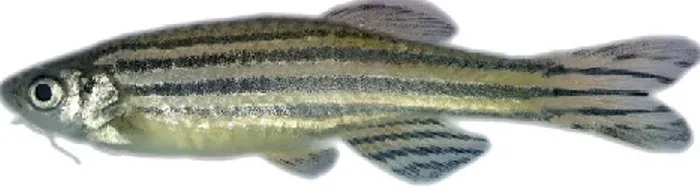
The Somalian cavefish Phreatichthys andruzzii (Ercolini & Berti, 1975) (Figure 6) is a tropical cyprinid and an inhabitant of the subterranean water systems in the Bud-Bud region of Somalia, under the central Somalian dessert, whose ancestors probably adapt to a subterranean life in the phreatic waters due to climate changes. Its habitat is formed by large phreatic layers that develop in Eocene horizontal limestone rock formations, which are accessible only through sink holes and shallow wells.
Figure 6. The Somalian cavefish Phreatichthys andruzzii. This extreme animal model shows complete eye
loss, no pigmentation, no scales, as well as strongly reduced metabolism and oxygen consumption (Sources: modified from Cavallari et al., 2011).
The main reason why we decided to study Phreatichthys andruzzii, and not the better-known Astyanax mexicanus, is that the Somalian cavefish presents a more extreme troglomorphic phenotype than the mexican cavefish Astyanax. The eye degeneration is much more rapid (Berti et al., 2001), while many Astyanax populations do not always show complete eye degeneration and, in some cases, they still have rudimental eyes. In P.