Doctoral programme of Biochemistry and Molecular Biology
-Bibim 2.0
HPLC-MS-MS analysis of thyroid
hormones and iodotyrosines in
knocked-out murine and natural human
DEHAL-1 deficiency
Candidate
Supervisor
Abstract
Thyroid hormones (TH), namely 3,5,3’-triiodothyronine (T3) and its precursor thyroxine (T4), are key regulators of growth processes and development, and crucially control the energy metabolism. A proper availability of iodine within the thyroid is crucial for the synthesis of TH in order to main-tain the homeostasis of circulating levels. The daily dietary iodine intake is not sufficient to susmain-tain their synthesis and, in fact, most of the intra-thyroidal iodine is recycled through the activity of the Iodotyrosine Dehalogenase-1 (DEHAL-1) enzyme. Mono-iodotyrosine (MIT) and di-iodotyrosine (DIT) precursors produced in excess during TH synthesis, are deiodinated by DEHAL-1, leading to the production of I- and Tyr that are recycled and reused within the thyroid. In the last decade, human mutations of DEHAL1 causing the impairment of its catalytic activity were reported. The clinical picture caused by these mutations recapitulates the phenotype of what is known as Iodoty-rosine deiodinase deficiency (ITDD), characterized by hypothyroidism, goitre and, if not treated during childhood, by intellectual impairments. A hallmark of ITDD resides in the increase of urinary and plasmatic MIT and DIT levels.
High-performance liquid chromatography coupled to tandem mass spectrometry (HPLC-MS-MS) is a powerful technique characterized by high specificity and sensitivity. Even if clinical testing of TH is still performed using immunoassays, various HPLC-MS-MS methods to assay TH at low concentrations have been proposed. HPLC-MS-MS is now emerging as a powerful technique that complements traditional testing overcoming some of their limitations. Hitherto, only one HPLC-MS-MS method have been proposed in order to assay MIT and DIT levels in urine that has been used in the discovery of one of the most recent discovered human DEHAL1 mutations.
The aim of my project was to develop a sensitive and robust HPLC-MS-MS method able to quan-tify MIT and DIT in urine and plasma, together with T3 and T4 in the latter case. The method was extensively validated and used to assay these molecules in urine and plasma collected from the novel Dehal1 knock-out mouse. Our method played a crucial role in the biochemical charac-terization of this first mammalian model of defective iodine recycling through DEHAL-1 deletion. We detected increased urinary and plasmatic levels of MIT and DIT in the knock-out mice and we demonstrated the presence of elevated concentrations of both molecules even at the first stage of life. The influence of iodine availability was tested, showing that mice were still euthyroid when levels of dietary iodine were sufficiently high. In the presence of scarce iodine availability, coupled to the impaired ability to recycle iodine through DEHAL-1, knock-out mice became hypothyroid.
Our results demonstrated the importance of iodine availability in triggering hypothyroidism in the presence of ITDD.
The importance of a powerful technique able to detect MIT and DIT was demonstrated assaying these molecules in human urine collected from a consanguineous family with a diagnosed DEHAL-1 deficiency. Our HPLC-MS-MS method was able to detect elevated urinary levels of MIT and DIT in the index patient that was clinically diagnosed with ITDD showing goitre and hypothyroidism. Remarkably, we detected a massive increase of both molecules in urine collected from one of the brothers that was healthy at the time of sample collection but that developed hypothyroidism and goitre several years later.
In conclusion, our findings demonstrated the ability of our validated HPLC-MS-MS method to detect MIT, DIT together with T3 and T4 in urine and plasma sample. We showed the potential of MIT and DIT assay, especially in urine, for the early detection of hypothyroidism in DEHAL-1 deficiency. Considering the presence from the beginning of life, the detection of iodotyrosines could be potentially included in the human new-born screening for hypothyroidism.
Abbreviations
3-T1 = 3-iodo-L-Thyronine AcK = Potassium acetate ACN = Acetonitrile AcOH = Acetic acid
AF-1 = Activation function 1
APCI = Atmospheric-pressure chemical ionization API = Atmospheric-pressure ionization
APPI = Atmospheric-pressure photo ionization BBB = Blood brain barrier
CAD = Collision gas
cAMP = Cyclic adenosine monophosphate CE = Collision energy
CH2Cl2= Methylene chloride
CI = Chemical ionization
CID = Collision-induced dissociation ClO4-= Perchlorate
CP = Choroid plexus CSF = Cerebrospinal fluid CUR = Curtain gas
CxP = Collision exit potential DBD = DNA binding domain
DEHAL = Iodotyrosine dehalogenase DESI = Desorption electrospray ionization DIO = Iodothyronine deiodinase
DIT = Di-iodotyrosine
DNT = 3,5-dinitro-L-Tyrosine DP = Declustering potential DUOX = Dual oxidase ED = Equilibrium dialysis EI = Electron ionization EP = Entrance potential
ERK = Extracellular related kinase ESI = Electrospray ionization
FA = Formic acid
FAB = Fast atom bombardment FMN = Flavin mononucleotide fTH = Free TH
GC = Gas chromatography
GC-MS = Gas chromatography coupled to mass spectrometry GH = Growth hormone
GLUT = Glucose transporter GS = Gas source
H2O2= hydrogen peroxide
HCl = Hydrochloric acid
HIF-1 = Hypoxia-inducible factor 1 HO = Homozygous
HPLC = High.performance liquid chromatography
HPLC-MS-MS = HPLC coupled to tandem mass spectrometry HPT = Hypothalamic-pituitary-thyroid axis
HT = Heterozygous I = Iodine
I-= Iodide I+= Iodinium
IQ1 = Focusing lens 1 IS = Internal standard ISV = Ion spray voltage
ITDD = Iodotyrosine deiodinase deficiency Ka = Association constant
KCN = Voltage-gated K+channels Km = Michaelis-Menten constant KO = Knock-out
KOMP = KO mouse project repository LAT = L-Type amino acid transporters LBD = Ligand binding domain
LC =Liquid Chromatography LID = Low iodine diet LOD = Limit of detection LOQ = Limit of quantification
L-T4 = Levothyroxine
MALDI = Matrix-assisted laser desorption ionization MAPK = Mitogen-activated protein kinase
MCT = Mono-carboxylate transporter MeOH = Methanol
MIT = Mono-iodo tyrosine
MRM = Multiple reaction monitoring MS = Mass spectrometry
MS-MS = Tandem mass spectrometry
NADPH = Nicotinamide adenine dinucleotide phosphate NH4OH = Ammonium hydroxide
NID = Normal iodine diet NIS = Sodium-Iodide symporter
NTCP = Na+/taurocholate co-transporter
OATP = Organic anion transporter polypeptide PIK-3 = Phosphatidylinositol 3-kinase
PTU = 6n-propyl-2-thiouracil
PVN = Hypothalamic paraventricular nucleus rT3 = 3,3’,5’-triiodothyronine or reverse T3 RXR = Retinoid X receptor
SAGE = Serial analysis of gene expression SCN-= Thiocyanate
SD = Standard deviation SEM = Standard error mean SLC = Solute carrier family SPE = Solid phase extraction
SRM = Selective reaction monitoring T0AM = Iodothyronamine T1AM = 3-iodothyronamine T2 = Di-iodothyronine T2AM = Di-iodothyronamine T3 = 3,5,3’-tri-iodothyronine T3AM = Tri-iodothyronamine T4 = 3,3’,5,5’-tetra-iodothyronine or Thyroxine TA = 3-iodo-thyroacetic acid
TAAR1 = Trace amine associated receptor 1 TBG = Thyroxine-binding globulin
TEM = Source temperature
Tetrac = 3,5,3’,5’-tetraiodothyroacetic acid TG = Thyroglobulin
TH = Thyroid hormones TPO = Thyroid peroxidase TR = Thyroid receptor
TRH = Thyrotropin-releasing factor Triac = 3,5,3’-triiodothyroacetic acid TSH = Thyroid-stimulating hormone TTH = Total TH
TTR = Transthyretin UF = Ultrafiltration
UHPLC = Ultra-high pressure liquid chromatography VLID = Very low iodine diet
Contents
I Introduction
12
1 Thyroid hormones 12 1.1 Synthesis . . . 12 1.2 Transport . . . 18 1.3 Mechanism of action . . . 21 1.4 Peripheral metabolism . . . 231.5 DEHAL-1: structure, function and clinical implications . . . 26
2 High-performance liquid chromatography coupled to tandem mass spectrometry 30 2.1 General introduction to the technique . . . 30
2.2 Importance of HPLC-MS-MS in TH analysis . . . 35
II Aim of the project
39
III Materials and Methods
40
3 Chemicals and reagents 40 4 Dehal1 knock-out mice samples collection 40 4.1 Iodine deficiency model . . . 414.2 Natural early-life model . . . 41
5 Human DEHAL-1 family samples collection 41 6 HPLC-MS-MS conditions 42 6.1 Instrumental layout and operative conditions . . . 42
6.2 Samples extraction . . . 44
6.3 Method validation . . . 45
7 Study approval 45
IV Results and Discussion
47
9 Method development and validation 47
10 Dehal1 knock-out mice 53
10.1 Iodine deficiency model . . . 53 10.2 Natural early-life model . . . 57
11 Human DEHAL-1 family 59
V Conclusions
64
VI Pubblications
66
List of Figures
1 Chemical structure of T4 and T3. . . 12
2 Schematic view of the HPT axis and its negative feedback control mechanism. . . 13
3 TH deiodination reactions. . . 15
4 Representation of the key proteins involved in the biosynthesis of thyroid hormones. 16 5 TH genomic mechanism of action . . . 21
6 Schematic overview of the classical pathways of TH. . . 23
7 Chemical structures of some of the main TH metabolites. . . 26
8 Conformational changes of the DEHAL-1 active site to accommodate iodotyrosines. 28 9 DEHAL1 genotypic investigation of the index patient’s family. . . 30
10 Schematic representation of the charged droplet disintegration in ESI. . . 33
11 Quadrupoles rail of the ABSciex API 4000 currently used in our laboratory. . . 34
12 Graphic description of the main scan modes used in MS/MS. . . 35
13 Pedigree of the Lebanon consanguineous family. . . 42
14 HPLC-MS-MS chromatogram of a standard solution containing butylated MIT, DIT, T3 and T4 and their relative ISs. . . 48
15 Representative chromatograms of plasmatic MIT and DIT from WT and KO mice. 51 16 Scatter plot with bar graphs showing levels of MIT, DIT and TH in mice plasma samples from the pilot experiment. (n=5; ** vs WT p< 0.01). . . 52
17 Scatter plot with bar graphs of mice urine levels of MIT, DIT from the pilot exper-iment. (n=5 for WT and n=3 for KO; * p< 0.05). . . 52
18 XY graphs showing levels of MIT, DIT and TH in mice plasma subjected for 28 days to NID. . . 53
19 XY graphs showing urine levels of MIT and DIT after 28 days under NID. . . 54
20 XY graphs showing levels of MIT, DIT and TH in mice plasma subjected for 28 days to LID. . . 55
21 XY graphs showing urine levels of MIT and DIT after 28 days under LID. . . 55
22 XY graphs showing levels of MIT, DIT and TH in mice plasma subjected for 28 days to VLID. . . 56
23 XY graphs showing urine levels of MIT and DIT after 28 days under VLID. . . 56
24 Scatter blot with bar graphs illustrating the plasmatic levels of MIT, DIT and TH in 10 days mice. . . 57
25 Scatter blot with bar graphs showing the concentration of MIT, DIT and TH found in plasma from juveniles. . . 58
26 Scatter blot with bar showing the concentration of MIT and DIT in juvenile urine. . 59 27 Representative chromatograms of DIT assay in human urine. . . 61 28 Column graphs with MIT and DIT levels found in urine from human patients. . . . 62
List of Tables
1 HPLC pumps gradient. . . 43
2 MS operative parameters. . . 43
3 Some of the method performances showing retention times and calibration curves equations. . . 48
4 Instrumental LOD and LOQ of iodotyrosines and TH. . . 49
5 Accuracy results for plasma and urine. . . 49
6 Recovery and matrix effect for plasma and urine. . . 50
7 Thyroid status parameters of the index patient before starting the replacement ther-apy with L-T4. . . 59
8 Thyroid status parameters of the index patient after 3 to 5 months without replace-ment therapy with L-T4. . . 60
9 Thyroid status parameters of the index patient after 6 months of reliable good ad-herence to the replacement therapy with L-T4. . . 60 10 Levels of TSH and TH of the index patient found in the last follow up in June 2015. 61 11 Clinical picture of the index patient’s family at the time of urine samples collection. 63 12 Thyroid parameters of the oldest brother of the index patient after clinical follow-up. 63
Part I
Introduction
1 Thyroid hormones
1.1 Synthesis
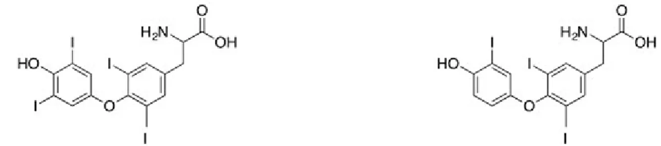
Thyroid hormones (TH) play a key role in regulating growth, development and energy metabolism. They have the crucial function to control tissues differentiation, brain development, cardiovascular and skeletal homeostasis. Moreover, they are critically involved in the control of carbohydrates and lipid metabolism through the regulation of insulin sensitivity in the liver and the suppression of glu-coneogenesis. TH regulate basal metabolic rate and adaptive thermogenesis, with a considerable effect on body weight [1]. With the term TH we refer to the iodinated tyrosine-based molecules 3,3’,5,5’-tetra-iodothyronine (T4 or Thyroxine) and 3,5,3’-tri-iodothyronine (T3) (Figure 1). In humans, the thyroid produces mainly T4, considered as prohormone, since it is converted in pe-ripheral tissues into the biologically active T3 which has a higher affinity for the nuclear thyroid hormone receptor (TR). The thyroid produces 20% of the circulating T3, in a ratio of 1:14 respect to T4 [2, 3].
Figure 1: Chemical structure of T4 and T3. The two tyrosinic rings containing 4 and 3 atoms of
iodine, respectively, are coupled together through an ether bridge.
Levels of circulating TH are controlled by central and peripheral highly sensitive negative-feedback loop mechanisms. The hypothalamus and the pituitary regulate the production of TH by the thyroid, creating a system fundamental in preserving TH homeostasis, known as Hypothalamic-Pituitary-Thyroid axis (HPT). Thyrotropin-Releasing Factor (TRH) is a tripeptide (pGlu-His-ProNH2) se-creted by hypothalamic paraventricular nucleus (PVN) in the hypothalamus [4]. The interaction between the secreted TRH and the G-protein coupled receptor TRH-1 on the pituitary thyrotrope
cells leads to an increase of intracellular cAMP that stimulates the synthesis and release of the pituitary Thyroid-Stimulating Hormone (TSH). Furthermore, TRH has also a role in the control of TSH activity, being important in its glycosylation [5]. TSH, known also as Thyrotropin, is a heterodimeric 28 KDa glycoprotein consisting ofα and β subunits, secreted by pituitary thyrotrope cells. An appropriate glycosylation of TSH subunits is important for the interaction with its G-protein coupled TSH receptor on the thyroid follicular cell, which triggers the synthesis of TH [6]. The increase of circulating T3 and T4 controls the HPT through a highly sensitive negative feed-back mechanism, decreasing the synthesis of TRH and TSH at hypothalamus and pituitary gland, respectively (Figure 2) [7].
Figure 2: Schematic view of the HPT axis and its negative feedback control mechanism. TSH synthesis is stimulated by hypothalamic TRH and inhibited by circulating TH, which are also re-sponsible for the pituitary inhibition of TSH synthesis. Conversely, a decrease of circulating TH rapidly triggers the synthesis and release of both TRH and TSH, in order to normalize levels of T3 and T4.
TH inhibits the synthesis of TRH and both subunits of TSH at a transcriptional level. Additionally, it has been reported the ability of TH to inhibit TSH post-transcriptional modifications and also its release [8]. Conversely, the reduction of circulating TH rapidly activates the secretion of TRH and, afterward, of TSH, leading to an increase TH synthesis and secretion in order to normalize circulating levels [9].
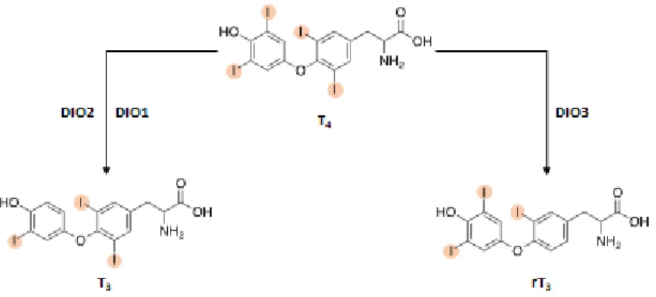
The other mechanism by which levels of TH are controlled takes place intracellularly and involves three members of the thioredoxin enzyme family, namely iodothyronine deiodinases (DIO) [3].
The iodotyrosine deiodinases type 1,2 and 3 (DIO1, DIO2 and DIO3, respectively) are responsible for the regulation of levels and the activity of thyroid hormones, through the removal of iodine from T4 precursors [10]. T4 enzymatic deiodination to form T3 mainly occurs in extrathyroidal tissues but, to a minor extent, also in the thyroid [11]. The three DIOs share an integral membrane nature and the common general structure comprehending a selenocysteine active site [12]. They comprise a single N-terminal, transmembrane segment connected to a larger globular cytosolic domain, containing the selenocysteine active site embedded in a thioredoxin-like fold [2]. The presence of this rare amino acid in the catalytic domain is critical for the reductive deiodination reaction: selenium provides the electrons needed for the elimination of iodine which is reduced to I- and increases substrate affinity and fast turnover rate. Finally, the oxidized enzyme is then reduced and regenerated through the protein thiols [13]. DIO1 and DIO2 catalyse the removal of the iodine at the outer ring (phenolic ring) of T4 to form T3, in a process resulting in its activation, whereas DIO3 is responsible for the removal of the iodine at the inner ring (tyrosyl ring) to form the inactive metabolite rT3 (3,3’,5’-triiodothyronine or reverse T3) (Figure 3). The three isoforms possess different biochemical and regulatory features, exhibiting different tissue localization [11]. DIO1 is located in the plasma membrane and is able to convert T4 into T3 with a Km for T4 in the μM range. It is mainly expressed in liver, thyroid and kidneys and, as a matter of fact, it is not expressed in the central nervous system. DIO2 is the isoform responsible for the formation of T3 in the brain and it is also expressed in thyroid and in other key thyroid-responsive tissues, namely pituitary gland, skeletal muscles and brown adipose tissue [14]. DIO2 is located in the endoplasmic reticulum and its Km for T4 is in the nM range, meaning that T4 is a better substrate for DIO2 rather than DIO1 in vitro. DIO3, responsible for the inactivation of both T3 and T4, is present in the plasma membrane and it is mainly expressed in brain, placenta and pancreas [3]. DIO3 possesses a high affinity for T3 with a Km in the nM range and its activity is stimulated by developmental and disease signals [15].
Furthermore, the three isoforms respond to different inhibitors: while iopanoate is a common in-hibitor of all three isoforms, 6n-propyl-2-thiouracil (PTU) is a selective DIO1 potent inin-hibitor and, from an historically point of view, played a fundamental role in the discovery of DIO2 [14, 16]. As already introduced above, most of the human circulating T3 is not produced by the thyroid gland, but it is the result of the deiodination activity in the extrathyroidal tissues, catalysed mainly by DIO2 and, with a minor extent, by DIO1 [17].
Figure 3: TH deiodination reactions. T4 is activated by the catalytic activity of DIO1 and DIO2 to
form T3 whereas the inactivation reaction leading to rT3 is catalysed by DIO3.
T3 itself, is an important regulator of DIOs activity, stimulating the activity of DIO1 while having an opposite effect on DIO2 [18]. Hyperthyroidism increases DIO1 and decreases DIO2 expression, while the opposite is observed in hypothyroidism, in which the peripheral conversion T4 to T3 is enhanced by the DIO2 induction [3]. Moreover, T4 decreases the activity of DIO2 through post translational mechanisms, i.e. ubiquitin conjugation, contrary to DIO1 and DIO3 that are not susceptible to these reactions. Notably, Dio1 and/or Dio2 knock-out (KO) animal models have shown the ability of the thyroid to preserve circulating concentration of T3. The HPT is involved in this adaptive mechanism to maintain normal T3, since an increase of TSH serum level in response to reduced serum T4 has been observed [15].
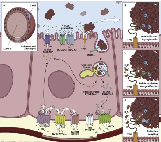
The synthesis of TH is a complex process that comprises a series of tissue specific biochemical reactions. The functional unit of the thyroid for de-novo biosynthesis of TH is represented by thyroid follicles which are formed by a monolayer of follicular cells, called thyrocytes. The apical membrane of thyrocytes encloses the interior follicle lumen known as colloid, while the basolateral membrane faces the bloodstream [19]. The interior part of the follicles contains a large number of proteins among which, the most highly expressed, is an iodoglycoprotein called Thyroglobulin (TG). TG is a protein composed by two homodimers of ~330 KDa each and is the primary source of TH. In fact, it contains ~ 70 tyrosine residues with an averaging iodine content of 2.5-50 atoms of iodine per mol of TG and it can be itself a source of this essential element. TSH stimulates TG endocytic uptake into thyrocytes and its lysosomal degradation, resulting in the release of TH from the TG polypeptide backbone [20] (Figure 4).
thyro-Figure 4: Representation of the key proteins involved in the biosynthesis of thyroid hormones. A.
Structure of thyroid follicles showing C cells involved in the production of calcitonin, important in the control of circulating Ca2+ levels; B,C and D. Main steps involved in TPO and TG activity leading to oxidative coupling and synthesis of TH (Adapted from Carvalho et al. [19]).
cytes driven by the sodium-iodide symporter (NIS). NIS is a 13-segments transmembrane glyco-protein belonging to the solute carrier family 5 (SLC5A5) which transports 2 ions of Na+per each
I-ion [21]. The active transport of I-against its electrochemical gradient inside the cytoplasm, relies
on the activity of another important transmembrane protein, namely the Na+/K+-ATPase pump.
With its activity, NIS is able to concentrate I -around 30-60 folds in the cytoplasm of thyrocytes.
This ensures that most of the ingested iodide, a scarce element in the environment, is accumulated in the thyroid and available for the synthesis of TH [22]. NIS transcription, translation and activa-tion are stimulated by TSH through the activaactiva-tion of the G-protein coupled TSH receptor [23, 24]. Conversely, the transport of I-inside the thyrocyte is blocked by competitor NIS inhibitors, i.e. per-chlorate (ClO4-) and thiocyanate (SCN-) [19]. I-itself is considered a main factor in controlling the accumulation of I-. In 1948, Wolff and Chaikoff reported that high levels of iodide were able to
reduce its transport and accumulation in rat thyroid. This effect became known as “Wolff-Chaikoff effect”[25].
However, even in presence of high levels of I -, TH biosynthesis can be restored after ~ 2 days
through an adaptive mechanism mediated by the inhibition of I -transport and known as “escape
from Wolff-Chaikoff effect” [26, 27]. The electrochemical equilibrium is then restored via the pres-ence on the basolateral membrane of two voltage-gated K+channels, namely KCNQ1 and KCNE2. These ion channels are responsible for the efflux of K+outside the cell and their key role in thyroid function has been reported [28].
Once inside the cytoplasm, I-is transported to the thyrocyte apical membrane and finally towards the
follicle lumen. This transport, known as “iodide efflux”, is mediated by a number of apical channels among which Pendrin was the first one discovered [29]. The rapid translocation of Pendrin toward the apical membrane is regulated by TSH through post-translational mechanisms, resulting in an increase of iodide efflux [30]. More recently, the important role in the process of iodide efflux of the chloride channel ClC5 and the calcium-activated chloride channel Anoctamin-1, has been demonstrated [31, 32].
I- ions inside the lumen need to be oxidized in order to be integrated and bound to the tyrosyl residues of TG [19]. This oxidation reaction is catalyzed by the key enzyme of the TH biosynthesis, namely thyroid peroxidase (TPO). This is a glycosylated oxidoreductase with the fundamental role in iodination and coupling of iodotyrosines residues on TG, leading to T3 and T4 synthesis. The heme-containing active site and most parts of TPO are situated in the follicular lumen and form the extracellular process responsible for the enzymatic activity, whereas the small transmembrane C-terminal region anchors it at the apical membrane of thyrocytes [33, 34, 35]. The oxidation reaction of iodide molecules makes use of hydrogen peroxide (H 2O2) generated by dual oxidase
enzymes, namely DUOX1 and DUOX2. These Ca2+-dependent glycoflavoproteins are members of the nicotinamide adenine dinucleotide phosphate (NADPH) oxidase family and they are located on the apical surfaces of thyrocytes, together with their activators DUOXA1 and DUOXA2 [36, 37]. H2O2 is responsible for the oxidation of TPO, so that it can oxidize I - to form iodinium (I+) and
hypo-iodite (IOH-) ions. These very reactive iodine species bind to tyrosyl residues on the TG
backbone to form mono-iodo tyrosine (MIT) and di-iodo tyrosine (DIT) [19].
The next step leads to the synthesis of T3 and T4, a process called thyroid hormonogenic coupling reaction. The exact mechanism of this process is still poorly understood, even if it has been
hypoth-esized the involvement of a free radical intermediate or an ionic interaction. However, hormono-genic coupling involves the donation of iodophenoxyl group from MIT or DIT called “donor”, to a DIT residue called “acceptor”, producing T3 and T4, respectively, and leaving a dehydroalanine in the “donor” position, as a side products of the reaction [38]. The analysis of kinetic models of iodination and hormonogenic coupling reactions revealed that only iodotyrosines properly oriented in the space can form T3 and T4. Moreover, they showed that iodinated tyrosyl residues preferen-tially admit the biosynthesis of T4 that, as already mentioned, is the main product of the thyroid [39]. Only 2 to 8 molecules of TH are formed from the 25-30 Tyr residues iodinated by TPO and, in normal conditions, TG roughly contains 2.5 residues of T4 and 0.7 of T3 [19]. Intrathyroidal T3 biosynthesis, formed by the coupling of a MIT and a DIT residue, follows the same pathway as T4. Noteworthy, most of the circulating T3 is not synthetized through T4 deiodination on TG, but it is the result of the DIO1 and DIO2 peripheral actions stimulated by TSH [10].
Synthetized TH bound onto TG are stored in the colloid until the increase of TSH levels promotes its internalization into thyrocytes via endocytosis processes. T3 and T4, together with uncoupled MIT and DIT, are released from the newly formed lysosomes by the proteolytic activity of en-dopeptidases. Free released MIT and DIT molecules, are enzymatically deiodinated to obtain I -and Tyr residues that can be reused in the biosynthesis of TH. This important recycling process is catalyzed by an enzyme known as iodotyrosine dehalogenase (DEHAL-1) [40] which will be extensively described in the Paragraph 1.5.
1.2 Transport
Synthetized TH need to be released into the bloodstream, with a process that, for a long time, was believed to occur by a passive diffusion mechanism considering the lipophilic structure of TH. Lately, it has been described that TH reach the bloodstream through a series of transporters present on the basolateral plasma membrane of thyrocytes.
TH transporters belong to different families of solute carriers, they are not specific for TH and are widely expressed among tissues other than the thyroid [41]. Mono-carboxylate transporters (MCT) are a family of 14 transporters, among which MCT8 and MCT10 are sodium and proton independent TH transporters that mediate their transport through the plasma membrane [42, 43]. MCT8 is a highly specific TH transporter whereas MCT10 is an aromatic amino acids transporter possessing a slightly higher efficiency at transporting T3, but lower efficiency at transporting T4 [41, 44]. MCT8 has a particular importance for TH uptake into the brain, as revealed by the
identi-fication of an X-linked mutation leading to a syndrome characterized by psychomotor retardation, known as Allan-Herndon-Dudley syndrome [45, 46]. Even if circulating T3 levels are elevated, this neurodevelopment disorder is characterized by low intracerebral TH concentration, leading to a cerebral hypothyroid state during brain development [47]. MCT8 is expressed in many tissues in addition to thyroid (liver, heart, kidney, placenta, intestine, thyroid, and brain) and in distinct areas within the brain with an important role for T3 transport across the blood-brain barrier (BBB). Pathogenic mutations in MCT8 encoding gene have been detected in several diseases, indicating a pathophysiological role for TH transport [41]. In the last two decades, KO models have been de-signed to better understand the role of MCT8 in different tissues and the pathological implication of its mutations (the reader can refer to some excellent reviews [48, 49]). Liver Na+/taurocholate co-transporter (NTCP) is a seven transmembrane domains glycoprotein involved in the enterohepatic circulation of bile acid. Belonging to the solute carrier gene family (SLC10A), these proteins rec-ognize TH sulphate derivatives which are transported to the liver where DIO1 is responsible for the deiodination and rapid degradation of iodothyronine sulphate [50]. The L-Type amino acid trans-porters (LAT) are heterodimeric protein comprised of a 12 transmembrane light chain domain and single transmembrane glycosylated heavy chain domain, generally involved in the transport of neu-tral amino acids. LAT1 and LAT2 are the two isoforms involved in TH transport, with differential affinity: in fact, LAT1 transports T3 more efficiently than LAT2 [51, 52]. LAT1 and LAT2 have dif-ferent tissue distribution, with the first one primarily distributed in brain, placenta and tumors, and the latter one primarily distributed in kidney, colon and intestine [53]. Organic anion transporter polypeptide (OATP) is a family of 12 transmembrane domain proteins involved in the transport of a series of amphipathic compounds, like steroids, anionic oligopeptides, bile salts and drugs. Up to seven members of this family are able to transport TH, among which OATP1C1 shows higher specificity to transport T3, rT3, T4 and sulphate T4 derivatives. It is expressed in several tissues but its physiological relevance is still unknown [41, 48]. Since TH are highly hydrophobic molecules, several carrier proteins are responsible for their transport and distribution through the bloodstream and cellular compartments [11]. Almost 95% of circulating TH are bound to thyroxine-binding globulin (TBG), transthyretin (TTR) and albumin, whereas the rest 5% is bound to minor carrier proteins, like lipoproteins or, with a minor extent, can be available in the circulation as free TH (fTH). Blood carrier proteins most probably have the function to ensure a constant availability of TH to the cells and tissues, avoiding urinary loss and controlling TH levels during abnormal production and degradation.
TBG is a glycoprotein synthetized in the liver made of a single 56 KDa polypeptide chain. It has only one binding site for TH, with an association constant Ka of 1x10¹⁰ M-1 and 4.6x10⁸M-1 for
T4 and T3, respectively. Notably, the binding of T4 to TBG induces conformational changes that increase its stability. Since it carries the major part of TH (~ 70%), qualitative and quantitative abnormalities of TBG have a significant impact on total circulating TH levels [54, 55].
TTR, formerly known as thyroxine-binding pre-albumin, is a 55 KDa protein that circulates in blood as a stable homotetramer of identical subunits of 127 amino acids each [56]. Subsequently to the discovery of its TH binding properties, it has been demonstrated that TTR exists also in part as a complex with retinol binding protein, with a role in the transport of Vitamin A [57]. Only one of two TH binding sites is occupied and it has a Ka of 2x10 8 M-1 and 1x10⁶ M-1 for T4 and T3, respectively. The lower affinity for TH respect to TBG, allows their rapid dissociation from TTR which, therefore, seems to be associated with the immediate delivery of T3 and T4 [55]. TTR carries 10 to 15 % of protein bound T4 and it is mostly synthetized in liver and, with a minor extent, in the central nervous system. It has an important role in the transport and delivery of T4 in the cerebrospinal fluid (CSF), where it is the main TH transporting protein [58].
Albumin binds ~5% of circulating TH with a lower affinity respect to TBG and TTR: in fact, its Ka 1.5x10⁶ M-1 and 2x10⁵ M-1 for T4 and T3, respectively [55]. Despite these low affinities, the relative contribution in TH transport is still significant because of the high amount of albumin that circulates in human serum. It has been proposed an albumin role as a fast TH resource during the rapid exchange in capillary transits [59].
TH play a crucial role in the brain development and maintenance of its adult functions. In fact, TH deficiency in fetal and neonatal periods results in cretinism characterized by mental retardation, deafness and ataxia, whereas in adult, can cause impairment of motor skills, intellectual defects and spasticity. For these reasons, adequate levels of TH in the brain are essential and can reach the brain by crossing the BBB, the highly selective permeability barrier which separates the bloodstream from the brain extracellular fluids. T4 is thought to be the primary TH crossing the BBB to be then locally converted into T3 by DIO2 in the hypothalamic astrocytes and tanycytes [60]. TH can cross the BBB via the already mentioned specific transporters such as MCT8 or OATP1C1 or can also indirectly reach the brain via the blood-CSF barrier. Choroid plexus (CP) is a highly vascularized structure part of the brain ventricular part, responsible for the synthesis of CSF and TTR, which is the main T4 transporter in this fluid. TTR plays a pivotal role in the delivery of T4 from plasma into CP and in its subsequent transport into CSF, offering an optimal exchange to deliver T4 into the brain [61, 62].
1.3 Mechanism of action
TH are essential for many diverse processes, like tissues development and the regulation of cellular metabolism. The effects of TH are mediated by two mechanisms: a genomic one, in which TH interacts with nuclear TR, and a nongenomic one, with a rapid onset, in which TH interacts with plasma membranes and other receptors [63].
TR are nuclear receptors that act as ligand-dependent transcription factors, able to modulate the transcription of target genes when activated by TH. In fact, the binding of T3 to TR can increase or decrease the transcription rate of target genes. In the nucleus, TR can constitutively bind to specific regions located in the promoter region of TH target genes, known as thyroid hormone response element (TRE) [64]. It has been shown that TR are still bound to TRE even in the absence of TH, repressing or silencing the basal transcription of the positive regulated target genes [65]. In the absence of its ligand, TR can be found as monomer, homodimers if coupled with another TR, or heterodimers, mainly coupled with retinoid X receptor (RXR) [66] (Figure 5).
Figure 5: TH genomic mechanism of action (Adapted from Ortiga-Carvalho et al. [64]) The heterodimer TR-RXR appears to be the favoured conformation to bind the DNA, even if the
in vivo functions and the roles of these complexes are still under investigation and remain to be
determined [67].
The two genes TRΑ and TRΒ encode for the two nuclear receptors, TRα and TRβ, respectively. Not all the mammalian TR protein act as a nuclear receptor and, in fact, other proteins can be synthetized from alternative splicing, but their physiological roles are still unknown. The four iso-forms acting as functional receptors are TRα1, TRβ1, TRβ2, and TRβ3. These functional isoiso-forms
showed different patterns of expression in development and in adult tissues: TRα1 and TRβ1 are widely expressed, with the first one mostly expressed in brain, heart, skeletal muscle, and brown adipose tissue, and the latter one in brain, liver, and kidney. TRβ2 is predominantly expressed in pituitary gland, hypothalamus, and cochlea, whereas TRβ3 is mainly expressed in kidney, liver, and lung, and seemed to be functional in rat [68, 69]. TRβ is the responsible of most of TH effects on metabolism and, from a pharmacological point of view, also an ideal target to treat metabolic disorders, mainly lipid-related, or brain diseases. An ideal thyromimetic drug, should possess a high TRβ selectivity, in order to avoid any adverse effects on bone and heart [70].
TR are member of the highly conserved nuclear receptor superfamily and consist of a single peptide that is folded into three different functional domains: an amino terminal domain (A/B) which also contains the activation function 1 region (AF-1) involved in transactivation; a central DNA binding domain (DBD) containing two “zinc fingers”, critically important in sequence-recognition on the TRE; and a carboxyl terminal ligand binding domain (LBD) which determines the specificity and high affinity of the receptor for TH ligands [63]. An additional linker region between DBD and LBD domains contributes to activation, repression and corepressors interaction and it is important for DNA and ligand binding [71].
Nongenomic effects of TH do not involve the classical concept of TR mediated TH action. They do not require gene transcription and protein synthesis but can include modulation of gene tran-scription. The mechanisms of several nongenomic actions depend upon signal transduction system and can involve novel TH membrane receptors, extranuclear TRβ or truncated isoforms of TR α [63, 72].
A novel receptor unrelated to classical TR exists on the integrin αVβ3, a plasma membrane struc-tural protein highly expressed in tumour cells and dividing endothelial cells. IntegrinαVβ3 has no structural homology with TR and TH binding takes place nearby the Arg-Gly-Asp recognition site where are located the two binding sites [73]. Binding of T3 activates phosphatidylinositol 3-kinase (PIK-3), specifically driving TRαinto the nucleus and promoting transcription of hypoxia-inducible factor 1 (HIF-1) gene. TH can also interact with the integrin receptor and activates the extracel-lular related kinase 1 and 2 (ERK-1 and ERK-2) responsible for the transduction of TH signals in cancer cells. Moreover, interaction of both T3 and T4 with integrin αVβ3 is involved in their demineralizing action [63].
with TRβ. The result is an activation of voltage-dependent potassium channels with a decrease of excitability and reduction of hormone secretion [74]. The activation of PIK-3 mediated by T3 has also direct and indirect effects on the transcriptional increase HIF-1, glucose transporter 1 (GLUT-1), and MCT4 transporter [65]. Both T3 and T4 can stimulate mitogen-activated protein kinase (MAPK) activity increasing angiogenesis. The activation of MAPK leads to phosphorylation of the tumour protein p53 with a final decrease of its transcriptional activity. In the nucleus, acti-vated kinase forms a complex with TR β and phosphorylate its serine. T3 is also responsible of many nongenomic actions on plasma membrane proteins, contributing to basal activity of some ion pumps such as Ca2+-ATPase, Na+/K+-ATPase and Na+/H+antiporter [75].
Recent studies on astrocyte cells, demonstrated the involvement of truncated TRαisoforms, namely TRΔα1 TR Δα2, in the regulation of actin cytoskeleton modelling which has a key role in the developmental program of the brain [72]. In response to T3, truncated TRα isoforms have shown to be imported into mitochondrial inner membrane where they are able to directly stimulate oxidative phosphorylation processes [65].
1.4 Peripheral metabolism
As already discussed above, the deiodination reaction of T4 to form the active T3 or the inactive rT3 is catalysed by DIO enzymes that are important in the control of TH levels. Once inside the cell, TH can undergo to tissue-specific metabolism, which can involve further deiodination, deamination, decarboxylation, sulfonation and conjugation reactions (Figure 6).
Figure 6: Schematic overview of the classical pathways of TH.
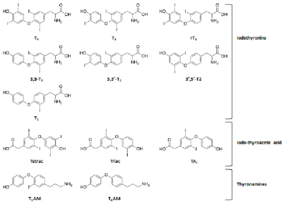
T3 and rT3 can be subjected to additional deiodination reactions, to form 3,5-diiodothyronine (3,5-T2), 3,3’-diiodothyronine (3,3’-(3,5-T2), 3’,5’-diiodothyronine (3’,5’-T2) and 3-iodo-L-Thyronine
(3-T1). 3,3’-T2 and 3’,5’-T2 demonstrated no remarkable activities, whereas 3,5-T2 proved the ability to bind to TR and have been studied for a long time by researchers. 3,5-T2 actions are mediated by the TR classical interaction but also through rapid effects at the cell membrane and mitochondria [76]. In vivo animal experimental models showed the ability of administered 3,5-T2 to alter the expression of canonical T3 regulated genes in various tissues, whereas, in pituitary models, it was able to suppress TSH and stimulate GH [77]. Animal models showed that the parental adminis-tration of 3,5-T2 was able to modulate energy metabolism, probably through the stimulation of liver fatty acid oxidation into mitochondria [78, 79]. Moreover, 3,5-T2 administration was able to prevent body weight gain, liver adiposity, hyperlipidaemia and insulin resistance, without any sign of thyrotoxicosis [76]. Acute 3,5-T2 exposition in an isolated rat heart model, showed the ability to increase oxidative metabolism without affecting cardiac contractility [80]. The physiological rele-vance of 3,5-T2 and 3,3’-T2 remains to be determined, however their presence in serum has been confirmed through the application of immunoassays and accurate high performance liquid chro-matography tandem mass spectrometry (HPLC-MS-MS) methods. Nowadays, their actual serum concentrations are still debated and no clinical applications of their detection have been reported [81, 82, 83].
DIO are also able to deiodinate deaminated TH metabolites, namely 3,5,3’,5’-tetraiodothyroacetic acid (Tetrac) and 3,5,3’-triiodothyroacetic acid (Triac). Tetrac is produced by deamination of T4 and its presence in human serum have been confirmed at low nM concentrations. Triac can be produced either by deiodination of Tetrac or by deamination of T3 and, despite its importance in pathophysiological cell-specific actions, distorted serum concentrations have been reported. They are both transported by TTR in serum and they act as thyromimetic compounds lowering TSH con-centrations. Tetrac binds to TR onto integrin αVβ3 and elevated concentrations of this compound have been found in Grave’s disease patients. Triac has a potent T3-mimetic activity showing a high affinity for TRβ and it has been described to be a substrate for MCT8 transporter [77].
Thyronamines are a novel class of endogenous iodothyronine-like signalling compounds that are structurally related to TH, differing only for the absence of the carboxylate group on the alanine side chain. Thyronamines differ for the number and position of iodine atoms, they are desig-nated with TxAM and their nomenclature is analogous to TH, where x is the number of iodine
atoms [84]. Hitherto, only two members of this class have been detected in human serum using HPLC-MS-MS, namely Thyronamine (T0AM) and 3-iodothyronamine (T1AM) [85, 86, 87, 88].
In 2004, the discovery of a transient hypothermia effects after the administration of T1AM in rats,
biosyn-thetic pathways, mechanisms of action and pathophysiological effects. In the pioneering paper by Scanlan et al., it was also demonstrated that the rapid onset of T 1AM effects was not compatible
with the classical TH nuclear mechanisms, but more suitable with a G-coupled receptor. The sug-gested receptor responsible of T1AM effects was identified in the trace amine associated receptor 1
(TAAR1). However, mechanisms of T1AM actions can include other membrane receptors and
in-tracellular targets which are still debated and not clear [86, 85, 77]. The mechanism of endogenous T1AM biosynthesis is still uncertain. One of the putative mechanisms involves the deiodination
of T3 or T4 to form T2, which is decarboxylated to form di-iodo-thyronamine (T 2AM) and, then,
further deiodinated to form T1AM [89]. Additionally to the already cited hypothermic effects, in
vivo and in vitro models showed the ability of administered T1AM to reduce cardiac inotropic and
chronotropic effects [90, 91, 92], to protect during in ischemia reperfusion models [93, 94], to in-duce acute metabolic responses with actions on carbohydrates and lipids [95, 96, 97, 98] and it is emerging as a possible modulator of noradrenergic, dopaminergic and histaminergic systems [99]. T1AM and thyronamines can undergo several metabolic reactions such as sulfonation,
glucuronida-tion and acetylaglucuronida-tion whose effects are still under investigaglucuronida-tion [100].
In most tissues, the major metabolite of exogenous T1AM and T3AM are the corresponding acidic
compounds, namely 3-iodo-thyroacetic acid (TA1) and the already mentioned Triac, respectively.
The mechanism for the biosynthesis of TA1is still under debate. A putative pathway was postulated
by Lorenzini et al. and involved the oxidative deamination of T1AM to form the corresponding acid
TA1through an aldehydic intermediate [101]. The biological role of TA1 is still unknown, even if
an activity on the histaminergic system has been reported [102, 103].
Moreover, the phenolic hydroxyl group of TH can undergo conjugation reactions with the formation of sulfonated and glucuronated metabolites. Sulfation reactions of T4, T3, T2 and thyronamines have been reported as well, catalysed by sulfotransferase enzymes. Interestingly, sulfonated TH have a high affinity for DIO1 and this modification is a critical step to enhance the deiodination and inactivation of T4, T3 and T2 [17]. Interestingly, sulphate T3 can represent a reservoir of inactive hormone and might undergo enterohepatic recycling, whereas sulphate T2 might be useful as a biomarker for fetal thyroid function considering its secretion into maternal circulation through the placenta. Sulphated thyronamines have been investigated as well, but their biological roles and relevance are still unclear [77]. Sulfonation and glucuronidation are part of the so-called phase II detoxification reactions and they are involved in the inactivation and excretion of TH. The general aim of these reactions is to increase the solubility of TH in water and facilitate their excretion through bile and/or urine [84].
Figure 7: Chemical structures of some of the main TH metabolites.
1.5 DEHAL-1: structure, function and clinical implications
All the steps involved in the synthesis of TH, starting from I- uptake from the bloodstream to
the final synthesis and release of T3 and T4, have been already extensively illustrated (Par.1.1). TH are the only mammalian hormones containing a halogen in their structure, iodide, which is present at a very low concentration in the environment. Accordingly, thyroid functions depend on the availability of this halogen in the thyroid for the synthesis of TH. Dietary iodide is mainly ingested from sea salt, where it is present as NaI, milk, meat and fish, but daily I -intake amounts
alone are not sufficient to sustain the production of TH. For this purpose, the enzyme DEHAL-1 is responsible for the enzymatic deiodination of MIT and DIT generated in excess during TH synthesis [104]. MIT and DIT are released six-seven times more than TH during proteolysis of TG but they are inactive at thyroid level. The catalytic activity of DEHAL-1 produces the intrathyroidal I-that, together with released Tyr molecules, can be recycled and reused for the synthesis of TH [105].
In the past, the existence of this deiodination activity has been already demonstrated even if the enzyme responsible for this reaction was still lacking. In 2003, Moreno et al., through the
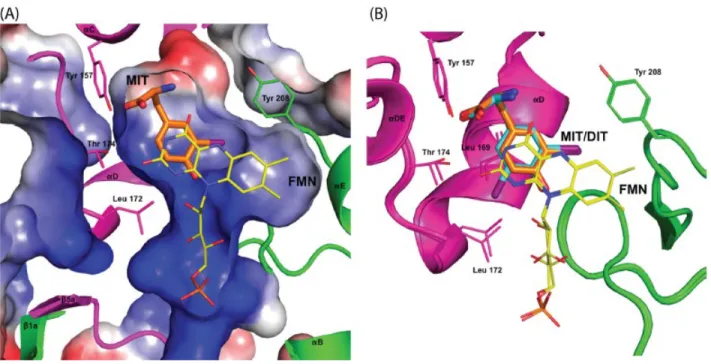
appli-cation of the serial analysis of gene expression technique (SAGE) to thyroid tissue in combination with a computational method, identified the DEHAL1 gene encoding for the protein responsible of the dehalogenation of TH. The gene was found to be mainly present in the thyroid gland and, with minor extent, in kidneys and liver [106]. Three isoforms of this enzyme were identified, namely DEHAL-1, DEHAL-1B and DEHAL-1C although only the first one showed dehalogenation activ-ity, whereas the other two isoforms resulted inactive [107]. The structure of DEHAL-1 became clear after that the first crystal structure was reported by Thomas et al. in 2009 [108]. It has a molecular weight of 33 KDa and belongs to the NADH oxidase/flavin reductase superfamily, with a flavin mononucleotide (FMN) as a prosthetic group which is essential for its catalytic activity. T3 and T4 are neither deiodinated by DEHAL-1, nor able to bind to this enzyme, which does not contain the catalytic selenocysteine responsible for DIOs activity. DEHAL-1 is a transmembrane protein located at the apical membrane of the thyrocytes: it consists of a single transmembrane domain, a short carboxyl terminal domain and an amino terminal domain containing most of the nitro-reductase activity. The active site residues Glu-153, Tyr-157 and Lys-178 are responsible for the recognition of the zwitterionic forms of MIT and DIT as well as for the redox characteristics of the FMN. The alignment of the larger DIT over the isoalloxazine portion of the FMN is achieved through minor shifts of Leu-169, Thr-174 and Leu-172 (Figure 8).
The catalysed deiodination reaction occurs at the apical membrane in close proximity to TG and takes place during TG proteolysis both before and after its endocytosis into the thyrocytes [109]. The exact mechanism of the dehalogenation reaction is still under investigation, even if the pre-viously proposed mechanism consisting in the simultaneous transfer of two electrons have been recently overcome by a single electron transfer mechanism. This putative process implicates a single electron transfer from the hydroquinone form of FMN and involves the biding of the phe-nolate form of iodotyrosines to DEHAL-1. The phephe-nolate form enables the coordination with two hydrogen bond donors leading to the protonation of the halogen containing α-carbon. The pro-tonated intermediate undergoes reductive dehalogenation through the electron donation from the hydroquinone form of FMN to form a transient semiquinone intermediate. The elimination of the halogen forms a stable phenoxy radical that can accept an electron from the semiquinone interme-diate of FMN regenerating the oxidized form of FMN. The final product of the reaction is Tyr that, together with I-, can be recycled to synthetize TH [110].
In vitro, DEHAL-1 activity showed to be dependent on NADPH levels and proportional to the
enzyme concentration. The reported Km for the deiodination of MIT and DIT were 1.35x10 -6 and 2.67x10⁻⁶, respectively, showing a higher affinity of DEHAL-1 for MIT respect to DIT.
Fur-Figure 8: Conformational changes of the DEHAL-1 active site to accommodate iodotyrosines.
A. MIT-DEHAL-1 co-crystal structure showing the surface characteristics: blue indicates positive charges whereas red indicates negative charges; B. Conformational changes required for the align-ment of DIT (cyan) to the active site of DEHAL-1 equally to MIT (orange) (Adapted from Thomas et al. [108]).
thermore, in vitro and in vivo models showed that nitro aromatic compounds can be substrate of this nitro-reductase, revealing that the nitro-analogue 3,5-dinitro-L-Tyrosine (DNT) is a power-ful DEHAL-1 inhibitor [40]. Moreover, DEHAL-1 is able to promote chlorination and de-bromination reactions of Tyr and bromo-Tyr, respectively.It was demonstrated that chloro-and bromo-Tyr were able to bind to the active site of DEHAL-1 with the same affinity as iodoty-rosines, even if the dichlorination reaction resulted up to 20-fold slower. Conversely, DEHAL-1 was unable to defluorinated fluoro-Tyr that showed also a weaker binding ability to the enzyme respect to iodotyrosines [111]. Further studies reported the up-regulation of Dehal1 mRNA in rats by TSH-stimulated cAMP and the down-regulation by acute and chronic administration of iodide. Moreover, the activity of DEHAL-1 was increased in hypothyroid rats [112].
In 2008, Moreno et al., reported the first human mutations of DEHAL1. Three different mutations were identified: two missense mutations (Arg101Trp and Ile116Thr) and one in-frame depletion of three base pair (Phe105-Ile106Leu). They were situated in the exon 2 of the gene encoding a FMN- binding site localized within the nitro-reductase domain of the protein. These mutations
drastically reduced the deiodination activity of DEHAL-1, leading to the release of MIT and DIT in plasma before their excretion through urine. The patients presented variable pathological phe-notypes that can be related to the time of expression of the disease or to environmental factors, such as iodine intake. The four analysed patients came from unrelated consanguineous families, all of them had sever goitrous hypothyroidism and two of them suffered from intellectual deficits due to late diagnosis and treatment [113]. Failure of DEHAL-1 leads to an already known disease called iodotyrosine deiodinase deficiency (ITDD), which is characterized by hereditary hypothy-roidism, goiter, psychomotor deficits and intellectual retardation when not properly treated in early stages of life. The identification of DEHAL1 mutations opened novel opportunities to comprehend ITDD, considering the lack of expression at the beginning of life where TH play a crucial role in the development. In fact, two of the four patients analysed by Moreno et al., were subjected to the neonatal screening for hypothyroidism and, worryingly, resulted normal, meaning that the disease may not be present at the beginning of life [114]. An additional missense homozygous (HO) mu-tation (Ala220Thr) has been reported by Afink et al. in 2008 from another consanguineous family. The mutation, situated on the exon 4, resulted in an inactive DEHAL-1 activity in vitro showing both catalytic impairment and protein degradation. The two homozygous index patients (mother and daughter) showed hypothyroidism, goiter and, one of them, had mental retardation. High con-centrations of MIT and DIT have been detected in urine from index patients. Interestingly, high levels of both iodotyrosines have been found also in one of the heterozygous sons at the age of 9, without any other symptoms. During the puberty (14 years old), clinical evidences of hypothy-roidism with a massive goiter were discovered, demonstrating that phenotype can emerge over time [115]. Hitherto, the diagnosis of ITDD is not included in the neonatal screening programmes even if consequences in the neurodevelopment of untreated patients have been reported [114].
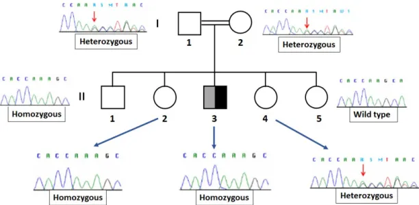
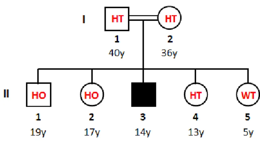
Recently, a novel DEHAL1 homozygous mutation (p.A57SfsX62) was identified in the offspring of a consanguineous family from Lebanon. The mutation consisted in the insertion of an adenosine between the position 168 and 169 of the exon 1, causing a frameshift that led to an early-stop codon at the amino acid 62 of the protein. This novel mutation resulted in an almost completely truncated enzyme with the consequent delete of the functional nitro reductase domain and an impaired activ-ity of DEHAL-1. The mutation was discovered in an index patient of 11 years old who consulted his physician because of a goitre developed in the last 3 weeks. Genetic investigation on DEHAL1 gene mutations was extended to several member of the index patient’s family (Figure 9). Genetic sequencing revealed the heterozygous condition of his parents and of the younger sister, whereas the older brother and the older sister resulted homozygous and the youngest sister was wild-type. At that time, phenotypic investigation identified goitre only in the index patient whereas none of
the other members showed any significant symptoms [116].
Figure 9: DEHAL1 genotypic investigation of the index patient’s family. The father (I-1) and the
mother (I-2) resulted heterozygous. Among the siblings, two members (II-1 and II-2) other than the index patient, exhibited the homozygous mutation whereas the other sisters were heterozygous (II-4) and WT (II-5), respectively.
2 High-performance liquid chromatography coupled to tandem
mass spectrometry
2.1 General introduction to the technique
Liquid chromatography (LC) coupled to mass spectrometry (MS) is a powerful and versatile tech-nique, widely used in analytical laboratories. In the chromatographic column the components of a mixture are separated based on their affinity for both the stationary phase and the mobile phase, and then ionized and detected by the coupled mass spectrometer.
With the term HPLC, we refer to a LC separation technique carried out at high operative pressure up to hundreds of bars or to thousands of bars in ultra-high pressure liquid chromatography (UH-PLC) instruments. This separation technique relies on two main elements, namely stationary phase and mobile phase. The analytes are dissolved in an appropriate liquid mobile phase and pumped through the stationary phase, which is the hearth of the HPLC system, where the separation takes place. The stationary phase is constituted by a chromatographic column tightly packed with a solid
adsorbent material. Each compound interacts differently with both the adsorbent material present in the chromatographic column and the eluent. So that, it is eluted with a different time, known as retention time. In general, the higher is the affinity for the stationary phase (or the lower is that for the mobile phase), the later a compound is eluted from the column. Conversely, an analyte with a lower affinity for the stationary phase will be eluted with shorter retention times from the chromatographic column. The type of interactions depends on the compound chemical structure and, as a consequence, on its own physical-chemical characteristics, such as polarity, and on the characteristics of both stationary and mobile phases. Accordingly, stationary phase is accurately selected in order to achieve the best selectivity to separate the different components of a mixture [117]. Among the numerous stationary phases commercially available, a large part of the HPLC separations are carried out by the so-called reverse phase stationary phases. Differently from the classical type, known as normal phases, in which the stationary phase was made of silica or alu-mina, the surface of the silica particles is coated with siloxane together with various alkyl (C8 or C18) or aryl groups. The end part of the group is the one responsible for the interaction between stationary phase and the analytes. Reverse phase chromatography is indicated for the separation of a wide variety of different compounds and it is preferable considering that a large part of the suitable solvents are compatible with mass spectrometry, which is often coupled to HPLC. Mobile phase is usually constituted of two solvents (typically an organic phase and an aqueous-phase) that are mixed together before being pumped into the system. Depending on the complexity of the mixture, the characteristics of analytes and the selected chromatographic column, the mobile phase is appropriately chosen. Its composition can be kept constant during the chromatographic analysis, creating what is known as isocratic condition. If the components of the mixture are poorly resolved under isocratic conditions, it is common to make use of a gradient condition where the composition of the mobile phase can vary during time. A typical gradient involves the gradual increase of the organic solvent with respect to water, up to reversing the relative concentration of the two solvents.
In the HPLC-MS technique, separated compounds eluted from the chromatographic column are detected with mass spectrometers. The coupling of the two techniques generates a sort of three dimensions chromatogram, known as ion chromatogram, which includes retention time, intensity, and mass spectra, where each peak identifies a different component of the mixture. In the last decades, mass spectrometry has rapidly evolved, and technological implementations led to the advent of more and more sensitive instruments.
The basic principle of MS is to generate ions from a compound by a proper ionization technique, to separate these ions according to their mass to charge ratio (m/z) and to detect them by their individual m/z and relative abundance. The basic scheme that all mass spectrometers follow implies an ion source, a mass analyser and a detector. The ions produced in the ionization step are directed to the high vacuum part of the mass spectrometer consisting of the mass analyser and the detector. Ions are accelerated through the mass analyser under magnetic and electric fields to be separated according to their m/z. Finally, the detector provides the ions abundances on the basis of their relative ion signal intensities, which are then plotted in order to obtain the mass spectra [118]. Ionization techniques are divided in two main groups, known as hard and soft techniques, that differs from each other based on the quantity of residual energy imparted to the molecules. Hard ionization techniques, such has electron ionization (EI), confer a high degree of fragmentation that is useful for structural identifications. Conversely, in soft ionization techniques the amount of residual energy is little and more suitable for the coupling with the HPLC technique. Examples of soft ionization techniques include, pressure photo ionization (APPI), atmospheric-pressure chemical ionization (APCI), electrospray (ESI), desorption electrospray ionization (DESI) and matrix-assisted laser desorption ionization (MALDI). Among all the ionization techniques, ESI interface was used for our experimental work and will be briefly described.
In the past, many of the existing ion sources were incompatible with a continuous liquid stream, such as the eluate of an HPLC. The situation changed when ESI was developed by Professor John Fenn in 1989 [119] and optimized by Prof. Andries Bruins. This ground-breaking interface, to-gether with APCI, enabled the effective coupling of mass spectrometry with HPLC and, more recently, UHPLC. Considering the great impact that this invention had on protein and peptide bio-chemistry, Prof. Fenn was awarded with the Nobel Prize in bio-chemistry, shared with Professor Koichi Tanaka, one of the developers of MALDI [120]. In ESI, the liquid is dispersed into a spray of elec-tric charged droplets created through a metal capillary exposed to a high electric field (several kV). The solvent included into this highly charged spray is evaporated until the droplets become unstable, reaching what is known as Rayleigh limit. At this stage, droplets undergo deformation and the more the volume of the droplets decreases, the more the charge density at the droplet surface increases. When the electrostatic repulsions become stronger than the surface tension hold-ing droplets together, they “explode” creathold-ing more stable and smaller droplets (Figure 10). The subsequent desolvation process causes the release of sample ions from the surface of the charged droplets that are then transported to the high vacuum part of the mass spectrometer to be analysed [121, 122].
Figure 10: Schematic representation of the charged droplet disintegration in ESI. The parent droplet
releases several offspring droplets that undergo size reduction through solvent evaporation until they finally release the sample ions (Adapted from Bruins et al. [121]).
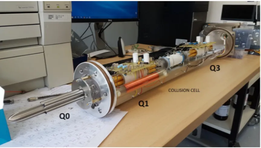
Once generated, ions are transported into the mass analyser which is the hearth of the mass spec-trometer, to be separated according to their m/z ratio. The aim of the analyser it to hold selected ions with specific masses and move them to the ion detector for counting. Different type of ions can be produced in the source: molecular ions with positive or negative charge (if the polarity in the ionization source is switched), components of the mobile phase and relative adducts from their combination with molecular ions, fragment ions if fragmentation is employed. There are more than a few types of analysers that can be employed for the separation of ions that use dif-ferent methods to separate them according to their m/z: quadrupole mass analyser, ion trap mass analyser, time-of-flight mass analyser, Fourier transform analyser, magnetic and electromagnetic sector mass analyser and ion cyclotron resonance analyser [123]. For our experimental work we used quadrupole analysers and, more specifically, a triple quadrupoles analyser generating what is known as tandem mass spectrometry (MS/MS). The quadrupole consists of four parallel rods on which a direct and varying (radio frequency) voltages are applied. By varying the voltages during time, it is possible to scan across a range of m/z and select the transmission of specific ions through the axis of the rods creating the mass spectrum. MS/MS is a particular configuration that increases the specificity and sensitivity of the analysis, obtained by placing in series a collision cell (q2) be-tween two quadrupoles (Q1 and Q3) (Figure 11). Fragmentation in the collision cell usually occurs with an inert collision gas, such as argon or nitrogen, and it known as collision-induced dissociation (CID).
Figure 11: Quadrupoles rail of the ABSciex API 4000 currently used in our laboratory. Q0 is a
quadrupole used as ion guide and allows the transmission of ions to the triple quadrupoles system. Rods of Q1 and Q3 are made of porcelain covered with a thin gold layer and are placed on opposite sides of the collision cell where ions fragmentation occurs.
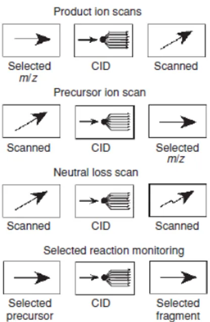
Q1 and Q3 can operate in different scan modes that are illustrated in Figure 12: the “product ion scan” consists of the selection of an ion with a specific m/z in Q1 which is fragmented in q2. All the produced fragment ions are then scanned and detected using Q3; in the “precursor ion scan” Q3 focuses on a selected fragment ion produced in q2 whereas Q1 is scanning all the masses to identify the precursor ion; another common mode is the so-called “neutral loss scan” where both Q1 and Q3 scan all the ions included in a selected m/z range with a constant mass offset between the two quadrupoles. It detects all the fragmentations leading to a specific neutral fragment; the most used is the so-called Selected Reaction Monitoring (SRM), known also as Multiple Reaction Monitoring (MRM). In this mode there is no scan, since both Q1 and Q3 are focused on selected masses. Importantly, ions selected in Q1 are only detected if they produce a give fragment by a selected reaction.
After the mass analyser, ions are then detected and converted into a usable signal by a detector, a de-vice able to transform incident ions into an electric current which is proportional to their abundance. Considering the minimal number of ions coming out from the mass analyser, an amplification of the signal is normally required in order to provide usable signals for data processing. At present, among the several available types of detectors, the electron multiplier is widely used. Briefly, pos-itive and negative ions strike a conversion dynode triggering the emission of numerous secondary particles. These are then converted into electrons and amplified by a cascade effect to produce an electric current that is then processed by the data system and generate the mass spectra [117, 123].
Figure 12: Graphic description of the main scan modes used in MS/MS (adapted from DeHofmann
et al. [117]).
2.2 Importance of HPLC-MS-MS in TH analysis
TH are commonly assayed in serum and, sporadically, in plasma. As already discussed in Par 1.2, more than 90 % of circulating TH are bound to serum carrier protein. This implies the inaccessibil-ity for tissues to the protein bound fraction which is considered inactive. In order to produce their genomic and non-genomic effects, TH dissociate from the carrier proteins and enter into the cells. However, a very small percentage (< 1%) is already present as free fraction (fT3 and fT4) which is directly accessible for peripheral tissues and, thus, considered as the biologically active fraction [124]. Considering the low concentration range of free TH (pM), it is evident how the quantifi-cation of the free fraction is more challenging respect to the total fraction (protein-bound + free). Moreover, the assay of the total fraction may not accurately reflect the thyroid status when protein concentrations or binding capacities are altered since this can affect the bound fraction without altering the levels of free TH. Because of these reasons, clinicians prefer to assess the levels of fT3 and fT4 on a routine basis [125].
The main methodologies that have been developed for the assay of serum fTH are divided in indirect and direct methods. The first one consisted in the mathematical estimation of the free fraction and involved the concomitant measurement of total TH (TTH) and TH binding proteins. The total concentration was then divided by TBG concentration or multiplied by an approximate value of
available TBG binding sites obtained through T3 and T4 uptake tests. This approach resulted in the evaluation of free T3 and T4 indexes that were usually requested by clinicians and were used in last decades [126]. Clearly, these indirect methods were dependent on the levels of TH binding proteins generating an underestimation or overestimation of TH levels. Moreover, the concentration of serum TH binding proteins can differ among the individuals and the method could only cover an ideal window of variability [127, 128].
Conversely, several direct immunoassays have been developed to assay fTH but require the physical separation of the free fraction prior the analysis. For this purpose, two main techniques are used to isolate the free fraction from the bound one, namely Equilibrium dialysis (ED) and Ultrafiltration (UF) that are not exempt from technical limitations. ED is subjected to dilution effects and to pos-sible interference of the buffer components on the equilibrium between fTH and bound TH [129]. Conversely, UF is not affected by dilution effects and the yield of measured fTH is higher than ED [130]. However, some drawbacks were identified also for UF, such as membrane adsorption, protein leakage and susceptibility to pH and temperature [129].
Nowadays, most of the clinical parameters of the thyroid are determined using automated immuno-metric methods. In particular, highly sensitive (LOQ 0.7 – 0.07 pM, depending on the analytical system used) and automated immunoassays that generally use chemiluminescence detection, are currently employed as gold standards to detect serum fT3 and fT4 [131]. However, the accuracy of immunoassays can be affected by the variation of serum-binding protein concentration that can oc-cur in case of pregnancy, genetic variations, medications that disrupt TH binding to serum proteins or certain medical conditions. Moreover, several studies have shown discrepancies in fTH results according to different immunoassays performed. This variability in fTH quantification may reflect differential assay susceptibility to the alteration in serum binding proteins [124]. Furthermore, the inverse log-linear relationship existing between fT4 (but also fT3) and TSH, poorly correlated when fTH were measured using immunoassays [132, 133].
Some of the above-mentioned weaknesses of the immunoassays, can be overcome with mass spec-trometry. HPLC-MS-MS is a highly specific and sensitive technique able to detect low concen-tration hormones. A valuable advantage of HPLC-MS-MS is related to the use of stable isotope-labelled internal standards (ISs) analogues of TH that can be used to monitor the analytical pro-cess and compensate for analytical errors. Isotope dilution methods are widely applied in clini-cal MS and are used routinely for the cliniclini-cal measurement of steroid hormones and Vitamin D [134, 135, 136]. HPLC-MS-MS is emerging as a powerful technique able to detect serum fTH