INTERNATIONAL JOURNAL OF IMMUNOPATHOLOGY AND PHARMACOLOGY Vol. 19, no. 4, 775-786 (2006)
EFFECT OF
BIFIDOBACTERIUM INFANTIS ON INTERFERON-
y-
INDUCED
KERATINOCYTE APOPTOSIS: A POTENTIAL THERAPEUTIC APPROACH TO SKIN
IMMUNE ABNORMALITIES
B.
CINQUE,
L.
DI MARZIOI, D.N. DELLA RICCIA,
F. BIZZINI,
M. GIULIANF, D. FANINI, C. DE SIMONE and M.G. CIFONE
Department ofExperimental Medicine, University ofL 'Aquila;
IDepartment ofDrug Science,
University
G.
d 'Annunzio, Chieti;
2Department ofSurgical Sciences, University ofL 'Aquila,
L 'Aquila, Italy
Received February 1,2006 -Accepted August 10,2006
Current management of atopic dermatitis is mainly directed to the reduction of cutaneous
inflammation. Since patients with atopic dermatitis show abnormalities in immunoregulation, a therapy
aimed to adjust their immune function could represent an alternative approach, particularly in the
severe form of the disease. Indeed, T-Iymphocytes constitute a large population of cellular infiltrate
in atopic/allergic inflammation and a dysregulated T-cell induced keratinocyte apoptosis appears to
be an important pathogenetic factor of the eczematous disease. In recent years, attention has been
focused on the interaction between host and probiotics which may have anti-inflammatory properties
and immunomodulatory activities. The aim of the present work is to investigate the effect of a selected
probiotic extract, the Bifidobacterium in/antis extract, on a human keratinocyte cell line (HaCaT)
abnormal apoptosis induced by activated-T-Iymphocyte. An
in vitro model of atopic dermatitis was used
to assess the ability of the probiotic extract to protect HaCaT from apoptosis induced by soluble factors
(IFN-y and CD95 ligand) released by human T-Iymphocytes
in vitro activated with anti-CD3/CD28
mAbs or Phytohemoagglutinin. Evidence is given that the bacterial extract treatment was able to totally
prevent T lymphocyte-induced HaCaT cell apoptosis
in vitro. The mechanism underlying this inhibitory
effect has been suggested to depend on the ability of the bacterial extract to significantly reduce
anti-CD3/CD28 mAbs and mitogen-induced T-cell proliferation, IFN-y generation and CD95 ligand release.
These preliminary results may represent an experimental basis for a potential therapeutic approach
mainly targeting the skin disorders-associated immune abnormalities.
T-cells represent a large population ofthe cellular
infiltrate and mediate a dysregulated cytokine
response in cutaneous inflammatory processes, thus
participating in the development of eczematous
reactions associated with several skin diseases,
including
allergic
contact
dermatitis
(ACD)
and atopic dermatitis (AD)
(1).A dendritic
cell-dependent T cell-mediated immune response can be
observed during contact hypersensitivity reactions
and it is induced by epicutaneous sensitisation with
hapten (2). Although AD is regarded as a Th2-type
disease (3),
IFN-y, a Thl type cytokine, has been
Key words: Bifidobacterium in/antis, Apoptosis, HaCaT, T-cell activation
Mailing address:Dr M. Grazia Cifone, PhD, Full Professor of General Pathology, Department of Experimental Medicine, University of L'Aquila,
Via Vetoio, 10 Coppito 2, 67100 L'Aquila, Italy Tel: +390862433503 Fax: +39 0862 433523
e-mail: [email protected]
775
0394-6320 (2006)
Copyright©by BIOLlFE, s.a.s. This publication and/or article is for individual use only and may not be further reproduced without written permission from the copyright holder. Unauthorized reproduction may result in financial and other penalties
776
B. CINQUE ET AL. reported to be expressed in the late stages of AD (4).Accordingly, a shift from Th2 in the acute phase to Th I in the chronic phase in AD has been proposed (5-7). In particular, both type 2 IL-4 and IL-5 cytokines and type I IFN-y cytokine have been suggested to play important roles in skin inflammation in a murine model of eczematous dermatitis (8). Furthermore, the injection of IFN-y into the skin of human volunteers demonstrated that this cytokine can induce transient skin inflammation (9).
Homeostasis in epidermis, as tissue renewing, is mainly guaranteed by keratinocytes through their capacity to proliferate and differentiate into epithelial cells forming the skin barrier (10). Trautmann et al. reported that an altered keratinocyte apoptosis represents a major mechanism in the pathogenesis of eczematous disorders and could be one of the causes for the damage of epidermal barrier. IFN-y release bIFN-y skin-infiltrating T-cells was indeed shown to be capable of upregulating CD95 on keratinocytes, thus rendering them susceptible to apoptosis by CD95 ligand (CD95L) expressed on and/or released by the T-cell surface (I). Recently, several therapeutic approaches have been employed in correcting specific immune abnormalities in AD (11-12). Skin inflammatory diseases can benefit from immunomodulators and/or immunosuppressive agent-based treatments (i.e. cyclosporine A, topical tacrolimus/FK506, rapamycine) which are able to prevent the abnormal keratinocyte apoptosis mainly associated to these pathological conditions (13-15).
Many species of lactic acid bacteria (LAB) have been reported to be immunomodulatory both in vitro and in vivo (16-18), and to have beneficial effects on animal and human health [i.e., protection against enteric infections, use as an oral adjuvant, immunopotentiation in malnutrition and prevention of chemically induced tumors (19-20)], thus they are frequently used as probiotics. In particular, LAB-induced immune regulation is considered as one of the potential causes of their antitumoral activity (16, 21-22).
In a previous investigation, we analysed the effects of six LAB strains on human normal and tumoral lymphocytes in vitro (23). All examined strains, with regard to efficacy, were able to induce apoptosis of tumor cell lines, without affecting normal cell viability. This effect could be associated with the presence, on bacterial extracts, of arginine
deiminase and/or neutral sphingomyelinase, which in turn were able, respectively, to prevent polyamine synthesis and to generate endogenous ceramide in tumor cells, thus leading to apoptotic cell death. Taken together, these findings led us to analyse, through an in vitro disease model of atopic dermatitis, the potential effect ofone probiotic extract (Bifidobacterium in/antis extract) on the abnormal T-cell activation which has been indicated to be mainly responsible for AD-associated apoptosis of keratinocytes. We therefore conducted a preliminary study on the effects of this bacterial extract on anti-CD3/CD28 mAbs or Phytohemoagglutinin-induced T-cell proliferation and IFN-y generation, as well as on activated T-cell induced HaCaT apoptosis in vitro. Our results indicate that the in vitro treatment with a particular probiotic extract led to a significant decrease of activated T-cell proliferation, IFN-y generation, CD95 ligand release and, consequently, of activated T cell-induced HaCaT apoptosis.
MATERIALS AND METHODS
Media and chemicals
Media and culture reagents were purchased from EuroClone (Ltd. United Kingdom). Anti-CD3 (clone HIT3a; mouse anti-human monoclonal antibody) and anti-CD28 (clone CD28.2) mAbs were provided by Becton Dickinson (San Diego, CA, USA) and Phytohemoagglutinin-P (PHA) was from Sigma (St. Louis, MO, USA). Anti-CD95 mAb (clone ZB4) was purchased from Upstate (New York, USA). Neutralizing anti-human IFN-y mAb (clone 25723) was from R&D Systems, Inc. (Minneapolis, USA). Fluorescein anti-mouse IgG I was obtained from Vector Laboratories (Burlingame, CA, U.S.A.). [methyPH]Thymidine I mCi mL-1 (specific activity 80 Ci mmol') was purchased from Amersham (Buckinghamshire, UK).
Cell culture
The spontaneously immortalized human keratinocyte cell line HaCaT (24) was grown in plastic culture dishes (Nunc, Wiesbaden, Germany) containing Dulbecco's modified Eagle's medium (DMEM) supplemented with 10% fetal calf serum (FCS), 2 mM L-glutamine, 100 U penicillin, 50 ug mL-1 streptomycin and 50 ug mL-1 gentamycin. Confluent cells were subcultured at day 3 after detaching the cells with a 0.1% trypsin/O.02% ethylenediamine tetraacetic acid solution.
Preparation ofbacterial extracts
Int. J. Immunopathol. Pharmacol.
777
(S244), Bifidobacterium longum, Lactobacillus acidophilus, Lactobacillus bulgaricus, Lactobacillus casei, Bifidobacterium infantis, Bifidobacterium breve), obtained
from VSL Pharmaceuticals (Gaithesburg, Maryland, USA) in a pure lyophilized form (l08 coiony-fonning units [CFU]
g'), were routinely grown in MRS broth medium under aerobic conditions (25-26). Bacterial cultures were washed and re-suspended in 10 ml of phosphate buffered solution (PBS), sonicated (l0 cycles on-off, alternating 45 sec. on and 2 min. oft) with a Vibracell sonicator (Sonic and Materials Inc., Danbury, CT). Bacterial homogenates were centrifuged at 28,000 g for 20 min at 4°C in order to remove cell wall fragments and intact bacteria from cytosol, and the supernatant fraction (cytoplasm) was then filtered (0.45 urn). The filtrate was stored at -80°C until use. The protein concentration of bacterial extracts was determined using the Bio-Rad assay with bovine serum albumin standards (Sigma, St. Louis, MO, USA). The extracts were added to cell cultures to a final concentration of 25, 125, or 250 ug protein mL-I.
T-lymphocyte isolation and culture
T-Iymphocytes from healthy donors were isolated from heparinized blood by density centrifugation over Ficoll-Hypaque and MACS system (Pan T-Cell Isolation kit II cod 130-091-156), according to the instructions of the manufacturer (Miltenyi Biotec, Bergisch Gladbach, Germany), This procedure routinely yielded a >97% CD3 positive population, as analysed by flow cytometry. The cells were resuspended in culture medium (RPMI 1640 supplemented with 10% FCS, 2 mM L-glutamine, 100 IV mL-I penicillin and 100 ug mL-I streptomycin). The cells were counted and their viability, as assessed by Trypan blue dye exclusion and cytometric analysis, was routinely greater than 98%. The lymphocyte cultures were run in 96-well tissue culture plates and incubated in a humidified 5% CO2atmosphere at 37°C. The T-cells (l06 mL-I) were stimulated with PHA (20 ug mL-I), anti-CD3 mAb (l ug mL-I) plus anti-CD28 mAb (l ug mL-I) in the presence or absence of bacterial extracts (final concentrations: 25, 125, or 250 ug protein mL-I) in a total volume of200 ul of culture medium. Experiments were performed according to the following schedule: a) untreated T-cell cultures; b) PHA- or anti-CD3/CD28 mAbs-stimulated T-cell cultures; c) bacterial extract treated T-Iymphocytes; d) T-cells treated with bacterial extracts in the presence of either PHA or anti CD3/CD28mAbs. Bacterial extracts and mAbs were added simultaneously. The viability and apoptosis of T-cells in the presence of different stimuli was tested by propidium iodide incorporation using flow cytometry (CELLquest software, Becton Dickinson). Cell proliferation was evaluated by pulsing the cell cultures with 1 /lCi of [3H]thymidine during the last 6 hrs of a
72 hr culture. Incorporation of [3H]TdR was measured by standard liquid scintillation counting techniques after harvesting with a Skatron harvester.
Analysis by flow cytometry of anti-CD3 mAb binding to T-cells
In order to test whether each probiotic extract may interact with CD3-crosslinking, we measured anti-CD3 binding by indirect immune fluorescence. Briefly, T-cells were incubated for 30 minutes at 4°C with 250 ug mL-I of each bacterial extract alone and with anti-CD3 mAb (1 ug mL-I) in the presence or absence of the bacterial extract. Following a washout with PBS, the FITC-conjugated anti-mouse IgG was added for 30 min at 4°C after which the cells were analysed by cytofluorimetry. FlTC-conjugated anti-mouse IgG was used as control isotype.
Culture ofHaCaT with T-cell conditioned medium
Conditioned medium was obtained from T-cells grown for 48 hrs in RPMI-I640 with or without PHA, or anti-CD3 plus anti-CD28 in the presence or absence of increasing concentrations of each bacterial extract. After centrifugation, the T-cells supernatants were collected, diluted 1/5and added to HaCaT (1.2x10⁵mL-I) in 6-well
plates. The culture was incubated for 72 hrs at 37°C in a humidified atmosphere containing 5% CO2, Where
indicated, the conditionsd medium was preincubated for 30 minutes with neutralizing anti-human IFN-y mAb (3 ug mL-I) before addition to the HaCaT. After 72 hrs of incubation, pellets were collected and used to evaluate apoptosis level as described below.
HaCaT apoptosis evaluation by propidium iodide solution
DNA flow cytometry was performed as described elsewhere (27). After the treatments the cells were harvested, washed twice with PBS and centrifuged. The pellets (106) were gently resuspended in 1 ml of hypotonic propidium iodide (PI) solution (50 ug mL-I in 0,1% sodium citrate
+
0,1 % Triton X-IOO; Sigma, St Louis, MO). The tubes were kept overnight at 4°C in the dark. The PI fluorescence of individual nuclei and the percentage of apoptotic cell nuclei (subdiploid DNA peak in the DNA fluorescence histogram) were measured by flow cytometry with standard fluorescein-activated cell scan equipment (FACScan Becton Dickinson, San Jose, CA). Where indicated, the HaCaT were pretreated with anti-CD95 mAb (250 ng mL-I), known to block induction of apoptosis via CD95 receptor in Jurkat T cells.Quantification ofcytokines
Cell cultures were prepared as above-described for the T-lymphocyte isolation and culture. The plates were incubated
778
B. CINQUE ET AL at 37°C in 5% CO2for 24, 48 and 72 hrs. Cell cultures werethen centrifuged and supernatants were collected and stored at -70°C. IFN-y and sCD95L levels in supernatants were determined by commercial ELISA kit (Human Interferon gamma ELISA Pierce Endogen; sCD95 Ligand ELISA MBL Co. Nagoya. Japan), as recommended by the manufacturer. The 48-h incubation was chosen since at this time the higher production for both cytokines was observed.
Statistical analysis
Statistical analysis of data was done by using one-way ANaYA followed by Student's t test or by the correlation analysis (Prism 3.0 GraphPad Sofwtware, San Diego). Data were expressed as mean± SD and P value <0.05 was used as the significance criterion.
RESULTS
Effect of bacterial extracts on anti-CD3/CD28 mAbs or PHA-induced proliferative response of human I-lymphocytes
In order to assess the possible effect of probiotics
on
anti-CD3/CD28
mAbs
and
PHA-induced
proliferative response, we firstly treated for 72 hrs
human T-lymphocytes with seven different strains
(250 ug protein ml,") of LAB
(Streptococcus thermophilus, Bifidobacterium longum, Lactobacillus acidophilus, Lactobacillus bulgaricus, Lactobacillus casei, Bifidobacterium infantisand
Bifidobacterium breve)extracts in the presence or
absence of PHA or anti-CD3/CD28 mAbs. All tested
bacterial extracts significantly (P<O.OO I) suppressed
T-cells proliferation (Fig. 1A), even if an intergroup
difference in terms of the proliferation decrease
obtained with the different bacterial extracts could
be observed (range % inhibition: about 50% to
almost 100%),
Bifidobacterium infantisbeing the
most efficient. Based on these results, all subsequent
experiments designed to analyse the underlying
mechanisms of the above reported effect were
performed with
Bifidobacterium infantisextracts.
To further delineate the effect of
Bifidobacterium infantisextracts on anti-CD3/CD28 mAbs and
PHA-induced proliferative response,
human
T-lymphocytes
were treated with three different
concentrations (25, 125, 250 ug protein ml.")
of bacterial extract in the presence or absence of
PHA or anti-CD3/CD28 mAbs. Fig. IB shows that
exposure to the bacterial extract caused a significant
and concentration-dependent decrease in activated
T-cells [3H]thymidine incorporation (% inhibition
range: 60-95%). Toxic effects potentially due to the
bacterial extract based treatment on T-lymphocytes
could be excluded since no significant influence
on cell viability was observed, as analysed by
both Trypan blue exclusion and propidium iodide
incorporation using flow cytometry. A significant
inverse correlation between the inhibitory effect
and bacterial extract concentration was observed
(R=-0.908, P<O.OOI and R=-0.7, P<0.03 for cells
treated with PHA and anti-CD3/CD28
mAbs,
respectively). On the other hand, bacterial extract
did not significantly affect spontaneous [3H]TdR
incorporation from lymphocytes at basal conditions
(without PHA or anti-CD3/CD28 mAbs).
Effect of Bifidobacterium infantis on anti-CD3 mAb binding to human T-lymphocytes
To exclude the possibility that the above reported
inhibitory effect could be due to the interference of
bacterial extract with the mAb induced-cross-linking
of surface receptors, the binding of anti-CD3 mAb in
the presence of the bacterial extract was evaluated.
As shown in Fig.2, the anti-CD3 mAb binding, as
analysed by cytofluorimetry, was not influenced
in the presence of the bacterial extract at higher
concentration (250 ug
ml,:').Effects of Bifidobacterium infantis on activated T lymphocyte-induced IFN-y production and sCD95 ligand release
The incubation ofT-lymphocytes with both PHA
and anti-CD3/CD28 mAbs for 48 hrs, as expected,
was followed by a significant increase (P<O.OO I) of
IFN-y secretion in the culture medium (Fig. 3A), as
assessed by a specific enzyme-linked immunosorbent
assay (ELISA). The addition of bacterial extract (25
ug ml,") in the culture medium was able to strongly
and significantly (P<O.OO I) inhibit either PHA- or
anti-CD3/CD28 mAbs-induced IFN-y generation.
At higher concentrations (125 and 250 ug ml,"), the
presence of the bacterial extract totally abrogated
PHA- or anti-CD3/CD28
mAbs-induced IFN-y
release which returned to basal levels. The extent
of inhibition of IFN-y secretion well correlated with
the impairment of cell proliferation observed when
activated T-cells were cultured in the presence of
the bacterial extract (R=0.953, P<O.OOI for
PHA-Int.J. Immunopalhol. Pharmacol.
779
activated cells; R=0.988, P<O.OOI for anti-CD3/
CD28 mAbs-activated cells).
The effect of the bacterial extract on soluble
CD95 ligand (sCD95L) release from PHA- or
anti-CD3/CD28 mAbs-activated T-Iymphocytes in
vitro
was also investigated. The levels of sCD95L
in the supernatants from T-lymphocytes, incubated
for 48 hrs with or without stimuli in the presence
or absence of bacterial extract, were measured by
ELISA. The results of these experiments showed
the ability of the stimuli to significantly (P<O.OOl)
increase the level of sCD95L (Fig. 3B).
Itis worthy
of note that the presence of Bifidobacterium extract
in the cell cultures led to a relevant and significant
(P<O.OOl) decrease of the sCD95L release by
PHA- or anti-CD3/CD28 mAbs-stimulated T-cells,
without affecting the basal release of sCD95L.
The extent of inhibition of sCD95L release was
concentration-dependent and well correlated either
with the impairment of cell proliferation (R=0.981,
P<O.OOI for PHA-activated cells; R=0.942, P<O.OOI
for anti-CD3/CD28 mAbs-activated cells) or IFN-y
generation (R=0.989, P<O.OOI for PHA-activated
cells; R=0.976, P<O.OOI for anti-CD3/CD28
mAbs-treated cells) observed when T-cells were cultured in
the presence of the bacterial extract.
Effect
0/
Bifidobacterium in/antis on T
cell-mediated HaCaT apoptosis
To confirm the ability of activated T-cells to
induce
HaCaT apoptosis,
we investigated the
effect of T-cell supernatants on HaCaT culture. To
eliminate the disadvantage of apoptotic bodies or
fragmented DNA being derived from T-cells, the
latter were incubated with the appropriate stimuli
for 48 hrs and then centrifuged. The conditioned
media were then added to HaCaT culture for three
days and the cells were subsequently analysed for
apoptosis by flow cytometry. This model allowed
us to verify that HaCaT apoptosis was induced by
soluble factors secreted from PHA- or anti-CD3/
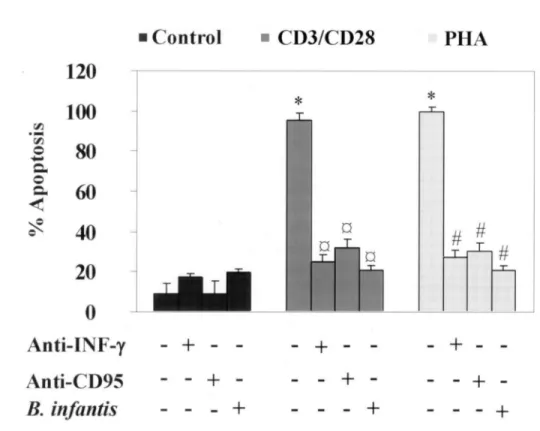
CD28 mAbs-activated T-Iymphocytes. In Fig. 4
the mean values (±SD) of data from 3 experiments
in duplicate are shown. As evident, the apoptosis
levels in HaCaT treated with supernatants from PHA
or anti-CD3/CD28 mAbs-activated T-Iymphocytes
appeared to be comparable. The presence of
neutralizing anti-human IFN-y in the conditioned
media or the preincubation of HaCaT with blocking
anti-CD95 mAb dramatically reduced the apoptosis
(approximately 65-80% and 70-80% reduction,
respectively), thus confirming previous observations
showing that this event could be totally due to the
IFN-y released from activated T-cells and CD95
system
(1).According to these results and as reported by other
authors (1), keratinocyte apoptosis being induced
by IFN-y and sCD95 ligand, to evaluate the effect
of the bacterial extract we used it at concentration
of 250 ug ml,:' since this concentration, in respect
to the other concentrations, completely abrogated
PHA- or anti-CD3/CD28 mAbs-induced IFN-y
production and sCD95 ligand release. Supernatants
from
both
anti-CD3/CD28
mAb-
and
PHA-stimulated T-Iymphocytes, cultured in the presence
of the bacterial extract, were able to almost totally
abrogate HaCaT apoptosis.
Itwas noted that when
the bacterial extract was added directly to HaCaT
cultures in the presence of the conditioned medium
derived from activated-T cells, the apoptosis level
did not change, thus suggesting that the inhibitory
effect of bacterial extract on HaCaT apoptosis was
not directed on HaCaT, being mainly attributed
to its suppressive action on T-cell activation and,
consequently, to IFN-y generation and sCD95L
release. Fig. 5 displays a representative experiment
showing that, as expected, while no effect could
be observed when HaCaT were incubated with
supernatant from untreated and unstimulated T-cells
(panel A), the conditioned medium from anti-CD3/
CD28 mAb- and PHA-stimulated T-cells was able
to induce relevant levels of HaCaT apoptosis (range:
80-100%) (panel Band C respectively). Similar
results were obtained when the bacterial extract was
added directly to HaCaT culture (panel D, E and F).
Itis of interest that the medium conditioned from
activated T-cells in the presence of the bacterial
extract was able to reduce the percentage number
of hypodiploid nuclei (panel H and I). Altogether,
these findings show the ability of probiotic extract
to strongly inhibit and prevent the HaCaT apoptosis
induced by soluble factors from activated-T-cells.
DISCUSSION
780
B. CINQUE ET AL.A
- - -
PHA
-++
++++++
300 l::: c 250 'C ~ 8 l:l. 200...
'""' c ..,=~
... >'i 150 CIlE
.5 l:l. ] ~ 100 >. ..c: f-50 :I: 0 CD3/CD 28 lkltreated PHA -IS400
*
'J:! t'<l...
Q"'"
300
...
~ 8'0=
....
- l', ., 8200
."<lS "'"...'
s
~ ~ I-100
::t:: ~*
*
I
III
§ CCortrd lJ5244 !3B.Iwgum oL.acicbphilus DL.bulgaricus IJL.caseiE,JB. irfantis
Ii!B.txeve
CD3/CD28
PHA
- 1
+
- L+
- "3+
-
4+
-
+
- -+
- -+
- -+
-25 J1gmIJI 125 J1gmIJI 250J1gmIJI
B!fitlobacterhutlhyalitisextracts
Fig.1.Effect ofbacterial extracts on anti-CD3/CD28 mAbs and PHA-inducedproliferation ofhuman lymphocytes. T-cell proliferation, measured by [3H]TdR incorporation, is expressed as counts per minute, values represent the mean±SD of three determinations from one experiment representative of three. (Panel A): proliferation was assessed in cultures containing isolated lymphocytes treated with different bacterial extracts used at the same protein concentration (250 Ilg mL-l), with anti-CD3/CD28 mAbs (l fig mL-I), or PHA (20 fig mL-I) in the presence or absence ofbacterial extracts.
*
= P<0.001 when compared to untreated Tcells; #= P<0.001 compared to anti-CD3/CD28-stimulated Tcells;§= P<0.001compared to PHA-stimulated T cells. (Panel B): T-cells were incubated for 72hrs with or without Bifidobacterium infantis (protein concentration of25, 125, 250 Ilg mL-I) in presence or absence ofPHA (20 fig mLI) or anti-CD3/CD28 mAbs (l fig mL-I).
*
= P<0.001 when compared to untreated cells; ##= P<0.001 and #= P=0.003 compared to PHA-stimulated T cells;00 = P<0.001 and0 =P=0.007 compared to anti-CD3/CD28-stimulated T-cells.Int. J.Immnnopathol.Pharmacol.
781
- = CD3+B.in/antis B •.•. =CD3 o 0&>8 ...---,
N A ...,=Control+
FITC-IgG- =Negative control+FITC-IgG
o 0&>
8
-r- - - -- - - - ---,
N o N VI~....
C ::3' o 'Uo 00 o v o N VI~ ~::3' o 'Uo 00 102 FL1-HFig. 2. Effect of Bifidobacterium infantis extracts on anti-CD3 mAb binding to human T-lymphocytes. T-cells were
culturedwith bacterial extractsalone(250IlgmL-J)orstimulatedwith anti-CD3 mAb(lllg mL-J) in presenceor absence
0/250IlgmL-J0/ bacterial extract.(A).Controlisotyp e stained(Fl'I'Csconjugated anti-mouse/gG) in presence (solid
line) or absence (dottedline) ofbacterial extracts.(B).Anti-CD3 mAbbindingin presence (solid line) or absence (dotted line) of'bacterial extracts. Thedatashown are from onerepresentativeofthree independent experiments.
* A # a # a 1200 ,... ~ 1000
..
"
.:
800...
Of' 600 .eo....
Z 400 f::l 200 0 CD3ICD28 PHA + + -- -- + -- + - +- - +-- +-- + - + -25~mL' 1125j.tgniL·1250~mlr! Bifidobacteriumill/all/isext rac tsB 25j.tgniL·1 125j.tgmL·1250j.tgmL' 1 Bijidobactt'n1l111hyalitis extr act s ,... 0.50 ~
..
"
0.40.:
...
oJ .s 0. 30 -e;
Of, 0.20 := IF) ~ 0.10 Q'd
0.00 CD3JC~ . u PHA * - - + - + --- + - - + -+- - +-# a - - + +-Fig. 3. Effect of Bifidobacterium infantis extracts on
IFN-y and sCD95 ligand production by activated
human T-cells. The IFN-y(A) andsCD95 ligand (B)
levelswereevaluatedby aspecific ELISA. The values
rep resent the mean values of duplicatedeterminations
and arerep resentativeof1of 3experiments.SD values
were ever lower than 5% ofmean values. *P<O.OOl
comp ared to untreatedT-cells. #p< O.OOl comparedto
PHA-stimulated T-cells. ap<0.001 compared toa
782
B. CINQUE ET AL.• C
ontrol
•
C
D3/C D28
PHA
120
*
*
100
rIJ.
-
rIJ80
0-
e, 0 c,60
~~
40
# #
#
20
0
A
nti - I
F-
y
-
+
-
-
+
-
-
+
-A
nti-C D95
-
+
-
-
+
-
- +
B
.
i
nfa ntis
-
+
-
+
- -
+
Fig. 4.HaCaT cell apoptosis levels in presence ofconditioned medium from unstimulated or stimulated T cells cultured in presence or absence of Bifidobacterium infantis extracts. The Y axis represents the % of HaCaT cells apoptosis induced by the supernatants of unstimulated or stimulated (with PHA or anti-CD3/CD28 mAbs) T-cells. Histograms show the difference between treatments with bacterial extracts (250 Ilg ml;'), neutralizing anti-human IFN-y mAb (3 Ilg ml:'], anti-CD95 mAb (clone ZB4; 250 Ilg ml;'} on the apoptotic behaviour. Values are expressed as the mean ±SD of2 . determinations. Shown data represent one out ofthree independent experiments. *P<O.OOI when compared to untreated, ap<O.OOI compared to anti-CD3/CD28 mAbs-stimulated T-cells, #P<O.OOI compared to PHA-stimulated T-cells.
in .cutaneous inflammatory processes (i.e. atopic
dermatitis), where they represent a large population
of the cellular infiltrate and mediate a dysregulated
cytokine response which, in tum, could be responsible
for abnormal keratinocyte apoptosis (1-7). Our study
was conducted to assess the ability of a
Bifidobacteriaextract to affect the immune response of human
T-lymphocytes in a disease model ofAD
in vitro.Our results demonstrate that all the analysed
probiotic extracts were able to significantly inhibit
human T-lymphocyte proliferation induced by
anti-CD3 plus anti-CD28 mAbs
in vitro,even ifat different
extents
(range
50-100%),
the
Bifidobacteriuminfantis
being the most efficient in this effect. No
effect was observed when cells were treated with
bacterial extracts only. The inhibitory effect on
activated T-cell proliferation was significant even at
the lowest concentration of
Bifidobacterium infantisextract and could not be attributed either to toxic
effects or to any interference of the bacterial extract
with membrane receptor crosslinking. Indeed, the
treatment with bacterial extract did not influence cell
viability analysed by Trypan blue exclusion as well
anti-CD3 binding, as assayed by cytofluorimetry.
The activation of T cells with both PHA and
anti-CD3/CD28 mAbs, as expected, led to the
induction of IFN-y generation (28). Taking into
account that IFN-y plays an important role in skin
inflammation (8-9), the influence of the bacterial
extract treatment on the generation and release of
this cytokine from stimulated T-lymphocytes was
also analysed. IFN-y levels generated by treatment
with PHA or anti-CD3/CD28 mAbs appeared to be
significantly inhibited in the presence of the bacterial
extract even at the lowest concentration of the latter,
and totally abrogated at 125 and 250 ug mL-J. Taken
101.J. Immunopathel, Pharmacol.
783
Unstimulated CD3/CD28-Stimulated PHA-Stimulated
~ 0 '" M
3.03
D
88.05
E
s
0 ~Bacterial extract added to~;;: ~N0
HaCaT culture
!!
§ 8~RI
I~
..
0('\1
MlMIl,
0 0 100 101 102 103 104 100 101 102 103 FL2-H FL2-H 0~
t-o 104 10 0 101 102 103 104 FL2-H 0 It> M89.5
F
0 ee N 0 ~N <: ~ 0 0 u ... 0I
t- M1 0.r-.
104 100 101 102 103 10' FL2-H 0 It> MB
..
086.4
C
N 0 "N l: ~ 0 0 j:?: ~...---,85.08
s
6.03
A
I
MlIII
,-....Without Bacterial extract ~~
§
8!
o
~N Bacterial extract added 8!§
to T-cell culture.
11.04
G
I
MlI
~
r - "17.5
H
I
MlItA
r-.-.. o..
N15.4
I
1 I MlV
f"W\Fig. 5. Effect ofBifidobacterium infantis extracts on HaCaT apoptosis induced by conditioned medium from unstimulated
or stimulated T cells, as determined through flow cytometry. Data represent the results ofone out of3 independent
experi-ments. HaCaT cells were incubated for3 days with conditioned medium from unstimulated T-cells (A, D and G),
anti-CD31CD28 stimulated T-cells (B, E and H) and PHA-stimulated T-cells (C. F and I). D. E and F show HaCaT apoptosis when the bacterial extracts (250 Ilg mL-1
) were directly added to cell culture;G, Hand [show HaCaT apoptosis when the
bacterial extracts (250 Ilg mL·I) were incubated with stimulated T-cells. MJ indicates the apoptotic peak representing the cells with a sub-diploid DNA content.
together, these results suggest that the bacterial extract treatment
in vitro
was able to significantly or totally prevent either T-cell proliferation or IFN-y generation. Previous studies reported that AD skin-infiltrating activated-T-cells upregulate the expression of CD95 receptor on keratinocytes through IFN-y and induce apoptosis by CD95 ligand which could be expressed on T-cell surface or released into the extracellular microenvironment (1, 29-30). Experiments designed to assess the effect of the bacterial extract on CD95L released from activated T-lymphocytes, showed thatthe treatment was able to strongly and concentration-dependently inhibit the levels of sCD95L released either from PHA or anti-CD3/CD28 mAb stimulated T-cells.
In our experimental
in vitro
model, activated T cell-derived soluble factors were able to induce HaCaT apoptosis. Of note, when stimulated, T-cells were cultured in the presence of the bacterial extract, the conditioned medium was unable to induce HaCaT apoptotic death, the effect being comparable to that observed either when blocking anti-CD95784
B.infantis extracts B. CINQUE ET AL. B.infantis extracts•
•
•
•
•
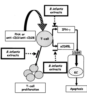
B.infantis extractsFig. 6. Proposed model ofthe effect ofBifidobacterium infantis extracts on prevention ofkeratinocyte apoptosis mediated
by activated T-cells.
mAb (about 70-80%) or neutralizing anti-IFN-y mAb (about 65-80%) were used. Overall, these results suggest that keratinocyte apoptosis could be mediated by T cell released IFN-y, and almost totally attributed to CD95 system, confirming previously reported results (I). The bacterial extract-induced inhibitory effect on HaCaT apoptosis appeared to be indirect, being mediated by the impaired ability of activated-T cells to release adequate levels of IFN-y and sCD95L. This hypothesis was further supported by the results from experiments indicating that no inhibitory effect could be observed on keratinocyte apoptosis when the bacterial extract was directly added to HaCaT cell cultures in the presence of conditioned medium from activated-T cells. activated-The proposed model summarising the observed effects of Bifidobacterium extract on T-cell induced keratinocyte apoptosis is shown in Fig. 6. Taken together, these results could represent the experimental basis for a new therapeutic approach to immune abnormalities-associated atopic dermatitis.
Judging from our findings, the components of Bifidobacterium infantis,considered to be responsible for the observed immunosuppression, could be
located in the cytoplasm and not in cell wall fractions. However, additional investigations are needed in order to identify the immunomodulator components and the biomolecular mechanisms by which these factors operate. Moreover, further research is also needed to assess the effect of an experimental Bifidobacteria extract in vivo treatment on the AD-associated T-cell activation and keratinocyte apoptosis. In the eczematous reactions, T-cells act as important mediators ofthe inflammatory processes, as supported by the observation that immunosuppressive drugs such as FK506 and glucocorticoids, which block T-cells activation, are effective in the treatment of eczematous disorders (1). Skin represents a functionally distinct immune compartment, and chronic inflammation of the skin is generally associated with tissue infiltration by T-cells (31).
Finally, since a reduced ratio of Bifidobacteria to Clostridia has been observed to precede the development of atopy, and patients with allergic diseases resulted in being less often colonized
by
Enterococci and Bifidobacteria but more often by Clostridia and Staphylococci compared to not allergic patients (32-34), our investigation could haveInt.J. Immunopalhol. Pharmacol.
785
a potential clinical significance also in this context.ACKNOWLEDGEMENTS
The Authors thank Gasperina De Nuntiis (Department of Experimental Medicine, University of L'Aquila, L'Aquila, Italy) for technical assistance and Miss Florence Pryen for careful language revision of the manuscript. The present work was financially supported by a grant "Progetto di Interesse Generale di Ateneo", University of L' Aquila, L'Aquila.
REFERENCES
1. Trautmann A., M. Akdis, D. Kleemann, F. Altznauer, H.V. Simon, T. Graeve, M. Noll, E.B. Brocker, K. Blaser and C.A. Akdis. 2000. T cell-mediated Fas-induced keratinocyte apoptosis plays a key pathogenetic role in eczematous dermatitis. 1. Clin. Invest. 106:25.
2. Akiba H., J. Kehren, M.T. Ducluzeau, M. Krasteva, F. Horand, D. Kaiserlian,F.Kaneko and J.F. Nicolas. 2002. Skin inflammation during contact hypersensitivity is mediated by early recruitment of C08+ T cytotoxic 1 cells inducing keratinocyte apoptosis.1. Immunol. 168:3079.
3. Cooper K.D. 1994. Atopic dermatitis: recent trends in pathogenesis and therapy. J. Invest. Dermato/. 102:128.
4. Grewe M., C.A. Bruijnzeel-Koomen, E. Schopf, T. Thepen, A.G. Langeveld-Wildschut, T. Ruzicka and J. Krutmann. 1998. A role for Thl and Th2 cells in the immunopathogenesis of atopic dermatitis.
Immunol. Today 19:359.
5. Grewe M., K. Gyufko, E. Schopf and J. Krutmann. 1994. Lesional expression of interferon-gamma in atopic eczema. Lancet 343:25.
6. Ohmen J.D., J.M. Hanifin, B.J. Nickoloff, T.H. Rea, R. Wyzykowski, J. Kim, D. Jullien, T. McHugh, A.S. Nassif and S.c. Chan. 1995. Overexpression of IL-I0 in atopic dermatitis. Contrasting cytokine patterns with delayed-type hypersensitivity reactions.1.Immuno/. 154:1956.
7. Hamid Q., T. Naseer, E.M. Minshall, Y.L. Song, M. Boguniewicz and D.Y. Leung. 1996. In vivo expression ofIL-12 and IL-13 in atopic dermatitis. 1.
Allergy. Clin. Immuno/. 98:225.
8. Spergel J.M., E. Mizoguchi, H. Oettgen, A.K. Bhan and R.S. Geha. 1999. Roles ofTHI and TH2 cytokines in a murine model of allergic dermatitis. 1.
C/in. Invest. 103: 1103.
9. Barker J.N., M.H. Allen and D.M. MacDonald. 1990. Alterations induced in normal human skin by
in vivointerferon-gamma. Br.1.Dermato/. 122:451.
10. Fuchs E. 1990. Epidermal differentiation: the bare essentials.1.Cell. Bio/. 111:2807.
II. Leung D.Y. 1999. Pathogenesis of atopic dermatitis.
J.Allergy Clin. Immunol. 104:99.
12. Leung D.Y. 2000. Atopic dermatitis: new insights and opportunities for therapeutic intervention. 1.
Allergy Clin. Immunol. 105:860.
13. Zaki I., R. Emerson and B.R. Allen. 1996. Treatment of severe atopic dermatitis in childhood with cyclosporin. Br.1.Dermatol. 135:21.
14. Zurbriggen B., B. Wuthrich, A.B. Cachelin, P.B. Wili and M.K. Kagi. 1999. Comparison of two formulations of cyclosporin A in the treatment of severe atopic dermatitis. Aa double-blind, single-centre, cross-over pilot study. Dermatology 198:56. 15. Bekersky I., W. Fitzsimmons, A. Tanase, R.M.
Maher, E. Hodosh and I. Lawrence. 2001. Nonclinical and early clinical development of tacrolimus ointment for the treatment of atopic dermatitis.J.Am. Acad. Dermatol. 44: 17.
16. Erickson K.L. and N.E. Hubbard. 2000. Probiotic immunomodulation in health and disease. 1. Nutr.
130:403.
17. Matsumoto S., N. Watanabe, A. Imaoka and Y. Okabe. 200I. Preventive effects of Bifidobacterium-and Lactobacillus-fermented milk on the development of inflammatory bowel disease in senescence-acceleratedmouse PINit strain mice. Digestion 64:92. 18. Schultz M., H.J. Linde, N. Lehn, K. Zimmermann, J. Grossmann, W. Falk and J. Scholmerich. 2003. Immunomodulatory consequences of oral administration of Lactobacillus rhamnosus strain GG in healthy volunteers.1.Dairy Res. 70:165.
19. Dugas B., A. Mercenier, I. Lenoir-Wijnkoop, C. Arnaud, N. Dugas and E. Postaire. 1999. Immunity and probiotics.lmmunol. Today 20:387.
20. Gorbach S.L. 2000. Probiotics and gastrointestinal health. Am.J.Gastroenterol. 95:2.
786
B. CINQUE ET AL. 21. Perdigon G., S. Alvarez, M. Rachid, G. Agueroand N. Gobbato. 1995. Immune system stimulation by probiotics. 1. Dairy Sci. 78:1597.
22. Watanabe T. 1996. Suppressive effects of Lactobacillus casei cells, a bacterial immunostimulant, on the incidence of spontaneous thymic lymphoma in AKR mice. Cancer 1mmunol.
1mmunother. 42:285.
23. Di Marzio L., F.P. Russo, S. D' Alo, L. Biordi, S. Ulisse, G. Amicosante, e. De Simone and M.G. Cifone. 200 I. Apoptotic effects of selected strains of lactic acid bacteria on a human T leukemia cell line are associated with bacterial arginine deiminase and! or sphingomyelinase activities. Nutr. Cancer 40:185. 24. Boukamp P., R.T. Petrussevska, D. Breitkreutz,
J. Hornung, A. Markham and N.E. Fusenig. 1988. Normal keratinization in a spontaneously immortalized aneuploid human keratinocyte cell line. 1. Cell Bio!. 106:761.
25. Dave R.1. and N.P. Shah. 1996. Evaluation of media for selective enumeration of Streptococcus thermophilus, Lactobacillus delbrueckii ssp. bulgaricus, Lactobacillus acidophilus, and bifidobacteria.1. Dairy Sci. 79:1529.
26. Gonzalez R., A. Blancas, R. Santillana, A. Azaola and e. Wacher. 2004. Growth and final product formation by Bifidobacterium infantis in aerated fermentations. Appl. Microbio!. Biotechnol. 65:606. 27. Nicoletti I., G. Migliorati, M.e. Pagliacci, F.
Grignani and e. Riccardi. 1991. A rapid and simple method for measuring thymocyte apoptosis by propidium iodide staining and flow cytometry. 1.
Immuno!. Methods 139:271.
28. Trautmann A., M.Akdis, P.Schmid-Grendelmeier, R. Disch, E.B. Brocker, K. Blaser and e.A. Akdis. 200 I. Targeting keratinocyte apoptosis in the treatment of atopic dermatitis and allergic contact dermatitis. 1. Allergy Cfin. Immuno!. 108:839. 29. Sayama K., S. Yonehara, Y. Watanabe and Y. Miki.
1994. Expression of Fas antigen on keratinocytes
in vivo and induction of apoptosis in cultured
keratinocytes.1. Invest. Dermato!' 103:330.
30. Matsue H., H. Kobayashi, T. Hosokawa, T. Akitaya and A. Ohkawara. 1995. Keratinocytes constitutively express the Fas antigen that mediates apoptosis in IFN gamma-treated cultured keratinocytes. Arch. Dermatol. Res. 287:315. 31. Tamaki K. and K. Nakamura. 200I. The role of
lymphocytes in healthy and eczematous skin. Curro
Opin. Allergy Clin. 1mmunol. 1:455.
32. Kalliomaki M., P. Kirjavainen, E. Eerola, P. Kero, S. Salminen and E. Isolauri. 200 I. Distinct patterns of neonatal gut microflora in infants in whom atopy was and was not developing.J.Allergy Clin. Immunol. 107:129.
33. Bjorksten B., E. Sepp, K. Julge, T. Voor and M. Mikelsaar. 200 I. Allergy development and the intestinal microflora during the first year of life. 1.
Allergy Cfin. Immuno!. 108:516.
34. Watanabe S., Y. Narisawa, S. Arase, H. Okamatsu, T. Ikenaga, Y. Tajiri and M. Kumemura. 2003. Differences in fecal microflora between patients with atopic dermatitis and healthy control subjects.